medstarfellows-application haiying bie · per asymmetric unit) has been refined to 2.2 Å...
TRANSCRIPT

nature chemical biology | VOL 9 | NOVEMBER 2013 | wwwnaturecomnaturechemicalbiology 739
articlePublished online 11 sePtember 2013thinsp|thinspdoi 101038nchembio1357
Lysosomal storage diseases (LSDs) are a broad class of devastat-ing genetic diseases that collectively represent over 70 disorders MPS I is an autosomal recessive LSD characterized by a defi-
ciency in the enzyme IDUA1 IDUA participates in the stepwise deg-radation of the glycosaminoglycans heparan sulfate and dermatan sulfate by removing a single a-l-iduronyl residue from the nonre-ducing ends of these complex polysaccharides Because of the wide-spread distribution of glycosaminoglycans in tissues and organs (in which they typically exist as proteoglycans) severely affected humans with no residual IDUA activity typically die in early child-hood because of profound skeletal cardiac and neurological dis-turbances1 MPS I patients are typically described as belonging to one of three clinical phenotypes Hurler Hurler-Scheie and Scheie for the severe intermediate and attenuated forms of the disease respectively2 MPS I is a rare disease it is estimated to affect 1 in every 100000 live births Collectively LSDs comprise a substantial proportion of childhood metabolic diseases with a combined fre-quency of about 1 in every 5000 live births The currently approved treatment for MPS I is enzyme replacement therapy (ERT) Owing to the high price associated with ERT (~$450000 per patient per year) alternative treatments such as the development of pharmaco-logical chaperones3ndash5 are being sought for many of the LSDs
Over 100 gene mutations causing the various forms of MPS I have been reported67 Despite the considerable details available concerning genotypes and the corresponding clinical phenotypes of MPS I a correlation between the two has been difficult owing to the lack of a detailed three-dimensional molecular structure of the enzyme Not only has the lack of a molecular structure contributed to the difficulty in interpreting the effects of these mutations but also there has been difficulty in deciphering a detailed enzymatic mecha-nism for IDUA The only other member of the family 39 glycoside hydrolases that has had its tertiary structure elucidated is the b-d-xylosidase (TsXy)89 from Thermoanaerobacterium saccharolyti-cum Those structural analyses contributed to the understanding of a bacterial version of the GH39 family but the marginal sequence identity between TsXy and the mammalian IDUA (~22 identity)10 has made it extremely difficult to predict accurately the correct structure of IDUA11 or to interpret the mechanism of IDUA
insights into mucopolysaccharidosis i from the structure and action of a-l-iduronidasehaiying bie14 Jiang yin14 Xu he24 allison r Kermode2 ethan d goddard-borger3 stephen g Withers3 amp michael n g James1
Mucopolysaccharidosis type I (MPS I) caused by mutations in the gene encoding a-l-iduronidase (IDUA) is one of approxi-mately 70 genetic disorders collectively known as the lysosomal storage diseases To gain insight into the basis for MPS I we crystallized human IDUA produced in an Arabidopsis thaliana cgl mutant IDUA consists of a TIM barrel domain containing the catalytic site a b-sandwich domain and a fibronectin-like domain Structures of IDUA bound to iduronate analogs illustrate the Michaelis complex and reveal a 25B conformation in the glycosyl-enzyme intermediate which suggest a retaining double dis-placement reaction involving the nucleophilic Glu299 and the general acidbase Glu182 Unexpectedly the N-glycan attached to Asn372 interacts with iduronate analogs in the active site and is required for enzymatic activity Finally these IDUA struc-tures and biochemical analysis of the disease-relevant P533R mutation have enabled us to correlate the effects of mutations in IDUA to clinical phenotypes
Here we present five comprehensive three-dimensional struc-tures for human IDUA including two apo-enzyme structures and three enzymendashinhibitor complexes These lsquosnapshotrsquo structures are instrumental in delineating the full course of catalytic events during substrate hydrolysis by IDUA We report a new finding of a high-mannose N-glycan at Asn372 contributing to the enzymatic activity of IDUA Taking P533R a relatively frequent missense mutation in the IDUA gene among MPS I patients as a case in point we discuss the implications of these protein structures for understanding MPS I disease pathophysiology particularly the enzyme mechanism and some of the phenotype-genotype correlations for predicting clinical phenotypes and the clinical outcomes of ERT Future design of active sitendashspecific inhibitors (pharmacological chaperones) as alternative disease therapeutics for MPS I will be greatly aided by our findings
resultscrystal structures of apo-iduaThe molecular structure of recombinant human IDUA produced in A thaliana has been determined in two crystal forms a rhom-bohedral form space group R3 and a monoclinic form space group P21 (Supplementary Results Supplementary Table 1) The R3 crystal form has two molecules in the asymmetric unit and has been refined to 21 Aring resolution the P21 crystal form (one molecule per asymmetric unit) has been refined to 22 Aring resolution Least-squares comparisons among these three molecules show that all three structural determinations agree very well with one another (Supplementary Table 2) As with many of the glycoside hydro-lases IDUA has three prominent domains a TIM barrel extending from Arg48 to Ala394 (Fig 1a) that contains the active site residues Glu182 and Glu299 a b-sandwich (Glu398 to Ala542) that contains the N terminus of the enzyme and the first b-strand b1 (His30 to Leu43) and a third domain (Thr552 to Glu640) that resembles the fold of a type III fibronectin domain and consists of the C-terminal residues of the enzyme (Supplementary Fig 1) The last residue that is visible in our electron density maps is Pro642 The remaining 11 residues are disordered in the present structures (from Val643 to Pro653)
1Department of Biochemistry University of Alberta Edmonton Alberta Canada 2Department of Biological Sciences Simon Fraser University Burnaby British Columbia Canada 3Department of Chemistry University of British Columbia Vancouver British Columbia Canada 4These authors contributed equally to this work e-mail michaeljamesualbertaca
npg
copy 2
013
Nat
ure
Am
eric
a In
c A
ll rig
hts
rese
rved
740 nature chemical biology | VOL 9 | NOVEMBER 2013 | wwwnaturecomnaturechemicalbiology
article NATURe cheMIcAl BIoloGy doi 101038nchembio1357
In human IDUA there are six N-linked glycosylation sites The oligosaccharide structures at each site of recombinant human IDUA secreted from a Chinese hamster ovary (CHO) cell line have been determined by MS12 The IDUA expressed in the seeds of a cgl mutant of Arabidopsis has much reduced complexity in these N-linked glycans13 the majority of which are nonmatured high-mannose N-glycans (~95) (Supplementary Note) The two crystal forms that have been determined in the present study reveal only three N-glycosylated asparagine residues Asn110 Asn372 and Asn415 There was no interpretable electron density that could be associated with carbohydrate residues at the other three sites Asn190 Asn336 and Asn451 The most well-defined N-glycan from all of the struc-tures determined is present on Asn372 in the three structures of the idopyranosyluronic acid (IdoA) analogs bound to molecule B of IDUA (Supplementary Fig 2)
There are two unique structural features of IDUA that are found on insertions in (i) the TIM barrel domain (b12 and b13) and (ii) the b-sandwich domain (a15 and a16) (Supplementary Fig 1) The glycosylation site Asn372 resides on the b-hairpin insertion between b11 and b14 of the TIM barrel Asn372 has been shown to be a high-mannose site in both forms of recombinant human IDUA (that produced in CHO cells12 and that produced in plants13) Two helices (a15 and a16) are inserted between strands b19 and b20 of the b-sandwich domain These helices and the loop of poly-peptide linking them are packed against the glycan-containing b- hairpin formed by b12 and b13 These two structural features are not observed in the TsXy structure9
Previous sequence alignments and biochemical studies on TsXy and IDUA have provided evidence via LCMS analysis of peptides from labeled enzymes that the nucleophilic carboxylate is Glu299 in IDUA (Glu277 in TsXy) and that the general acidbase in IDUA is Glu182 (Glu160 in TsXy)814 Subsequently the crystal structure of a covalently bound 2-deoxy-2-fluoro-a-d-xylosyl-xylosidase inter-mediate complex confirmed that the nucleophile is indeed Glu277 in TsXy9 The relative disposition and surrounding environments of Glu299 and Glu182 in human IDUA are shown in Figure 1b The carboxylates of Glu299 and Glu182 are separated by 52 Aring a distance that is typically observed in retaining glycoside hydrolases8915 The environment of Glu299 is consistent with its carboxyl group being
ionized it is the recipient of two hydrogen bonds one from Nz of Lys264 and the other from Nh1 of Arg89 In contrast the carboxyl group of Glu182 is partially buried and does not make hydrogen bonds to other groups on IDUA It is predicted to have a pKa of 56 consistent with a catalytic mechanism that requires it to be proto-nated at the pH of the lysosome16
structures of iduandashinhibitor complexesThrough crystal soaking and co-crystallization we obtained com-plexes of IDUA with each of the three substrate analog inhibi-tors 5-fluoro-a-l-idopyranosyluronic acid fluoride (5F-IdoAF) 2-deoxy-2-fluoro-a-l-idopyranosyluronic acid fluoride (2F-IdoAF) and [2R 3R 4R 5S]-2-carboxy-345-trihydroxy-piperidine (IdoADNJ) (Fig 2 and Supplementary Table 1) Both 5F-IdoAF and 2F-IdoAF were designed as reagents to trap the glycosyl-enzyme intermediate though in both cases turnover of this inter-mediate appeared to be fast8 Through flash-cooling of the crystals it was anticipated that either a Michaelis complex or the covalent intermediate might be trapped IdoADNJ was synthesized as a potential transition state analog inhibitor but was found to bind with only weak affinity (half-maximum inhibitory concentration ~3 mM) The refined sA-weighted 2||Fo|-|Fc|| electron density maps clearly revealed that both 5F-IdoAF and IdoADNJ are bound in a 2So skew-boat conformation in the active site of IDUA most likely mimicking an enzyme-substrate Michaelis complex in the normal catalytic pathway (Fig 3a) The 2F-IdoAF molecule forms a cova-lent adduct with the carboxylate group of Glu299 and the result-ing 2F-IdoA adduct adopts a distorted 25B conformation thereby illuminating the key glycosyl-enzyme intermediate on the catalytic pathway (Fig 3) The protein structures in all three complexes align very well with one another as well as with the native enzyme struc-tures rms deviation values range from 014 Aring to 036 Aring and there are no detectable interdomain movements consistent with these molecular structures representing the catalytically competent con-formation of the enzyme (Supplementary Table 2)
In total the noncovalently bound 5F-IdoAF forms 11 hydrogen bonds and ~70 van der Waals contacts (4-Aring cutoff) with the enzyme (Fig 2a) 2F-IdoA and IdoADNJ bind the active site of IDUA with similar numbers of hydrogen bonds and van der Waals interactions
a bTIM barrel
TIM barrel
β-sandwich
β-sandwich
Fibronectin Fibronectin
C
CCCCCCCCCCCCCC
N
N
Asn415 Asn415
Asn110Asn110
D223
E182
E299
H185
E178N181
H91H262D298
R89K264
D349D301
L303R363
Asn372Asn372
Figure 1 | overview of the idua molecule and close-up view of the catalytic domain (a) Two views (related by a 90deg rotation) of the complete apo-IDUA molecule from the P21 crystal form The TIM barrel is in slate blue with the central eight strands of the b-barrel in yellow Three of the six possible N-glycosylation sites have electron density for the attached sugar residues Asn110 has a single N-acetylglucosamine (NAG) Asn372 has five saccharide residues (Man3NAG2) and Asn415 has a single NAG (supplementary Fig 2) The b-sandwich domain is represented in green The C-terminal type III fibronectin-like domain is represented in red (b) A close-up view of the active site of IDUA The carbon atoms of the nucleophile Glu299 and the general acidbase Glu182 are in magenta Other residues that are proposed to be of importance in substrate binding and the catalytic mechanism have the following color scheme yellow carbon atoms red oxygen atoms blue nitrogen atoms The residues involved in substrate binding are Arg363 Asp349 His91 and Asn181 Arg89 and Lys264 provide a positively charged environment that ensures a depressed pKa for the carboxyl group of Glu299
npg
copy 2
013
Nat
ure
Am
eric
a In
c A
ll rig
hts
rese
rved
nature chemical biology | VOL 9 | NOVEMBER 2013 | wwwnaturecomnaturechemicalbiology 741
articleNATURe cheMIcAl BIoloGy doi 101038nchembio1357
(Fig 2bc) All of the hydroxyl groups of the IdoA analogs participate in hydrogen bond interactions with IDUA Furthermore O3 as seen in molecule B in the asymmetric unit is involved in a polar interaction with a man-nose residue from the oligosaccharide chain extending from the N-glycosylation site Asn372 (Figs 2 and 3ab)
role of glycosylation at asn372The observation that the N-glycan on Asn372 participates in substrate binding was unexpected and to our knowledge a new finding for glycosidases It was previously reported that the major N-glycan on Asn372 of CHO-produced IDUA is of a high- mannose type (Man9NAG2)12 Similarly high-mannose structures were found at this position in our plant-produced enzyme13 We traced Man7NAG2 in molecule B of the two IDUA molecules in the asymmetric unit of the R3 crystal form Mannose 7 (Fig 3 and Supplementary Fig 2) makes polar and van der Waals contacts (lt4 Aring) with atoms O3 C1 C2 C3 C4 and C5 of the IdoA ana-logs bound in the active site of IDUA (Figs 2 and 3ab and Supplementary Table 3) In addition this mannose is situated approxi-mately 43 Aring on top of the C5 carboxylate of the IdoA analogs The active site of IDUA is relatively more open to the solvent compared to that of TsXy however it seems that IDUA effectively uses this high-mannose chain to extend the lsquoback wallrsquo of the IdoA bind-ing pocket upward thereby separating the bound IdoA from solvent at the a face We have determined that the enzymatic activ-ity of the deglycosylated IDUA (IDUA treated with carbohydrate- binding module-peptide N-glycosidase F (CBM-PNGase F)) was 112 of that of untreated (glycosylated) IDUA The Km and Vmax of deglycosylated IDUA were roughly three times and 50 of the Km and Vmax of the untreated IDUA respectively suggesting that N-glycosylation is critical for IDUA activity (Fig 4) Further sup-port of the involvement of Asn372 N-glycan in catalysis came from a report published online while a revised version of this manuscript was being reviewed that showed a correlation between the level of N-glycosylation at Asn372 and the enzymatic activity of IDUA17 A vital role for Asn372 glycosylation in substrate binding or catalysis is a tantalizing prospect mutation of the N-glycan sequon at posi-tion 372 followed by a determination of the kinetic consequences (Km) would be required to assess this thoroughly
enzyme-ligand interactions and substrate specificityOn the basis of kinetics data a previous study18 postulated that two cationic binding sites exist in IDUA one of which might interact with the C5 carboxylate group of IdoA Evidence for the carboxylate- binding site is now found in our structures The C5 carboxylates of 5F-IdoAF 2F-IdoA and IdoADNJ are all bound snugly in a positively charged pocket formed by the side chains of Arg363 and Lys264 and the main chain NH groups of Gly305 and Trp306 of IDUA (Figs 2d and 3ab) The Nh2 of Arg363 donates a hydrogen bond of 27 Aring and 29 Aring to one of the carboxylate oxygens (O6A) of 5F-IdoAF in molecules A and B in the asymmetric unit respectively This same carboxylate oxygen of 5F-IdoAF also forms hydrogen bonds with the Nz of Lys264 and the main chain amide nitrogen of Gly305 The other carboxylate oxygen (O6B) of 5F-IdoAF is involved in a
hydrogen bond interaction with the main chain nitrogen of Trp306 Such a concentration of hydrogen bonds to the C5 carboxylate sug-gests that this site may be the anchor point for initial substrate bind-ing Consistent with this hypothesis Arg363 is required for IDUA activity in tissue culture (transformed fibroblasts) and an R363C mutation in IDUA is associated with a clinical manifestation of the Hurler-Scheie (intermediate) MPS I phenotype19 Furthermore the structural recognition of both the presence and proper configura-tion of the C5 carboxylate of IdoA by IDUA is mutual an IdoA analog without the l-configured C5 carboxylate moiety is not an active substrate for IDUA20 indicating an essential role for the spe-cific interactions between IdoA and the carboxylate-binding pocket in substrate recognition and turnover by IDUA
The O2 O3 and O4 hydroxyl groups of IdoA are also involved in multiple hydrogen bonds with IDUA (Fig 2) Additionally F1 and by inference the glycosidic oxygen atoms of substrate molecules accepts a hydrogen bond from the Oe2 of Glu182 Overall an exten-sive network of polar and nonpolar interactions exists between these IdoA analogs and IDUA involving several of the key resi-dues in the active site The clinical findings that mutations at some of the above residues are associated with MPS I phenotypes also substantiate the importance of these residues in substrate binding catalysis or both6
implications for the catalytic mechanismIDUA is an a-retaining glycoside hydrolase with strict substrate specificity for l-IdoA8 As shown for many other configuration-retaining glycosidases the hydrolysis of polysaccharide substrates by IDUA most likely proceeds through a double displacement
Glu299
N
NHHis91
NH2
O
Asn181
O
HO
Glu182
NH3
Lys264
Arg363
NH
NH
O
O
Gly305Trp306
NH2
H2N NH
O
HO HOAsp349
OHO
HO
OH
O
OH
Man7
N
NH
O
O NH3
NH
NH
O
O
NH2
H2N NH
OHOH
OHO
HO
OH
O
OH
Man7
NH2
O
Glu299
His91
Asn181
Glu182Lys264
Arg363
Gly305Trp306
Asp349
N
NH
NH2
O
O
HO NH3
NH
NH
O
O
NH2
H2N N
O
HO
OHO
HO
OH
O
OH
Man7
Glu299
His91
Asn181
Glu182Lys264
Arg363
Gly305Trp306
Asp349
a
c
b
d
Figure 2 | schematic views of the polar interactions between idoa analogs and idua (a) The interactions between 5F-IdoAF and IDUA (b) The interactions between 2F-IdoA and IDUA (c) The interactions between IdoADNJ and IDUA In a-c the ligands and the nucleophile Glu299 are shown in the conformations observed in the crystal structures with the surrounding sA-weighted 2||Fo|-|Fc|| electron density contoured at 1 s shown in purple mesh (d) The electrostatic potential surface representation of the IDUA active site Blue and red indicate positive and negative potentials respectively
npg
copy 2
013
Nat
ure
Am
eric
a In
c A
ll rig
hts
rese
rved
742 nature chemical biology | VOL 9 | NOVEMBER 2013 | wwwnaturecomnaturechemicalbiology
article NATURe cheMIcAl BIoloGy doi 101038nchembio1357
mechanism2122 Our crystal structures have confirmed that the gen-eral acidbase in this reaction is Glu182 (ref 11) Furthermore the covalent complex of IDUAndash2F-IdoA is consistent with the general catalytic mechanism of retaining glycosidases23
The two key snapshots of IdoA analogs bound to IDUA have allowed us to envisage the full course of events along the catalytic pathway Figure 3a shows a view of an analog (5F-IdoAF) of the Michaelis complex with the anomeric carbon (C1) of IdoA poised to receive the nucleophilic attack by the carboxylate of Glu299 this ideal spatial placement results from the 2So conformation of the IdoA pyranose ring The general acid carboxyl group of Glu182 is also ideally positioned to donate a proton to the glycosidic oxygen of a substrate (Fig 3d) In the IDUAndash5F-IdoAF complex the car-boxyl group of Glu182 makes a hydrogen bond (30 Aring) with the F1 substituent of the pyranose ring (Fig 3a) The inferred spatial orientation of the general acid and the glycosidic oxygen of a poly-saccharide substrate is fully consistent with the lateral protonation mechanism reported for the clan GHA glycosidases24
By superposing the IDUAndash5F-IdoAF complex and the IDUAndash2F-IdoA adduct it is possible to appreciate the conformational changes that the pyranose ring undergoes as a result of the electrophilic migration during catalysis2122 The major change is in the position of C1 as it moves ~12 Aring to form the covalent bond to the nucleophilic carboxylate of Glu299 (Fig 3c) As a consequence of this movement through the transition state the pyranose ring converts from the 2So skew-boat conformation in the Michaelis complex to a 25B boat
conformation in the glycosyl-enzyme intermediate The equivalent 25B conformation has been seen previously in the glycosyl-enzyme intermediates of the GH11 family xylanases with substrates having no C5 substituents2526
In addition to the conformational changes taking place in the substrate as one traverses the catalytic pathway there are confor-mational adjustments in the side chain of Glu299 notably the c1 angle of Glu299 changes from the gauche (ndash) (ndash77deg) to the gauche (+) (54deg) conformation (Fig 3c) This results in only a minor differ-ence in the position adopted by the nucleophilic oxygen atom (Oe1) and it remains in an ideal orientation to carry out the nucleophilic attack on the anomeric carbon atom (C1) of the substrate in the Michaelis complex The Nz of Lys264 is located about 30 Aring from the ring oxygen in the IDUAndash2F-IdoA complex a distance sug-gestive of a hydrogen bond In the glycosylated intermediate this potential hydrogen bond would involve one of the lone pair elec-trons of the ring oxygen in an ideal geometry that can only be satisfied by a 25B conformation of the IdoA molecule A similar orientation and distance (~30 Aring) between the endocyclic oxygen of the xylose substrate and the Oh of Tyr85 of Bacillus agaradhaer-ens family 11 xylanase was reported previously2526
A water molecule was observed ~32 Aring from the carboxylate of Glu182 and about 47 Aring from the C1 of the covalently attached 2F-IdoA molecule (Fig 2b) This is consistent with the role of Glu182 in the second half of the hydrolytic reaction in which a nucleophilic water molecule assisted by the general base Glu182
O
OHOOOC
HOOH
HOOOC
HOOH
O
O
Glu182Acidbase
OH
O O
Glu299
R HOR
Glu182
OO
Glu299
O
HOOOC
HOOH
OH
O
Glu182
H
O O
Glu299
O
O
NucleophileGlycosyl-enzyme intermediate
25B2So2SoO
OH
H
H262
E299K264
D301
G305
R363
Man7
Man6Man6
H91
N181
E182
R89
5F-IdoAFN181
E182
H262
R89 E299 K264
D301
G305
W306
2F-IdoA
Man7
R363
H91
D349 D349
5F-IdoAF
2F-IdoA
E299E299
E182W306
b
d
ca
Figure 3 | the catalytic pathway for human idua and supporting crystallographic evidence (a) A model of the Michaelis complex with 5F-IdoAF bound in the active site of IDUA The enzyme residues are distinguished by carbon atoms highlighted in green and the ligand carbon atoms in gray Hydrogen bonds are represented by dashed lines (b) The glycosyl-enzyme intermediate as seen in the active site of IDUA with Glu299 covalently modified by 2F-IdoAF The carbon atoms of the enzyme residues are highlighted in cyan and those of the ligand are shown in yellow (c) A comparative lsquofrontrsquo view of the 5F-IdoAF and covalently bound 2F-IdoA in the active site of IDUA with the C2-C5 axis of the IdoA ring running almost perpendicular to the plane of the paper The anomeric carbon lsquotravelsrsquo ~12 Aring downward from its position in the IDUAndash5F-IdoAF complex to that in the IDUAndash2F-IdoA adduct (d) The proposed catalytic pathway of IDUA Acid catalysis by Glu182 may not be important for fluoride departure but is crucial for the natural substrate of IDUA
npg
copy 2
013
Nat
ure
Am
eric
a In
c A
ll rig
hts
rese
rved
nature chemical biology | VOL 9 | NOVEMBER 2013 | wwwnaturecomnaturechemicalbiology 743
articleNATURe cheMIcAl BIoloGy doi 101038nchembio1357
carries out an in-line attack at the C1 atom to release the second product iduronic acid from the enzyme
correlation of clinical phenotypes with idua mutantsMore than 100 mutations in the gene encoding IDUA that cause dis-eases of variable severity in MPS I patients have been reported719 Many of these mutations are missense mutations whose structural implications are difficult to predict without a crystal structure of IDUA (Supplementary Fig 3) Our present structures of human IDUA offer for what is to our knowledge the first time insights into the association between the clinical phenotypes and defects in the IDUA structure caused by the corresponding missense mutations In Supplementary Table 4 we list our analysis of the structural conse-quences of each mutation As an initial effort to validate these predic-tions we analyzed the effects of the P533R mutation on the IDUA structure and enzymatic activity This is a relatively frequent missense mutation in the IDUA gene that is associated with intermediate to
severe MPS I phenotypes and has shown relatively high prevalence in Brazilian Tunisian and Moroccan MPS I patients27ndash30 Being ~25 Aring from the catalytic Glu299 Pro533 is located on a loop that connects strands b23 and b24 of the b-sandwich domain this loop interacts with helix a13 of the TIM barrel and with a helical loop leading to a15 Through a core of hydrophobic interactions with b23 b24 and a16 helix a15 is intimately involved in positioning the N-glycan chain on Asn372 which lies above the active site of IDUA and makes contact with the substrate (Fig 5ab and Supplementary Fig 1) Pro533 is surrounded by the side chains of Ala448 Leu535 and Val316 (Fig 5c)
ba
c
IDU
A a
ctiv
ity (μ
mol
min
ndash1 m
gndash1)
Rate
(μm
ol m
inndash1 m
gndash1)
1ra
te (m
in m
g μm
olndash1)
Untreatedcgl IDUA
7
6
5
4
3
2
1
0
10
8
6
4
2
00
0 002 004 006 008 010
3
2
1
0
150 450 750300 600
Deglycosylatedcgl IDUA
Untreatedcgl IDUA
Deglycosylatedcgl IDUA
4-MUI (μM)
1[4-MUI] (μM-1)
Figure 4 | marked effects of deglycosylation by cbm-Pngase F on the hydrolytic activity of cgl idua and kinetic parameters (a) There is an almost tenfold reduction in enzymatic activities after deglycosylation of cgl IDUA by CBM-PNGase F (bc) Michaelis-Menten (b) and Lineweaver-Burk (c) plots of the reaction rates versus concentration of the fluorescent substrate (4-methylumbelliferyl a-l-idopyranosid)uronic acid (4-MUI) All of the assays were carried out in triplicate and the results are presented as mean plusmn sd The kinetic values Km and Vmax obtained by fitting the raw data to the standard Michaelis-Menten equation are 270 plusmn 23 mM and 77 plusmn 015 mmol minndash1 mgndash1 for untreated cgl IDUA and 770 plusmn 120 mM and 36 plusmn 018 mmol minndash1 mgndash1 for deglycosylated cgl IDUA respectively
ba
c e
dRa
te (μ
mol
min
-1 m
g-1)
4-MUI (μM)
WT IDUAP533R
WT IDUAP533R
Time (min)
10
8
6
4
2
0
120
100
80
60
40
20
0
0
0 10 20 30 40 50 60 70 80 90
150 300 450 750600
Resi
dual
act
ivity
()
5F-IdoAF
Pro533
N372F495
V371
V316
L535
N478
P533
L532
A448
S443
T317 A320
V494
F369
R359
F360
P496
F501
M504
W487
Figure 5 | structural and biochemical analyses of the P533r mutation in idua (a) A full-molecule view of the P533R mutation relative to the active site the Asn372-linked N-glycan and the unique insertions in the IDUA protein (magenta b23 and b24 cyan a15 and a16) The domains of IDUA are colored as in Figure 1 The Asn372-linked N-glycan and the bound 5F-IdoAF are represented as sticks with gray and green carbon atoms respectively Pro533 is shown as space-filling spheres with salmon-colored carbon atoms (b) Hydrophobic packing among a15 a16 b23 and b24 contributes to stabilizing the conformation of the Asn372 N-glycan (c) The side chain of Pro533 is involved in multiple van der Waals interactions with several nearby residues (d) Michaelis-Menten kinetics of the wild-type and P533R mutant enzymes The assays were carried out in triplicate and results are presented as mean plusmn sd The Km and Vmax values obtained by fitting the raw data to the standard Michaelis-Menten equation are 362 plusmn 29 mM and 88 plusmn 018 mmol minndash1 mgndash1 for wild-type IDUA and 353 plusmn 366 mM and 45 plusmn 015 mmol minndash1 mgndash1 for the P533R mutant respectively (e) Time course of the effect of heat inactivation at 50 degC on the catalytic activities of the wild-type and the P533R mutant enzymes measured at 37 degC For each enzyme three samples were assayed at each time point and results are shown as mean plusmn sd The level of decrease in enzyme activity is significantly greater (P lt 005) for the P533R mutant than for the wild-type enzyme
npg
copy 2
013
Nat
ure
Am
eric
a In
c A
ll rig
hts
rese
rved
744 nature chemical biology | VOL 9 | NOVEMBER 2013 | wwwnaturecomnaturechemicalbiology
article NATURe cheMIcAl BIoloGy doi 101038nchembio1357
replacement of the proline by the much bulkier and positively charged arginine at position 533 is likely to cause serious structural perturbations owing to both space limitation and the charge on the arginine By affecting the conformation of the TIM barrel as well as the position of a15 hence the conformation of the Asn372-linked N-glycan the P533R mutation may impair catalysis and possibly also the ability of IDUA to bind its natural substrates Such a structural perturbation could also affect protein folding interfering with the ability of the IDUA enzyme to pass endoplasmic reticulum quality control and thus undergo maturation and trafficking to the lysosome as intimated by a previous report27 In further examining the effects of this mutation on IDUA activity by in vitro assays performed on the purified recombinant enzymes it seemed that the Km of IDUAP533R for the artificial substrate 4-MUI was relatively unaffected whereas the Vmax decreased by twofold when compared to that of the wild-type enzyme (Fig 5d) Furthermore heat inactivation experiments indicated that the P533R mutant has a lower thermodynamic stability than the wild-type enzyme (Fig 5e) This largely supports the data in two previous reports2731 and the conclusions therein regarding the IDUAP533R mutant as not only having lowered specific activity but also showing substantial impairment in processing and trafficking
DIScUSSIoNMPS I like many other LSDs is associated with varying degrees of clinical severity and substantial symptom heterogeneity in patients who may carry one or two types of debilitating mutations in the IDUA alleles The only current US Food and Drug Administrationndashapproved therapy for MPS I is ERT this type of therapy has draw-backs in both efficacy and cost Our crystal structures of IDUA provide what is to our knowledge the first glimpse into the molecu-lar organization of the enzymersquos active site in both the apo- and the ligand-bound conformations Such structural information has sub-stantially enhanced our understanding of the catalytic mechanism of IDUA and related enzymes involved in a family of important human disorders Although our structural models cannot directly predict how mutations will affect the rates of IDUA protein folding or the in situ half-life of IDUA in different human tissues they nonethe-less provide a very valuable basis for casting viable predictions that can later be tested It will be imperative to follow up this work with a detailed characterization of how the various missense mutations directly affect the catalytic ability of the IDUA enzyme impede the maturation pathway leading to the functional IDUA enzyme within the lysosome or both Our findings provide a solid basis for clinical prognosis of outcomes of ERT and for future structure-based drug design and synthesis aimed at developing alternative MPS I thera-peutics via the lsquopharmacological chaperonersquo approach
received 3 May 2013 accepted 5 September 2013 published online 11 September 2013 corrected online 20 September 2013
MeThoDSMethods and any associated references are available in the online version of the paper
Accession codes PDB coordinates and structure factors of the R3 and P21 native crystal forms and 2F-IdoAF 5F-IdoAF and IdoADNJ complexes have been deposited under accession codes 4JXO 4JXP 4KH2 4KGJ and 4KGL
references1 Clarke LA The mucopolysaccharidoses a success of molecular medicine
Expert Rev Mol Med 10 e1 (2008)2 Roubicek M Gehler J amp Spranger J The clinical spectrum of a-l-iduronidase
deficiency Am J Med Genet 20 471ndash481 (1985)3 Valenzano KJ et al Identification and characterization of pharmacological
chaperones to correct enzyme deficiencies in lysosomal storage disorders Assay Drug Dev Technol 9 213ndash235 (2011)
4 Tropak MB et al Identification of pharmacological chaperones for Gaucher disease and characterization of their effects on b-glucocerebrosidase by hydrogendeuterium exchange mass spectrometry ChemBioChem 9 2650ndash2662 (2008)
5 Yu Z Sawkar AR amp Kelly JW Pharmacologic chaperoning as a strategy to treat Gaucher disease FEBS J 274 4944ndash4950 (2007)
6 Neufeld EF amp Muenzer J The Metabolic and Molecular Bases of Inherited Disease Vol III (eds Scriver CR et al) 3421ndash3452 (McGraw-Hill New York 2001)
7 Terlato NJ amp Cox GF Can mucopolysaccharidosis type I disease severity be predicted based on a patientrsquos genotype A comprehensive review of the literature Genet Med 5 286ndash294 (2003)
8 Nieman CE et al Family 39 a-l-iduronidases and b-d-xylosidases react through similar glycosyl-enzyme intermediates identification of the human iduronidase nucleophile Biochemistry 42 8054ndash8065 (2003)
9 Yang JK et al Crystal structure of b-d-xylosidase from Thermoanaerobacterium saccharolyticum a family 39 glycoside hydrolase J Mol Biol 335 155ndash165 (2004)
10 Altschul SF Gish W Miller W Myers EW amp Lipman DJ Basic local alignment search tool J Mol Biol 215 403ndash410 (1990)
11 Rempel BP Clarke LA amp Withers SG A homology model for human a-l-iduronidase insights into human disease Mol Genet Metab 85 28ndash37 (2005)
12 Zhao KW Faull KF Kakkis ED amp Neufeld EF Carbohydrate structures of recombinant human a-l-iduronidase secreted by Chinese hamster ovary cells J Biol Chem 272 22758ndash22765 (1997)
13 He X et al Characterization and downstream mannose phosphorylation of human recombinant alpha-L-iduronidase produced in Arabidopsis complex glycan-deficient (cgl) seeds Plant Biotechnol J doi101111pbi12096 (31 July 2013)
14 Vocadlo DJ MacKenzie LF He S Zeikus GJ amp Withers SG Identification of glu-277 as the catalytic nucleophile of Thermoanaerobacterium saccharolyticum b-xylosidase using electrospray MS Biochem J 335 449ndash455 (1998)
15 Armand S et al Stereochemical course and reaction products of the action of b-xylosidase from Thermoanaerobacterium saccharolyticum strain B6A-RI Eur J Biochem 236 706ndash713 (1996)
16 Rostkowski M Olsson MH Sondergaard CR amp Jensen JH Graphical analysis of pH-dependent properties of proteins predicted using PROPKA BMC Struct Biol 11 6 (2011)
17 Maita N et al Human a-l-iduronidase uses its own N-glycan as a substrate-binding and catalytic module Proc Natl Acad Sci USA 110 14628ndash14633 (2013)
18 Hopwood JJ amp Muller V Diagnostic enzymology of a-l-iduronidase with special reference to a sulfated disaccharide derived from heparin Clin Sci (Lond) 62 193ndash201 (1982)
19 Yogalingam G et al Identification and molecular characterization of a-l-iduronidase mutations present in mucopolysaccharidosis type I patients undergoing enzyme replacement therapy Hum Mutat 24 199ndash207 (2004)
20 Clements PR Muller V amp Hopwood JJ Human a-l-iduronidase 2 Catalytic properties Eur J Biochem 152 29ndash34 (1985)
21 Vocadlo DJ Davies GJ Laine R amp Withers SG Catalysis by hen egg-white lysozyme proceeds via a covalent intermediate Nature 412 835ndash838 (2001)
22 Davies GJ Planas A amp Rovira C Conformational analyses of the reaction coordinate of glycosidases Acc Chem Res 45 308ndash316 (2012)
23 Koshland DE Stereochemistry and the mechanism of enzymatic reactions Biol Rev Camb Philos Soc 28 416ndash436 (1953)
24 Heightman TD amp Vasella AT Recent insights into inhibition structure and mechanism of configuration-retaining glycosidases Angew Chem Int Edn Engl 38 750ndash770 (1999)
25 Sabini E et al Catalysis and specificity in enzymatic glycoside hydrolysis a 25B conformation for the glycosyl-enzyme intermediate revealed by the structure of the Bacillus agaradhaerens family 11 xylanase Chem Biol 6 483ndash492 (1999)
26 Sidhu G et al Sugar ring distortion in the glycosyl-enzyme intermediate of a family G11 xylanase Biochemistry 38 5346ndash5354 (1999)
27 Matte U et al Identification and characterization of 13 new mutations in mucopolysaccharidosis type I patients Mol Genet Metab 78 37ndash43 (2003)
28 Alif N et al Mucopolysaccharidosis type I characterization of a common mutation that causes Hurler syndrome in Moroccan subjects Ann Hum Genet 63 9ndash16 (1999)
29 Laradi S et al Mucopolysaccharidosis I a-l-iduronidase mutations in three Tunisian families J Inherit Metab Dis 28 1019ndash1026 (2005)
30 Scott HS et al a-l-Iduronidase mutations (Q70X and P533R) associate with a severe Hurler phenotype Hum Mutat 1 333ndash339 (1992)
31 Bunge S et al Genotype-phenotype correlations in mucopolysaccharidosis type I using enzyme kinetics immunoquantification and in vitro turnover studies Biochim Biophys Acta 1407 249ndash256 (1998)
acknowledgmentsThis paper is dedicated to the memory of John Colter (1923ndash2013) chair of the Department of Biochemistry University of Alberta from 1961 to 1987 We thank S Khan for his technical assistance during in-house data collection and the staff at the Canadian Light Source in Saskatoon and the Stanford Synchrotron Radiation Lightsource for their
npg
copy 2
013
Nat
ure
Am
eric
a In
c A
ll rig
hts
rese
rved
nature chemical biology | VOL 9 | NOVEMBER 2013 | wwwnaturecomnaturechemicalbiology 745
articleNATURe cheMIcAl BIoloGy doi 101038nchembio1357
assistance in the data collection We are grateful for the initial work done by K Bateman on the growth of the monoclinic form of IDUA crystals for synthetic work performed by A Wong and for the expression and purification of CBM-PNGase-F by E Kwan We also thank J Hopwood for the monoclonal antibody to human IDUA MNGJ and ARK are grateful for the funding support from the Canadian Institutes for Health Research (grant no MOP123222) HB thanks Alberta Innovates Health Solution for the fellowship support EDG-B thanks the Canadian Institute of Health Research for a postdoctoral fellowship ARK is grateful for funding support from the Canadian Society for Mucopolysaccharide and Related Diseases and to the Michael Smith Foundation for Health Research for the Senior Scholar Award (award no CI-SSH-01915(07-1))
author contributionsXH and ARK performed protein production and purification and the comparative
kinetics work related to IDUA deglycosylation and IDUAP533R EDG-B performed chemical synthesis and other enzyme kinetics HB crystallized the protein HB and JY collected the diffraction data and carried out structure determination HB JY and MNGJ analyzed the data and wrote the paper with contributions from XH ARK SGW and EDG-B MNGJ and ARK supervised the project
competing financial interestsThe authors declare no competing financial interests
additional informationSupplementary information is available in the online version of the paper Reprints and permissions information is available online at httpwwwnaturecomreprintsindexhtml Correspondence and requests for materials should be addressed to MNGJ
npg
copy 2
013
Nat
ure
Am
eric
a In
c A
ll rig
hts
rese
rved
nature chemical biology doi101038nchembio1357
oNlINe MeThoDSSynthesized IDUA inhibitors [2R 3R 4R 5S]-2-carboxy-345-trihydroxy-piperidine (IdoADNJ nojirimycin) was prepared according to the procedure previously reported32 1H and 13C NMR spectra of this material were in agree-ment with previously reported data32 2-deoxy-2-fluoro-α-L-idopyranosyluronic acid fluoride (2F-IdoAF) and 5-fluoro-α-L-idopyranosyluronic acid fluoride (5F-IdoAF) were prepared according to the procedures reported previously8 1H and 13C NMR spectra of these two compounds were in agreement with previously reported data8
Transgenic Arabidopsis cgl seeds and affinity purification of recombinant human IDUA Arabidopsis cgl line A47 (Supplementary Note) was used in the present study in which the T2 generation seeds are characterized by IDUA activity of 745 plusmn 75 unitsmg TSP (total soluble protein) and IDUA protein 17 TSP or 27 mgmg dry seeds33 The IDUA yield was further improved by selfing of plants and further selection Briefly T2 seeds from line A47 were germinated on selection medium (25 mgL kanamycin in half-strength Murashige and Skoog medium)1333 Ten to fifteen transgenic seedlings from each line were grown to maturity and selfed to obtain T3 seeds Protein was extracted from each seed stock and the expression levels were determined by activity assays and western blot analyses (data not shown) In the T3 seeds the recombinant IDUA reached 72 plusmn 06 total solu-ble protein (98 mgmg dry seeds)
Human recombinant IDUA was purified to homogeneity from the T3 seeds using concanavalin AndashSepharose and anti-IDUA affinity chromatography as described previously3435
Crystallization Prior to crystallization trials IDUA was further purified by a HiLoad 1660 Superdex 75 size-exclusion chromatography column to remove any impurities it was then concentrated to 10 mgml for crystallization The IDUA protein was crystallized using the sitting-drop vapor diffusion method Two dis-tinct morphologies of crystals were observed after 5ndash7 d of growth at room tem-perature The best-isolated needle-shaped crystals were grown from condition 84 of the Index Screen (02 M MgCl2 01 M HEPES pH 75 25 PEG 3350 Hampton Research Aliso Viejo CA) together with semicrystalline spherulites also reported previously36 The rhomboid plate-shaped crystals were grown from condition 86 of the Index Screen (02 M sodium potassium tartrate 20 PEG 3350 Hampton Research Aliso Viejo CA) The crystals were further optimized by vapor diffusion in a hanging-drop tray and the best rhomboid-plate-shaped crystals resulted from solutions of 001M HEPES pH 75 026M sodium potassium tartrate and 20 PEG 3350 at room temperature
Initially we tried to solve the IDUA structure by the molecular replacement method using several different search models TsXy (PDB code 1PX8)9 which shares the highest sequence identity of 22 (101465) with IDUA and the homol-ogy model of IDUA8 but no definitive solution was obtained Subsequently we carried out a number of heavy atomndashderivative screening trials using Heavy Atom Screen (Pt Hg Au and M2) from Hampton Research (Aliso Viejo CA) We pre-pared a number of native rhomboid plate-shaped crystals and soaked them with different heavy atoms at 2-mM and 5-mM concentrations The ethyl-mercuric phosphate (EMP)- and K2PtCl4-soaked crystals produced fluorescence signals but the soaking procedure tended to damage the crystals and the diffraction spots were smeared out To obtain high-quality heavy-atom diffraction data containing a strong anomalous signal we prepared the co-crystals of IDUA in the presence of 2 mM and 5 mM Pt or Hg compounds The crystal structure was ultimately solved by SAD phasing using the data collected from a co-crystal of IDUA grown in 5 mM EMP
The 2F-IdoAndashIDUA and 5F-IdoAFndashIDUA complexes were obtained by soaking native R3 crystals in a 20 mM concentration of inhibitors for 15 min and 25 min respectively To grow the IdoADNJndashIDUA complex the inhibitor IdoADNJ at 5 mM was added to the protein solution and incubated at room temperature for 30 min before setting up the crystallization trays The IdoADNJndashIDUA co-crystals were grown in the same condition as the native R3 crystal All crystals were cryoprotected in mother liquor containing 30 glycerol and then flash cooled in liquid nitrogen
Data collection X-ray diffraction data of all of the native crystals were collected on beamlines 08B1 and 08ID at the Canadian Light Source and the data sets of three inhibitorndashIDUA complexes were collected on beamline 11-1 at the Stanford Synchrotron Radiation Lightsource The R3 Hg-derivative SAD data set was collected at the Hg peak energy of 10064 Aring We collected 1000 frames with an oscillation angle of 12deg per image and a crystal-to-detector distance of 400 mm to produce a high-redundancy data set for SAD phasing The R3 native data set was collected with a crystal-to-detector distance of 320 mm and a wavelength of 09793 Aring We collected 360 frames with an oscillation angle of 1deg The P21 native data set was collected using a Mar325 detector on beamline 08ID with a crystal-to-detector distance of 300 mm and an oscillation angle of 05deg per frame (covering a total oscillation range of 180deg) Three inhibitorndashIDUA complex data sets were collected at a wavelength of 097945 Aring and 180 images were collected with an oscillation angle of 1deg
The raw data of R3 native R3 Hg-SAD and 5F-IdoAFndashIDUA complex were indexed integrated and scaled with the XDS suite of programs37 and the data of native P21 2F-IdoAFndashIDUA and IdoADNJndashIDUA complexes were processed with HKL-2000 (ref 38)
Structure solution and refinement The heavy-atom coordinates phases and den-sity modification map were calculated by autosol of the Phenix program suite39 Four distinct Hg atoms were observed per asymmetric unit The resultant phasing figure of merit was 027 After the density modification the resulting high-quality electron den-sity map was used for the model-building with ARPwARP40 After ARPwARP 90 of the amino acid residues were in density and the remaining amino acids and the carbohydrate residues were manually built using the program Coot41 and refined with the program REFMAC5 (refs 4243) The correct side chain orientations of histidine asparagine and glutamine have been checked by REDUCE44 The high-resolution P21 native structure was solved by molecular replacement using the program Phaser45 and the R3 structure solution as the search model TLS refinement46 was performed using 12 TLS groups determined by the TLS Motion Determination (TLSMD) server47 The stereochemical quality of the protein model was checked using PROCHECK48 In the P21 crystal form 994 of the residues were in the favored regions of the Ramachandran plot without any outliers In the R3 crystal form 994 of the residues were in favored regions of the Ramachandran plot with 04 outliers
Deglycosylation of cgl IDUA and effect of deglycosylation on enzyme activity Approximately 3 mg of purified cgl IDUA was deglycosylated by incubating the cgl IDUA with 30 mg CBM-PNGase F in a buffer containing 20 mM Tris (pH 70) and 05 M NaCl at 22 degC overnight The resultant protein mixture was fractionated and checked by 10 SDS-PAGE to ensure complete deglycosylation (data not shown) Removal of CBM-PNGase F was achieved by applying the protein mixture to a 50 mg Avicel slurry The great majority of cgl IDUA molecules have N-glycans that are in a high-mannose form35 these were removed by CBM-PNGase F A small fraction of cgl IDUA molecules however have complex N-glycans containing a-13-fucose residues (~42)35 This small fraction of cgl IDUA molecules is resistant to deglycosylation by CBM-PNGase F and were removed from the high-mannose form using Con A Sepharose chromatography Enzyme activities of cgl IDUA and deglycosylated cgl IDUA were determined in triplicate using an in vitro assay in which 1 mM of the sub-strate (4-methylumbelliferyl a-l-idopyranosid)uronic acid and 002 microg of the IDUA enzymes were incubated at 37 degC for 10 min in a total volume of 15 microL containing 01 M dimethylglutarate buffer pH 45 2 mM sodium metabisulfite and 035 bovine serum albumin as described previously13
Expression of recombinant human (wild-type) IDUA and P533R-IDUA genes in tobacco BY-2 cells and purification of the recombinant proteins The P533R mutation was engineered into the cDNA encoding the wild-type (WT) human IDUA (GenBank accession number M74715) using the QuikChange site-directed mutagenesis kit (Stratagene Santa Clara USA) The primers used for mutagenesis were 5ʹ-CGCTGCGGCTGCGGTCGCTTTTGCT-3ʹ (forward) and 5ʹ-AGCAAAAGCGACCGCAGCCGCAGCG-3ʹ (reverse) To allow for efficient synthesis and secretion of the recombinant IDUA proteins a sequence encoding the signal peptide of proaleurain (MAHARVLLLALAVLATAAVAVA) was used to replace the sequence encoding the signal peptide of human IDUA using the following prim-ers 5ʹ-CTCGCCGTCCTGGCCACGGCCGCCGTCGCCGTCGCCGAGGCCCCGCACCTGGTGCAGGTG (SPp-IDUA-F1) 5ʹ-CACCATGGCCCACGCCCGCGTCCTCCTCCTGGCGCTCGCCGTCCT GGCCACGGCCG (SPp-IDUA-F2) and 5ʹ-TCATGGATTGCCCGGGGATGG (IDUA-R) The PCR products were cloned into a Gateway entry vector pENTRD-TOPO (Invitrogen Burlington Canada) and the DNA sequences were determined The resulting constructs containing sequences encoding the WT IDUA and IDUAP533R were subcloned into a Gateway expression vector (pSITE-0B)49 which carries the neomycin phosphotransferase II gene (for kanamycin resistance) and the cauliflower mosaic virus (CaMV) 35S promoter (with a duplicated enhancer element 2X35S) to drive expression of the WT and mutant IDUA genes
Transformation of tobacco BY-2 cells was carried out as described previously50 After screening the resulting cell lines by IDUA activity assays and western blot analysis two stable high-expressing lines for WT and IDUAP533R were identi-fied for IDUA protein purification For this the BY-2 cells expressing WT-IDUA or IDUAP533R were ground in liquid nitrogen with a mortar and pestle The powder was then extracted in buffer A (20 mM Tris pH 70 05 M NaCl 05 mM PMSF and 002 sodium azide) After centrifugation for 20 min at 3500 rpm the supernatant was passed through three layers of Miracloth (EMD Biosciences Inc La Jolla CA USA) and centrifuged at 30000 rpm at 4 degC for 45 min in an ultracentrifuge (Beckman Coulter Optima L-100 K) The supernatant was loaded onto a column containing Affi-Gel bound to monoclonal anti-IDUA at 10 mlh for overnight (recycling) at 4 degC After washing the unbound protein with buffer A the IDUA was eluted with buffer B (50 mM sodium citrate pH 40 2 M NaCl 002 sodium azide) and the eluate was concentrated with Amicon 30-kDa centrifuge filters Protein concentrations were determined using the Bio-Rad DC (Bio-Rad Laboratories Mississauga Canada) protein assay kit and bovine serum albumin as standard
Determination of enzyme kinetic parameters of IDUA IDUA was purified to homogeneity as indicated by SDS-PAGE analysis Michaelis-Menten kinetics for plant-derived IDUA were determined at 37 degC and pH 45 using a fluorometric assay as described previously35 Reactions were performed in a total volume of 100 microL with ~35 ng of purified plant-derived IDUA in a buffer containing 01 M dimethylglutarate
npg
copy 2
013
Nat
ure
Am
eric
a In
c A
ll rig
hts
rese
rved
doi101038nchembio1357 nature chemical biology
(pH 45) 2 mM sodium metabisulfite and 035 bovine serum albumin and substrate (4-methylumbelliferyl a-l-idopyranosid)uronic acid (cat no M334701 Toronto Research Chemicals Toronto Ontario Canada) at concentrations of 1ndash750 microM All measurements were made in triplicate Rates of reaction were determined by dividing by the reaction time and concentration of the enzyme and were fit to a Michaelis-Menten curve using GraphPad Prism version 601 for Windows (GraphPad Software La Jolla California USA httpwwwgraphpadcom)
Heat inactivation Equal amounts of the purified P533R and wild-type enzymes were incubated at 50 degC in the storage buffer (20 mM dimethylglutaric acid pH 60 02 M NaCl 5 (vv) glycerol 5 (vv) ethanol) and their catalytic activities on the synthetic substrate 4-MUI were monitored over time Enzyme activity assay condition is the same as described above using 4-MUI (Toronto Research Chemicals Toronto Ontario Canada) at 750 μM For each enzyme three replicates were taken at each time point and chilled on ice followed by determination of enzyme activities Residual activity was taken as the percentage of enzyme activity at time zero Data were plotted as mean plusmn sd To evaluate the decrease of residual enzymatic activity of the P533R mutant IDUA relative to that of the wild-type IDUA enzyme simultaneous multiple comparison of the measurements at all of the time points except for time zero was performed using the Bonferroni correction (R package version 300 httpwwwr-projectorg) The significance level α for the overall comparison was set to be 005 and the null hypothesis of no difference between IDUAP533R and wild-type IDUA was strongly rejected
32 Bashyal BP Chow H-F Fellows LE amp Fleet GWJ The synthesis of polyhydroxylated amino acids from glucuronolactone enantiospecific synthesis of 2S3R4R5S-trihydroxypipecolic acid 2R3R4R5S-trihydroxypipecolic acid and 2R3R4R-dihydroxyproline Tetrahedron 43 415ndash422 (1987)
33 Downing WL et al Synthesis of enzymatically active human a-l-iduronidase in Arabidopsis cgl (complex glycan-deficient) seeds Plant Biotechnol J 4 169ndash181 (2006)
34 He X et al Production of a-l-iduronidase in maize for the potential treatment of a human lysosomal storage disease Nat Commun 3 1062 (2012)
35 He X et al Influence of an ER-retention signal on the N-glycosylation of recombinant human a-l-iduronidase generated in seeds of Arabidopsis Plant Mol Biol 79 157ndash169 (2012)
36 Ruth L Eisenberg D amp Neufeld EF a-l-iduronidase forms semi-crystalline
spherulites with amyloid-like properties Acta Crystallogr D Biol Crystallogr 56 524ndash528 (2000)
37 Kabsch W Xds Acta Crystallogr D Biol Crystallogr 66 125ndash132 (2010) 38 Otwinowski Z amp Minor W Processing of X-ray diffraction data collected in
oscillation mode Methods Enzymol 276 307ndash326 (1997) 39 Adams PD et al PHENIX a comprehensive Python-based system for
macromolecular structure solution Acta Crystallogr D Biol Crystallogr 66 213ndash221 (2010)
40 Langer G Cohen SX Lamzin VS amp Perrakis A Automated macromolecular model building for X-ray crystallography using ARPwARP version 7 Nat Protoc 3 1171ndash1179 (2008)
41 Emsley P amp Cowtan K Coot model-building tools for molecular graphics Acta Crystallogr D Biol Crystallogr 60 2126ndash2132 (2004)
42 Winn MD et al Overview of the CCP4 suite and current developments Acta Crystallogr D Biol Crystallogr 67 235ndash242 (2011)
43 Murshudov GN et al REFMAC5 for the refinement of macromolecular crystal structures Acta Crystallogr D Biol Crystallogr 67 355ndash367 (2011)
44 Word JM Lovell SC Richardson JS amp Richardson DC Asparagine and glutamine using hydrogen atom contacts in the choice of side-chain amide orientation J Mol Biol 285 1735ndash1747 (1999)
45 McCoy AJ et al Phaser crystallographic software J Appl Crystallogr 40 658ndash674 (2007)
46 Winn MD Murshudov GN amp Papiz MZ Macromolecular TLS refinement in REFMAC at moderate resolutions Methods Enzymol 374 300ndash321 (2003)
47 Painter J amp Merritt EA Optimal description of a protein structure in terms of multiple groups undergoing TLS motion Acta Crystallogr D Biol Crystallogr 62 439ndash450 (2006)
48 Laskowski RA Macarthur MW Moss DS amp Thornton JM PROCHECK a program to check the stereochemical quality of protein structures J Appl Crystallogr 26 283ndash291 (1993)
49 Chakrabarty R et al PSITE vectors for stable integration or transient expression of autofluorescent protein fusions in plants probing Nicotiana benthamianandashvirus interactions Mol Plant Microbe Interact 20 740ndash750 (2007)
50 Babajani G Tropak MB Mahuran DJ amp Kermode AR Pharmacological chaperones facilitate the post-ER transport of recombinant N370S mutant b-glucocerebrosidase in plant cells evidence that N370S is a folding mutant Mol Genet Metab 106 323ndash329 (2012)
npg
copy 2
013
Nat
ure
Am
eric
a In
c A
ll rig
hts
rese
rved
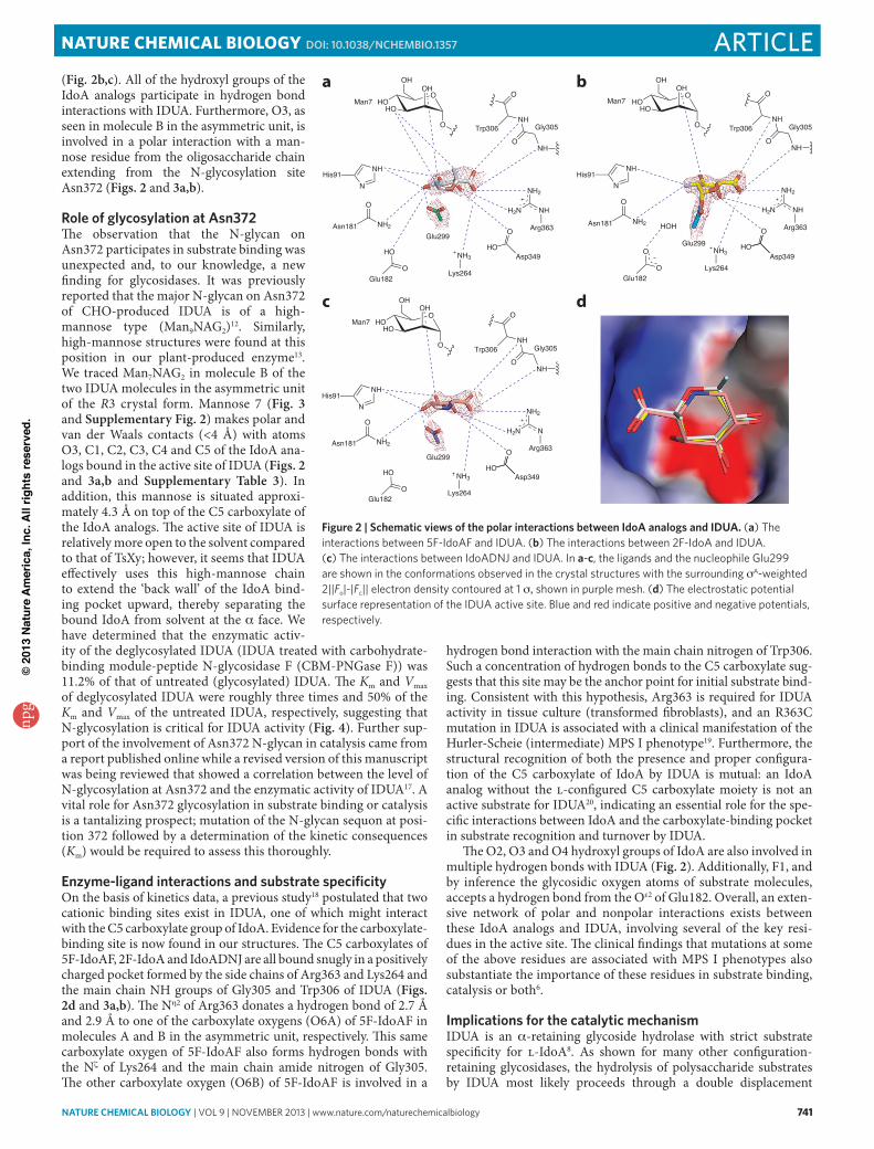
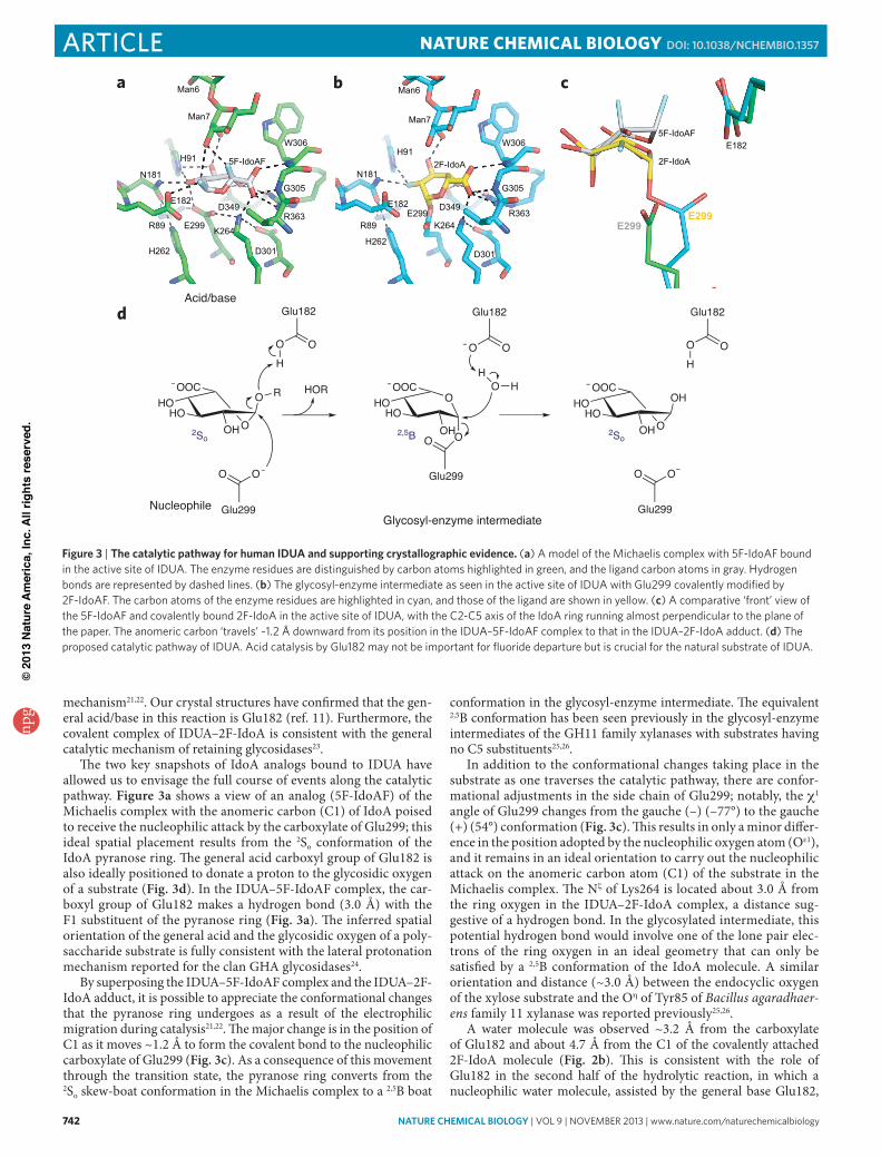