management of severe traumatic intrusion in the permanent
TRANSCRIPT
1Rovira- Wilde A, et al. BMJ Case Rep 2021;14:e235676. doi:10.1136/bcr-2020-235676
Management of severe traumatic intrusion in the permanent dentitionAlex Rovira- Wilde , Nick Longridge, Sarah McKernon
Case report
To cite: Rovira- Wilde A, Longridge N, McKernon S. BMJ Case Rep 2021;14:e235676. doi:10.1136/bcr-2020-235676
Institute of Clinical Sciences, University of Liverpool Faculty of Health and Life Sciences, Liverpool, UK
Correspondence toAlex Rovira- Wilde; alex_ rovira- wilde@ hotmail. co. uk
Accepted 28 November 2020
© BMJ Publishing Group Limited 2021. Re- use permitted under CC BY- NC. No commercial re- use. See rights and permissions. Published by BMJ.
SUMMARYTraumatic intrusion is considered one of the most severe luxation injuries to the permanent dentition. There are limited studies based on minimal evidence supporting suggested management protocols, owing to the rare occurrence of intrusion. The following case report details the multidisciplinary management and 18- month follow- up, in line with current UK guidelines, of a 23- year old adult male who sustained severe intrusion injuries to both permanent maxillary central incisor teeth. Timely, accurate diagnosis and subsequent appropriate management correlates with improved outcomes for traumatic injuries and it is therefore imperative those involved with the acute and long- term management of dentoalveolar trauma are aware of current guidelines.
BACKGROUNDFive per cent of all injuries requiring acute treatment are related to dental trauma.1 2 Intrusion injuries have a reported incidence of 0.5%–2% of all dental trauma with a predilec-tion towards males.1 By definition, intrusion presents as apical displacement of a tooth into alveolar bone. The resulting damage to peri-odontal ligament, root cementum, neurovas-cular bundle and surrounding alveolar bone can severely compromise the long- term prognosis of the affected permanent dentition.3 Of these cases, 97.2%occur in the anterior maxilla (upper canine- canine teeth).1
Given the relatively rare presentation of dental intrusion injuries, management protocols are based on limited evidence and several forms of guidance exist.4–6 Short- term management of intrusion injuries may include surgical or ortho-dontic repositioning of traumatised teeth and provision of a flexible trauma splint. Improved outcomes of treatment have correlated with timely and appropriate management.7 In the permanent dentition, pulp necrosis may occur in 88%–98% of intruded teeth with complete root development and subsequent endodontic treatment is necessary.5 8 Even with optimal immediate and long- term management, root resorption can occur in 51%–73% of cases, leaving affected teeth with a guarded long- term prognosis.8–10 This may ultimately result in early loss of teeth.
CASE PRESENTATIONA fit and well 23- year old man presented to a dental hospital after sustaining dental trauma falling onto the back support of a dining room
chair. The patient presented to an accident and emergency (A+E) department where he was assessed by oral and maxillofacial (OMFS) clinicians. An orthopantomogram (OPG) was requested to assess facial fractures was ordered by our OMFS colleagues at the time of presen-tation to the accident and emergency depart-ment. He was discharged without treatment and advised to seek further management by his general dental practitioner. He presented to a dental hospital 36 hours following injury. His medical history was unremarkable.
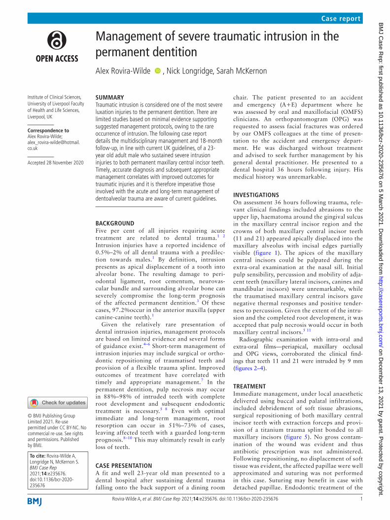
INVESTIGATIONSOn assessment 36 hours following trauma, rele-vant clinical findings included abrasions to the upper lip, haematoma around the gingival sulcus in the maxillary central incisor region and the crowns of both maxillary central incisor teeth (11 and 21) appeared apically displaced into the maxillary alveolus with incisal edges partially visible (figure 1). The apices of the maxillary central incisors could be palpated during the extra- oral examination at the nasal sill. Initial pulp sensibility, percussion and mobility of adja-cent teeth (maxillary lateral incisors, canines and mandibular incisors) were unremarkable, while the traumatised maxillary central incisors gave negative thermal responses and positive tender-ness to percussion. Given the extent of the intru-sion and the completed root development, it was accepted that pulp necrosis would occur in both maxillary central incisors.5 11
Radiographic examination with intra- oral and extra- oral films—periapical, maxillary occlusal and OPG views, corroborated the clinical find-ings that teeth 11 and 21 were intruded by 9 mm (figures 2–4).
TREATMENTImmediate management, under local anaesthetic delivered using buccal and palatal infiltrations, included debridement of soft tissue abrasions, surgical repositioning of both maxillary central incisor teeth with extraction forceps and provi-sion of a titanium trauma splint bonded to all maxillary incisors (figure 5). No gross contam-ination of the wound was evident and thus antibiotic prescription was not administered. Following repositioning, no displacement of soft tissue was evident, the affected papillae were well approximated and suturing was not performed in this case. Suturing may benefit in case with detached papillae. Endodontic treatment of the
on Decem
ber 13, 2021 by guest. Protected by copyright.
http://casereports.bmj.com
/B
MJ C
ase Rep: first published as 10.1136/bcr-2020-235676 on 5 M
arch 2021. Dow
nloaded from

2 Rovira- Wilde A, et al. BMJ Case Rep 2021;14:e235676. doi:10.1136/bcr-2020-235676
Case report
teeth 11 and 21 was initiated 2 weeks following trauma. An interappointment non- setting calcium hydroxide dressing was provided to prevent the development of inflamma-tory (infection- related) external resorption.11 Seven weeks following trauma, the splint was removed and orthograde endodontic treatment was completed using gutta percha via the warm vertical condensation obturation technique (figure 6). While there was no evidence of complete alve-olar fracture through both the palatal and the labial alve-olar plates with a mobile alveolar segment, the positioning of the apices in relation to the labial bone suggested labial plate fracture, and at initial review, it was decided to extend splinting to a total of 7 weeks.
OUTCOME AND FOLLOW-UPClinical and radiographic review followed published guid-ance.5 11 Pulp sensibility, percussion and mobility testing of adjacent teeth (maxillary canines and lateral incisors) remained unremarkable. Clinically, a high- pitched metallic sound, suggestive of external replacement root resorption
(ERRR), was exhibited on percussion testing of the left maxil-lary central incisor from 29 weeks post trauma (figure 7). There was minimal radiographic evidence suggestive of replacement resorption (figure 8A,B) over an 18- month review period. In light of the ankylotic sound exhibited on percussion, further annual clinical and radiographic review including cone- beam CT (CBCT) is planned to monitor ERRR progression. The position of both central incisors is acceptable to the patient; the patient had an existing ante-rior open bite prior to injury. Mild incisal edge discrepancies that persist following repositioning can be managed restor-atively with composite edge bonding. Large discrepancies or other malocclusions may require orthodontic intervention.
Figure 1 Initial presentation 36 hours following injury. Incisal edge of maxillary central incisors partially visible.
Figure 2 Standard maxillary occlusal radiograph identifying apical displacement of the maxillary central incisors (36 hours following injury).
Figure 3 Periapical radiographs suggesting apical displacement of the maxillary central incisors (36 hours following injury).
Figure 4 An orthopantomogram confirming severe apical displacement of the maxillary central incisors and ruled out facial fractures taken within the accident and emergency department following trauma (radiolucency suggestive of caries to be managed by the general dental practitioner).
on Decem
ber 13, 2021 by guest. Protected by copyright.
http://casereports.bmj.com
/B
MJ C
ase Rep: first published as 10.1136/bcr-2020-235676 on 5 M
arch 2021. Dow
nloaded from

3Rovira- Wilde A, et al. BMJ Case Rep 2021;14:e235676. doi:10.1136/bcr-2020-235676
Case report
DISCUSSIONEpidemiologyIntrusion is a rare form of dental trauma. An epidemiological study involving 216 intruded teeth displayed a prevalence of intrusion in 1.9% of all traumatic dental injuries over a 50- year period in Europe.1 The main aetiological factor appeared to be falling, as in this case, resulting in impacts acting axially along affected teeth. Coincident dental inju-ries occurred in 66.5% of teeth, predominately involving crown fractures.1 Of these cases, 53.7% involved intrusions of more than one tooth.1
PresentationClinical presentations include crown submergence, gingival lacerations, lack of mobility and a metallic sound on percus-sion of the intruded tooth/teeth. Intrusion may present radiographically as displacement of a root apex (this may present as foreshortening of roots should there be displace-ment in an anteroposterior direction). Radiographically, the periodontal ligament space around a root apex may appear obliterated. The majority of intrusion injuries are displaced
Figure 5 Provision of titanium trauma splint following surgical repositioning of maxillary central incisors (36 hours following injury).
Figure 6 Periapical radiographs showing maxillary central incisors immediately following endodontic treatment (7 weeks following injury).
Figure 7 Eighteen- month postoperative clinical views, evidence of marginal bone loss around maxillary central incisors present interproximally.
Figure 8 Periapical radiographs showing maxillary central incisors at 6- month (left) and 18- month (right) review following endodontic treatment.
on Decem
ber 13, 2021 by guest. Protected by copyright.
http://casereports.bmj.com
/B
MJ C
ase Rep: first published as 10.1136/bcr-2020-235676 on 5 M
arch 2021. Dow
nloaded from

4 Rovira- Wilde A, et al. BMJ Case Rep 2021;14:e235676. doi:10.1136/bcr-2020-235676
Case report
within a range of 1–8 mm.7 10 Accurate and comprehensive diagnosis may be aided by radiographs at several angles such as a combination of occlusal and periapical views of the affected teeth.1 2 Parallax views may also reveal dentoal-veolar fractures or signs of further damage to traumatised teeth outside the plane of a single view.4 While readily avail-able in a hospital setting, extra- oral films, such as an OPG, may not show sufficient detail to identify root or alveolar fractures. Therefore, use of adjunctive intra- oral films, such as standard maxillary occlusal and periapical radiographs, is recommended.
Management and complicationsAcute management protocols aim to restore intruded teeth to their original position, either via orthodontic or surgical positioning, in the hope of re- establishing a pre- existing relationship between the tooth and bone.12 13 Given the sparsity of evidence comparing the efficacy of different treatment options, there is diverging opinion on a superior management option.4–6 However, recent UK- based guidance, following thorough review of published literature, provides a consensus on accepted management protocols and is summarised in table 1.5 Allowing spontaneous re- eruption, otherwise known as passive repositioning, may be advised in cases of mild intrusion (<3 mm) with mature apices.5 Active repositioning via orthodontic means is advised should no coronal movement occur 2–3 weeks following trauma.5 Moderately displaced (3–6 mm) permanent teeth with mature apices require ‘active repositioning’ either surgically or orthodontically. Surgical repositioning, seen in this case, is justified when managing severely intruded teeth (>6 mm).5 Since the management of this case, inter-national guidance has been published advocating identical management protocols with ranges of ‘moderate’ displace-ment amended to 3–7 mm.11
Acute management adjuncts may involve splinting and/or antibiotic prescription. The splinting of repositioned teeth using a non- rigid, flexible splint has been strongly recom-mended in recent guidance.4 5 One sound tooth, either side of the traumatised teeth should be splinted.14 Splinting for a period of 6–8 weeks has been recommend by Andreasen et al.15 However, splinting for 10 days has also been shown to reduce mobility sufficiently enough to allow function.12 There is limited evidence corroborating the administration of systemic antibiotics to aid pulpal or periodontal healing. However, clinical judgement may be exercised in cases with contamination of associated soft tissue injuries.5
Subsequent collateral damage to dentine, cementum, peri-odontal ligament and alveolar bone may result in significant long- term complications such as pulp necrosis, infection of the root canal system, marginal bone loss, root resorption
and subsequent tooth loss.16 17 The prevalence of pulp necrosis in intruded teeth with complete root development is estimated to occur in 88%–98% of cases.5 Early endodontic treatment is advised and should be initiated within 2 weeks of trauma or as soon as the position of the tooth allows.11 Thus, long- term follow- up of teeth is advised in multiple guidance documents.1 4 5 The three- dimensional capability of CBCT is reported to be more sensitive than periapical radiographs in identifying apical changes and is useful when assessing teeth with suspected root resorption.18 19
Pulp necrosis, as a result of apical displacement of the tooth, may occur as the pulp’s main blood supply, via the apical foramen, is severed.20 21 Resorption, which is defined as ‘progressive loss of cementum and dentine through continued osteoclastic activity’, may be initiated and/or maintained following substantial damage to cementum and the periodontal ligament.17 22–24 Injury severity correlates with the extent of resorption.25–27 Younger patients may be at greater risk of rapid resorption.28 Substantial damage may result in complete resorption of the root. In adults, this may take up to 20 years; however, in children, tooth loss as a result of this resorptive process may occur in 3–7 years.26 29 In light of the extremely high risk of pulpal necrosis and resorption in cases with moderate or severe intrusion, prompt onward referral for further specialist dental assess-ment is required.
Marginal bone loss is a commonly documented sequalae of traumatic intrusion, especially in cases of multiple intru-sion injuries.30 31 Marginal bone loss following intrusion injuries appears to occur as a result of the crushing of the surrounding alveolar bone as well as exposure of bone to the oral cavity. As Tarnow et al highlights, the position of crestal bone relative to the contact point greatly influences the posi-tion of the papillae.32 This would explain the loss of papillae interproximal to the maxillary central incisors in this case (figure 7). This was an acceptable aesthetic outcome for the patient, which would otherwise require restorative treat-ment, orthodontics or periodontal surgery to correct.33
Delays in the acute management of intrusion injuries may complicate repositioning and further increase prevalence of pulp necrosis, resorption and compromise aesthetics.7 16 In this case, immediate referral to a specialty dental setting may have further optimised treatment outcome by enabling rapid surgical repositioning and commencement of root
Table 1 Summary of treatment recommendations for intruded teeth based on UK guidance5
Severity of intrusion Management
Mild (<3 mm) Passive repositioning*
Moderate (3–6 mm) Surgical or orthodontic repositioning†
Severe (>6 mm) Surgical repositioning
*Start orthodontic repositioning after 3 weeks should movement not occur with passive repositioning.†Orthodontic and surgical repositioning both appropriate. A surgical approach often involves fewer.
Learning points
► The occurrence of dental intrusion is rare. ► Given the large forces required to illicit such an injury, damages to tooth and surrounding structures can be extensive and may have profound complications that may significantly compromise the long- term prognosis of intruded teeth.
► Acute management, in the form of surgical repositioning and the provision of a flexible trauma splint, is simple and should be accessible to those working in acute care settings.
► Given the poor prognosis of dental intrusion injuries, immediate onward referral for multidisciplinary specialist input may optimise long- term treatment outcomes.
► Awareness of up- to- date guidance on the management of intruded teeth is imperative among clinicians working in acute care settings.
on Decem
ber 13, 2021 by guest. Protected by copyright.
http://casereports.bmj.com
/B
MJ C
ase Rep: first published as 10.1136/bcr-2020-235676 on 5 M
arch 2021. Dow
nloaded from

5Rovira- Wilde A, et al. BMJ Case Rep 2021;14:e235676. doi:10.1136/bcr-2020-235676
Case report
canal treatment within the recommended 2- week window following trauma.5 Delaying endodontic treatment longer than this time has been correlated with a higher prevalence of root resorption.34 This may increase the risk of subse-quent tooth loss, which is heavily implicated in quality of life.35
Twitter Nick Longridge @LongridgeNick
Contributors The following authors were involved in the submitted article: AR- W, NL, SMK. SMK led the initial management (initial assessment and surgical repositioning and provision of trauma splint for patient). NL provided subsequent root canal therapy of both central incisors. SMK and NL were both involved in the follow- up. AR- W was involved in the inquisition, reporting and analysis of data, and drafting of the submitted article.
Funding The authors have not declared a specific grant for this research from any funding agency in the public, commercial or not- for- profit sectors.
Competing interests None declared.
Patient consent for publication Obtained.
Provenance and peer review Not commissioned; externally peer reviewed.
Open access This is an open access article distributed in accordance with the Creative Commons Attribution Non Commercial (CC BY- NC 4.0) license, which permits others to distribute, remix, adapt, build upon this work non- commercially, and license their derivative works on different terms, provided the original work is properly cited and the use is non- commercial. See: http:// creativecommons. org/ licenses/ by- nc/ 4. 0/.
ORCID iDAlex Rovira- Wilde http:// orcid. org/ 0000- 0001- 8217- 0975
REFERENCES 1 Andreasen JO, Bakland LK, Matras RC, et al. Traumatic intrusion of permanent teeth.
Part 1. An epidemiological study of 216 intruded permanent teeth. Dent Traumatol 2006;22:83–9.
2 Petersson EE, Andersson L, Sörensen S. Traumatic oral vs non- oral injuries. Swed Dent J 1997;21:55–68.
3 Wigen TI, Agnalt R, Jacobsen I. Intrusive luxation of permanent incisors in Norwegians aged 6-17 years: a retrospective study of treatment and outcome. Dent Traumatol 2008;24:612–8.
4 Diangelis AJ, Andreasen JO, Ebeleseder KA, et al. International association of dental Traumatology guidelines for the management of traumatic dental injuries: 1. fractures and luxations of permanent teeth. Dent Traumatol 2012;28:2–12.
5 Albadri S, Zaitoun H, Kinirons MJ, et al. Uk national clinical guidelines in paediatric dentistry: treatment of traumatically intruded permanent incisor teeth in children. Int J Paediatr Dent 2010;20 Suppl 1:1–2.
6 Flores MT, Andersson L, Andreasen JO, et al. Guidelines for the management of traumatic dental injuries. I. fractures and luxations of permanent teeth. Dent Traumatol 2007;23:66–71.
7 Andreasen JO, Bakland LK, Andreasen FM. Traumatic intrusion of permanent teeth. Part 3. A clinical study of the effect of treatment variables such as treatment delay, method of repositioning, type of splint, length of splinting and antibiotics on 140 teeth. Dent Traumatol 2006;22:99–111.
8 Al- Badri S, Kinirons M, Cole B, et al. Factors affecting resorption in traumatically intruded permanent incisors in children. Dent Traumatol 2002;18:73–6.
9 Chaushu S, Shapira J, Heling I, et al. Emergency orthodontic treatment after the traumatic intrusive luxation of maxillary incisors. Am J Orthod Dentofacial Orthop 2004;126:162–72.
10 Kinirons MJ, Sutcliffe J. Traumatically intruded permanent incisors: a study of treatment and outcome. Br Dent J 1991;170:144–6.
11 Bourguignon C, Cohenca N, Lauridsen E, et al. International association of dental Traumatology guidelines for the management of traumatic dental injuries: 1. fractures and luxations. Dent Traumatol 2020;36:314–30.
12 Humphrey J, Kenny D, Barrett E. Clinical outcomes for permanent incisor luxation in a paediatric population I. intrusion. Dent Traumatol 2003;29:266–73.
13 Turley PK, Joiner MW, Hellstrom S. The effect of orthodontic extrusion on traumatically intruded teeth. Am J Orthod 1984;85:47–56.
14 Brown CL, Mackie IC. Splinting of traumatized teeth in children. Dent Update 2003;30:78–82.
15 Andreasen JO, Andreasen FM, Andersson L. Text book and color atlas of traumatic injuries to the teeth. 4th edn. Copenhagen: Blackwell, 2007.
16 Andreasen JO. Luxation of permanent teeth due to trauma. A clinical and radiographic follow- up study of 189 injured teeth. Scand J Dent Res 1970;78:273–86.
17 Yu CY, Abbott PV. Responses of the pulp, periradicular and soft tissues following trauma to the permanent teeth. Aust Dent J 2016;61 Suppl 1:39–58.
18 Tsai P, Torabinejad M, Rice D, et al. Accuracy of cone- beam computed tomography and periapical radiography in detecting small periapical lesions. J Endod 2012;38:965–70.
19 Patel S, Foschi F, Mannocci F, et al. External cervical resorption: a three- dimensional classification. Int Endod J 2018;51:206–14.
20 Andreasen FM, Pedersen BV, Vestergaard- Pederesen B. Prognosis of luxated permanent teeth--the development of pulp necrosis. Endod Dent Traumatol 1985;1:207–20.
21 Andreasen FM, Kahler B. Pulpal response after acute dental injury in the permanent dentition: clinical implications- a review. J Endod 2015;41:299–308.
22 Fuss Z, Tsesis I, Lin S. Root resorption--diagnosis, classification and treatment choices based on stimulation factors. Dent Traumatol 2003;19:175–82.
23 Patel S, Ford TP. Is the resorption external or internal? Dent Update 2007;34:218–29. 24 Ebeleseder KA, Santler G, Glockner K, et al. An analysis of 58 traumatically intruded
and surgically extruded permanent teeth. Endod Dent Traumatol 2000;16:34–9. 25 Andreasen JO. Relationship between cell damage in the periodontal ligament after
Replantation and subsequent development of root resorption. Acta Odontol Scand 1981;39:15–25.
26 Trope M. Root resorption due to dental trauma. Endod Topics 2002;1:79–100. 27 Andersson L, Bodin I, Sörensen S. Progression of root resorption following
Replantation of human teeth after extended extraoral storage. Endod Dent Traumatol 1989;5:38–47.
28 Heithersay GS. Management of tooth resorption. Aust Dent J 2007;52:S105–21. 29 Barrett EJ, Kenny DJ. Survival of avulsed permanent maxillary incisors in children
following delayed Replantation. Endod Dent Traumatol 1997;13:269–75. 30 Andreasen JO, Bakland LK, Andreasen FM. Traumatic intrusion of permanent teeth.
Part 2. A clinical study of the effect of preinjury and injury factors, such as sex, age, stage of root development, tooth location, and extent of injury including number of intruded teeth on 140 intruded permanent teeth. Dent Traumatol 2006;22:90–8.
31 Andreasen JOVE, Vinding TR, Christensen SSA. Predictors for healing complications in the permanent dentition after dental trauma. Endod Topics 2006;14:20–7.
32 Tarnow DP, Magner AW, Fletcher P. The effect of the distance from the contact point to the crest of bone on the presence or absence of the interproximal dental papilla. J Periodontol 1992;63:995–6.
33 Prato GPP, Rotundo R, Cortellini P. Interdental papilla management: a review and classification of the therapeutic approaches. Int J Periodontics Restorative Dent 2004;24:246–55.
34 Stewart CJ, Elledge RO, Kinirons MJ, et al. Factors affecting the timing of pulp extirpation in a sample of 66 replanted avulsed teeth in children and adolescents. Dent Traumatol 2008;24:625–7.
35 Gerritsen AE, Allen PF, Witter DJ, et al. Tooth loss and oral health- related quality of life: a systematic review and meta- analysis. Health Qual Life Outcomes 2010;8:126.
on Decem
ber 13, 2021 by guest. Protected by copyright.
http://casereports.bmj.com
/B
MJ C
ase Rep: first published as 10.1136/bcr-2020-235676 on 5 M
arch 2021. Dow
nloaded from

6 Rovira- Wilde A, et al. BMJ Case Rep 2021;14:e235676. doi:10.1136/bcr-2020-235676
Case report
Copyright 2021 BMJ Publishing Group. All rights reserved. For permission to reuse any of this content visithttps://www.bmj.com/company/products-services/rights-and-licensing/permissions/BMJ Case Report Fellows may re-use this article for personal use and teaching without any further permission.
Become a Fellow of BMJ Case Reports today and you can: ► Submit as many cases as you like ► Enjoy fast sympathetic peer review and rapid publication of accepted articles ► Access all the published articles ► Re-use any of the published material for personal use and teaching without further permission
Customer ServiceIf you have any further queries about your subscription, please contact our customer services team on +44 (0) 207111 1105 or via email at [email protected].
Visit casereports.bmj.com for more articles like this and to become a Fellow
on Decem
ber 13, 2021 by guest. Protected by copyright.
http://casereports.bmj.com
/B
MJ C
ase Rep: first published as 10.1136/bcr-2020-235676 on 5 M
arch 2021. Dow
nloaded from