objectives historical perspectives anesthesia and severe …€¦ · anesthesia and severe tbi 1....
TRANSCRIPT
1
Care of the Head Trauma Patient
Julin F. Tang, MD, MS
ProfessorDepartment of Anesthesia and
Critical Care MedicineUCSF/San Francisco Gen Hospital
ObjectivesAnesthesia and severe TBI
1. Historical perspective in traumatic brain injury patients
2. Intraoperative hemodynamic changes during emergent decompressive craniotomy/craniectomy
3. Literature reviews and future studies
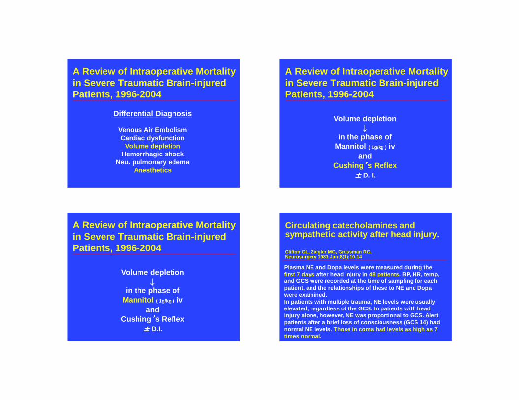
Monro-Kellie doctrine (1824)
Intracranial volume (constant)
= v. Brain + v. Blood
+ v. CSF + v. Mass lesion
Historical Perspectives
Trepanation 1488-1516
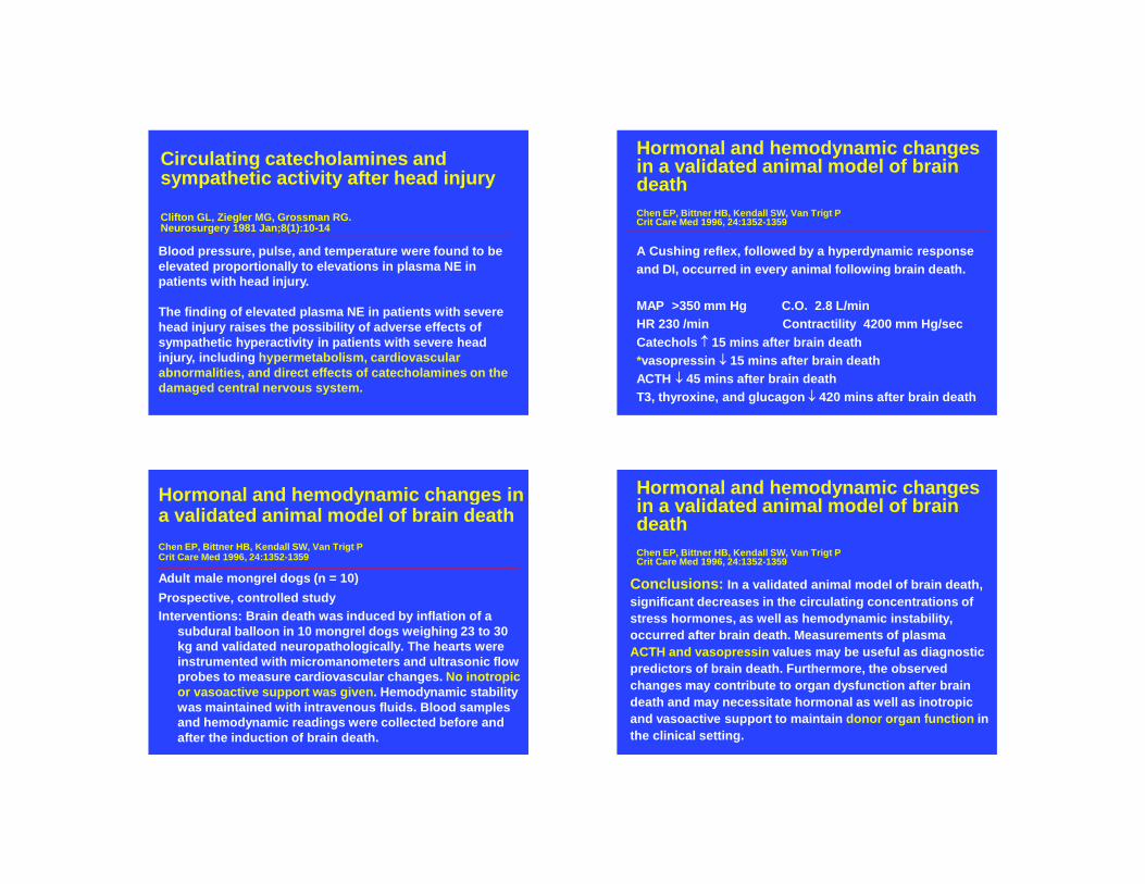
Monro-Kellie doctrine (1824)
Intracranial volume (constant)
= v. Brain + v. Blood
+ v. CSF + v. Mass lesion
Historical Perspectives
Trepanation skull Iron age

2
Historical Perspectives
1881 Naunyn and Schreiber1902 Cushing
Lundberg N, Kjallquist A, Bien C. Reduction of increased intracranial pressure by hyperventilation. Acta Psy chiatr Scand 1959; 34: 4-64
Galicich JH, French LA. Use of dexamethasone in the treatment of cerebral edema resulting from brain tu mors and brain surgery. Am Pract 1961; 12: 212-223
Lundberg N, Troupp H, Lorin H. Continuous recording of the ventricular fluid pressure in patients with sev ere acute traumatic brain damage. J. Neurosurg 1965; 22: 581-590
Jennett B, Teasdale G. Aspect of coma after severe head injury. Lancet 1977; 1: 878-881.
Historical Perspectives
Traumatic Coma Data Bank - Began collecting data in 1979
Bowers SA, Marshall LF. Outcome in 200 consecutive cases of severe head injury treated in San Diego County: a p rospective analysis. Neurosurgery 1980; 6: 237-242
Secondary brain injury - Experimental research began in 1980
Marmarou A, Anderson RI, Ward JD, et al . Impact of ICP instability and hypotension on outcome in patients with severe head trauma. J Neurosurg 1991; 75: S59-S66
Historical Perspectives
Traumatic Brain Injury(TBI)
TBI is a non-degenerative, non-congenitalinsult to the brain from an externalmechanical force , possibly leading topermanent or temporary impairments of cognitive, physical, and psychosocialfunctions with an associated diminished or altered state of consciousness.

3
Traumatic Brain Injury
Physical mechanisms of brain injuryimpact loadingimpulsive loadingstatic or quasistatic loading
Types of tissue deformitycompressivetensileshear
Traumatic Brain Injury
Types of intracranial hemorrhages
epidural hematomasubdural hematoma subarachnoid hemorrhageintracerebral hemorrhageintraventricular hemorrhage
Neurological scale of TBI
GCS = E4 + M6 + V5
Severe TBI - 3 to 8Moderate TBI - 9 to 12Mild TBI - 13 to 15
Epidemiology
Annually, 250,000 TBI need hospitalization.
Approximately 50,000 US deaths per year.
Mortality rate in severe TBI - 33 to 55%
Overall Mortality rate in TBI - 20 to 25%
TBI accounts for ~ 35% of all deaths from
acute injuries in the United States.
TCDB

4
Primary vs. SecondarySevere TBI
Primary injury is from bruising or
penetrating objects
Secondary injury is from hypoxia or
decrease cerebral perfusion
Peri-operative Care of Head Trauma patient
EMT → ED → Rad → ICU
EMT→ ED→ Rad→ OR→ ICU
Hypotension • Hypoxemia
OR Room 1 Cases @ SFGH Trauma center
• 100 - 120 cases per year; 8 - 10 cases per month• 20% of patients required BT > 20 units for initial
resuscitation (<12 o)• 25% of patients required activation of MTP• 33% mortality rate; ~ 1/3 of patients had severe
traumatic brain injury.• ~ 30% traumatic brain injury; (10 % OR mortality)• Transfused 21 - 39 u pRBC with a 25% mortality
Transfused > 40 u pRBC with a 52% mortality• Complications from massive fluid resuscitation

5
Preoperative predictors of reduction in arterial blood pressure following dural opening during surgical evacuation of acute subdural hematoma
Kawaguchi M, Sakamoto T, Ohnishi H, Karasawa J, Fur uya HJ Neurosurg Anesthesiol. 1996 Apr;8(2):117-22
*Retrospective chart review56 patients with traumatic acute subdural hematoma
Preoperative variables: clinical profile, hemodynam ic parameters, neurological findings, and Cranial CT s can
5 min before opening the dura vs dural openingGroup A (n = 18) MAP reduction > 20%Group B (n = 38) MAP within +/- 20% of baseline valu es
Preoperative predictors of reduction in arterial blood pressure following dural opening during surgical evacuation of acute subdural hematoma
Kawaguchi M, Sakamoto T, Ohnishi H, Karasawa J, Fur uya HJ Neurosurg Anesthesiol. 1996 Apr;8(2):117-22
Preoperative predictors of reduction in arterial blood pressure following dural opening during surgical evacuation of acute subdural hematoma
Kawaguchi M, Sakamoto T, Ohnishi H, Karasawa J, Fur uya HJ Neurosurg Anesthesiol. 1996 Apr;8(2):117-22
Low GCS score, absence of the mesencephalic cistern , and bilaterally dilated pupils were particularly strong predictors of this amount of blood pressure reducti on. The clinical outcomes of patients in group A follow ing dural opening during surgery were significantly poo rer.
Findings suggest that blood pressure reduction foll owing opening of the dura in patients undergoing surgical evacuation of hematoma for traumatic ASDH may be predicted by careful preoperative assessment of neurological and CT scan findings.

6
The Incidence & Risk Factors forHypotension During Emergent Decompressive Craniotomy in Children with Traumatic Brain InjuryPatrick Miller, MD*, Christ D. Mack, MS, Marla Sa mmer, MD, and Monica S. Vavilala, MD*Anesth Analg 2006;103:869-875
CONCLUSION: Between 1994 and 2004, a retrospective cohort study in children (108) <13 yr with traumatic brain injury (TBI) at a Level 1 pediatric trauma center to describe risk factors for intraoperative hypotension (IH) during emergent decompressivecraniotomy. Overall, 56 (52%) patients had IH. ED hypotension , blood loss , CT lesion volume , and CT midline shift predicted IH. Anesthesiologists can expect children with preoperative CT midline shift 4 mm to have IH during this procedure.
↓
20
40
60
80
100
120
140
160
-20 -10 0 +10 +20
TIME (minutes)
MA
P (
mm
Hg)
DURA OPEN
Intraoperative Mortality in Severe Traumatic Brain-injured Patients
1. What is the pathophysiology of this deleterious event ?
2. Can we improve the operative outcome by EGDT?
7
A Review of Intraoperative Mortality in Severe Traumatic Brain-injured Patients, 1996-2004
Differential Diagnosis
Venous Air EmbolismCardiac dysfunction
Volume depletionHemorrhagic shock
Neu. pulmonary edema Anesthetics
A Review of Intraoperative Mortality in Severe Traumatic Brain-injured Patients, 1996-2004
Volume depletion
in the phase ofMannitol ( 1g/kg ) iv
andCushing ’’’’s Reflex
±±±± D.I.
↓
A Review of Intraoperative Mortality in Severe Traumatic Brain-injured Patients, 1996-2004
Volume depletion
in the phase ofMannitol ( 1g/kg ) iv
andCushing ’’’’s Reflex
±±±± D. I.
↓
Circulating catecholamines and sympathetic activity after head injury.
Clifton GL, Ziegler MG, Grossman RG.Neurosurgery 1981 Jan;8(1):10-14
Plasma NE and Dopa levels were measured during the first 7 days after head injury in 48 patients . BP, HR, temp, and GCS were recorded at the time of sampling for e ach patient, and the relationships of these to NE and D opa were examined. In patients with multiple trauma, NE levels were us ually elevated, regardless of the GCS. In patients with h ead injury alone, however, NE was proportional to GCS. Alert patients after a brief loss of consciousness (GCS 1 4) had normal NE levels. Those in coma had levels as high as 7 times normal.
8
Circulating catecholamines and sympathetic activity after head injury
Clifton GL, Ziegler MG, Grossman RG.Neurosurgery 1981 Jan;8(1):10-14
Blood pressure, pulse, and temperature were found t o be elevated proportionally to elevations in plasma NE in patients with head injury.
The finding of elevated plasma NE in patients with severe head injury raises the possibility of adverse effec ts of sympathetic hyperactivity in patients with severe h ead injury, including hypermetabolism, cardiovascular abnormalities, and direct effects of catecholamines on the damaged central nervous system.
Hormonal and hemodynamic changes in a validated animal model of brain death Chen EP, Bittner HB, Kendall SW, Van Trigt PCrit Care Med 1996, 24:1352-1359
Adult male mongrel dogs (n = 10)
Prospective, controlled studyInterventions: Brain death was induced by inflation of a
subdural balloon in 10 mongrel dogs weighing 23 to 30 kg and validated neuropathologically. The hearts we re instrumented with micromanometers and ultrasonic fl ow probes to measure cardiovascular changes. No inotropic or vasoactive support was given . Hemodynamic stability was maintained with intravenous fluids. Blood sampl es and hemodynamic readings were collected before and after the induction of brain death.
Hormonal and hemodynamic changes in a validated animal model of brain death Chen EP, Bittner HB, Kendall SW, Van Trigt PCrit Care Med 1996, 24:1352-1359
A Cushing reflex, followed by a hyperdynamic respon se and DI, occurred in every animal following brain de ath.
MAP >350 mm Hg C.O. 2.8 L/min HR 230 /min Contractility 4200 mm Hg/secCatechols ↑ 15 mins after brain death*vasopressin ↓ 15 mins after brain deathACTH ↓ 45 mins after brain deathT3, thyroxine, and glucagon ↓ 420 mins after brain death
Hormonal and hemodynamic changes in a validated animal model of brain death Chen EP, Bittner HB, Kendall SW, Van Trigt PCrit Care Med 1996, 24:1352-1359
Conclusions: In a validated animal model of brain death, significant decreases in the circulating concentrat ions of stress hormones, as well as hemodynamic instability , occurred after brain death. Measurements of plasma ACTH and vasopressin values may be useful as diagnostic predictors of brain death. Furthermore, the observe d changes may contribute to organ dysfunction after b rain death and may necessitate hormonal as well as inotr opic and vasoactive support to maintain donor organ function in the clinical setting.
9
Perioperative practice changes in Severe Traumatic Brain-injured Patients, 2004-
Volume Depletion Mannitol iv is routinely givenHct is ~ normal or above normalBD is always within less than -10Minimize anesthetics
Cushing ’’’’s reflexHemodynamic monitoringCVP measurement
Perioperative practice changes in Severe Traumatic Brain-injured Patients, 2004-
EDtrauma Intubated with Etomidate - avoid ↓ BPIV access - C-line, if possibleSent labs and blood samples for T&CETCO2 monitoring ( ~ 30 )Hemodynamic monitoring - a-line, if possible
RadCTContinue monitoringMaintain ““““hemodynamic ””””
Perioperative practice changes in Severe Traumatic Brain-injured Patients, 2004-
OR (before dural opening) Established IJ or subclavian c-line, a-lineHemodynamic monitoring - pressors*Fluid resuscitation - CVP 10 or TEE 2009
Avoid fix anesthetic agentsO2 and muscle relaxant
OR (during dural opening)Well communication Slow decompression - fenestration technique
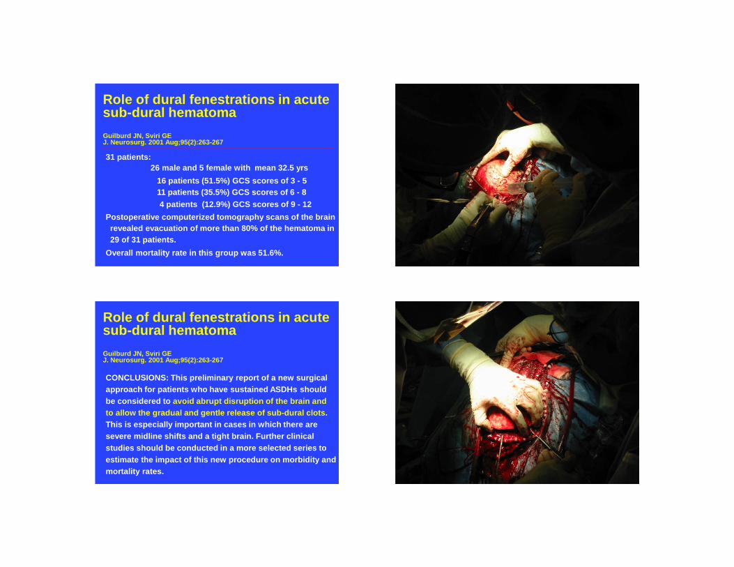
Role of dural fenestrations in acute sub-dural hematoma
Guilburd JN, Sviri GEJ. Neurosurg. 2001 Aug;95(2):263-267
One of the factors contributing to poor outcomes in cases of ASDHs could be rapid surgical decompression, owi ng to the severe extrusion of the brain through the crani otomy defect in response to acute brain swelling. To avoi d the deleterious consequences of abrupt decompression of the sub-dural space with disruption of brain tissue. Th is procedure consists of creating multiple fenestratio ns of the dura (MFD) in a mesh-like fashion and removing clots through the small dural openings that are left open , avoiding the creation of a wide dural opening and t he disruption of and additional damage to brain tissue .
10
Role of dural fenestrations in acute sub-dural hematoma
Guilburd JN, Sviri GEJ. Neurosurg. 2001 Aug;95(2):263-267
31 patients: 26 male and 5 female with mean 32.5 yrs
16 patients (51.5%) GCS scores of 3 - 511 patients (35.5%) GCS scores of 6 - 8
4 patients (12.9%) GCS scores of 9 - 12
Postoperative computerized tomography scans of the brain revealed evacuation of more than 80% of the hematom a in 29 of 31 patients.
Overall mortality rate in this group was 51.6%.
Role of dural fenestrations in acute sub-dural hematoma
Guilburd JN, Sviri GEJ. Neurosurg. 2001 Aug;95(2):263-267
CONCLUSIONS: This preliminary report of a new surgi cal approach for patients who have sustained ASDHs shou ld be considered to avoid abrupt disruption of the brain and to allow the gradual and gentle release of sub-dura l clots . This is especially important in cases in which ther e are severe midline shifts and a tight brain. Further cl inical studies should be conducted in a more selected seri es to estimate the impact of this new procedure on morbid ity and mortality rates.
11
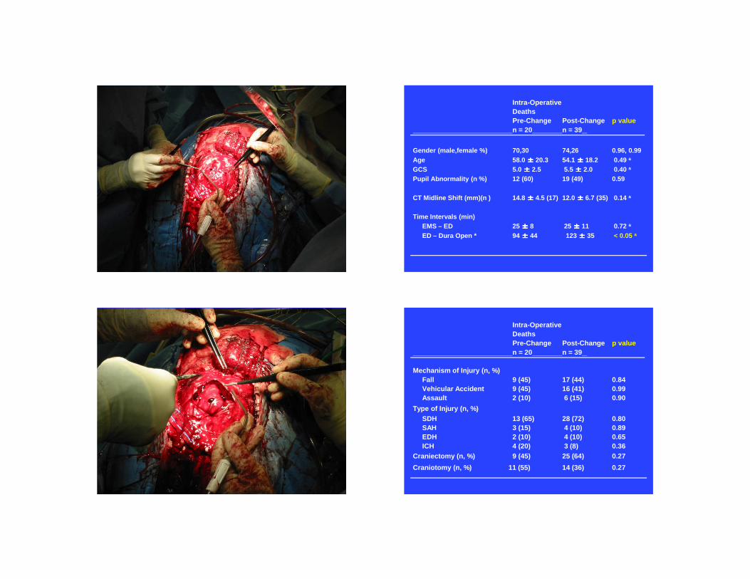
Intra-OperativeDeathsPre-Change Post-Change p valuen = 20 n = 39
Gender (male,female %) 70,30 74,26 0.96, 0.99Age 58.0 ±±±± 20.3 54.1 ±±±± 18.2 0.49 a
GCS 5.0 ±±±± 2.5 5.5 ±±±± 2.0 0.40 a
Pupil Abnormality (n %) 12 (60) 19 (49) 0.59
CT Midline Shift (mm)(n ) 14.8 ±±±± 4.5 (17) 12.0 ±±±± 6.7 (35) 0.14 a
Time Intervals (min)EMS – ED 25 ±±±± 8 25 ±±±± 11 0.72 a
ED – Dura Open * 94 ±±±± 44 123 ±±±± 35 < 0.05 a
Intra-OperativeDeathsPre-Change Post-Change p valuen = 20 n = 39
Mechanism of Injury (n, %)Fall 9 (45) 17 (44) 0.84Vehicular Accident 9 (45) 16 (41) 0.99Assault 2 (10) 6 (15) 0.90
Type of Injury (n, %)SDH 13 (65) 28 (72) 0.80SAH 3 (15) 4 (10) 0.89EDH 2 (10) 4 (10) 0.65ICH 4 (20) 3 (8) 0.36
Craniectomy (n, %) 9 (45) 25 (64) 0.27
Craniotomy (n, %) 11 (55) 14 (36) 0.27

12
Intra-OperativeDeathsPre-Change Post-Change p valuen = 20 n = 39
Volume Resuscitation (L) 6.3 ±±±± 3.6 9.9 ±±±± 5.2 < 0.01
Crystalloids/Colloids 2.8 ±±±± 1.4 4.0 ±±±± 1.8 < 0.01Blood Products 1.1 ±±±± 0.9 1.9 ±±±± 1.7 < 0.05
WB (n) 1.7 ±±±± 0.6 (3) 0 (0) ns a
PRBC (n) 1.0 ±±±± 0.5 (15) 1.1 ±±±± 0.8 (35) nsFFP (n) 0.6 ±±±± 0.4 (5) 1.3 ±±±± 0.9 (22) < 0.05PLTS (n) 0 (0) 0.4 ±±±± 0.2 (12) < 0.05 a
Volume Loss (L) (n) 3.9 ±±±± 2.6 (16) 6.8 ±±±± 3.4 (39) < 0.05UO (n) 0.8 ±±±± 0.7 (10) 2.6 ±±±± 1.5 (38) < 0.001EBL (n) 1.2 ±±±± 0.7 (15) 1.5 ±±±± 1.0 (36) ns
Fluid Balance (L) (n) * 1.3 ±±±± 2.9 (9) 2.8 ±±±± 4.4 (35) nsMannitol Dose (g) (n) 81.1 ±±±± 29.0 (19) 98.5 ±±±± 30.6 (33) < 0.05CVP (mmHg) (n) 0 (0) 15.1 ±±±± 5.8 (20) < 0.001 a
Intra-OperativeDeathsPre-Change Post-Change p valuen = 20 n = 39
Isoflurane (n) * 31.4 ±±±± 19.0 (13) 17.2 ±±±± 13.3 (20) < 0.05
Desflurane (n) * 60 ±±±± 0 (1) 155 ±±±± 67 (3) nsSevoflurane (n) * 10.0 ±±±± 0.0 (1) 74.5 ±±±± 97.4 (5) nsFentanyl (n) ** 237.5 ±±±± 182.7 (8) 178.6 ±±±± 128.6 (7) nsVersed (n) ** 0 (0) 2 ±±±± 0 (2) nsPropofol (n) ** 500 ±±±± 0.0 (1) 0 (0) nsMuscle Relaxants † a 65% 100% < 0.001Vasopressor Support † a 95% 92% ns
Phenylephrine 80% 74% ns Epinephrine 85% 69% ns Nor-Epinephrine 0% 10% nsDopamine 20% 5% ns Vasopressin 20% 10% ns Ephedrine 10% 8% ns
20
40
60
80
100
120
140
160
-20 -10 0 +10 +20
TIME (minutes)
MA
P (
mm
Hg)
DURA OPEN
MAPpre = 120 mmHg
MAPpost = 98 mmHg
∆MAP = ICP
13
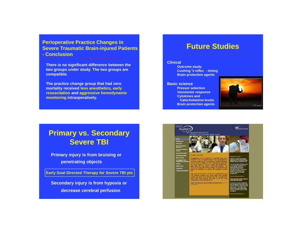
Perioperative Practice Changes in Severe Traumatic Brain- injured Patients - Conclusion
There is no significant difference between the two groups under study. The two groups are compatible.
The practice change group that had zero mortality received less anesthetics , early resuscitation and aggressive hemodynamic monitoring intraoperatively.
Primary vs. SecondarySevere TBI
Primary injury is from bruising or
penetrating objects
Secondary injury is from hypoxia or
decrease cerebral perfusion
Early Goal Directed Therapy for Severe TBI pts
Future Studies
ClinicalOutcome study Cushing ’’’’s reflex - timingBrain protection agents
Basic sciencePressor selectionVasomotor responseCytokines and
Catecholamine levelsBrain protection agents
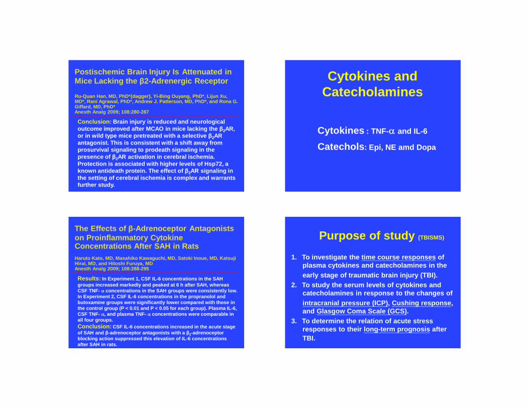
Postischemic Brain Injury Is Attenuated in Mice Lacking the β2-Adrenergic Receptor
Ru-Quan Han, MD, PhD*{dagger}, Yi-Bing Ouyang, PhD* , Lijun Xu, MD*, Rani Agrawal, PhD*, Andrew J. Patterson, MD, P hD*, and Rona G. Giffard, MD, PhD*Anesth Analg 2009; 108:280-287
Conclusion: Brain injury is reduced and neurological outcome improved after MCAO in mice lacking the β2AR, or in wild type mice pretreated with a selective β2AR antagonist. This is consistent with a shift away fr om prosurvival signaling to prodeath signaling in the presence of β2AR activation in cerebral ischemia. Protection is associated with higher levels of Hsp7 2, a known antideath protein. The effect of β2AR signaling in the setting of cerebral ischemia is complex and war rants further study.
14
Postischemic Brain Injury Is Attenuated in Mice Lacking the β2-Adrenergic Receptor
Ru-Quan Han, MD, PhD*{dagger}, Yi-Bing Ouyang, PhD* , Lijun Xu, MD*, Rani Agrawal, PhD*, Andrew J. Patterson, MD, P hD*, and Rona G. Giffard, MD, PhD*Anesth Analg 2009; 108:280-287
Conclusion: Brain injury is reduced and neurological outcome improved after MCAO in mice lacking the β2AR, or in wild type mice pretreated with a selective β2AR antagonist. This is consistent with a shift away fr om prosurvival signaling to prodeath signaling in the presence of β2AR activation in cerebral ischemia. Protection is associated with higher levels of Hsp7 2, a known antideath protein. The effect of β2AR signaling in the setting of cerebral ischemia is complex and war rants further study.
The Effects of β-Adrenoceptor Antagonists on Proinflammatory Cytokine Concentrations After SAH in RatsHaruto Kato, MD, Masahiko Kawaguchi, MD, Satoki Ino ue, MD, KatsujiHirai, MD, and Hitoshi Furuya, MDAnesth Analg 2009; 108:288-295
Results: In Experiment 1, CSF IL-6 concentrations in the SAH groups increased markedly and peaked at 6 h after S AH, whereas CSF TNF- α concentrations in the SAH groups were consistently low. In Experiment 2, CSF IL-6 concentrations in the pro pranolol and butoxamine groups were significantly lower compared with those in the control group (P < 0.01 and P < 0.05 for each g roup). Plasma IL-6, CSF TNF- α, and plasma TNF- α concentrations were comparable in all four groups.Conclusion: CSF IL-6 concentrations increased in the acute stag e of SAH and β-adrenoceptor antagonists with a β2-adrenoceptor blocking action suppressed this elevation of IL-6 c oncentrations after SAH in rats.
Cytokines and Catecholamines
Cytokines : TNF-α and IL-6
Catechols : Epi, NE amd Dopa
Purpose of study (TBISMS)
1. To investigate the time course responses of plasma cytokines and catecholamines in the early stage of traumatic brain injury (TBI).
2. To study the serum levels of cytokines and catecholamines in response to the changes of intracranial pressure (ICP), Cushing response, and Glasgow Coma Scale (GCS).
3. To determine the relation of acute stress responses to their long-term prognosis after TBI.
15
Summary and Recommendations
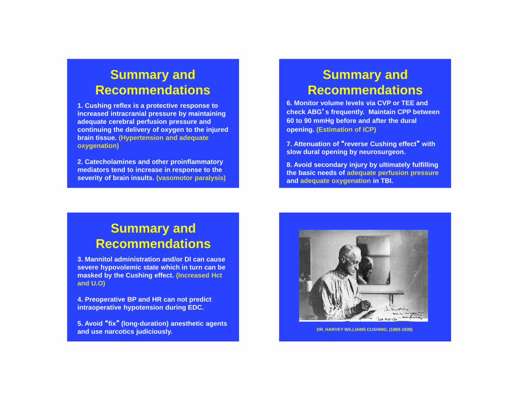
1. Cushing reflex is a protective response to increased intracranial pressure by maintaining adequate cerebral perfusion pressure and continuing the delivery of oxygen to the injured brain tissue. (Hypertension and adequate oxygenation)
2. Catecholamines and other proinflammatory mediators tend to increase in response to the severity of brain insults. (vasomotor paralysis)
Summary and Recommendations
3. Mannitol administration and/or DI can cause severe hypovolemic state which in turn can be masked by the Cushing effect. (Increased Hct and U.O)
4. Preoperative BP and HR can not predict intraoperative hypotension during EDC.
5. Avoid ““““fix ”””” (long-duration) anesthetic agents and use narcotics judiciously.
Summary and Recommendations
6. Monitor volume levels via CVP or TEE and check ABG ’’’’s frequently. Maintain CPP between 60 to 90 mmHg before and after the dural opening. (Estimation of ICP)
7. Attenuation of ““““reverse Cushing effect ”””” with slow dural opening by neurosurgeon.
8. Avoid secondary injury by ultimately fulfilling the basic needs of adequate perfusion pressureand adequate oxygenation in TBI.
DR. HARVEY WILLIAMS CUSHING. (1869-1939 )

16
↓