neurointensive care monitoring for severe traumatic brain ...cdn.intechopen.com/pdfs/33538.pdf ·...
TRANSCRIPT
11
Neurointensive Care Monitoring for Severe Traumatic Brain Injury
Zamzuri Idris, Muzaimi Mustapha and Jafri Malin Abdullah Universiti Sains Malaysia
Malaysia
1. Introduction
Worldwide, traumatic brain injury is one of the leading cause of permanent disability and death. Contextually, close monitoring and immediate therapy for any accompanying abnormalities are crucial in order to reduce the rate of mortality or morbidity associated with this acquired brain injury. For this purpose, an isolated neurointensive care (NIC) specialised for brain injury is highly warranted, manned by appropriately trained staff who understand the current pathophysiology of brain injury and equipped with various modes of therapy to tackle any diagnosed abnormalities. To date, we are witnessing progress in managing severely injured brain from no specific monitoring to specific ones, and from a single intracranial pressure monitoring to future trend of multiple cranial monitoring alongside availability of various mode of therapies. The multimodal brain monitoring as it is commonly known is a concept whereby, intracranial pressure as well as various other important cerebral parameters can readily be monitored. Monitoring alone will not alter the outcomes of severely injured brain patients. Prompt recognition of any abnormality from the monitoring and availability of therapy to correct the diagnosed abnormality are some of the factors that are recognised to influence better outcome score. However, at present, two obvious limitations in the multimodality brain monitoring are our incomplete understanding of the underlying pathophysiology of the severely injured brain and the limited availability of mode of therapy to the neurosurgeon or neurointensivist to treat abnormalities detected from the state-of-the-art monitoring. Research are ongoing in these areas and in this chapter, we discuss in details the current pathophysiology of traumatic brain injury, roles of multimodality brain monitoring in NIC and the prospects of brain hypothermia to correct commonly associated abnormal monitored-parameters.
2. Pathophysiology underlying severely injured brain
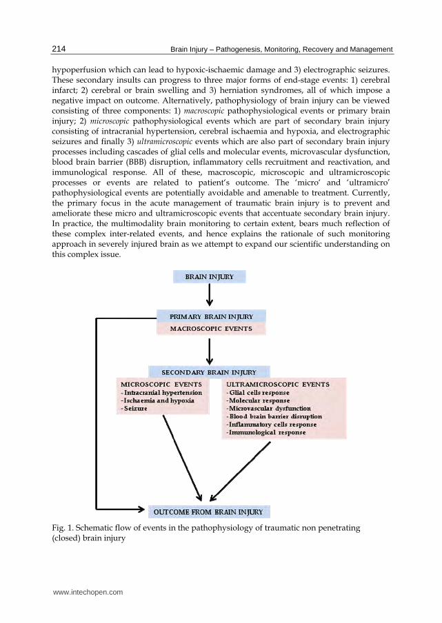
Closed brain injury processes can be divided into two phases, primary and secondary brain injury. The primary brain injury occurs during the initial insult. The severity and type of impact incurred during the initial insult will substantially influence the structural lesions that ensue. This severely injured brain is vulnerable to further insult which is known as the secondary brain injury (figure 1). In this phase, the commonly associated forms of insult can be broadly categorised into three: 1) intracranial hypertension; 2) cerebral hypoxia and
www.intechopen.com
Brain Injury – Pathogenesis, Monitoring, Recovery and Management
214
hypoperfusion which can lead to hypoxic-ischaemic damage and 3) electrographic seizures. These secondary insults can progress to three major forms of end-stage events: 1) cerebral infarct; 2) cerebral or brain swelling and 3) herniation syndromes, all of which impose a negative impact on outcome. Alternatively, pathophysiology of brain injury can be viewed consisting of three components: 1) macroscopic pathophysiological events or primary brain injury; 2) microscopic pathophysiological events which are part of secondary brain injury consisting of intracranial hypertension, cerebral ischaemia and hypoxia, and electrographic seizures and finally 3) ultramicroscopic events which are also part of secondary brain injury processes including cascades of glial cells and molecular events, microvascular dysfunction, blood brain barrier (BBB) disruption, inflammatory cells recruitment and reactivation, and immunological response. All of these, macroscopic, microscopic and ultramicroscopic processes or events are related to patient‘s outcome. The ‘micro‘ and ‘ultramicro’ pathophysiological events are potentially avoidable and amenable to treatment. Currently, the primary focus in the acute management of traumatic brain injury is to prevent and ameliorate these micro and ultramicroscopic events that accentuate secondary brain injury. In practice, the multimodality brain monitoring to certain extent, bears much reflection of these complex inter-related events, and hence explains the rationale of such monitoring approach in severely injured brain as we attempt to expand our scientific understanding on this complex issue.
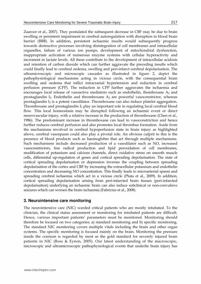
Fig. 1. Schematic flow of events in the pathophysiology of traumatic non penetrating (closed) brain injury
www.intechopen.com
Neurointensive Care Monitoring for Severe Traumatic Brain Injury
215
2.1 Primary and secondary brain injury Primary brain injury occurs at the time of impact with an instantaneous clinical effect. Pathological classification for this form of injury is largely evident either clinically or by imaging (macroscopic events) which include focal-epidural, subdural, intracerebral haematomas, focal contusion and laceration, focal or diffused subarachnoid haemorrhage and focal or diffused axonal and vascular injuries. Surgery plays a major role to treat some of these pathologies. On the other hand, secondary brain injury or damage occurs at some stage after the initial impact and therefore can aggravate the primary brain injury. From figure 1, it is apparent that the microscopic and ultramicroscopic events contribute to the secondary brain injury pathogenesis. However, currently there are limited treatment options available in treating the secondary brain injury processes or damages. In 2002, Narayan reviewed clinical trials in head injury and concluded that most trials assessing pharmacological neuroprotective agents failed to show clinically robust efficacy (Narayan et al., 2002). Surgically, the typical available options are burr hole and cerebrospinal fluid (CSF) drainage and a wide decompressive craniectomy. The latter has regained therapeutic interest in the past years with beneficial effects shown for massive brain infarcts, brain swelling and brain herniation that constitute the end results of secondary brain injury processes as discussed above (Carandang & Krieger, 2008; Cooper et al., 2008; Eberle et al., 2010; Figaji et al., 2007). Other possible alternatives to treat secondary brain damage include therapeutic cerebral hypothermia (Dietrich & Bramlett, 2010; Gluckman et al., 2005), barbiturates therapy (Marshall et al., 2010), raising cerebral blood flow and perfusion by using pressors (Lewis et al., 1998) and rheological agents such as mannitol and hypertonic saline therapy (Berger et al., 1995; Oddo et al., 2009).
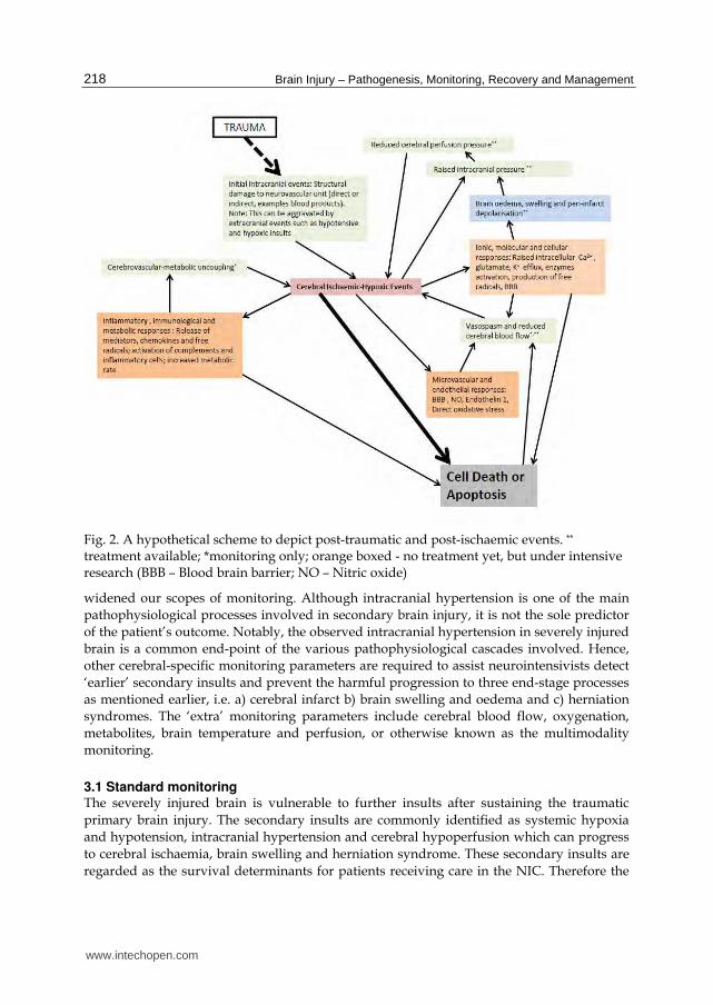
2.1.1 Ultramicroscopic events Trauma will induce a significant and protracted inflammatory, molecular, microvascular and immunological responses. These response cascades following direct damage to the neurovascular unit which incite ischaemic-hypoxic events. Systematically, four major mechanisms may contribute to cerebral ischaemia or hypoxia: a) direct or indirect structural damage to intracerebral arteries, neurons and glial cells (neurovascular unit); b) cerebrovascular-metabolic uncoupling; c) intracranial hypertension and, d) vasospasms of cerebral artery. These mechanisms are the interrelated cascades which underlie the pathophysiological processes that propagate the brain injury (figure 2). Damage to the neurovascular unit which comprises cerebral endothelial cells, astrocytes and neurons as well as extracellular matrix plays significant role in these cascades (Dirnagl et al., 1999). The cascades involved opening of voltage dependent and agonist-gated ion and calcium channels causing intracellular calcium and sodium overload and efflux of potassium. Intracellular sodium overload causes cytotoxic oedema while efflux of potassium leads to peri-infarcted brain tissue depolarisation and seizures. Influx of calcium is further promoted by the release of large amount of excitatory amino acids particularly glutamate. Intracellular calcium and glutamate act in a vicious circle which augments the amount of intracellular calcium. Excess intracellular calcium can induce damage to the organelles such as mitochondria, intracellular second messengers, cellular membrane and activation of numerous intracellular enzymes system which finally lead to apoptosis. Apoptosis is a form of cell death in which a programmed sequence of events leads to the elimination of cells without releasing much harmful substances. When brain cells experienced an injury or episode of ischaemia, they either become dead and the area involved is better known as
www.intechopen.com
Brain Injury – Pathogenesis, Monitoring, Recovery and Management
216
infarcted area, or the cells may survive the injuries and in time recover completely or partially, or alternatively the cells enter the apoptotic pathway. Interestingly, during the early stages of injury, the process of apoptosis can still be reversed. This reversal is made possible by correcting the cellular energy metabolism or cerebrovascular metabolic coupling or by lowering the brain temperatures (Liou et al., 2003). In addition to the events described above, damage to the neurovascular unit can also cause production of free radicals. Free radicals are atomic or molecular species with unpaired electrons. They are highly reactive and unstable, therefore capable of oxidizing and damaging all cellular components when their amounts exceed the anti-oxidant defence mechanisms as seen in episode of ischaemia/reperfusion brain injury (Lewen et al., 2000). Other harmful events that occur at ultramicroscopic levels when the brain is injured are inflammatory and immunological responses. Increased levels of pro-inflammatory cytokines such as interleukin-6 (IL-6), interleukin-8 (IL-8), interleukin-10 (IL-10) and tumour necrosis factor alpha (TNF-α) have been demonstrated in cerebrospinal fluid, serum and brain tissue following head injury (Csuka et al., 1999; Is et al., 2007; Kossmann et al., 1997). The underlying dynamics of inflammation in traumatic brain injury are complex, encompassing cytokines, chemokines, adhesion molecules, complement factors and even the free radicals. The lead triggers for inflammatory activation are multi-factorial and thought to involve factors such as extravasated blood products, intracellular components, reactive free oxygen and nitrogen radicals by brain resident microglia and astrocytes sensing perturbation of tissue homeostasis (Juliet et al., 2008; Mathew et al., 1994). These initial events fuel further release of inflammatory mediators which facilitate influx of extracerebral inflammatory cells by vascular endothelial changes and chemotaxis. Studies have also shown the role of adhesion molecules such as vascular cell adhesion molecule, P-selectin, intracellular adhesion molecule-1 and gradient of chemotactic chemokines in upregulating these events (Hausmann et al., 1998). The ensuing extracellular oedema is triggered by vascular dilatation and leakage as a result of vasoactive inflammatory molecules such as nitric oxide (NO) and vascular endothelial growth factor, as well as activations of complement system by dying cells to aggravate the initial inflammation and secondary tissue damage (Grzybicki et al., 1998; Stahel et al., 1998). Currently, monitoring of neuroinflammation is only made possible with techniques such as brain microdialysis, whilst others such as free radical imaging and monitoring remains under intensive research (Vergely et al., 2003).
2.1.2 Microscopic events Typically in normal subjects, the cerebral blood flow (CBF) and metabolism are a coupled phenomenon. Cerebrovascular metabolic uncoupling is reported to occur in over half of brain injured patients (Obrist et al., 1984). Uncoupling, which impairs the CBF and metabolism following traumatic severe brain injury is related to unfavourable functional outcome because of the correlation with ischaemic-hypoxic events. Reduction in CBF is obviously affecting the status of cellular metabolism and hence promoting cellular death. Interestingly, uncoupling is also expected with an excessive increased in CBF (luxury perfusion or hyperaemia) which is harmful to the injured brain by causing brain swelling (congested brain) or oedema. In 2007, Menashe Zaaroor noted significant reduction in CBF within 24 hours of brain injury and then increased at the second day after the injury. Further analysis revealed patients with good outcome after head injury had stable and normal CBF after initial period of significant reduction, whereas in patients with poor outcome, the course of CBF is triphasic with a secondary decrease on the third day post trauma (Menashe
www.intechopen.com
Neurointensive Care Monitoring for Severe Traumatic Brain Injury
217
Zaaroor et al., 2007). They postulated the subsequent decrease in CBF may be due to brain swelling or persistent impairment in cerebral autoregulation with disruption in blood brain barrier (BBB). In addition, persistent ischaemic insults would subsequently progress towards destructive processes involving disintegration of cell membranes and intracellular organelles, failure of various ion pumps, development of mitochondrial dysfunction, inappropriate activation of numerous enzyme systems with cellular hyperactivity and increment in lactate levels. All these contribute to the development of intracellular acidosis and retention of carbon dioxide which can further aggravate the preceding insults which could finally lead to cerebral oedema, swelling and peri-infarct cerebral depolarisation. The ultramicroscopic and microscopic cascades as illustrated in figure 2, depict the pathophysiological mechanisms acting in vicious circle, with the consequential brain swelling and oedema that inflict intracranial hypertension and reduction in cerebral perfusion pressure (CPP). The reduction in CPP further aggravates the ischaemia and encourages local release of vasoactive mediators such as endothelin, thomboxane A2 and prostaglandin I2. Endothelin and thromboxane A2 are powerful vasoconstrictor, whereas prostaglandin I2 is a potent vasodilator. Thromboxane can also induce platelet aggregation. Thromboxane and prostaglandin I2 play an important role in regulating local cerebral blood flow. This local homeostasis may be disrupted following an ischaemic event or direct neurovascular injury, with a relative increase in the production of thromboxane (Chen et al., 1986). The predominant increase in thromboxane can lead to vasoconstriction and hence further reduces cerebral perfusion and also promotes local thrombus formation. Aside from the mechanisms involved in cerebral hypoperfusion state in brain injury as highlighted above, cerebral vasospasm could also play a pivotal role. An obvious culprit to this is the presence of blood products such as haemoglobin that act through multiple mechanisms. Such mechanisms include decreased production of a vasodilator such as NO, increased vasoconstrictors, free radical production and lipid peroxidation of cell membranes, modification of potassium and calcium channels, direct oxidative stress on smooth muscle cells, differential up-regulation of genes and cortical spreading depolarisation. The state of cortical spreading depolarisation or depression inverses the coupling between spreading depolarisation of the cortex and CBF by increasing the extracellular potassium and endothelin concentration and decreasing NO concentration. This finally leads to microarterial spasm and spreading cerebral ischaemia which act in a vicious circle (Pluta et al., 2009). In addition, cortical spreading depolarisation arising from peri-infarcted brain tissues (peri-infarcted depolarisation) underlying an ischaemic brain can also induce subclinical or non-convulsive seizures which can worsen the brain ischaemia (Fabricius et al., 2008).
3. Neurointensive care monitoring
The neurointensive care (NIC) warded critical patients who are mostly intubated. To the
clinician, the clinical status assessment or monitoring for intubated patients are difficult.
Hence, various important patients’ parameters must be monitored. Monitoring should
therefore be focused on two categories; a) standard monitoring and b) specific monitoring.
The standard NIC monitoring covers multiple vitals including the brain and other organ
systems. The specific monitoring is focused mainly on the brain. Monitoring the pressure
inside the cranium is regarded by most as the gold standard for severely injured brain
patients in NIC (Ross & Eynon, 2005). Our latest understanding of the macroscopic,
microscopic and ultramicroscopic pathophysiological events that underlie brain injury has
www.intechopen.com
Brain Injury – Pathogenesis, Monitoring, Recovery and Management
218
Fig. 2. A hypothetical scheme to depict post-traumatic and post-ischaemic events. **
treatment available; *monitoring only; orange boxed - no treatment yet, but under intensive research (BBB – Blood brain barrier; NO – Nitric oxide)
widened our scopes of monitoring. Although intracranial hypertension is one of the main
pathophysiological processes involved in secondary brain injury, it is not the sole predictor
of the patient’s outcome. Notably, the observed intracranial hypertension in severely injured
brain is a common end-point of the various pathophysiological cascades involved. Hence,
other cerebral-specific monitoring parameters are required to assist neurointensivists detect
‘earlier’ secondary insults and prevent the harmful progression to three end-stage processes
as mentioned earlier, i.e. a) cerebral infarct b) brain swelling and oedema and c) herniation
syndromes. The ‘extra’ monitoring parameters include cerebral blood flow, oxygenation,
metabolites, brain temperature and perfusion, or otherwise known as the multimodality
monitoring.
3.1 Standard monitoring The severely injured brain is vulnerable to further insults after sustaining the traumatic
primary brain injury. The secondary insults are commonly identified as systemic hypoxia
and hypotension, intracranial hypertension and cerebral hypoperfusion which can progress
to cerebral ischaemia, brain swelling and herniation syndrome. These secondary insults are
regarded as the survival determinants for patients receiving care in the NIC. Therefore the
www.intechopen.com
Neurointensive Care Monitoring for Severe Traumatic Brain Injury
219
aim of standard monitoring is to prevent or to recognize these potential threats which could
originate from other body systems besides the brain. Head injured patients in NIC should be
monitored for nine key body systems: a) central nervous system (discussed specifically
under brain monitoring); b) cardiovascular system which includes parameters such as heart
rate and rhythm, blood and venous pressure, stroke volume, cardiac output and total
peripheral resistance; c) pulmonary system with regular monitoring of arterial blood gases,
chest radiographs, and breathing and ventilator parameters; d) renal and metabolic system
with regular blood, urine and metabolic profiles monitoring; e) gastrointestinal and
nutritional status pertaining to body calories; f) hematological profiles; g) microbiologic and
sepsis profiles; h) peripheries and skin condition and lastly i) endocrine and hormonal
profiles which are frequently overlooked, except for features of diabetes insipidus or
syndrome of inappropriate antidiuretic hormone hypersecretion. Recent studies have
demonstrated 75% incidence of endocrine dysfunction at 6 months post trauma in children
and 21% incidence of hypothalamic-pituitary dysfunction in adults (Kaulfers et al., 2010;
Krahulik et al., 2010). These findings prompted debates as to whether hormonal profile
evaluation should be routinely checked in all head injured patients and treated accordingly.
3.2 Specific monitoring Since limitations exist in bedside neurological findings as a monitoring tool for various
pathophysiological processes described in previous sections, specific central nervous system
monitoring is needed for severe brain injured patients in the NIC. The use of serial
computed tomography (CT) brain scanning protocol to predict the presence or absence of
abnormality or raised ICP has not been well received in practice, partly due to poor
correlations between CT findings and raised ICP, and because of undesirable effects from
repeated radiation exposure and frequent mobilization to the patients (Kouvarellis et al.,
2011; Miller et al., 2004). Therefore, specific brain monitoring seems necessary. Currently,
the assessment of intracranial status for severely injured brain is commonly done with gold
standard ICP monitoring alone. Lately, as our understanding on the complex
pathophysiology underlying severely injured brain expands, new modalities of brain
monitoring emerge, incorporating various brain parameters known as the multimodality
monitoring (Idris et al., 2007; Isa et al., 2003; Stuart et al., 2010).
4. Multimodality monitoring
The development of new neuromonitoring techniques has been particularly important
because typically standard cranial monitoring techniques such as ICP and CPP
measurements seem insufficient in detecting subtle manifestations of brain injury or poor
surrogates for physiologic parameters of interest. For these reasons, multimodality
monitoring which monitors more brain parameters is required. Multimodality monitoring
includes monitoring of the following parameters:
i. Intracranial pressure (ICP) ii. Cerebral perfusion pressure (CPP) iii. Cerebral blood flow (CBF parameters) iv. Focal and global cerebral tissue oxygenation (PtiO2 and SjVO2) v. Cerebral tissue carbon dioxide tension (PtiCO2) vi. Cerebral pH
www.intechopen.com
Brain Injury – Pathogenesis, Monitoring, Recovery and Management
220
vii. Cerebral temperature viii. Cerebral metabolic parameters (brain microdialysis) ix. Cerebral EEG (EEG and BIS) x. Cerebral evoked potentials (somatosensory, brainstem and visual evoked potentials:
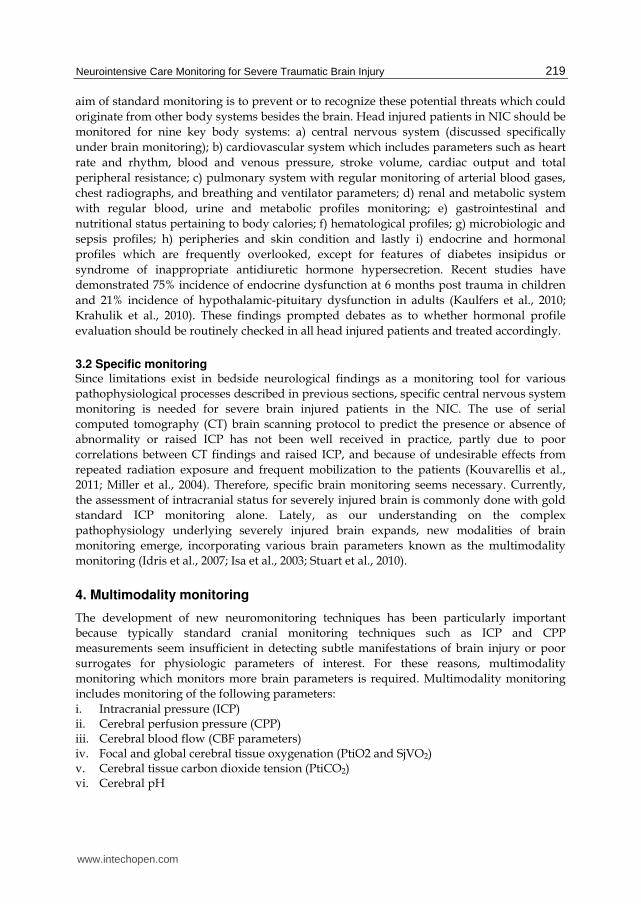
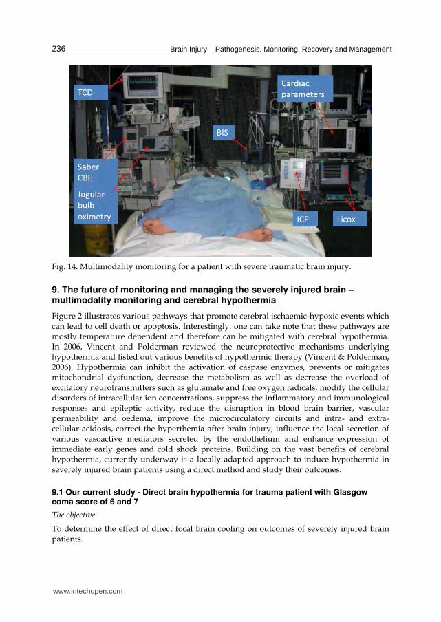
SSEP/BAEP/VEP) Figure 3 shows the role of multimodality monitoring for the injured brain. Intracranial pressure is used to monitor the cranial pressure, Licox or Neurotrends system is used to monitor status of regional brain oxygenation, carbon dioxide tension, pH and brain temperature, while jugular bulb oximetry is used to monitor global brain oxygenation. Saber cerebral blood flow, transcranial doppler, cerebral EEG and evoked potentials can be used to monitor cerebral blood flow and therefore identifying ischaemic insults, and cerebral microdialysis can monitor the cellular events. Furthermore, continuous EEG monitoring in NIC can also diagnose the subclinical seizures which play significant role in secondary brain injury and one of the parameters that determine the prognosis (Kazibutowska et al., 1992).
Fig. 3. Multimodality monitoring and secondary brain injury.
5. Two major parameters to consider during multimodality monitoring: ICP and CBF
Intracranial hypertension causes a reduction in cerebral perfusion pressure which can lead to ischaemia, brain swelling and ultimately infarction. In addition, persistent increased in
www.intechopen.com
Neurointensive Care Monitoring for Severe Traumatic Brain Injury
221
ICP could cause a shift in intracranial structures (midline shift or mass effect) and finally herniation syndromes. Therefore, monitoring ICP is considered a standard part of head injury management. Nonetheless, monitoring the ICP alone seems inadequate, because raised in ICP in head injured patients can be associated with both an increment and a reduction in CBF (Menashe Zaaroor, 2007). In 1992, Bouma noted about one-third of head injured patients had low CBF and therefore showed an early cerebral blood flow-metabolism mismatch (Bouma et al., 1992). Figure 2 highlights the importance of CBF monitoring to prevent or reduce the degree of cerebral ischaemic-hypoxic insults. Whilst the optimal method for measuring CBF remains to be established, there are numerous alternatives when used together can estimate the CBF. Those alternatives include: a) CPP; b) blood flow velocity by transcranial doppler; c) focal and global cerebral oxygenation with Licox or Neurotrends systems or jugular bulb oximetry; d) regional CBF with Saber 2000; e) cerebral metabolic parameters with brain microdialysis; f) cerebral perfusion imaging techniques and g) cerebral evoked potentials and EEG. These are in fact features of multimodality neuromonitoring. The CBF and CPP are two variables that are inter-related. The CBF is proportional to CPP and inversely related to the cerebral vascular resistance (CVR). The CPP represents the pressure gradient driving CBF and hence oxygen and metabolite delivery, i.e. in other words, the CPP is the perfusion pressure gradient across cerebral vascular bed whereby the incoming systemic arterial pressure minus the opposing force from venous outflow pressure. In traumatic brain injury cases, the elevated ICP is the opposing force to the incoming mean systemic arterial pressure (MAP). Therefore, CPP in equation form can be stated as CPP = MAP – ICP. In theory, CPP is equivalent to the transmural pressure across the cerebral vessel walls. In other words, at the arteriolar level, CPP is the stimulus for autoregulatory response and at the capillary, it is the driving force for fluid exchange. The normal brain autoregulates its blood flow to provide a constant flow regardless of blood pressure by altering the CVR to obtain a maintained flow within an autoregulatory range of 60 - 160 mmHg of MAP or CPP of 50 – 150 mmHg. These homeostatic mechanisms are often lost in severely injured brain due to impairment in cerebral autoregulation, implying that cerebral autoregulation is a homeostatic mechanism that minimizes deviations in CBF when CPP or MAP changes. It acts through vasomotor effectors that control CVR. Vasomotor effectors are controlled by a) CPP; b) metabolites; c) oxygen pressure; d) carbon dioxide pressure and e) temperature. In NIC setting, the level of changes in cerebral autoregulation among patients is individualised and therefore the optimum CPP may vary considerably. Clinically, continuous methods of autoregulation tests or monitoring rely on the observation of spontaneous responses of CBF to spontaneous fluctuations in MAP or CPP (Zweifel et al., 2008), arterial partial pressure of CO2 (Puppo et al., 2008) or metabolites (Lee et al., 2001). Study by Lee in 2001 showed that during the first 2 weeks after head injury, CO2 reactivity remains relatively intact, pressure autoregulation is variably impaired and metabolic suppression reactivity remains severely impaired and they further noted that unlike hypotension, hypoxia or haemorrhagic brain lesions, elevated ICP appears to affect all components of vasoreactivity that were tested (Lee et al., 2001). In similar year, Czosnyka also noted that autoregulation is not only impaired when associated with a high ICP or low arterial blood pressure, but it can also be disturbed by an excessive CPP (Czosnyka et al., 2001). Studies have shown that impairment or failure of autoregulation can contribute to unfavorable outcome (Czosnyka et al., 2001; Lam et al., 1997; Smielewski et al., 1997). This could possibly be explained by a disturbance in the pressure-volume (elastance) curve
www.intechopen.com
Brain Injury – Pathogenesis, Monitoring, Recovery and Management
222
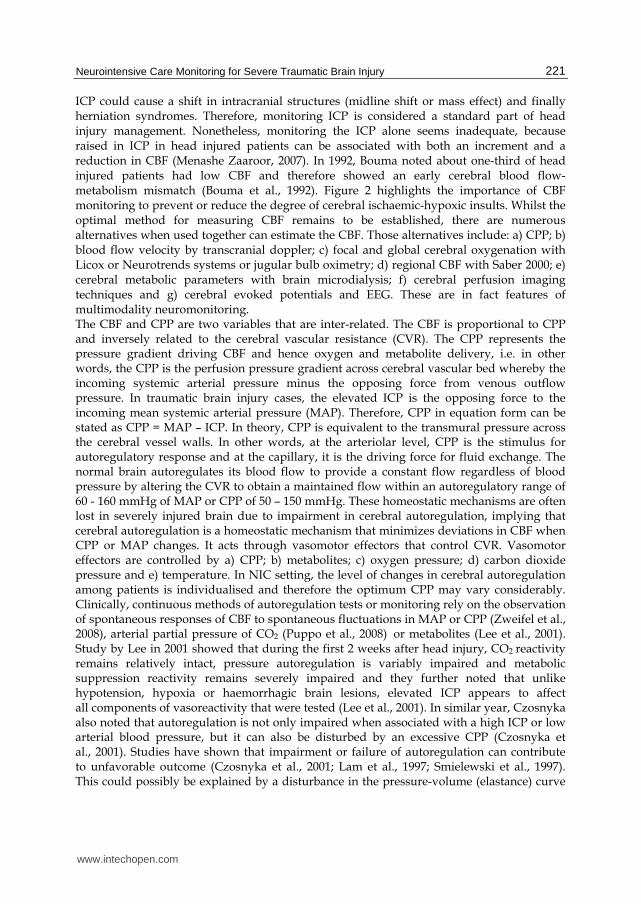
that is steeper suggesting poor compliance of the brain (figure. 4). Since elevated ICP and changes in CBF are two key factors to consider in all severely injured brain patients (figure. 2), besides cerebral autoregulation, the concept of cerebral pulsation is also thought to play a significant role in head injury pathogenesis.
Fig. 4. Pressure-volume curves of craniospinal contents. (A) is normal pressure-volume curve of compliant system and (B) is the pressure-volume curve when brain compliance is reduced.
5.1 Monro-Kellie doctrine, cerebral autoregulation, cerebral pulsation, pulsatility and resistive index and brain compliance According to Monro-Kellie doctrine, the sum of the intracranial volumes of blood, brain and
CSF is constant and that an increase in any one component must be offset by an equal decrease in another or else ICP rises. For traumatic brain injury, a pathological increase in
brain volume due to cerebral oedema or an increase in blood volume would induce an initial reduction in CSF volume response. This has been traditionally viewed as CSF being
forced out into the spinal dural sac to maintain ICP. This anatomical compensatory response
can quickly become exhausted and further rise in ICP is expected. Since raised ICP can lead to reduction in CPP and therefore compromising CBF, the body finally has to utilise the
second defence mechanism, functional compensatory response (augment the CBF). This type of compensatory response is to ensure the brain receives an adequate blood flow and hence
counteracts the effect of raised ICP. Once the body compensatory responses (anatomical and functional) become overstretched, that is the only time we note a fast increase in ICP and
striking reduction in brain compliance (the steep part of the elastance curve). This decompensatory phase is related to poor brain compliance, raised ICP and reduction in CBF
(figure. 5, labelled as A,B and C). Besides cerebral autoregulation, increased in intracranial vessels pulsation is one of the mechanisms that could augment the CBF during ischaemic-
hypoxic events (Chan et al., 1992; Egnor et al., 2002; Muttaqin et al., 1993). Since both seem to be important, monitoring level of autoregulation and assessing the intracranial vessels
pulsation are two parameters that should be done in NIC for multimodality monitoring. Cerebral autoregulation is normally tested or monitored with transcranial doppler by using
static (vasopressor) or dynamic (Aaslid leg-cuff, carotid artery compression or tilt table declination) provoking test or with cerebrovascular reactivity index (PRx) whereby digitised
ICPs is divided with digitised MAPs and focusing on their trends or values; positive values
www.intechopen.com
Neurointensive Care Monitoring for Severe Traumatic Brain Injury
223
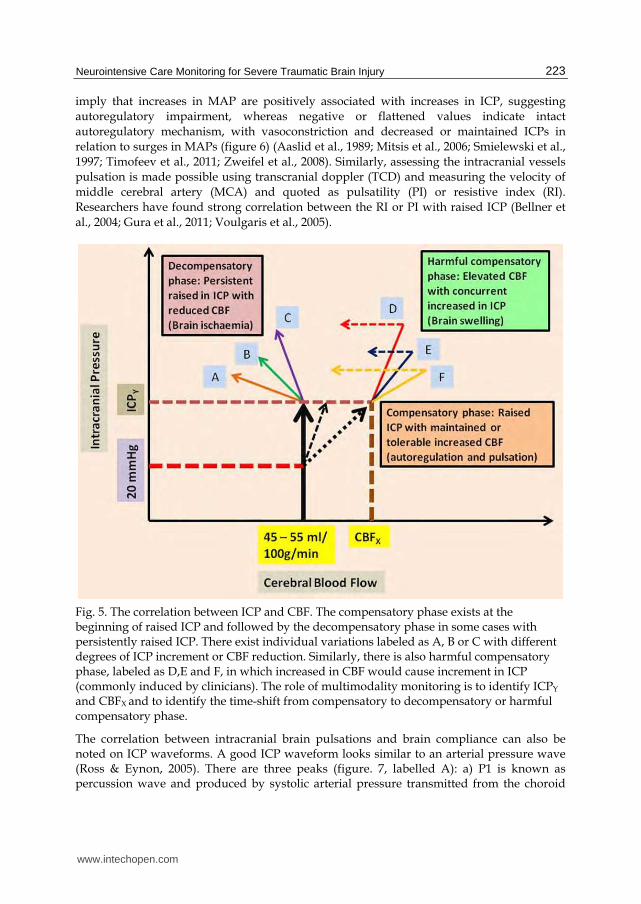
imply that increases in MAP are positively associated with increases in ICP, suggesting autoregulatory impairment, whereas negative or flattened values indicate intact
autoregulatory mechanism, with vasoconstriction and decreased or maintained ICPs in relation to surges in MAPs (figure 6) (Aaslid et al., 1989; Mitsis et al., 2006; Smielewski et al.,
1997; Timofeev et al., 2011; Zweifel et al., 2008). Similarly, assessing the intracranial vessels pulsation is made possible using transcranial doppler (TCD) and measuring the velocity of
middle cerebral artery (MCA) and quoted as pulsatility (PI) or resistive index (RI). Researchers have found strong correlation between the RI or PI with raised ICP (Bellner et
al., 2004; Gura et al., 2011; Voulgaris et al., 2005).
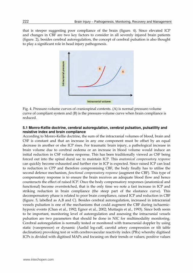
Fig. 5. The correlation between ICP and CBF. The compensatory phase exists at the beginning of raised ICP and followed by the decompensatory phase in some cases with persistently raised ICP. There exist individual variations labeled as A, B or C with different degrees of ICP increment or CBF reduction. Similarly, there is also harmful compensatory phase, labeled as D,E and F, in which increased in CBF would cause increment in ICP (commonly induced by clinicians). The role of multimodality monitoring is to identify ICPY
and CBFX and to identify the time-shift from compensatory to decompensatory or harmful compensatory phase.
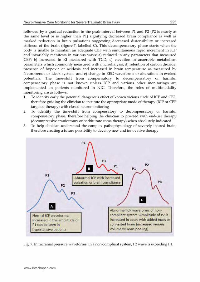
The correlation between intracranial brain pulsations and brain compliance can also be noted on ICP waveforms. A good ICP waveform looks similar to an arterial pressure wave (Ross & Eynon, 2005). There are three peaks (figure. 7, labelled A): a) P1 is known as percussion wave and produced by systolic arterial pressure transmitted from the choroid
www.intechopen.com
Brain Injury – Pathogenesis, Monitoring, Recovery and Management
224
Fig. 6. Positive and negative trends for the cerebrovascular pressure reactivity index (PRx). Positive trends signify impaired cerebral autoregulation whereas, negative and flattened trends denote intact autoregulation.
plexus to the ventricle; b) P2 is the tidal wave, its shape is more variable and originated from central venous wave from the right atrium and correlated with venous volume or venous congestion, sinus pressure and brain pulsations in injured brain. P2 may also stand for overall brain compliance; c) P3 is the dicrotic wave and is due to closure of the aortic valves. In between P2 and P3, there is dicrotic notch which is also thought arising from the effect of aortic valves closure. In 1965, Lundberg described three time-dependent patterns of pressure waves in patients with raised ICP (Lundberg et al., 1965). Lundberg A or plateau waves are 50 – 100 mmHg high and last for 5 to 20 minutes. This type of pressure waves demonstrate an early functional compensatory response between raised ICP and CBF. Cerebral vasculatures start to dilate in response to intracranial hypertension and critically low CBF, however, this autoregulatory vasodilatory response can dangerously lead to further increment in ICP via increased in intracranial blood volume in some brain injured patients with impaired autoregulation (figure. 5, labelled as D,E and F). Lundberg B waves occur at frequency of 0.5 – 2/min and are up to 50 mmHg in amplitude. It is detectable in normal and some individuals with impaired cerebral autoregulation (Balestreri et al., 2004). Lundberg C waves last 4 to 5 minutes and are up to 20 mmHg in amplitude and are of little clinical significance. Studies on ICP waveforms and Lundberg patterns of pressure waves have demonstrated that in the presence of raised ICP and intact cerebral autoregulation, the brain is able to regulate or optimise its CBF through compensatory mechanisms. Maintainance of CBF can be made via autoregulatory cerebral vasculature dilatations and increased in cerebral vasculature pulsations. Since P2 indicates overall compliance of the brain, at an early stage of compensation the peak-interval between P1 and P2 is increased which signifies increased cerebral vasculature pulsations and increased brain compliance (figure.7, labelled B). In the presence of persistently raised ICP, this initial response is
www.intechopen.com
Neurointensive Care Monitoring for Severe Traumatic Brain Injury
225
followed by a gradual reduction in the peak-interval between P1 and P2 (P2 is nearly at the same level or is higher than P1) signifying decreased brain compliance as well as marked reduction in brain pulsations suggesting decreased distensibility or increased stiffness of the brain (figure.7, labelled C). This decompensatory phase starts when the body is unable to maintain an adequate CBF with simultaneous rapid increment in ICP and invariably manifests in various ways: a) reduced in any parameters that measured CBF; b) increased in RI measured with TCD; c) elevation in anaerobic metabolism parameters which commonly measured with microdialysis; d) retention of carbon dioxide, presence of hypoxia or acidosis and increased in brain temperature as measured by Neurotrends or Licox system and e) change in EEG waveforms or alterations in evoked potentials. The time-shift from compensatory to decompensatory or harmful compensatory phase is not known unless ICP and various other monitorings are implemented on patients monitored in NIC. Therefore, the roles of multimodality monitoring are as follows: 1. To identify early the potential dangerous effect of known vicious circle of ICP and CBF,
therefore guiding the clinician to institute the appropriate mode of therapy (ICP or CPP
targeted therapy) with closed neuromonitoring
2. To identify the time-shift from compensatory to decompensatory or harmful
compensatory phase, therefore helping the clinician to proceed with end-tier therapy
(decompressive craniectomy or barbiturate coma therapy) when absolutely indicated
3. To help clinician understand the complex pathophysiology of severely injured brain,
therefore creating a future possibility to develop new and innovative therapy
Fig. 7. Intracranial pressure waveforms. In a non-compliant system, P2 wave is exceeding P1.
www.intechopen.com
Brain Injury – Pathogenesis, Monitoring, Recovery and Management
226
5.1.1 Vicious circle of ICP and CBF: ICP and CPP targeted therapy versus multimodality monitoring In previous sections, we recognised that an increase in ICP sets in an autoregulatory response to compensate for a reduction in CBF. Since CPP is proportional to CBF, increment in ICP would cause decrement in CPP. This is easily understood from the equation: CPP = MAP – ICP, whereby raised ICP causes reduction in CPP when MAPs are kept constant. Reduced CPP can be corrected by raising the MAP or by reducing the ICP. Reduction in ICP is normally called ICP targeted therapy, whilst CPP targeted therapy corrects the reduced CPP by raising the MAP. In 2003, Young recommended an aggressive CPP management for patients with elevated ICP (Young et al., 2003). Four patients in their study had comparatively good neurological outcome after receiving such management for extremely high ICP. Nonetheless, in some severely injured brain patients who have high CPP values or those received an aggressive CPP management appeared to develop cerebral oedema or brain swelling and had poor outcomes. This phenomenon is thought to arise from impairment in cerebral autoregulatory mechanisms (Dickman et al., 1991; Nujaimin et al., 2009; Zweifel et al., 2008). In patients with an impaired cerebral autoregulation, ICP targeted therapy seems more appropriate than CPP targeted therapy. This is mainly because of the vicious circle that can occur in patients with impaired cerebral autoregulation whereby ICP is further elevated by elevating the CPP (figure 5, labelled D,E and F). Many studies have shown a strong association between an elevated ICP and poor patient outcome in head injury (Cooper et al., 2008; Eberle et al., 2010; Idris et al., 2007). In most cases with poor outcome, both, elevated ICP and low CPP values appeared common features. Therefore it seems prudent to conclude that in such patients, the emphasis should not only on raising CPP but also on lowering the ICP to gain better outcome. In fact, the combination of ICP and CPP therapy forms the basis of the guidelines or protocols proposed by the US Brain Trauma Foundation (Bullock et al., 1996), European Brain Injury Consortium (EBIC) (Maas et al., 1997), Addenbrooke protocol from the Cambridge (Patel et al., 2002) and Lund protocol (that is still considering the importance of reducing ICP and improving the CPP) (Asgeirsson et al., 1994). In relation to figure 5, it is a daunting task to speculate which patient will benefit from CPP or ICP targeted therapy. Additional tests or monitoring are obviously needed to facilitate this difficult task in deciding which patients are likely to tolerate CPP targeted therapy. Since multimodality monitoring does monitor various cerebral parameters (ICP, CPP and CBF) and at the same time allowing cerebral autoregulation test or monitoring to be done, therefore it serves as an appropriate monitoring approach. Multimodality monitoring can detect not only further increment in ICP values but also the presence of any compromise in CBF in patients who already have elevated, extremely elevated or lethal levels of ICP and still undergoing CPP targeted therapy.
5.1.2 Shift from compensatory to decompensatory or harmful compensatory phase – Role of multimodality monitoring Intracranial hypertension (high ICP/low CPP) and reduction in CBF are two important
parameters that determine the outcome of severely injured brain patients. Figure 2
illustrates cerebral ischaemic-hypoxic insults are the core events that trigger various vicious
circles, including intracranial hypertension. Therefore, CBF monitoring should be
incorporated into the standard care for severely injured brain patients in the NIC. The
multimodality monitoring for the ICP and CBF offers crucial information on the status of
www.intechopen.com
Neurointensive Care Monitoring for Severe Traumatic Brain Injury
227
cerebral oxygenation, nutrition and function. The combination of various monitored
parameters can cross-validate one another and can give indications as to the phase the brain
is in for the managing clinicians (figure 5). Patients who are in the compensatory phase with
raised ICP and undergoing CPP targeted therapy may not yet be a candidate for
decompressive craniectomy or barbiturate therapy as long as the ICP is kept stable and CBF
parameters are within an acceptable range. However, those with deteriorating CBF
parameters (figure 5, labelled A,B and C) and/or persistently raised ICP (figure 5, labelled
A,B,C,D,E and F) may be considered for above mentioned end-tier therapy. In summary,
besides its potential to safe guard the CPP targeted therapy, multimodality monitoring of
the severely injured brain patients aids clinicians to decide on the appropriate timing for
end-tier therapy, decomperessive craniectomy or barbiturate therapy.
6. Measuring the two major parameters: ICP and CBF
The multimodality monitoring can be viewed as way of monitoring two important parameters that play significant roles in the pathogenesis of ischaemic-hypoxic events, namely, the ICP and CBF.
6.1 Intracranial pressure The ICP is normally monitored until an acceptable upper limit of 20 mmHg (Balestreri et al.,
2006). This is attained by using either ventricular catheter if the ventricles are opened or a
compliance monitor (Spiegelberg GmbH & Co., Hamburg, Germany) or an
intraparenchymal ICP, using Codman (Codman, Randolph, MA, USA) or Camino
transducers (Camino Laboratories, San Diego, CA)(Idris et al., 2007; Isa et al., 2003). If the
ICP values persistently read more than 20 mmHg and/or direct calculation of cerebral
perfusion pressure (CPP) appears less than 50 mmHg, draining of CSF is warranted. If the
ICP remains high, other modes of therapy such as adding muscle relaxants, giving mannitol
or hypertonic saline, temporary hyperventilation, hypothermia with cooling blanket and
fans are usually considered. At this stage, further investigation with CT brain is justifiable
provided that these additonal steps fail to reduce the ICP values. The subsequent treatment
should be based on the new imaging and monitored parameters. Generally, if the CT brain
revealed no new surgical lesion but the brain appeared swollen, the ICP values remained
high (or CPP values remained low) and/or presence of abnormal cerebral blood flow
parameters, the patient should then be treated with either one of the following:
1. Bifrontal decompressive craniectomy and dura augmentation for diffuse brain swelling without midline shift, or
2. Unilateral wide decompressive craniectomy and dura augmentation for diffuse brain swelling and presence of midline shift, or
3. Barbiturate therapy until burst suppression attained on continuous EEG monitoring. Nonetheless, if the ICP values remain persistently high, decompressive craniectomy can still be considered.
For patients with persistently low CPP values without a corresponding raised ICP,
decompressive craniectomy surgery or barbiturate therapy is not yet indicated. Instead,
these patients should be treated with inotropes and/or hypervolumic therapy to increase
CPP values. If by implementing CPP targeted therapy and the ICP started to increase, then
the end-tier therapy should now be considered.
www.intechopen.com
Brain Injury – Pathogenesis, Monitoring, Recovery and Management
228
6.2 Cerebral blood flow CBF can be measured in various ways. Measuring CBF can be classified into 4 ways: a. Direct measurement of CBF b. Indirect measurement of CBF c. Quantitative measurement of CBF d. Qualitative measurement of CBF Quantitative and qualitative measurements are employed mainly for research purposes and hardly used during monitoring of severely brain injured patients in NIC. Examples include using radioisotope or tracer to measure CBF with imaging modality such as PET or SPECT scan.
A) Direct measurement of CBF
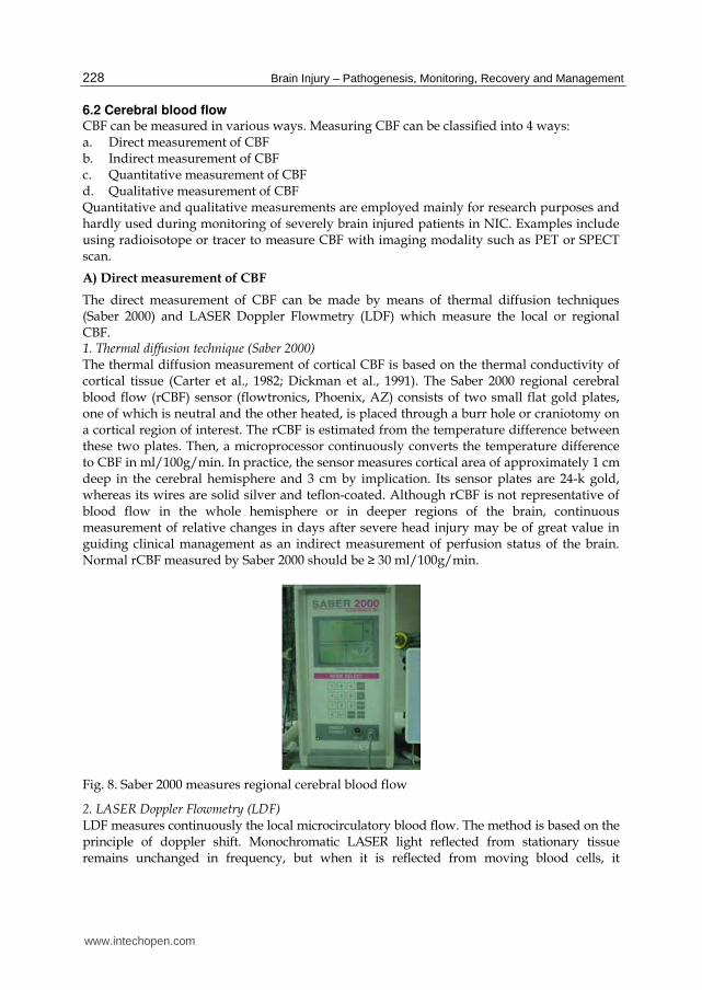
The direct measurement of CBF can be made by means of thermal diffusion techniques (Saber 2000) and LASER Doppler Flowmetry (LDF) which measure the local or regional CBF. 1. Thermal diffusion technique (Saber 2000) The thermal diffusion measurement of cortical CBF is based on the thermal conductivity of cortical tissue (Carter et al., 1982; Dickman et al., 1991). The Saber 2000 regional cerebral blood flow (rCBF) sensor (flowtronics, Phoenix, AZ) consists of two small flat gold plates, one of which is neutral and the other heated, is placed through a burr hole or craniotomy on a cortical region of interest. The rCBF is estimated from the temperature difference between these two plates. Then, a microprocessor continuously converts the temperature difference to CBF in ml/100g/min. In practice, the sensor measures cortical area of approximately 1 cm deep in the cerebral hemisphere and 3 cm by implication. Its sensor plates are 24-k gold, whereas its wires are solid silver and teflon-coated. Although rCBF is not representative of blood flow in the whole hemisphere or in deeper regions of the brain, continuous measurement of relative changes in days after severe head injury may be of great value in guiding clinical management as an indirect measurement of perfusion status of the brain. Normal rCBF measured by Saber 2000 should be ≥ 30 ml/100g/min.
Fig. 8. Saber 2000 measures regional cerebral blood flow
2. LASER Doppler Flowmetry (LDF) LDF measures continuously the local microcirculatory blood flow. The method is based on the principle of doppler shift. Monochromatic LASER light reflected from stationary tissue remains unchanged in frequency, but when it is reflected from moving blood cells, it
www.intechopen.com
Neurointensive Care Monitoring for Severe Traumatic Brain Injury
229
undergoes a frequency shift and therefore light reflected from the microcirculation measures the movements of red blood cells (Meyerson et al., 1991). The light is carried by a fibreoptic probe and the reflected light is converted into an electrical signal from which several values are derived. The flux (flow unit) represents the movement and concentrations of blood cells through the microvasculature. It is derived from the concentration of moving blood cells (CMBC) in the measured volume multiplied by the mean velocity. All three values, flux, CMBC and velocity are continuously recorded and displayed by the computer (Bolognese et al., 1993). LDF measures the local flow within a very small sample volume of approximately 1 mm3. LDF can also be used to measure the state of cerebral autoregulation via CO2 reactivity.
B) Indirect measurement of CBF
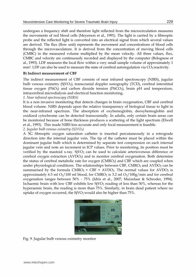
The indirect measurement of CBF consists of near infrared spectroscopy (NIRS), jugular bulb venous oximetry (SjVO2), transcranial doppler sonography (TCD), cerebral interstitial tissue oxygen (PtiO2) and carbon dioxide tension (PtiCO2), brain pH and temperature, intracerebral microdialysis and electrical function monitoring. 1. Near infrared spectroscopy (NIRS) It is a non invasive monitoring that detects changes in brain oxygenation, CBF and cerebral blood volume. NIRS depends upon the relative transparency of biological tissue to light in the near-infrared spectrum. The absorption of oxyhemoglobin, deoxyhemoglobin and oxidized cytochrome can be detected transcranially. In adults, only certain brain areas can be monitored because of bone thickness produces a scattering of the light spectrum (Elwell et al., 1993). This made NIRS less accurate and only focal measurement is feasible. 2. Jugular bulb venous oximetry (SjVO2) A 5G fibreoptic oxygen saturation catheter is inserted percutaneously in a retrograde direction into the internal jugular vein. The tip of the catheter must be placed within the dominant jugular bulb which is determined by separate test compression on each internal jugular vein and note an increment in ICP values. Prior to monitoring, its position must be verified by the mastoid x-ray. SjVO2 can be used to calculate arteriovenous difference or cerebral oxygen extraction (AVDO2) and to monitor cerebral oxygenation. Both determine the status of cerebral metabolic rate for oxygen (CMRO2) and CBF which are coupled when under physiological conditions. The relationships between CBF, CMRO2 and AVDO2 can be summarised by the formula CMRO2 = CBF × AVDO2. The normal values for AVDO2 is approximately 6.5 ml O2/100 ml blood, for CMRO2 is 3.2 ml O2/100g/min and for cerebral oxygenation ranges between 50% - 75% (Idris et al., 2007; Muizelaar & Schroder, 1994). Ischaemic brain with low CBF exhibits low SjVO2 reading of less than 50%, whereas for the hyperaemic brain, the reading is more than 75%. Similarly, in brain dead patient where no uptake of oxygen occurred, the SjVO2 would also be higher than 75%.
Fig. 9. Jugular bulb venous oximetry monitor
www.intechopen.com
Brain Injury – Pathogenesis, Monitoring, Recovery and Management
230
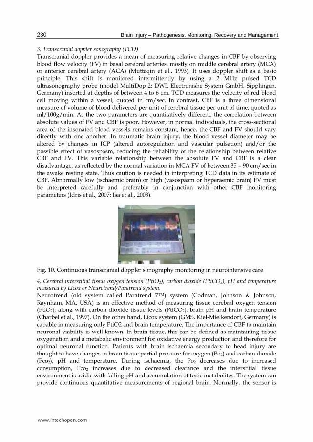
3. Transcranial doppler sonography (TCD) Transcranial doppler provides a mean of measuring relative changes in CBF by observing blood flow velocity (FV) in basal cerebral arteries, mostly on middle cerebral artery (MCA) or anterior cerebral artery (ACA) (Muttaqin et al., 1993). It uses doppler shift as a basic principle. This shift is monitored intermittently by using a 2 MHz pulsed TCD ultrasonography probe (model MultiDop 2; DWL Electronishe System GmbH, Sipplingen, Germany) inserted at depths of between 4 to 6 cm. TCD measures the velocity of red blood cell moving within a vessel, quoted in cm/sec. In contrast, CBF is a three dimensional measure of volume of blood delivered per unit of cerebral tissue per unit of time, quoted as ml/100g/min. As the two parameters are quantitatively different, the correlation between absolute values of FV and CBF is poor. However, in normal individuals, the cross-sectional area of the insonated blood vessels remains constant, hence, the CBF and FV should vary directly with one another. In traumatic brain injury, the blood vessel diameter may be altered by changes in ICP (altered autoregulation and vascular pulsation) and/or the possible effect of vasospasm, reducing the reliability of the relationship between relative CBF and FV. This variable relationship between the absolute FV and CBF is a clear disadvantage, as reflected by the normal variation in MCA FV of between 35 – 90 cm/sec in the awake resting state. Thus caution is needed in interpreting TCD data in its estimate of CBF. Abnormally low (ischaemic brain) or high (vasospasm or hyperaemic brain) FV must be interpreted carefully and preferably in conjunction with other CBF monitoring parameters (Idris et al., 2007; Isa et al., 2003).
Fig. 10. Continuous transcranial doppler sonography monitoring in neurointensive care
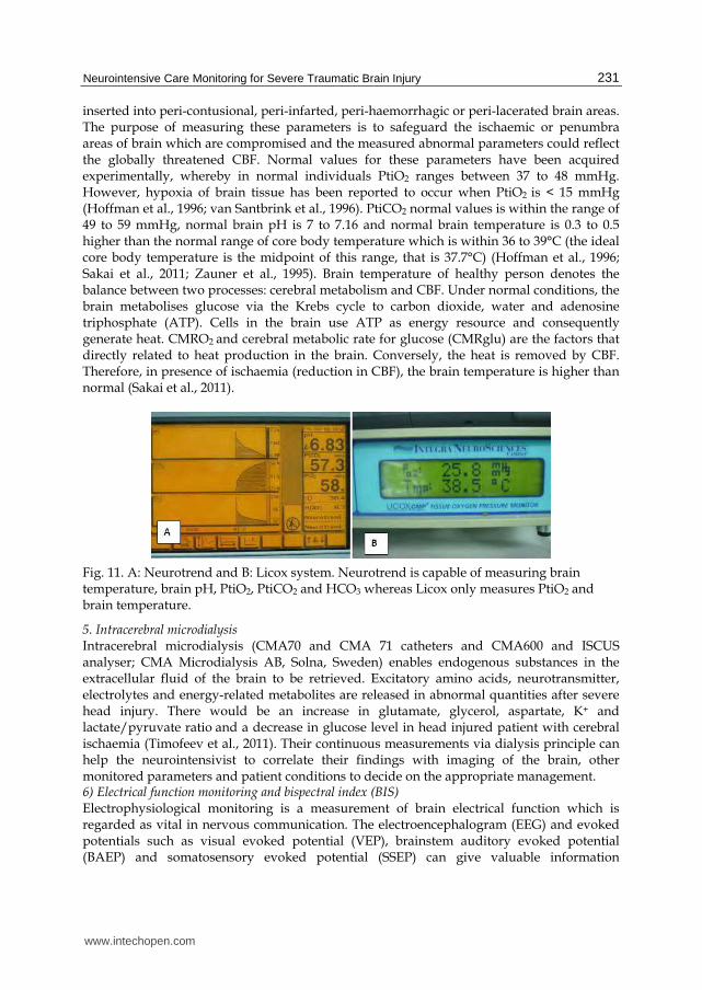
4. Cerebral interstitial tissue oxygen tension (PtiO2), carbon dioxide (PtiCO2), pH and temperature measured by Licox or Neurotrend/Paratrend system. Neurotrend (old system called Paratrend 7TM) system (Codman, Johnson & Johnson, Raynham, MA, USA) is an effective method of measuring tissue cerebral oxygen tension (PtiO2), along with carbon dioxide tissue levels (PtiCO2), brain pH and brain temperature (Charbel et al., 1997). On the other hand, Licox system (GMS, Kiel-Mielkendorf, Germany) is capable in measuring only PtiO2 and brain temperature. The importance of CBF to maintain neuronal viability is well known. In brain tissue, this can be defined as maintaining tissue oxygenation and a metabolic environment for oxidative energy production and therefore for optimal neuronal function. Patients with brain ischaemia secondary to head injury are thought to have changes in brain tissue partial pressure for oxygen (Po2) and carbon dioxide (Pco2), pH and temperature. During ischaemia, the Po2 decreases due to increased consumption, Pco2 increases due to decreased clearance and the interstitial tissue environment is acidic with falling pH and accumulation of toxic metabolites. The system can provide continuous quantitative measurements of regional brain. Normally, the sensor is
www.intechopen.com
Neurointensive Care Monitoring for Severe Traumatic Brain Injury
231
inserted into peri-contusional, peri-infarted, peri-haemorrhagic or peri-lacerated brain areas. The purpose of measuring these parameters is to safeguard the ischaemic or penumbra areas of brain which are compromised and the measured abnormal parameters could reflect the globally threatened CBF. Normal values for these parameters have been acquired experimentally, whereby in normal individuals PtiO2 ranges between 37 to 48 mmHg. However, hypoxia of brain tissue has been reported to occur when PtiO2 is < 15 mmHg (Hoffman et al., 1996; van Santbrink et al., 1996). PtiCO2 normal values is within the range of 49 to 59 mmHg, normal brain pH is 7 to 7.16 and normal brain temperature is 0.3 to 0.5 higher than the normal range of core body temperature which is within 36 to 39°C (the ideal core body temperature is the midpoint of this range, that is 37.7°C) (Hoffman et al., 1996; Sakai et al., 2011; Zauner et al., 1995). Brain temperature of healthy person denotes the balance between two processes: cerebral metabolism and CBF. Under normal conditions, the brain metabolises glucose via the Krebs cycle to carbon dioxide, water and adenosine triphosphate (ATP). Cells in the brain use ATP as energy resource and consequently generate heat. CMRO2 and cerebral metabolic rate for glucose (CMRglu) are the factors that directly related to heat production in the brain. Conversely, the heat is removed by CBF. Therefore, in presence of ischaemia (reduction in CBF), the brain temperature is higher than normal (Sakai et al., 2011).
Fig. 11. A: Neurotrend and B: Licox system. Neurotrend is capable of measuring brain temperature, brain pH, PtiO2, PtiCO2 and HCO3 whereas Licox only measures PtiO2 and brain temperature.
5. Intracerebral microdialysis Intracerebral microdialysis (CMA70 and CMA 71 catheters and CMA600 and ISCUS analyser; CMA Microdialysis AB, Solna, Sweden) enables endogenous substances in the extracellular fluid of the brain to be retrieved. Excitatory amino acids, neurotransmitter, electrolytes and energy-related metabolites are released in abnormal quantities after severe head injury. There would be an increase in glutamate, glycerol, aspartate, K+ and lactate/pyruvate ratio and a decrease in glucose level in head injured patient with cerebral ischaemia (Timofeev et al., 2011). Their continuous measurements via dialysis principle can help the neurointensivist to correlate their findings with imaging of the brain, other monitored parameters and patient conditions to decide on the appropriate management. 6) Electrical function monitoring and bispectral index (BIS) Electrophysiological monitoring is a measurement of brain electrical function which is regarded as vital in nervous communication. The electroencephalogram (EEG) and evoked potentials such as visual evoked potential (VEP), brainstem auditory evoked potential (BAEP) and somatosensory evoked potential (SSEP) can give valuable information
www.intechopen.com
Brain Injury – Pathogenesis, Monitoring, Recovery and Management
232
regarding the status of CBF and metabolism. The normal CBF is 45 to 55 ml/100g/min, and the electrical function is impaired (isoelectric silence) if CBF is < 23 ml/100g/min with irreversible damage occurs if CBF is < 8 ml/100g/min. Deterioration in their values correspond to reduced levels of transcranial oxygen extraction and hence CBF (Jordan, 1993; Procaccio et al., 2001). Besides, continuous bedside EEG monitoring is the best method for detecting non-convulsive seizures and presence of significant amount of background slowing frequencies informs the neurointensivist regarding the depth of coma and prognosis of the severely injured brain patients.
Fig. 12. Intracerebral microdialysis.
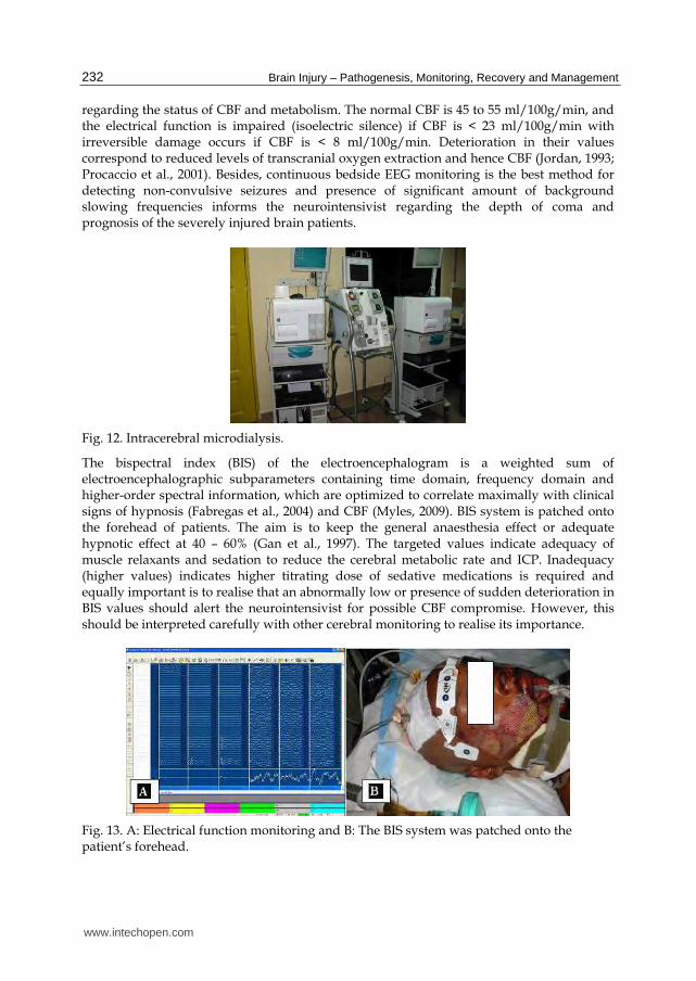
The bispectral index (BIS) of the electroencephalogram is a weighted sum of electroencephalographic subparameters containing time domain, frequency domain and higher-order spectral information, which are optimized to correlate maximally with clinical signs of hypnosis (Fabregas et al., 2004) and CBF (Myles, 2009). BIS system is patched onto the forehead of patients. The aim is to keep the general anaesthesia effect or adequate hypnotic effect at 40 – 60% (Gan et al., 1997). The targeted values indicate adequacy of muscle relaxants and sedation to reduce the cerebral metabolic rate and ICP. Inadequacy (higher values) indicates higher titrating dose of sedative medications is required and equally important is to realise that an abnormally low or presence of sudden deterioration in BIS values should alert the neurointensivist for possible CBF compromise. However, this should be interpreted carefully with other cerebral monitoring to realise its importance.
Fig. 13. A: Electrical function monitoring and B: The BIS system was patched onto the patient’s forehead.
www.intechopen.com
Neurointensive Care Monitoring for Severe Traumatic Brain Injury
233
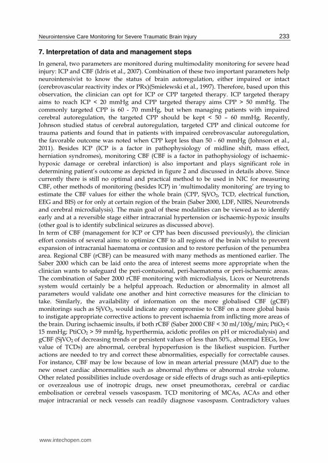
7. Interpretation of data and management steps
In general, two parameters are monitored during multimodality monitoring for severe head injury: ICP and CBF (Idris et al., 2007). Combination of these two important parameters help neurointensivist to know the status of brain autoregulation, either impaired or intact (cerebrovascular reactivity index or PRx)(Smielewski et al., 1997). Therefore, based upon this observation, the clinician can opt for ICP or CPP targeted therapy. ICP targeted therapy aims to reach ICP < 20 mmHg and CPP targeted therapy aims CPP > 50 mmHg. The commonly targeted CPP is 60 - 70 mmHg, but when managing patients with impaired cerebral autoregulation, the targeted CPP should be kept < 50 – 60 mmHg. Recently, Johnson studied status of cerebral autoregulation, targeted CPP and clinical outcome for trauma patients and found that in patients with impaired cerebrovascular autoregulation, the favorable outcome was noted when CPP kept less than 50 - 60 mmHg (Johnson et al., 2011). Besides ICP (ICP is a factor in pathophysiology of midline shift, mass effect, herniation syndromes), monitoring CBF (CBF is a factor in pathophysiology of ischaemic-hypoxic damage or cerebral infarction) is also important and plays significant role in determining patient’s outcome as depicted in figure 2 and discussed in details above. Since currently there is still no optimal and practical method to be used in NIC for measuring CBF, other methods of monitoring (besides ICP) in ‘multimodality monitoring’ are trying to estimate the CBF values for either the whole brain (CPP, SjVO2, TCD, electrical function, EEG and BIS) or for only at certain region of the brain (Saber 2000, LDF, NIRS, Neurotrends and cerebral microdialysis). The main goal of these modalities can be viewed as to identify early and at a reversible stage either intracranial hypertension or ischaemic-hypoxic insults (other goal is to identify subclinical seizures as discussed above). In term of CBF (management for ICP or CPP has been discussed previously), the clinician effort consists of several aims: to optimize CBF to all regions of the brain whilst to prevent expansion of intracranial haematoma or contusion and to restore perfusion of the penumbra area. Regional CBF (rCBF) can be measured with many methods as mentioned earlier. The Saber 2000 which can be laid onto the area of interest seems more appropriate when the clinician wants to safeguard the peri-contusional, peri-haematoma or peri-ischaemic areas. The combination of Saber 2000 rCBF monitoring with microdialysis, Licox or Neurotrends system would certainly be a helpful approach. Reduction or abnormality in almost all parameters would validate one another and hint corrective measures for the clinician to take. Similarly, the availability of information on the more globalised CBF (gCBF) monitorings such as SjVO2, would indicate any compromise to CBF on a more global basis to instigate appropriate corrective actions to prevent ischaemia from inflicting more areas of the brain. During ischaemic insults, if both rCBF (Saber 2000 CBF < 30 ml/100g/min; PtiO2 < 15 mmHg; PtiCO2 > 59 mmHg, hyperthermia, acidotic profiles on pH or microdialysis) and gCBF (SjVO2 of decreasing trends or persistent values of less than 50%, abnormal EEGs, low value of TCDs) are abnormal, cerebral hypoperfusion is the likeliest suspicion. Further actions are needed to try and correct these abnormalities, especially for correctable causes. For instance, CBF may be low because of low in mean arterial pressure (MAP) due to the new onset cardiac abnormalities such as abnormal rhythms or abnormal stroke volume. Other related possibilities include overdosage or side effects of drugs such as anti-epileptics or overzealous use of inotropic drugs, new onset pneumothorax, cerebral or cardiac embolisation or cerebral vessels vasospasm. TCD monitoring of MCAs, ACAs and other major intracranial or neck vessels can readily diagnose vasospasm. Contradictory values
www.intechopen.com
Brain Injury – Pathogenesis, Monitoring, Recovery and Management
234
between TCDs and the stated parameters discussed in earlier sections would alert the clinician towards cerebral vasospasm as the aetiology for cerebral hypoperfusion (high TCD but reduce CBF parameters). Triple-H therapy (includes expanding the plasma volume or hypervolemia, hemodilution and importantly hypertension), nimodipine or urgent interventional angiography for vasospasm should then be attempted to treat the identified problem. If only rCBF is impaired, steps should be taken to augment blood flow to those compromised areas only. However, currently, there is limited choice available for clinician to optimize rCBF. Most of the corrective measures for rCBF are in fact lending to optimize the gCBF. By augmenting the gCBF, the clinician may face notorious effect of ‘hyperaemic insult’ which in cases with markedly impaired cerebral autoregulation can be associated with brain swelling or odema and further elevation in ICPs that can aggravate the ischaemic-hypoxic circle. Therefore, multimodality monitoring offers more parameters to be closely considered when practicing ‘CPP targeted therapy’. By pushing the CPP, the clinician should look at SjVO2 which can become more than 75% suggesting cerebral hyperaemia and patterns of TCDs values which can become abnormally high. The aggressive CPP targeted therapy should not stop unless there is simultaneous elevation in ICPs, especially with parallel gradual emergence of deterioration in other monitored parameters (especially from Licox or Neurotrends and cerebral microdialysis), suggesting presence of markedly impaired cerebral autoregulation and evolving brain swelling which finally ends up with reduction in CBF in ‘that particular patient’. If ICPs become elevated (with other abnormal parameters suggesting/confirming reliable ICPs), the CPP targeted therapy should be stopped and measures such as lowering the MAP, reduction in plasma volume or inducing diuresis (with mannitol or diuretics), ICP lowering measures (such as mannitol or hypertonic saline, draining of CSF, adding or increasing sedative agent or muscle relaxant) should be taken. Decompressive craniectomy or barbiturate therapy should be reserved for patients who failed to respond to the initial measures and displayed concurrent features of time shift from compensatory to harmful compensatory or decompensatory phase.
8. Malaysia experience in multimodality monitoring
Our initial study published in 2003 on outcome of severe traumatic brain injury comparing the 3 monitoring approaches (multimodality, ICP alone and no cranial monitoring) revealed a statistically significant difference in the proportions of good outcomes between the multimodality group compared with the group of patients that underwent a single intracranial-based monitoring method and the group that received no monitoring (p = 0.003) based on a disability rating scale after a follow up of 12 months. Death was the focus of outcome in this study in which the multimodality approach to monitoring had superior results (Isa et al., 2003). The subsequent studies done in our centre were prognostic and cost effectiveness analysis studies of using different monitoring modalities in treating severe traumatic brain injury which were completed in 2004 and published in 2007 (Ibrahim et al., 2007; Idris et al., 2007).
8.1 Prognostic and cost effectiveness analysis studies of using different monitoring modalities in treating severe traumatic brain injury Between June 2002 and March 2004, 52 adult patients with severe traumatic non penetrating
head injury were recruited into our prospective randomised study at our University
www.intechopen.com
Neurointensive Care Monitoring for Severe Traumatic Brain Injury
235
Hospital, Universiti Sains Malaysia. The aim of the study was to investigate whether
multimodality monitoring in severe traumatic brain injury would alter the outcome score.
All adult patients, age of more than 14 years old with traumatic non penetrating head injury
who had GCS of less than 9 and CT brain without infratentorial pathology were included.
We excluded those who had a unilateral or bilateral fixed and dilated pupils resulted from
an on-going herniation, brain dead patient and patient known to have any condition that
lowering his or her functional status score. The randomisation process was made to allocate
to either multimodality monitoring or to standard ICP monitoring. In the multimodality
group, we monitored basic intensive care parameters and multiple cerebral parameters. In
the standard ICP monitoring group, only ICP was monitored together with basic intensive
care parameters. Monitoring proceeded for at least 3 days if uneventful. We recorded the
outcome at 6 months post treatment using the Barthel index score. The sixth month’s outcome between those groups (26 patients in each group) was not statistically significant (p < 0.48). However, the percentage of cases independent at 6 months was higher in the multimodality group compared with single modality group, 21.2% and 17.3% respectively. The multimodality monitoring group had also lower percentage of dependent cases at 6 months in comparison to the single ICP modality group, 28.8% and 32.7% respectively. The univariate analysis revealed, age (p < 0.03), GCS on arrival (p < 0.01), 24 hours fluid balance at day 2 of monitoring (p < 0.01), serum sodium (p < 0.03) and intracranial pressure at day 3 of monitoring (p < 0.01) were correlated with the outcomes. The trends of the parameters disclosed younger, higher GCS, lower injury severity score and Marshall grade would have a higher chance to be independent at 6 months post trauma. Tachycardic, hyperthermic, hypo- and hyper-volumic patients during the first 3 days of monitoring tend to be dependent at 6 months post injury. The dependent patients also tend to be acidotic or alkalotic for their arterial blood gases, have Paco2 of < 20 mmHg or Paco2 > 45 mmHg, blood haemoglobin level of < 8 g/dl, high blood urea and sodium, have persistently high ICP of more than 20 mmHg, CPP of < 55 mmHg, SjVO2 of < 50% or > 75%, rCBF < 35 ml/100g/min, TCD MCA flow velocity of < 35 cm/sec and PtiO2 of < 15 mmHg. Despite no significant statistical difference between the multimodality and single modality group, a possiblity of better outcome obtained with multimodality monitoring technique remains because of higher cases with independent status observed in that group. Obviously, this requires larger sample size for future study (Idris et al., 2007). We also did a study on cost effectiveness analysis of using multiple neuromodalities in treating severe traumatic brain injury. Despite a higher cost in multimodality monitoring, the application of multimodality monitoring for severe traumatic brain injury was more cost-effective (considering the cost of the equipment and the clinical outcome of the studied patients) than baseline neuromonitoring (p < 0.001)(Ibrahim et al., 2007).
8.2 Limitation of multimodality monitoring Despite various available cerebral monitoring, the current limitation in managing severely
injured brain is the therapy to optimize all monitored parameters. Except for high ICP, we
are yet to have any accepted standard for treating abnormalities detected by CBF/CPP
monitoring. Future works should focus on improving these diagnostic monitoring tools as
well as ways to treat or correct those detected abnormalities. Research on therapy is
continuing and one of the potential therapies is cerebral hypothermia.
www.intechopen.com
Brain Injury – Pathogenesis, Monitoring, Recovery and Management
236
Fig. 14. Multimodality monitoring for a patient with severe traumatic brain injury.
9. The future of monitoring and managing the severely injured brain – multimodality monitoring and cerebral hypothermia
Figure 2 illustrates various pathways that promote cerebral ischaemic-hypoxic events which can lead to cell death or apoptosis. Interestingly, one can take note that these pathways are mostly temperature dependent and therefore can be mitigated with cerebral hypothermia. In 2006, Vincent and Polderman reviewed the neuroprotective mechanisms underlying hypothermia and listed out various benefits of hypothermic therapy (Vincent & Polderman, 2006). Hypothermia can inhibit the activation of caspase enzymes, prevents or mitigates mitochondrial dysfunction, decrease the metabolism as well as decrease the overload of excitatory neurotransmitters such as glutamate and free oxygen radicals, modify the cellular disorders of intracellular ion concentrations, suppress the inflammatory and immunological responses and epileptic activity, reduce the disruption in blood brain barrier, vascular permeability and oedema, improve the microcirculatory circuits and intra- and extra-cellular acidosis, correct the hyperthemia after brain injury, influence the local secretion of various vasoactive mediators secreted by the endothelium and enhance expression of immediate early genes and cold shock proteins. Building on the vast benefits of cerebral hypothermia, currently underway is a locally adapted approach to induce hypothermia in severely injured brain patients using a direct method and study their outcomes.
9.1 Our current study - Direct brain hypothermia for trauma patient with Glasgow coma score of 6 and 7
The objective
To determine the effect of direct focal brain cooling on outcomes of severely injured brain patients.
www.intechopen.com
Neurointensive Care Monitoring for Severe Traumatic Brain Injury
237
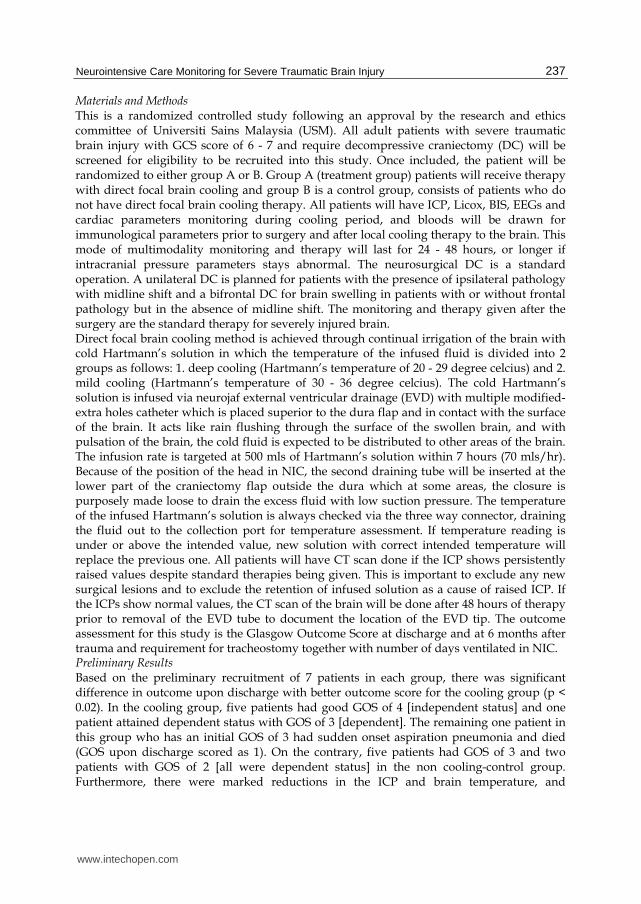
Materials and Methods This is a randomized controlled study following an approval by the research and ethics committee of Universiti Sains Malaysia (USM). All adult patients with severe traumatic brain injury with GCS score of 6 - 7 and require decompressive craniectomy (DC) will be screened for eligibility to be recruited into this study. Once included, the patient will be randomized to either group A or B. Group A (treatment group) patients will receive therapy with direct focal brain cooling and group B is a control group, consists of patients who do not have direct focal brain cooling therapy. All patients will have ICP, Licox, BIS, EEGs and cardiac parameters monitoring during cooling period, and bloods will be drawn for immunological parameters prior to surgery and after local cooling therapy to the brain. This mode of multimodality monitoring and therapy will last for 24 - 48 hours, or longer if intracranial pressure parameters stays abnormal. The neurosurgical DC is a standard operation. A unilateral DC is planned for patients with the presence of ipsilateral pathology with midline shift and a bifrontal DC for brain swelling in patients with or without frontal pathology but in the absence of midline shift. The monitoring and therapy given after the surgery are the standard therapy for severely injured brain. Direct focal brain cooling method is achieved through continual irrigation of the brain with cold Hartmann’s solution in which the temperature of the infused fluid is divided into 2 groups as follows: 1. deep cooling (Hartmann’s temperature of 20 - 29 degree celcius) and 2. mild cooling (Hartmann’s temperature of 30 - 36 degree celcius). The cold Hartmann’s solution is infused via neurojaf external ventricular drainage (EVD) with multiple modified-extra holes catheter which is placed superior to the dura flap and in contact with the surface of the brain. It acts like rain flushing through the surface of the swollen brain, and with pulsation of the brain, the cold fluid is expected to be distributed to other areas of the brain. The infusion rate is targeted at 500 mls of Hartmann’s solution within 7 hours (70 mls/hr). Because of the position of the head in NIC, the second draining tube will be inserted at the lower part of the craniectomy flap outside the dura which at some areas, the closure is purposely made loose to drain the excess fluid with low suction pressure. The temperature of the infused Hartmann’s solution is always checked via the three way connector, draining the fluid out to the collection port for temperature assessment. If temperature reading is under or above the intended value, new solution with correct intended temperature will replace the previous one. All patients will have CT scan done if the ICP shows persistently raised values despite standard therapies being given. This is important to exclude any new surgical lesions and to exclude the retention of infused solution as a cause of raised ICP. If the ICPs show normal values, the CT scan of the brain will be done after 48 hours of therapy prior to removal of the EVD tube to document the location of the EVD tip. The outcome assessment for this study is the Glasgow Outcome Score at discharge and at 6 months after trauma and requirement for tracheostomy together with number of days ventilated in NIC. Preliminary Results Based on the preliminary recruitment of 7 patients in each group, there was significant difference in outcome upon discharge with better outcome score for the cooling group (p < 0.02). In the cooling group, five patients had good GOS of 4 [independent status] and one patient attained dependent status with GOS of 3 [dependent]. The remaining one patient in this group who has an initial GOS of 3 had sudden onset aspiration pneumonia and died (GOS upon discharge scored as 1). On the contrary, five patients had GOS of 3 and two patients with GOS of 2 [all were dependent status] in the non cooling-control group. Furthermore, there were marked reductions in the ICP and brain temperature, and
www.intechopen.com
Brain Injury – Pathogenesis, Monitoring, Recovery and Management
238
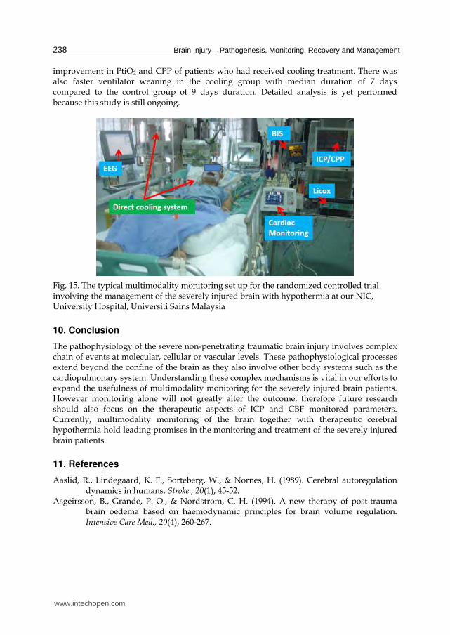
improvement in PtiO2 and CPP of patients who had received cooling treatment. There was also faster ventilator weaning in the cooling group with median duration of 7 days compared to the control group of 9 days duration. Detailed analysis is yet performed because this study is still ongoing.
Fig. 15. The typical multimodality monitoring set up for the randomized controlled trial involving the management of the severely injured brain with hypothermia at our NIC, University Hospital, Universiti Sains Malaysia
10. Conclusion
The pathophysiology of the severe non-penetrating traumatic brain injury involves complex chain of events at molecular, cellular or vascular levels. These pathophysiological processes extend beyond the confine of the brain as they also involve other body systems such as the cardiopulmonary system. Understanding these complex mechanisms is vital in our efforts to expand the usefulness of multimodality monitoring for the severely injured brain patients. However monitoring alone will not greatly alter the outcome, therefore future research should also focus on the therapeutic aspects of ICP and CBF monitored parameters. Currently, multimodality monitoring of the brain together with therapeutic cerebral hypothermia hold leading promises in the monitoring and treatment of the severely injured brain patients.
11. References
Aaslid, R., Lindegaard, K. F., Sorteberg, W., & Nornes, H. (1989). Cerebral autoregulation dynamics in humans. Stroke., 20(1), 45-52.
Asgeirsson, B., Grande, P. O., & Nordstrom, C. H. (1994). A new therapy of post-trauma brain oedema based on haemodynamic principles for brain volume regulation. Intensive Care Med., 20(4), 260-267.
www.intechopen.com
Neurointensive Care Monitoring for Severe Traumatic Brain Injury
239
Balestreri, M., Czosnyka, M., Hutchinson, P., Steiner, L. A., Hiler, M., Smielewski, P., et al. (2006). Impact of intracranial pressure and cerebral perfusion pressure on severe disability and mortality after head injury. Neurocrit Care., 4(1), 8-13.
Balestreri, M., Czosnyka, M., Steiner, L. A., Schmidt, E., Smielewski, P., Matta, B., et al. (2004). Intracranial hypertension: what additional information can be derived from ICP waveform after head injury? Acta Neurochir (Wien). 146(2), 131-141. Epub 2004 Feb 2002.
Bellner, J., Romner, B., Reinstrup, P., Kristiansson, K. A., Ryding, E., & Brandt, L. (2004). Transcranial Doppler sonography pulsatility index (PI) reflects intracranial pressure (ICP). Surg Neurol., 62(1), 45-51; discussion 51.
Berger, S., Schurer, L., Hartl, R., Messmer, K., & Baethmann, A. (1995). Reduction of post-traumatic intracranial hypertension by hypertonic/hyperoncotic saline/dextran and hypertonic mannitol. Neurosurgery., 37(1), 98-107; discussion 107-108.
Bolognese, P., Miller, J. I., Heger, I. M., & Milhorat, T. H. (1993). Laser-Doppler flowmetry in neurosurgery. J Neurosurg Anesthesiol., 5(3), 151-158.
Bouma, G. J., Muizelaar, J. P., Stringer, W. A., Choi, S. C., Fatouros, P., & Young, H. F. (1992). Ultra-early evaluation of regional cerebral blood flow in severely head-injured patients using xenon-enhanced computerized tomography. J Neurosurg., 77(3), 360-368.
Bullock R, C. R., Clifton C,, Ghajar J, M. D., Narayan RK,, Newell DW, P. L., Rosner MJ,, & JW, W. (1996). Guidelines for the management of severe head injury. Brain Trauma Foundation, American Association of Neurological Surgeons, Joint Section on Neurotrauma and Critical Care. J Neurotrauma., 13(11), 641-734.
Carandang, R., & Krieger, D. (2008). Decompressive Hemicraniectomy and Durotomy for Malignant Middle Cerebral Artery Infarction. Neurocritical Care, 8(2), 286-289.
Carter, L. P., Erspamer, R., White, W. L., & Yamagata, S. (1982). Cortical blood flow during craniotomy for aneurysms. Surg Neurol., 17(3), 204-208.
Chan, K. H., Miller, J. D., Dearden, N. M., Andrews, P. J., & Midgley, S. (1992). The effect of changes in cerebral perfusion pressure upon middle cerebral artery blood flow velocity and jugular bulb venous oxygen saturation after severe brain injury. J Neurosurg., 77(1), 55-61.
Charbel, F. T., Hoffman, W. E., Misra, M., Hannigan, K., & Ausman, J. I. (1997). Cerebral interstitial tissue oxygen tension, pH, HCO3, CO2. Surg Neurol., 48(4), 414-417.
Chen, S. T., Hsu, C. Y., Hogan, E. L., Halushka, P. V., Linet, O. I., & Yatsu, F. M. (1986). Thromboxane, prostacyclin, and leukotrienes in cerebral ischemia. Neurology., 36(4), 466-470.
Cooper, D. J., Rosenfeld, J. V., Murray, L., Wolfe, R., Ponsford, J., Davies, A., et al. (2008). Early decompressive craniectomy for patients with severe traumatic brain injury and refractory intracranial hypertension--A pilot randomized trial. Journal of Critical Care, 23(3), 387-393.
Csuka, E., Morganti-Kossmann, M. C., Lenzlinger, P. M., Joller, H., Trentz, O., & Kossmann, T. (1999). IL-10 levels in cerebrospinal fluid and serum of patients with severe traumatic brain injury: relationship to IL-6, TNF-alpha, TGF-beta1 and blood-brain barrier function. J Neuroimmunol., 101(2), 211-221.
Czosnyka, M., Smielewski, P., Piechnik, S., Steiner, L. A., & Pickard, J. D. (2001). Cerebral autoregulation following head injury. J Neurosurg., 95(5), 756-763.
www.intechopen.com
Brain Injury – Pathogenesis, Monitoring, Recovery and Management
240
Dickman, C. A., Carter, L. P., Baldwin, H. Z., Harrington, T., & Tallman, D. (1991). Continuous regional cerebral blood flow monitoring in acute craniocerebral trauma. Neurosurgery., 28(3), 467-472.
Dietrich, W., & Bramlett, H. (2010). The evidence for hypothermia as a neuroprotectant in traumatic brain injury. Neurotherapeutics, 7(1), 43-50.
Dirnagl, U., Iadecola, C., & Moskowitz, M. A. (1999). Pathobiology of ischaemic stroke: an integrated view. Trends in Neurosciences, 22(9), 391-397.
Eberle, B. M., Schnüriger, B., Inaba, K., Peter Gruen, J., Demetriades, D., & Belzberg, H. (2010). Decompressive craniectomy: Surgical control of traumatic intracranial hypertension may improve outcome. Injury, 41(9), 894-898.
Egnor, M., Zheng, L., Rosiello, A., Gutman, F., & Davis, R. (2002). A model of pulsations in communicating hydrocephalus. Pediatr Neurosurg., 36(6), 281-303.
Elwell, C. E., Owen-Reece, H., Cope, M., Wyatt, J. S., Edwards, A. D., Delpy, D. T., et al. (1993). Measurement of adult cerebral haemodynamics using near infrared spectroscopy. Acta Neurochir Suppl (Wien). 59, 74-80.
Fabregas, N., Gambus, P. L., Valero, R., Carrero, E. J., Salvador, L., Zavala, E., et al. (2004). Can bispectral index monitoring predict recovery of consciousness in patients with severe brain injury? Anesthesiology., 101(1), 43-51.
Fabricius, M., Fuhr, S., Willumsen, L., Dreier, J. P., Bhatia, R., Boutelle, M. G., et al. (2008). Association of seizures with cortical spreading depression and peri-infarct depolarisations in the acutely injured human brain. Clin Neurophysiol., 119(9), 1973-1984. Epub 2008 Jul 1914.
Figaji, A., Fieggen, A., Sandler, S., Argent, A., Le Roux, P., & Peter, J. (2007). Intracranial pressure and cerebral oxygenation changes after decompressive craniectomy in a child with traumatic brain swelling. Child's Nervous System, 23(11), 1331-1335.
Gan, T. J., Glass, P. S., Windsor, A., Payne, F., Rosow, C., Sebel, P., et al. (1997). Bispectral index monitoring allows faster emergence and improved recovery from propofol, alfentanil, and nitrous oxide anesthesia. BIS Utility Study Group. Anesthesiology., 87(4), 808-815.
Gluckman, P. D., Wyatt, J. S., Azzopardi, D., Ballard, R., Edwards, A. D., Ferriero, D. M., et al. (2005). Selective head cooling with mild systemic hypothermia after neonatal encephalopathy: multicentre randomised trial. The Lancet, 365(9460), 663-670.
Grzybicki, D., Moore, S. A., Schelper, R., Glabinski, A. R., Ransohoff, R. M., & Murphy, S. (1998). Expression of monocyte chemoattractant protein (MCP-1) and nitric oxide synthase-2 following cerebral trauma. Acta Neuropathol., 95(1), 98-103.
Gura, M., Elmaci, I., Sari, R., & Coskun, N. (2011). Correlation of pulsatility index with intracranial pressure in traumatic brain injury. Turk Neurosurg, 21(2), 210-215.
Hausmann, E. H., Berman, N. E., Wang, Y. Y., Meara, J. B., Wood, G. W., & Klein, R. M. (1998). Selective chemokine mRNA expression following brain injury. Brain Res., 788(1-2), 49-59.
Hoffman, W. E., Charbel, F. T., & Edelman, G. (1996). Brain tissue oxygen, carbon dioxide, and pH in neurosurgical patients at risk for ischemia. Anesth Analg., 82(3), 582-586.
Ibrahim, M. I., Abdullah, M., Naing, L., Abdullah, J. M., Idris, Z., & Aljunid, S. M. (2007). Cost effectiveness analysis of using multiple neuromodalities in treating severe traumatic brain injury in a developing country like Malaysia. Asian J Surg., 30(4), 261-266.
www.intechopen.com
Neurointensive Care Monitoring for Severe Traumatic Brain Injury
241
Idris, Z., Ghani, R. I., Musa, K. I., Ibrahim, M. I., Abdullah, M., Nyi, N. N., et al. (2007). Prognostic Study of Using Different Monitoring Modalities in Treating Severe Traumatic Brain Injury. Asian Journal of Surgery, 30(3), 200-208.
Is, M., Coskun, A., Sanus, G. Z., Tanriverdi, T., Kafadar, A. M., Hanimoglu, H., et al. (2007). High-sensitivity C-reactive protein levels in cerebrospinal fluid and serum in severe head injury: relationship to tumor necrosis factor-alpha and interleukin-6. J Clin Neurosci., 14(12), 1163-1171. Epub 2007 Sep 1119.
Isa, R., Wan Adnan, W. A., Ghazali, G., Idris, Z., Ghani, A. R., Sayuthi, S., et al. (2003). Outcome of severe traumatic brain injury: comparison of three monitoring approaches. Neurosurg Focus., 15(6), E1.
Johnson, U., Nilsson, P., Ronne-Engstrom, E., Howells, T., & Enblad, P. (2011). Favorable outcome in traumatic brain injury patients with impaired cerebral pressure autoregulation when treated at low cerebral perfusion pressure levels. Neurosurgery., 68(3), 714-721; discussion 721-712.
Jordan, K. G. (1993). Continuous EEG and evoked potential monitoring in the neuroscience intensive care unit. J Clin Neurophysiol., 10(4), 445-475.
Juliet, P. A., Mao, X., & Del Bigio, M. R. (2008). Proinflammatory cytokine production by cultured neonatal rat microglia after exposure to blood products. Brain Res., 1210, 230-239. Epub 2008 Mar 2018.
Kaulfers, A. M., Backeljauw, P. F., Reifschneider, K., Blum, S., Michaud, L., Weiss, M., et al. (2010). Endocrine dysfunction following traumatic brain injury in children. J Pediatr, 157(6), 894-899. Epub 2010 Aug 2024.
Kazibutowska, Z., Stelmach-Wawrzyczek, M., & Majchrzak, R. (1992). [Results of prospective 24-hour EEG studies of patients after cranio-cerebral injuries]. Neurol Neurochir Pol., 26(3), 304-310.
Kossmann, T., Stahel, P. F., Lenzlinger, P. M., Redl, H., Dubs, R. W., Trentz, O., et al. (1997). Interleukin-8 released into the cerebrospinal fluid after brain injury is associated with blood-brain barrier dysfunction and nerve growth factor production. J Cereb Blood Flow Metab., 17(3), 280-289.
Kouvarellis, A. J., Rohlwink, U. K., Sood, V., Van Breda, D., Gowen, M. J., & Figaji, A. A. (2011). The relationship between basal cisterns on CT and time-linked intracranial pressure in paediatric head injury. Childs Nerv Syst, 27 (7), 1139-1144.
Krahulik, D., Zapletalova, J., Frysak, Z., & Vaverka, M. (2010). Dysfunction of hypothalamic-hypophysial axis after traumatic brain injury in adults. J.Neurosurg, 113(3), 581-584.
Lam, J. M., Hsiang, J. N., & Poon, W. S. (1997). Monitoring of autoregulation using laser Doppler flowmetry in patients with head injury. J Neurosurg., 86(3), 438-445.
Lee, J. H., Kelly, D. F., Oertel, M., McArthur, D. L., Glenn, T. C., Vespa, P., et al. (2001). Carbon dioxide reactivity, pressure autoregulation, and metabolic suppression reactivity after head injury: a transcranial Doppler study. J Neurosurg., 95(2), 222-232.
Lewen, A., Matz, P., & Chan, P. H. (2000). Free radical pathways in CNS injury. J Neurotrauma., 17(10), 871-890.
Lewis, S., Wong, M., Myburgh, J., & Reilly, P. (1998). Determining cerebral perfusion pressure thresholds in severe head trauma. Acta Neurochir Suppl., 71, 174-176.
Liou, A. K., Clark, R. S., Henshall, D. C., Yin, X. M., & Chen, J. (2003). To die or not to die for neurons in ischemia, traumatic brain injury and epilepsy: a review on the stress-
www.intechopen.com
Brain Injury – Pathogenesis, Monitoring, Recovery and Management
242
activated signaling pathways and apoptotic pathways. Prog Neurobiol., 69(2), 103-142.
Lundberg, N., Troupp, H., & Lorin, H. (1965). Continuous recording of the ventricular-fluid pressure in patients with severe acute traumatic brain injury. A preliminary report. J Neurosurg., 22(6), 581-590.
Maas, A. I., Dearden, M., Teasdale, G. M., Braakman, R., Cohadon, F., Iannotti, F., et al. (1997). EBIC-guidelines for management of severe head injury in adults. European Brain Injury Consortium. Acta Neurochir (Wien). 139(4), 286-294.
Marshall, G. T., James, R. F., Landman, M. P., O'Neill, P. J., Cotton, B. A., Hansen, E. N., et al. (2010). Pentobarbital coma for refractory intra-cranial hypertension after severe traumatic brain injury: mortality predictions and one-year outcomes in 55 patients. J, 69(2), 275-283.
Mathew, P., Graham, D. I., Bullock, R., Maxwell, W., McCulloch, J., & Teasdale, G. (1994). Focal brain injury: histological evidence of delayed inflammatory response in a new rodent model of focal cortical injury. Acta Neurochir Suppl (Wien). 60, 428-430.
Menashe Zaaroor, E. M., Venyamin Shik, Jean F Soustiel. (2007). Course of cerebral blood flow and metabolism following severe brain injury. Correlation with neurological function and outcome. Indian Journal of Neurotrauma (IJNT) 4, 25-29.
Meyerson, B. A., Gunasekera, L., Linderoth, B., & Gazelius, B. (1991). Bedside monitoring of regional cortical blood flow in comatose patients using laser Doppler flowmetry. Neurosurgery., 29(5), 750-755.
Miller, M. T., Pasquale, M., Kurek, S., White, J., Martin, P., Bannon, K., et al. (2004). Initial head computed tomographic scan characteristics have a linear relationship with initial intracranial pressure after trauma. J Trauma., 56(5), 967-972; discussion 972-963.
Mitsis, G. D., Zhang, R., Levine, B. D., & Marmarelis, V. Z. (2006). Cerebral hemodynamics during orthostatic stress assessed by nonlinear modeling. J Appl Physiol., 101(1), 354-366. Epub 2006 Mar 2002.
Muizelaar, J. P., & Schroder, M. L. (1994). Overview of monitoring of cerebral blood flow and metabolism after severe head injury. Can J Neurol Sci., 21(2), S6-11.
Muttaqin, Z., Uozumi, T., Kuwabara, S., Arita, K., Kurisu, K., Ohba, S., et al. (1993). Hyperaemia prior to acute cerebral swelling in severe head injuries: the role of transcranial Doppler monitoring. Acta Neurochir (Wien). 123(1-2), 76-81.
Myles, P. S. (2009). Bispectral index monitoring in ischemic-hypoxic brain injury. J Extra Corpor Technol., 41(1), P15-19.
Narayan, R. K., Michel, M. E., Ansell, B., Baethmann, A., Biegon, A., Bracken, M. B., et al. (2002). Clinical trials in head injury. J Neurotrauma, 19(5), 503-557.
Nujaimin, U., Saufi, A., Rahman, A. G., Badrisyah, I., Sani, S., Zamzuri, I., et al. (2009). Post traumatic cerebral oedema in severe head injury is related to intracranial pressure and cerebral perfusion pressure but not to cerebral compliance. Asian J Surg., 32(3), 157-162.
Obrist, W. D., Langfitt, T. W., Jaggi, J. L., Cruz, J., & Gennarelli, T. A. (1984). Cerebral blood flow and metabolism in comatose patients with acute head injury. Relationship to intracranial hypertension. J Neurosurg., 61(2), 241-253.
Oddo, M., Levine, J. M., Frangos, S., Carrera, E., Maloney-Wilensky, E., Pascual, J. L., et al. (2009). Effect of mannitol and hypertonic saline on cerebral oxygenation in patients
www.intechopen.com
Neurointensive Care Monitoring for Severe Traumatic Brain Injury
243
with severe traumatic brain injury and refractory intracranial hypertension. J Neurol Neurosurg Psychiatry., 80(8), 916-920. Epub 2009 Mar 2016.
Patel, H. C., Menon, D. K., Tebbs, S., Hawker, R., Hutchinson, P. J., & Kirkpatrick, P. J. (2002). Specialist neurocritical care and outcome from head injury. Intensive Care Med., 28(5), 547-553. Epub 2002 Feb 2014.
Pluta, R. M., Hansen-Schwartz, J., Dreier, J., Vajkoczy, P., Macdonald, R. L., Nishizawa, S., et al. (2009). Cerebral vasospasm following subarachnoid hemorrhage: time for a new world of thought. Neurol Res., 31(2), 151-158.
Procaccio, F., Polo, A., Lanteri, P., & Sala, F. (2001). Electrophysiologic monitoring in neurointensive care. Curr Opin Crit Care., 7(2), 74-80.
Puppo, C., Farina, G., Lopez, F. L., Caragna, E., & Biestro, A. (2008). Cerebral CO2 reactivity in severe head injury. A transcranial Doppler study. Acta Neurochir Suppl., 102, 171-175.
Ross, N., & Eynon, C. A. (2005). Intracranial pressure monitoring. Current Anaesthesia & Critical Care, 16(4), 255-261.
Sakai, K., Yamada, K., Mori, S., Sugimoto, N., & Nishimura, T. (2011). Age-dependent brain temperature decline assessed by diffusion-weighted imaging thermometry. NMR Biomed, 28(Jan).
Smielewski, P., Czosnyka, M., Kirkpatrick, P., & Pickard, J. D. (1997). Evaluation of the transient hyperemic response test in head-injured patients. J Neurosurg., 86(5), 773-778.
Stahel, P. F., Morganti-Kossmann, M. C., & Kossmann, T. (1998). The role of the complement system in traumatic brain injury. Brain Res Brain Res Rev., 27(3), 243-256.
Stuart, R. M., Schmidt, M., Kurtz, P., Waziri, A., Helbok, R., Mayer, S. A., et al. (2010). Intracranial multimodal monitoring for acute brain injury: a single institution review of current practices. Neurocrit, 12(2), 188-198.
Timofeev, I., Carpenter, K. L., Nortje, J., Al-Rawi, P. G., O'Connell, M. T., Czosnyka, M., et al. (2011). Cerebral extracellular chemistry and outcome following traumatic brain injury: a microdialysis study of 223 patients. Brain., 134(Pt 2), 484-494. Epub 2011 Jan 2018.
van Santbrink, H., Maas, A. I., & Avezaat, C. J. (1996). Continuous monitoring of partial pressure of brain tissue oxygen in patients with severe head injury. Neurosurgery., 38(1), 21-31.
Vergely, C., Maupoil, V., Clermont, G., Bril, A., & Rochette, L. (2003). Identification and quantification of free radicals during myocardial ischemia and reperfusion using electron paramagnetic resonance spectroscopy. Arch Biochem Biophys., 420(2), 209-216.
Vincent, J.-L., & Polderman, K. H. (2006). Induced Hypothermia for Neuroprotection: Understanding the Underlying Mechanisms. In Intensive Care Medicine (pp. 328-346): Springer New York.
Voulgaris, S. G., Partheni, M., Kaliora, H., Haftouras, N., Pessach, I. S., & Polyzoidis, K. S. (2005). Early cerebral monitoring using the transcranial Doppler pulsatility index in patients with severe brain trauma. Med Sci Monit., 11(2), CR49-52.
Young, J. S., Blow, O., Turrentine, F., Claridge, J. A., & Schulman, A. (2003). Is there an upper limit of intracranial pressure in patients with severe head injury if cerebral perfusion pressure is maintained? Neurosurg Focus., 15(6), E2.
www.intechopen.com

Brain Injury – Pathogenesis, Monitoring, Recovery and Management
244
Zauner, A., Bullock, R., Di, X., & Young, H. F. (1995). Brain oxygen, CO2, pH, and temperature monitoring: evaluation in the feline brain. Neurosurgery., 37(6), 1168-1176; discussion 1176-1167.
Zweifel, C., Lavinio, A., Steiner, L. A., Radolovich, D., Smielewski, P., Timofeev, I., et al. (2008). Continuous monitoring of cerebrovascular pressure reactivity in patients with head injury. Neurosurg Focus., 25(4), E2.
www.intechopen.com
Brain Injury - Pathogenesis, Monitoring, Recovery andManagementEdited by Prof. Amit Agrawal
ISBN 978-953-51-0265-6Hard cover, 522 pagesPublisher InTechPublished online 23, March, 2012Published in print edition March, 2012
InTech EuropeUniversity Campus STeP Ri Slavka Krautzeka 83/A 51000 Rijeka, Croatia Phone: +385 (51) 770 447 Fax: +385 (51) 686 166www.intechopen.com
InTech ChinaUnit 405, Office Block, Hotel Equatorial Shanghai No.65, Yan An Road (West), Shanghai, 200040, China
Phone: +86-21-62489820 Fax: +86-21-62489821
The present two volume book "Brain Injury" is distinctive in its presentation and includes a wealth of updatedinformation on many aspects in the field of brain injury. The Book is devoted to the pathogenesis of braininjury, concepts in cerebral blood flow and metabolism, investigative approaches and monitoring of braininjured, different protective mechanisms and recovery and management approach to these individuals,functional and endocrine aspects of brain injuries, approaches to rehabilitation of brain injured and preventiveaspects of traumatic brain injuries. The collective contribution from experts in brain injury research area wouldbe successfully conveyed to the readers and readers will find this book to be a valuable guide to furtherdevelop their understanding about brain injury.
How to referenceIn order to correctly reference this scholarly work, feel free to copy and paste the following:
Zamzuri Idris, Muzaimi Mustapha and Jafri Malin Abdullah (2012). Neurointensive Care Monitoring for SevereTraumatic Brain Injury, Brain Injury - Pathogenesis, Monitoring, Recovery and Management, Prof. AmitAgrawal (Ed.), ISBN: 978-953-51-0265-6, InTech, Available from: http://www.intechopen.com/books/brain-injury-pathogenesis-monitoring-recovery-and-management/neurointensive-care-monitoring-for-severe-traumatic-brain-injury