list-mode proton ct reconstruction using their most …srit/biblio/rit2015a.pdfcontinuous...
TRANSCRIPT

1
List-mode proton CT reconstruction using theirmost likely paths via the finite Hilbert transform of
the derivative of the backprojectionSimon Rit, Rolf Clackdoyle, Jan Hoskovec and Jean Michel Letang
Abstract—Modern prototypes of proton computed tomography(CT) scanners can measure the energy, the position and thedirection of each proton, before and after the scanned object,in a list mode. Each detected proton contributes to an estimateof a line integral along an estimated curved proton path. Inthis work, we propose a backproject first algorithm based onthe two-step Hilbert transform to reconstruct proton CT images.The algorithm takes into account the estimated curved paths.A pixel-specific backprojection is computed from the averagemeasurements of protons which traverse the pixel with the samedirection according to the proton path estimates. Our simulationsstudies show that the algorithm has similar spatial resolution toa previous filtered backprojection (FBP) algorithm for protonCT using most likely paths while being computationally moreefficient and able to handle truncated data.
I. INTRODUCTION
The concept of proton CT was proposed early in the historyof CT [1]. From measurements of the energy loss of protonsthrough matter, a reconstructed image can be formed of therelative stopping power (RSP) map of tissues. The maindisavantadge of proton CT is its poor spatial resolution whichis caused by multiple Coulomb scattering that generates quasi-continuous deflections of the protons in matter. However, thepotential of proton CT to improve proton therapy treatmentplanning has lead to new hardware and software developmentsto combat the spatial resolution issues.
Much improvement of the spatial resolution in proton CThas been achieved thanks to the use of pairs of position sensi-tive detectors that record the position and the direction of eachproton, once before the scanned object and once afterwards[2]. The acquired list-mode information has the advantagethat the most likely path of each proton can be estimated[3] and incorporated into the reconstruction algorithm. Thistechnique substantially improves the spatial resolution overmethods that use straight-line models, and several groups arenow developing such scanner prototypes. Incorporating the
S. Rit and J.M. Letang are with the Universite de Lyon, CREATIS; CNRSUMR5220; Inserm U1044; INSA-Lyon; Universite Lyon 1; Centre LeonBerard, France (e-mail: [email protected]).R. Clackdoyle is with the laboratoire Hubert Curien, CNRS and UniversiteJean Monnet (UMR 5516), Saint Etienne, France.J. Hoskovec is with the laboratoire Hubert Curien, CNRS and UniversiteJean Monnet (UMR 5516), Saint Etienne, France and Universite de Lyon,CREATIS; CNRS UMR5220; Inserm U1044; INSA-Lyon; Universite Lyon1; Centre Leon Berard, France.This work was partially supported by grants ANR-12-BS01-0018 (DROITEproject) and ANR-13-IS03-0002-01 (DEXTER project) from the AgenceNationale de la Recherche (France) and by a grant from la Region Rhone-Alpes.
most likely path in an iterative reconstruction algorithm isstraightforward since it can be accounted for in the projectionmatrix (see, e.g., [4]). However, the practical applicabilityof iterative reconstruction is limited by the computationaltime, particularly for some clinical applications such as imageguided proton therapy.
Use of proton list-mode data in FBP algorithms is typicallyperformed after binning the data into a set of projectionimages. It has long been considered impossible to accountfor curved proton paths since all protons binned together havefollowed different paths. Therefore, the most likely path wasfirst used with FBP to only include the protons that hadfollowed close to straight paths [5] but it is preferable to makeuse of all acquired data.
There is no exact analytic reconstruction from integralsalong randomly curved lines, but heuristic use of most likelypaths of protons in FBP algorithms has recently been demon-strated. First, Rit et al. proposed the distance-driven binning[6] where multiple projection images per source position arebinned using different approximations of proton paths bystraight lines. For each of these projections, the straight linesare defined by the same source point but a different pointalong the most likely proton paths, at the intersection withplanes parallel to the detector at different distances from thesource. The most likely path of each proton is then indirectlyaccounted for by using a different set of input projection im-ages for the reconstruction of each voxel using the Feldkampalgorithm [7]. The improvement of the spatial resolution ofreconstructed images is similar to what is obtained with aniterative least-square algorithm using curved most likely pathsat a much lower computational cost [4].
More recently, Poludniowski et al. have proposed anotherFBP algorithm that more directly uses the most likely pathsby switching the order of the filtering and the backprojection[8]. The list-mode data are used to compute the discretebackprojection by averaging the measurements of protons thatwent through the same voxel with the same direction. Theproblem of this switching is that the backprojection shouldtheoretically be computed on an infinite support. Since thebackprojection array must inevitably be truncated in practice,some low frequency artifacts might be introduced by thisprocedure.
In this work, we follow a similar approach to that of [8]using another backproject first algorithm. Zeng derived a two-dimensional (2D) reconstruction algorithm [9] from the two-step Hilbert transform method [10] that starts with weighted
The 13th International Meeting on Fully Three-Dimensional Image Reconstruction in Radiology and Nuclear Medicine
324

2
backprojections. As he pointed out, this property is useful withlist-mode data such as those acquired by proton CT scanners.We applied this algorithm to proton CT and investigated itsperformance on phantom and patient simulations.
II. METHOD
We start with a brief description of Zeng’s reconstructionmethod before deriving an algorithm for its use in proton CT.
A. Backproject first reconstruction algorithm
Let p(s, θ) be parallel projections of an unknown 2Dfunction f(x, y) where the coordinates s ∈ R and θ ∈ [0, π)define a line by its signed distance to the origin and its anglewith the x-axis, respectively. Noo et al. have shown [10] thatf can be recovered from p using
f(x, y) = H
∫ π
0
∂
∂sp(s, θ)
∣∣∣∣s=−x sin θ+y cos θ
dθ (1)
with H the inverse Hilbert transform. The resulting algorithminvolves (1) taking the derivative of the projections p along s,(2) backprojecting the result and (3) taking the finite inverseHilbert transform of the backprojection along the x direction.
Zeng proposed inverting the order of the derivative and thebackprojection in this formula to obtain a backproject first typeof algorithm [9]. Zeng’s formula is
f(x, y) = H
{∂
∂xbs(x, y) +
∂
∂ybc(x, y)
}(2)
with the weighted backprojections
bs(x, y) =−∫ π
0
p(−x sin θ + y cos θ, θ) sin θ dθ (3)
bc(x, y) =
∫ π
0
p(−x sin θ + y cos θ, θ) cos θ dθ. (4)
Zeng obtains a reconstruction algorithm consisting of thefollowing steps: (1) calculate two weighted backprojectionimages bs and bc, (2) take the sum of the derivative inorthogonal directions of each backprojection bs and bc, and(3) take its inverse Hilbert transform. The next section focuseson how to obtain the two backprojection images bs and bc inthe context of proton CT.
B. Backprojection for proton CT
We consider an ideal proton CT scanner made of two pairsof flat trackers that record in a list the position and direction ofeach proton, before entering and after leaving the target object.The energy at the trackers is assumed to be known before theobject and is measured after the object. The incidence angleof the protons is varied by rotating the cone-beam source andtwo pairs of detectors along a circular trajectory.
Protons are deflected many times when going through mat-ter so their overall path is slightly curved. We let Γi(t) ∈ R3
denote the proton path, with time t ∈ R used to parameterizethe curved lines and i ∈ {1, ..., I} the proton index in the list-mode data. Using the Bethe-Bloch equation, one can relate
the energy loss of the proton to the sum gi of the three-dimensional (3D) relative stopping power map f of tissuesalong the proton path Γi
gi ≡∫ Eout
Ein
1
dE/dxw(E)dE '
∫ touti
tini
f(Γi(t)) dΓi(t) (5)
with Ein and Eout the energy before and after the object,dE/dxw the stopping power of water, and tini and touti thetimes at which the proton is measured entering and leaving thepatient, respectively. The proton CT reconstruction problemis to obtain an image of f from the path integrals gi and anestimate Γi of each proton path, both of which can be obtainedfrom the measurements.
Γi
Γi(touti )
Γi
Θi,l ' θk
Γi(tini )
Γi(ti,l)ζj
Fig. 1. Illustration of the notation used for the backprojection. The protonfollows the path Γi and its most likely path Γi is estimated from itsmeasured positions Γi(t
ini ) and Γi(t
outi ) and the corresponding directions
before entering and after leaving the object, respectively. A discrete set ofpositions (indexed by l, from tini = ti,0 to touti = ti,L) regularly samplesΓi to evaluate which pixels are reached by the most likely path and thecorresponding angle with the horizontal line. For example, here, the positionΓi(ti,l) falls in the indicator function ζj of the j-th pixel with an angleΘi,l that is binned to the closest angle θk and contributes to the averagemeasurement bj,k for the j-th pixel and the k-th direction.
Our solution to this reconstruction problem uses Zeng’salgorithm which starts by computing the two backprojectionimages bs and bc from the proton data. Only the 2D centralslice is considered here but the 3D trajectories of protons areused. Our approach to compute the weighted backprojectionsis based on [8], i.e., a discretization of Equations 3 and 4.First, we discretize space with j ∈ {1, ..., J} the pixel indexof the reconstructed slice. The variable θ is also discretized andwe let k ∈ {1, ...,K} be the index of the discrete values θk.Finally, the curved most-likely path of each proton is evaluatedat a discrete number of time positions ti,l between tini and touti
with l ∈ {1, ..., L} the time indices, tini = ti,0 and touti = ti,L(Figure 1). At each of the positions Γi(ti,l) along the protonpaths, we estimate the angle Θi,l between the x-axis and thedirection of the proton using the proton velocity dΓi/dt(ti,l).Using these discretizations, we compute a pixel-specific anddirection-specific average of the measurements with
bj,k =
∑i,l ζj(Γi(ti,l))ξk(Θi,l)gi∑i,l ζj(Γi(ti,l))ξk(Θi,l)
(6)
where ζj and ξk are basis functions for the j-th pixel and thek-th angle θk respectively. In this work, the basis functionsζj and ξk are indicator functions such that each measurement
The 13th International Meeting on Fully Three-Dimensional Image Reconstruction in Radiology and Nuclear Medicine
325
3
gi contributes to the the nearest pixels and directions alongthe proton path. Therefore, bj,k is the average value of themeasurements gi of protons whose most likely paths traversepixel j with a direction θk.
Recalling that p(−x sin θ + y cos θ, θ) is the measurementthat corresponds to the line that goes through point (x, y)with an angle θ, images of bs and bc are obtained from theaverage measurements bj,k by discretizing Equations 3 and 4and summing over the measurements for the same pixel, i.e.,
bsj =−∑k
bj,k sin θk∆θ (7)
bcj =∑k
bj,k cos θk∆θ (8)
with ∆θ the angular gap between consecutive θk. From thetwo discrete images bsj and bcj , one can reconstruct an imagef of the relative stopping power map of tissues using Zeng’salgorithm (Equation 2).
III. SIMULATIONS
Proton CT data simulated in previous studies were usedto validate the new algorithm. All simulations used Geant4Monte Carlo simulations [11] performed via Gate [12], thedetails of which (versions, geometry, physics parameters, etc.)are specified in [6] and [13].
Backproject firstDistance-driven binning
Position (mm)0 5 10 15R
ela
tiv
e s
top
pin
g p
ow
er
(no
un
it)
0.8
1
1.2
1.4
1.6
1.8
2
Backproject firstDistance-driven binning
Fig. 2. Slices and profiles of the CTP528 high-resolution module (window[0.8, 2.0]). Left: proton CT image obtained with the distance driven binning[6]. Right: proton CT image obtained with the new backprojection-firstalgorithm. Bottom: profiles along the lines displayed in each reconstruction.The green boxes are zooms in square regions-of-interest.
Two phantoms from [6] were used to evaluate spatialresolution. The first one was a virtual version of the CTP528high-resolution module of the Catphan phantom (The Phan-tom Laboratory, Salem, NY) made of a 20 cm-diameter wa-ter cylinder with various resolution features along a 10 cm-diameter circle (Figure 2). The second phantom was also a
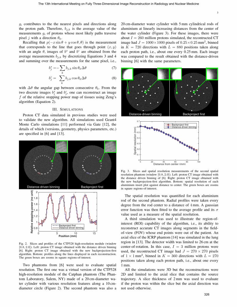
20 cm-diameter water cylinder with 5 mm cylindrical rods ofaluminium at linearly increasing distances from the center ofthe water cylinder (Figure 3). For these images, there wereabout I = 260 million protons simulated, the recontructed CTimage had J = 1000×1000 pixels of 0.25×0.25 mm2, binnedin K = 720 directions with L = 880 positions taken alongeach proton path, i.e., about one every 0.25 mm. Each imagewas compared to the result obtained with the distance-drivenbinning [6] with the same parameters.
Backproject firstDistance-driven binning
Distance from center (mm)0 20 40 60 80 100
Spatial re
solu
tion (
mm
)
0
0.1
0.2
0.3
0.4
0.5
0.6
0.7
Backproject firstDistance-driven binning
Fig. 3. Slices and spatial resolution measurements of the second spatialresolution phantom (window [0.8, 2.0]). Left: proton CT image obtained withthe distance driven binning of [6]. Right: proton CT image obtained withthe new backprojection-first algorithm. Bottom: spatial resolution of eachaluminium insert plot against distance to center. The green boxes are zoomsin square regions-of-interest.
The spatial resolution was quantified for each aluminiumrod of the second phantom. Radial profiles were taken everydegree from the rod center to a distance of 4 mm. A gaussianerror function was then fitted to the average profile and its σvalue used as a measure of the spatial resolution.
A third simulation was used to illustrate the region-of-interest (ROI) capability of the algorithm, i.e., its ability toreconstruct accurate CT images along segments in the field-of-view (FOV) whose end points were out of the patient. Anaxial slice of the ICRP phantom [14] was simulated in the lungregion in [13]. The detector width was limited to 26 cm at thecenter-of-rotation. In this case, I = 3 million protons wereused, the recontructed CT image had J = 270 × 270 pixelsof 1 × 1 mm2, binned in K = 360 directions with L = 270positions taken along each proton path, i.e., about one every1 mm.
All the simulations were 3D but the reconstructions were2D and limited to the axial slice that contains the sourcetrajectory. A slice thickness of 2 mm was used to evaluateif the proton was within the slice but the axial direction wasnot used otherwise.
The 13th International Meeting on Fully Three-Dimensional Image Reconstruction in Radiology and Nuclear Medicine
326
4
IV. RESULTS
The new backprojection-first algorithm gave very similarresults to the distance-driven binning method of [6]. Theprofiles of Figure 2 show different noise patterns but theresolution features are equally visible with both reconstruc-tion algorithms. The quantification of the spatial resolutionconfirmed these observations (Figure 3). Also, the spatialresolution was better for the rods that were closer to thephantom surface, a behavior that has been observed withiterative reconstruction as well [4] and which can be linked tothe difference between the estimated proton path Γi and itsreal path Γi. This difference increased with the distance to thepairs of position-sensitive detectors and was maximal near thecenter of the scanned object.
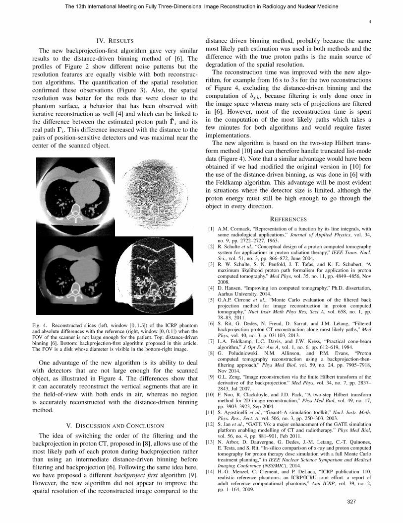
Fig. 4. Reconstructed slices (left, window [0, 1.5]) of the ICRP phantomand absolute differences with the reference (right, window [0, 0.1]) when theFOV of the scanner is not large enough for the patient. Top: distance-drivenbinning [6]. Bottom: backprojection-first algorithm proposed in this article.The FOV is a disk whose diameter is visible in the bottom-right image.
One advantage of the new algorithm is its ability to dealwith detectors that are not large enough for the scannedobject, as illustrated in Figure 4. The differences show thatit can accurately reconstruct the vertical segments that are inthe field-of-view with both ends in air, whereas no regionis accurately reconstructed with the distance-driven binningmethod.
V. DISCUSSION AND CONCLUSION
The idea of switching the order of the filtering and thebackprojection in proton CT, proposed in [8], allows use of themost likely path of each proton during backprojection ratherthan using an intermediate distance-driven binning beforefiltering and backprojection [6]. Following the same idea here,we have proposed a different backproject first algorithm [9].However, the new algorithm did not appear to improve thespatial resolution of the reconstructed image compared to the
distance driven binning method, probably because the samemost likely path estimation was used in both methods and thedifference with the true proton paths is the main source ofdegradation of the spatial resolution.
The reconstruction time was improved with the new algo-rithm, for example from 16 s to 3 s for the two reconstructionsof Figure 4, excluding the distance-driven binning and thecomputation of bj,k, because filtering is only done once inthe image space whereas many sets of projections are filteredin [6]. However, most of the reconstruction time is spentin the computation of the most likely paths which takes afew minutes for both algorithms and would require fasterimplementations.
The new algorithm is based on the two-step Hilbert trans-form method [10] and can therefore handle truncated list-modedata (Figure 4). Note that a similar advantage would have beenobtained if we had modified the original version in [10] forthe use of the distance-driven binning, as was done in [6] withthe Feldkamp algorithm. This advantage will be most evidentin situations where the detector size is limited, although theproton energy must still be high enough to go through theobject in every direction.
REFERENCES
[1] A.M. Cormack, “Representation of a function by its line integrals, withsome radiological applications,” Journal of Applied Physics, vol. 34,no. 9, pp. 2722–2727, 1963.
[2] R. Schulte et al., “Conceptual design of a proton computed tomographysystem for applications in proton radiation therapy,” IEEE Trans. Nucl.Sci., vol. 51, no. 3, pp. 866–872, June 2004.
[3] R. W. Schulte, S. N. Penfold, J. T. Tafas, and K. E. Schubert, “Amaximum likelihood proton path formalism for application in protoncomputed tomography.” Med Phys, vol. 35, no. 11, pp. 4849–4856, Nov2008.
[4] D. Hansen, “Improving ion computed tomography,” Ph.D. dissertation,Aarhus University, 2014.
[5] G.A.P. Cirrone et al., “Monte Carlo evaluation of the filtered backprojection method for image reconstruction in proton computedtomography,” Nucl Instr Meth Phys Res, Sect A, vol. 658, no. 1, pp.78–83, 2011.
[6] S. Rit, G. Dedes, N. Freud, D. Sarrut, and J.M. Letang, “Filteredbackprojection proton CT reconstruction along most likely paths,” MedPhys, vol. 40, no. 3, p. 031103, 2013.
[7] L.A. Feldkamp, L.C. Davis, and J.W. Kress, “Practical cone-beamalgorithm,” J Opt Soc Am A, vol. 1, no. 6, pp. 612–619, 1984.
[8] G. Poludniowski, N.M. Allinson, and P.M. Evans, “Protoncomputed tomography reconstruction using a backprojection-then-filtering approach,” Phys Med Biol, vol. 59, no. 24, pp. 7905–7918,Nov 2014.
[9] G.L. Zeng, “Image reconstruction via the finite Hilbert transform of thederivative of the backprojection.” Med Phys, vol. 34, no. 7, pp. 2837–2843, Jul 2007.
[10] F. Noo, R. Clackdoyle, and J.D. Pack, “A two-step Hilbert transformmethod for 2D image reconstruction,” Phys Med Biol, vol. 49, no. 17,pp. 3903–3923, Sep 2004.
[11] S. Agostinelli et al., “Geant4-A simulation toolkit,” Nucl. Instr. Meth.Phys. Res., Sect. A, vol. 506, no. 3, pp. 250–303, 2003.
[12] S. Jan et al., “GATE V6: a major enhancement of the GATE simulationplatform enabling modelling of CT and radiotherapy.” Phys Med Biol,vol. 56, no. 4, pp. 881–901, Feb 2011.
[13] N. Arbor, D. Dauvergne, G. Dedes, J.-M. Letang, C.-T. Quinones,E. Testa, and S. Rit, “In-silico comparison of x-ray and proton computedtomography for proton therapy dose simulation with a full Monte Carlotreatment planning,” in IEEE Nuclear Science Symposium and MedicalImaging Conference (NSS/MIC), 2014.
[14] H.-G. Menzel, C. Clement, and P. DeLuca, “ICRP publication 110.realistic reference phantoms: an ICRP/ICRU joint effort. a report ofadult reference computational phantoms,” Ann ICRP, vol. 39, no. 2,pp. 1–164, 2009.
The 13th International Meeting on Fully Three-Dimensional Image Reconstruction in Radiology and Nuclear Medicine
327