liposuction is an effective treatment for lipedemaresults...
TRANSCRIPT

© The Authors • Journal compilation © Blackwell Verlag GmbH, Berlin • JDDG • 1610-0379/2011/0901 JDDG | 1˙2011 (Band 9)
JDDG; 2011 • 9:33–40 Submitted: 3.5.2010 | Accepted: 29.6.2010
Keywords• lipedema• lipolymphedema• lipohypertrophy• liposuction• life quality• surgery• lipomatosis
SummaryBackground: Lipedema is a painful, genetically induced abnormal deposition ofsubcutaneous fat in the extremities of almost exclusively women. The pathogen-esis is unknown and no curative treatment is available. Conservative therapy con-sisting of lymphatic drainage and compression stockings is often recommended,but is only effective against the edema. Some patients show a short-termimprovement when treated in this way. The removal of the increased fat tissue oflipedema has become possible by employing advanced liposuction techniqueswhich utilize vibrating microcannulas under tumescent local anesthesia. Theeffectiveness of this approach to lipedema is the subject of this study. Patients and Methods: 25 patients were examined before liposuction and sixmonths thereafter. The survey included the measurement of the volume of thelegs and several parameters of typical pain and discomfort. The parameterswere measured using visual analogue scales (VAS, scale 0–10). Results: The volume of the leg was reduced by 6.9 %. Pain, as the predominantsymptom in lipedema, was significantly reduced from 7.2 ± 2.2 to 2.1 ± 2.1(p < 0.001). Quality of life as a measure of the psychological strain caused bylipedema improved from 8.7 ± 1.7 to 3.6 ± 2.5 (p < 0.001). Other parametersalso showed a significant improvement and the over-all severity scoreimproved in all patients.Conclusion: Liposuction reduces the symptoms of lipedema significantly.
Liposuction is an effective treatment for lipedema –results of a study with 25 patientsStefan Rapprich, Anne Dingler, Maurizio PoddaDepartment of Dermatology, Darmstadt Hospital, Germany
IntroductionLipedema is a painful, hereditary disorderusually affecting women that involves ac-cumulation of excess fatty tissue on theextremities [1–3]. Characteristic symp-toms include pain as well as sensitivity totouch and pressure. Patients also tend tobruise easily after minimal trauma. Overtime, the disorder progressively worsens.Synonyms for lipedema include lipalgia,adiposalgia, adipositas dolorosa, lipomato-sis dolorosa, lipohypertrophia dolorosa,and painful fat syndrome.The diagnosis is based on clinical ap-pearance. Lipedema should be differenti-ated from lipohypertrophy and lym-
phedema. Lipedema may be divided intothree types: whole leg, thigh, and lowerleg lipedema. In about 30 % of patients,there is also involvement of the arms [4].The disorder occurs in three stages:• Stage I: Thickening and softening of
the subcutis with small nodules; skin issmooth.
• Stage II: Thickening and softening ofthe subcutis with larger nodules; skintexture is uneven.
• Stage III: Thickening and hardening of the subcutis with large nodules, disfiguring lobules of fat on the inner thighs and inner aspects of theknees.
The cause of lipedema is unknown. Hormones are certainly one factor, as lipedema occurs virtually exclusively inwomen. In addition, early signs of diseasetend to appear with the onset of pubertyor after pregnancy. During these stages,the disease may also be referred to as lipohypertrophy which may develop into lipedema. Full-blown symptomatic diseaseusually manifests in the third or fourthdecade of life. In addition to hormonalfactors, a genetic disposition may be pre-sumed, as the disease often affects severalwomen in the same family (Figure 1).An important factor in the pathophysiol-ogy of lipedema is increased capillary
DOI: 10.1111/j.1610-0387.2010.07504.x Original Article 33

34 Original Article Liposuction in lipedema
JDDG | 1˙2011 (Band 9) © The Authors • Journal compilation © Blackwell Verlag GmbH, Berlin • JDDG • 1610-0379/2011/0901
permeability leading to orthostaticedema. This, and not the amount of adi-pose tissue, is responsible for the in-creased sensitivity of the tissue to touchand pressure. The increased capillaryfragility also explains the tendency tohematoma development.Lymph drainage is undisrupted. Indeed,it is even increased in the early stages oflipedema. In later stages, the capacity ofthe lymphatic system is exhausted andcan no longer ensure adequate drainage.This results in dynamic insufficiency.With decompensation of the lymphaticsystem, secondary lymphedema devel-ops. In clinical terms this is known aslipolymphedema – with all related se-quelae including leg ulcers. There are nocharacteristic histological changes associ-ated with the disease. The incidence of disease among women isestimated at 11 %. Among patients hospi-talized for lymphatic disease, the propor-tion is reportedly 8–17 % [3, 5, 6].Complex physical therapy (CPT),which is widely recommended, is onlyeffective against the edema. Only somepatients actually experience an improve-ment in symptoms, and then only for ashort period of time following each treat-ment session [6–10].The removal of excess fatty tissue usingliposuction has been made possible bymicrocannulae and – in a more advancedform – with vibrating cannulae undertumescent local anesthesia (Figure 2) [6,11–17]. This method, which has beenused for more than 15 years at the De-partment of Dermatology at DarmstadtHospital, is the focus of this study. Vari-ous measurement parameters were usedto assess the status of disease before lipo-suction and 6 months after to evaluate itseffectiveness in lipedema.
Patients and methodsBetween April 2006 and July 2008 (27 months), 105 patients with lipedemawere treated in our dermatology unit.The diagnosis of lipedema was con-firmed in all patients included in thestudy on the basis of guideline criteria[2]. Of the 105 patients, 25 could be followed-up at 6 months after the last liposuction procedure and the resultsevaluated.Among the remaining 80 patients, vari-ous reasons made it impossible to in-clude their results in the final evaluation.In some, therapy had not been con-cluded at the time of evaluation, or the
Figure 1: Mother and her daughters with lipedema.
Figure 2: Lower legs after tumescent anaesthesia and before liposuction by vibrating cannulas with4 mm diameter.
Figure 3: Volume of the legs measured by Image-3D-system (Fa. Bauerfeind). A digital camera takesphotos of the leg at different points. A computer calculates an exact 3D model of the leg with the cor-responding measurements.

follow-up visit at 6 months after the procedure had not yet taken place, or liposuction therapy was not performeddue to insurance coverage issues.At the time of the first liposuction session, all patients were between theages of 22 and 65 years (mean 38.0 ±12.5 years, median 34.0 years). Twentypatients had lipedema affecting thewhole leg, 3 had lipedema of the thigh,and 2 had lower leg involvement only [4, 5, 18].Clinical examination included height,weight, waist circumference, leg volumemeasurement using 3D imaging (Figure 3),and a self-assessment of symptoms (Figure 4). The self-assessment was basedon a quality of life survey for patientswith lymphatic diseases (FLQA-l survey
Liposuction in lipedema Original Article 35
© The Authors • Journal compilation © Blackwell Verlag GmbH, Berlin • JDDG • 1610-0379/2011/0901 JDDG | 1˙2011 (Band 9)
on quality of life in patients with lym-phatic diseases, Version 1.1, Augustin,Zschocke et al. 1998). This was modifiedto include 15 criteria that were assessedby the patient using a visual analoguescale (VAS) of 0 to 10. The survey wascompleted prior to beginning therapyand again at 6 months after the final liposuction treatment.Liposuction was performed with undertumescent local anesthesia with vibratingcannulae with a 4 mm diameter (Figure 2) and a handpiece (VibraSat®,Möller Medical, Fulda). The tumescencesolution used was based on Sattler [17]as follows: prilocaine: 1 % solution 50 ml,suprarenin: 1 : 1 000 1 ml, sodium bicar-bonate: 8.4 % 6 ml, triamquinolone:10 mg/ml 1 ml, sodium chloride solution
0.9 % 1 000 ml. The prilocaine concentra-tion in this preparation was about 0.05 %.In most patients, about 6 000 ml tumes-cence solution were infiltrated per ses-sion, with a maximum of 7 000 ml, anda minimum of 2 000 ml. The mean was5 155 ± 1 304 ml. The infiltration waswith a roll pump with a closed tube sys-tem (LipoSat®, Möller Medical, Fulda)and an adaptor for 6 cannulae (No. 1,long). The rate of infiltration was, de-pending on the region, severity, and indi-vidual tolerance to pain, between 120and 200 ml per minute. Sedation wasused if necessary, and consisted of5–10 mg diazepam i.v. For aspiration, vibrating cannulae with 3 blunt open-ings at the tip in a Mercedes star shape(Figure 2) were used.
Figure 4: Questionnaire for measuring disorders of lipedema by visual analogue scales and results.

Patients were treated in 1 to 5 sessions(mean 2.5 ± 1.1, median 2).The following regions on the body werecombined and treated symmetrically:• Medial aspects of the thighs and inner
aspects of the knees• Lateral aspects of the thighs and hip in
the same or an additional session • For larger-volume thighs, the anterior
aspects were also treated• Lower legsThree sessions at 4-week intervals weregenerally needed. For larger-volumethighs, 4 sessions (3 patients) or 5 ses-sions (1 patient) were required. Thenumber of necessary sessions was deter-mined by the mass of adipose tissue that
could be treated in a single session with6 000 ml tumescence solution in relationto the entire affected area.Therapy usually began with the medialaspects of the thighs and knees or withthe area that was causing the greatest dis-comfort. Two patients received inpatient treat-ment, and 23 patients were treated on anoutpatient basis. For each session, the aspiration volume was an average of2 482 ± 968 ml and the pure fat compo-nent was on average 1 909 ± 874 ml, or 77 %.Following liposuction, patients weregiven antibiotic prophylaxis for 3 daysconsisting of ciprofloxacin 2 � 250 mg
or cefuroxime 2 � 250 mg as well asthrombosis prophylaxis consisting ofenoxaparin 1 � 40 mg injected subcuta-neously for 5 days. For patients who un-derwent treatment of the lower legs, aswell as those with a history or family his-tory of thrombophilia, a 10-day regimenof enoxaparin 1 � 40 mg was adminis-tered (subcutaneous injection).Compression was performed during thefirst 7 days after liposuction for 24 hours.Afterward, compression therapy continuedduring the daytime only for 4–6 weeks.This consisted of wearing compressionstockings which the majority of patientsalready owned (76 % of patients; see below). The remainder were providedwith stockings prior to the operation.For liposuction of the lower legs, duringthe first 2–3 days a circumferential com-pression dressing was applied. Startingon the third day after liposuction, manuallymph drainage was performed 2–3-timesper week for at least 6 weeks.
Statistical methodsStatistical analysis was performed withSPSS for Windows, Version 15.0 (SPSSInc., U.S.A.). Continuous variables weredisplayed as averages; standard deviationwas used for dispersion.The continuous variables were tested fornormal distribution using the Shapiro-Wilk tests. The vast majority of testedvariables did not have a normal distribu-tion (Shapiro-Wilk test: p < 0.05). Whencomparing the samples, non-parametrictests were chosen in every case for samples that did not have normal distribution.For comparing more than two independ-ent samples with abnormal distributionwe used the H-test after Kruskal and Wal-lis. Wilcoxon tests were used fo assess sta-tistically significant differences in pairedsamples with abnormal distribution. Thecorrelation between 2 parameters was calculated with the correlation coefficientafter Spearman-Rho. The correlation co-efficient was evaluated as follows: r < 0.2:very low correlation; r = 0.2–0.5: low cor-relation; r = 0.5–0.7: moderate correla-tion; r = 0.7–0.9: high correlation; r = >0.9: very high correlation.In all of the tests performed, we also didtwo-sided significance testing. A p-value< 0.05 for statistical significance wasused in all tests.In the graphics, which were also createdusing SPSS, error bars were used to show
36 Original Article Liposuction in lipedema
JDDG | 1˙2011 (Band 9) © The Authors • Journal compilation © Blackwell Verlag GmbH, Berlin • JDDG • 1610-0379/2011/0901
Figure 5: Significant reduction of pain before and 6 month post liposuction.
Figure 6: Significant improvement of quality of life before and 6 month post liposuction.

© The Authors • Journal compilation © Blackwell Verlag GmbH, Berlin • JDDG • 1610-0379/2011/0901 JDDG | 1˙2011 (Band 9)
Liposuction in lipedema Original Article 37
corresponds to an average reduction ofleg volume of 1.2 ± 1.0 l, or 6.9 %. Thestarting values for leg volume rangedwidely from 13.0 to 28.7 l. The reduc-tion in volume was between 0.16–4.0 l,corresponding to a relative volume re-duction of 0.9 to 19.8 %.
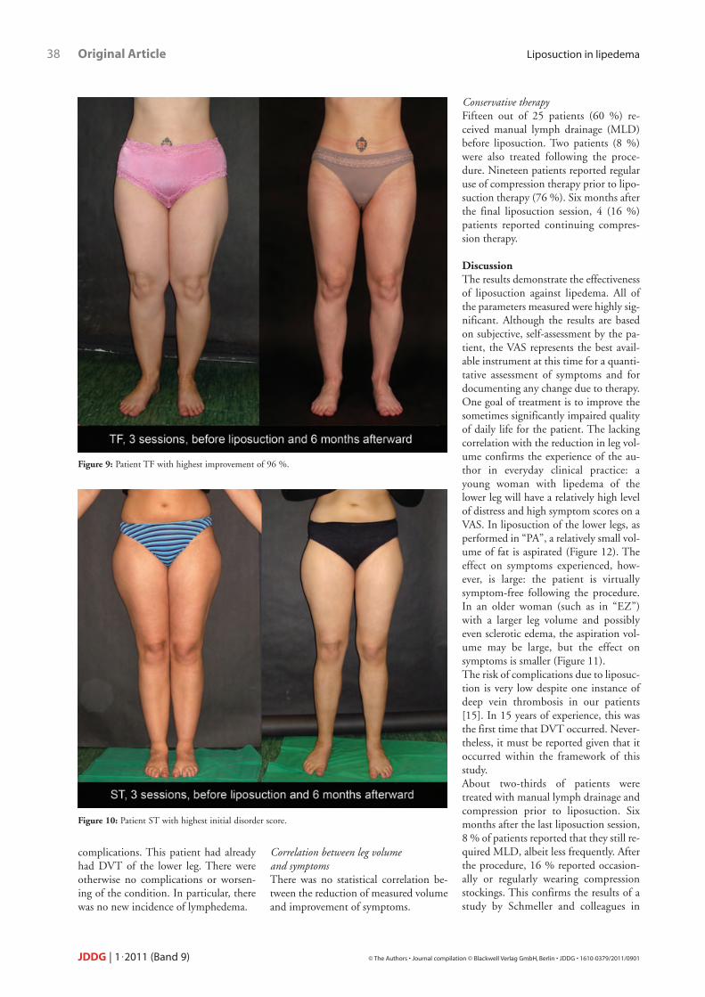
SymptomsThe results of self-assessment of symp-toms (Figure 4) indicate a significant orhighly significant improvement in all ar-eas. With regard to pain, the chief symp-tom of lipedema, there was an improve-ment of 7.2 ± 2.2 to 2.1 ± 2.1. Atp < 0.001 the improvement is highly sig-nificant (Figure 5). There was also signif-icant improvement in sensitivity to pres-sure, which is typical of lipedema, andbruising.The patient burden was described in partusing the question on quality of life impairment (Figure 6). Results showed a highly significant improvement from8.7 ± 1.7 to 3.6 ± 2.5. Patient satisfac-tion with the appearance of the legs alsoimproved notably.The 15 symptoms parameters were com-bined for a total score (highest value of150). The averages before liposuctionwere 92.0 ± 21.3, and 6 months after-ward they were 39.0 ± 23.2 (p < 0.001).This corresponds to about a 58 % im-provement (Figure 7).In the individual analysis (Figure 8),the patient we will refer to as “TF”(Figure 9) experience the greatest im-provement (96 %) in symptoms. Theseverest symptoms (baseline score of133) were reported by “ST” (Figure 10)who had about a 64 % improvementafter liposuction. The smallest im-provement was reported by “EZ” (Figure 11) who had a score of 21 %.The greatest reduction in pain, from 8 to 0 on the VAS, was reported by“PA” (Figure 12) with a relatively smallaspiration volume of 1 700 ml from thelower legs.
Results and complicationsAll patients reported improvement insymptoms after liposuction therapy. Dis-proportion of the legs also improved.Despite preventive measures one patientexperienced deep vein thrombosis(DVT) of the lower leg one week afterthe procedure. This was treatedpromptly and there were no further
the box. They are depicted in the graph-ics as circles, while extremes that aremore than 3 box lengths outside of thebox are shown as small crosses. The datawere displayed using simple and groupedbar graphs.
ResultsLeg volumeIn 25 patients studied, 3D imaging of legvolume showed a reduction in leg vol-ume of 18.0 ± 3.8 to 16.8 ± 3.5 l. This
the means in samples with normal distri-bution. Given the large distributionrange, the standard error was used as thedistribution measure. Box plots wereused to show the median values andquartiles in samples with abnormal dis-tribution. In the boxes, the median aswell as 25th-75th percentile is shown.The T-bars correspond to the smallestand largest values (as long as they werenot outliers or extremes). Outliers areany values 1½–3 box lengths outside of
Figure 7: Significant improvement of the disorder score before and 6 month post liposuction.
Figure 8: Different improvement oft he disorder score: analysis of exemplary cases.
complications. This patient had alreadyhad DVT of the lower leg. There wereotherwise no complications or worsen-ing of the condition. In particular, therewas no new incidence of lymphedema.
Correlation between leg volume and symptomsThere was no statistical correlation be-tween the reduction of measured volumeand improvement of symptoms.
Conservative therapyFifteen out of 25 patients (60 %) re-ceived manual lymph drainage (MLD)before liposuction. Two patients (8 %)were also treated following the proce-dure. Nineteen patients reported regularuse of compression therapy prior to lipo-suction therapy (76 %). Six months afterthe final liposuction session, 4 (16 %)patients reported continuing compres-sion therapy.
DiscussionThe results demonstrate the effectivenessof liposuction against lipedema. All ofthe parameters measured were highly sig-nificant. Although the results are basedon subjective, self-assessment by the pa-tient, the VAS represents the best avail-able instrument at this time for a quanti-tative assessment of symptoms and fordocumenting any change due to therapy.One goal of treatment is to improve thesometimes significantly impaired qualityof daily life for the patient. The lackingcorrelation with the reduction in leg vol-ume confirms the experience of the au-thor in everyday clinical practice: ayoung woman with lipedema of thelower leg will have a relatively high levelof distress and high symptom scores on aVAS. In liposuction of the lower legs, asperformed in “PA”, a relatively small vol-ume of fat is aspirated (Figure 12). Theeffect on symptoms experienced, how-ever, is large: the patient is virtuallysymptom-free following the procedure.In an older woman (such as in “EZ”)with a larger leg volume and possiblyeven sclerotic edema, the aspiration vol-ume may be large, but the effect onsymptoms is smaller (Figure 11).The risk of complications due to liposuc-tion is very low despite one instance ofdeep vein thrombosis in our patients[15]. In 15 years of experience, this wasthe first time that DVT occurred. Never-theless, it must be reported given that itoccurred within the framework of thisstudy.About two-thirds of patients weretreated with manual lymph drainage andcompression prior to liposuction. Sixmonths after the last liposuction session,8 % of patients reported that they still re-quired MLD, albeit less frequently. Afterthe procedure, 16 % reported occasion-ally or regularly wearing compressionstockings. This confirms the results of astudy by Schmeller and colleagues in
38 Original Article Liposuction in lipedema
JDDG | 1˙2011 (Band 9) © The Authors • Journal compilation © Blackwell Verlag GmbH, Berlin • JDDG • 1610-0379/2011/0901
Figure 9: Patient TF with highest improvement of 96 %.
Figure 10: Patient ST with highest initial disorder score.

2007 [10], which found that the majorityof patients no longer require prolongedfurther therapy after liposuction.After liposuction, combination therapywith MLD and compression therapy [2, 6–8, 10] is an essential part of thehealing process and treatment success.
Without a doubt, many lipedema pa-tients benefit from compression therapyif the edema is symptomatic. The effectonly persists as long as it is used, however, and this means patients requirelifelong therapy. Liposuction offers thepossibility of being able to go without
CPT for longer periods of time, as it re-duces both the edema and pathologicallyaltered adipose tissue [19]. Still, liposuction can only reduce theamount of fatty tissue, but not completelyremove it. Given that lipedema is achronic disease, depending on the amountof remaining fatty tissue, the disease maycontinue to progress. Nevertheless, lipo-suction for lipedema is the only therapyknown until now that succeeds in reduc-ing edema and symptoms such as pain.It is still too soon to assess long-term recurrence. The follow-up period of 6 months in this study is too short forthis. Further studies are needed to address this issue. In our own experience[14] the results may be expected to lastfor at least 8 years.
Conclusions for clinical practiceWhen performed by an experiencedpractitioner, tumescent liposuction is asafe and effective method of treatmentfor lipedema. The results of therapy arebetter in younger patients with early-stage disease compared with more severedisease in older patients. CPT, beforeand after liposuction, is an importantpart of therapy. <<<
Conflict of interestNone.
Correspondence to
Dr. med. Stefan RapprichHautklinikKlinikum DarmstadtHeidelberger Landstraße 379D-64297 DarmstadtTel.: +49-6151-107-955-4133Fax: +49-6151-107-4150E-mail: [email protected]
References1 Allen EV, Hines EA. Lipedema of the
legs. Proc Mayo Clin 1940; 15: 184–7.
Liposuction in lipedema Original Article 39
© The Authors • Journal compilation © Blackwell Verlag GmbH, Berlin • JDDG • 1610-0379/2011/0901 JDDG | 1˙2011 (Band 9)
Figure 11: Patient EZ with lowest improvement of 21 %.
Figure 12: Patient PA with maximum improvement of pain from 8 to 0.

2 AWMF. Leitlinien der Deutschen Gesellschaft für Phlebologie (DGP):Lipödem. AWMF-Leitlinien-RegisterNr. 037/012. 2009.
3 Schmeller W, Meier-Vollrath I. Lipödem – Aktuelles zu einem weitge-hend unbekannten Krankheitsbild. AktDermatol 2007; 33: 1–10.
4 Herpertz U. Ödeme und Lymphdrai-nage. Diagnose und Therapie vonÖdemkrankheiten. Schattauer, Stutt-gart, New York, 2003.
5 Herpertz U. Krankheitsspektrum desLipödems an einer LymphologischenFachklinik – Erscheinungsformen,Mischbilder und Behandlungsmöglich-keiten. Vasomed 1997; 5: 301–7.
6 Meier-Vollrath I, Schneider W, Schmeller W. Lipödem: VerbesserteLebensqualität durch Therapiekombi-nation. Deutsches Ärzteblatt 2005;102: A-1061–67.
7 Schmeller W, Meier-Vollrath I. Erfolg-reiche operative Therapie des Lipödems
mittels Liposuktion. Phlebologie 2004;33: 23–9.
8 Schmeller W, Meier-Vollrath I. ModerneTherapie des Lipödems: Kombinationvon konservativen und operativen Maß-nahmen. Lymph-Forsch 2004; 8: 22–6.
9 Schmeller W, Meier-Vollrath I. Tumes-cent liposuction: a new and successfultherapy for lipedema. J Cutan MedSurg 2006; 10: 7–10.
10 Schmeller W, Meier-Vollrath I. DasLipödem: neue Möglichkeiten der Therapie. Schweiz Med Forum 2007;7: 150–5.
11 Kaufmann R, Landes E, Podda M.Dermatologische Operationen. 3. Auf-lage, Thieme Verlag, Stuttgart, NewYork, 2005.
12 Rompel R, Petres J. Operative Derma-tologie. 2. Auflage, Springer Verlag,Heidelberg, 2007.
13 Cornely ME. Lipödem und die Diffe-rentialdiagnosen. Journal für Lympho-logie 2001; 1: 32–3.
14 Rapprich S, Loehnert M, Hagedorn M.Therapy of lipoedema syndrome by li-posuction under tumescent local ana-esthesia. Ann Dermatol Venereol 2002;129: 1S711.
15 Sattler G, Bergfeld D, Sommer B. [Lipo-suction]. Hautarzt 2004; 55: 599–604.
16 Sattler G, Hasche E, Rapprich S et al.Neue operative Behandlungsmöglich-keiten bei benignen Fettgewebserkran-kungen. Z Hautkr 1997; 72: 579–82.
17 Sattler G, Rapprich S, Hagedorn M.Tumeszenz-Lokalanästhesie – Untersu-chung zur Pharmakokinetik von Prilo-cain. Z Hautkr 1997; 7: 522–5.
18 Herpertz U. Die häufigsten Beinö-deme. Differenzierung zwischen Phle-bödem, Lymphödem und Lipödem.Phlebologie 2001; 30: 48–52.
19 Langendoen SI, Habbema L, NijstenTE, Neumann HA. Lipoedema: fromclinical presentation to therapy. A re-view of the literature. Br J Dermatol2009; 161(5): 980–6.
40 Original Article Liposuction in lipedema
JDDG | 1˙2011 (Band 9) © The Authors • Journal compilation © Blackwell Verlag GmbH, Berlin • JDDG • 1610-0379/2011/0901