tumescent liposuction - elsevier health
TRANSCRIPT
HISTORY AND SCIENCE
Background and history
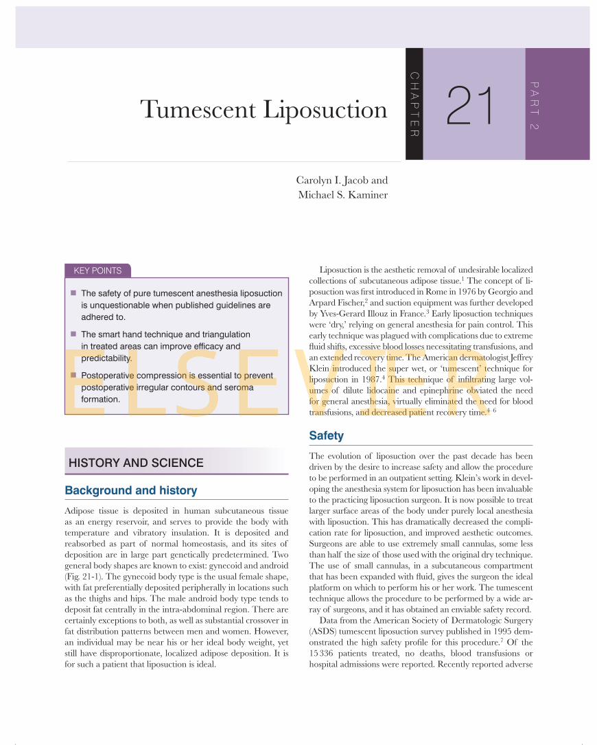
Adipose tissue is deposited in human subcutaneous tissue
as an energy reservoir, and serves to provide the body with
temperature and vibratory insulation. It is deposited and
reabsorbed as part of normal homeostasis, and its sites of
deposition are in large part genetically predetermined. Two
general body shapes are known to exist: gynecoid and android
( Fig. 21-1 ). The gynecoid body type is the usual female shape,
with fat preferentially deposited peripherally in locations such
as the thighs and hips. The male android body type tends to
deposit fat centrally in the intra-abdominal region. There are
certainly exceptions to both, as well as substantial crossover in
fat distribution patterns between men and women. However,
an individual may be near his or her ideal body weight, yet
still have disproportionate, localized adipose deposition. It is
for such a patient that liposuction is ideal.
Liposuction is the aesthetic removal of undesirable localized
collections of subcutaneous adipose tissue. 1 The concept of li-
posuction was fi rst introduced in Rome in 1976 by Georgio and
Arpard Fischer, 2 and suction equipment was further developed
by Yves-Gerard Illouz in France. 3 Early liposuction techniques
were ‘dry,’ relying on general anesthesia for pain control. This
early technique was plagued with complications due to extreme
fl uid shifts, excessive blood losses necessitating transfusions, and
an extended recovery time. The American dermatologist Jeffrey
Klein introduced the super wet, or ‘tumescent’ technique for
liposuction in 1987. 4 This technique of infi ltrating large vol-
umes of dilute lidocaine and epinephrine obviated the need
for general anesthesia, virtually eliminated the need for blood
transfusions, and decreased patient recovery time. 4 – 6
Safety
The evolution of liposuction over the past decade has been
driven by the desire to increase safety and allow the procedure
to be performed in an outpatient setting. Klein’s work in devel-
oping the anesthesia system for liposuction has been invaluable
to the practicing liposuction surgeon. It is now possible to treat
larger surface areas of the body under purely local anesthesia
with liposuction. This has dramatically decreased the compli-
cation rate for liposuction, and improved aesthetic outcomes.
Surgeons are able to use extremely small cannulas, some less
than half the size of those used with the original dry technique.
The use of small cannulas, in a subcutaneous compartment
that has been expanded with fl uid, gives the surgeon the ideal
platform on which to perform his or her work. The tumescent
technique allows the procedure to be performed by a wide ar-
ray of surgeons, and it has obtained an enviable safety record .
Data from the American Society of Dermatologic Surgery
(ASDS) tumescent liposuction survey published in 1995 dem-
onstrated the high safety profi le for this procedure. 7 Of the
15 336 patients treated, no deaths, blood transfusions or
hospital admissions were reported. Recently reported adverse
Tumescent Liposuction
Carolyn I. Jacob and
Michael S. Kaminer C
HA
PT
ER
21P
AR
T 2
KEY POINTS
■ The safety of pure tumescent anesthesia liposuction is unquestionable when published guidelines are adhered to.
■ The smart hand technique and triangulation in treated areas can improve effi cacy and predictability.
■ Postoperative compression is essential to prevent postoperative irregular contours and seroma formation.
324 Chapter 21 Cosmetic surgery procedures and techniques
outcomes in the New England Journal of Medicine were reported
to be due to lidocaine toxicity, but careful inspection of the
data suggests that is likely to be an erroneous conclusion. 8 In
2002, a national survey reported no deaths in 66 570 liposuc-
tion procedures performed by dermasurgeons between 1994
and 2000. The overall rate of serious adverse events (SAE) was
0.68 per 1000 cases. The SAE rates were higher for hospitals
and ambulatory surgery centers than for non-accredited offi ce
settings. 9 A second study, performed by the Accreditation
Association for Ambulatory Health Care Institute for Quality
Improvement examined 688 cases from 39 organizations, and
found a major complication rate of 0.14%, with one patient
requiring hospitalization. 10 In 2005, Coldiron et al. examined
4 years of Florida data and found no adverse events with the
use of tumescent anesthesia alone in liposuction. 11
Ultrasonic liposuction, both internal and external, has
recently been developed and hailed by some to be superior to
the standard tumescent technique. However, experience has
shown that although ultrasound can be a useful adjunct to
tumescent liposuction, it has not replaced it as the treatment
of choice. The high cost of the ultrasound equipment, larger
incisions needed for internal ultrasound assisted liposuction
(UAL), and the steep learning curve have put UAL out of the
reach of many surgeons. For these reasons it will not be dis-
cussed further in this chapter. New machinery and techniques
are certain to evolve in time, but tumescent liposuction
remains the standard for liposuction surgery
Basic science
Lidocaine toxicity is the most signifi cant factor that limits the
amount of anesthesia used in tumescent liposuction. Tradi-
tional dosing of lidocaine with epinephrine for dermal or
local infusion is 7 mg/kg. 12 Using the tumescent technique,
lidocaine doses of 35 mg/kg were shown to be safe and
effective, 13 and a second study has shown that dosages up to
55 mg/kg can be used with minimal risk of lidocaine toxicity. 14
That same study also suggests that even higher doses of lido-
caine can be used, and some liposuction surgeons around the
country have safely used lidocaine in doses of 70 – 80 mg/kg
and higher. The very dilute nature of the lidocaine (0.05 –
0.1%) in the tumescent solution, the slow rate of infi ltration,
the relatively avascular subcutaneous fat compartment, the
vasoconstrictive effect of epinephrine, the high lipid solubil-
ity of lidocaine and its strong binding affi nity to adipose tis-
sue, and the vascular compression due to tissue tumescence
all combine to delay systemic uptake of lidocaine. 13,15 – 17 The
peak plasma concentration has been shown to be 8 – 12 hours
after infusion, and estimated by the following formula: 14
Peak plasma lidocaine concentration ( g/ml)= [dose (mg)/1000
μ]] 1.25−
Lillis initially reported on the safe use of lidocaine doses of
60 – 90 mg/kg; 5 however, this was based on plasma lidocaine
levels monitored over a 60-minute period. Ostad et al. have
shown in 60 patients that liposuction with a lidocaine dose of
55 mg/kg is safe, with no evidence of lidocaine toxicity over a
24-hour period. 14 The peak plasma lidocaine concentrations
obtained from these patients was below 5 µ g/ml, under the
threshold for when recognizable signs of lidocaine toxicity
develop.
Lidocaine is an amide anesthetic which blocks sodium
channels, thus inhibiting the propagation of the neural
impulse. It is rapidly and effi ciently eliminated by hepatic
metabolism via the cytochrome P450 enzyme CYP3A4.
Patients should be screened for concomitant use of medi-
cations which are known P450 CYP3A4 inhibitors, such as
erythromycin or ketoconazole. 18 Lidocaine toxicity has been
reported in one patient taking sertraline hydrochloride (Zoloft)
and Flurazepam (Dalmane) who had liposuction performed
using 58 mg/kg tumescent lidocaine. At 10 hours, she had
clinical symptoms of nausea, vomiting, anxiety and impaired
short term memory with total blood plasma lidocaine concen-
tration of 6.3 µ g/mL. 18 Zoloft and other serotonin reuptake
inhibitors have been shown in vitro to inhibit the activity of
cytochrome P450 3A4 and 2D6. 19 In general, some physicians
recommend decreasing the lidocaine dose by 30 – 40% in
patients concurrently taking medications which interfere with
the P450 complex 18 ( Appendix A ). Whether this is actually
necessary in clinical practice has not been defi nitively deter-
mined.
The initial clinical manifestations of lidocaine toxicity are
perioral numbness or tingling ( Table 21-1 ). As lidocaine levels
rise, the patient may develop slurred speech, tinnitus, become
somnolent or confused. At terminally high levels, the patient
may have cardiac collapse. Lidocaine suppresses the myocar-
dium at a cellular level, depressing diastolic depolarization
and automaticity in the ventricles. 20
Fig. 21-1 Android (left) and gynecoid (right) body types. Pink areas denote zones of adipose deposition.

325Tumescent liposuction • Chapter 21
The advent of tumescent liposuction has eliminated the
need for intravenous fl uid replacement during the proce-
dure, and substantial fl uid shifts are not common with this
technique. Saline is used as the foundation for liposuction an-
esthesia, and when placed in the subcutaneous space is ab-
sorbed slowly into the microvasculature. This form of volume
replacement, known as hypodermoclysis, is the mechanism
for volume replacement during tumescent liposuction surgery.
Since the fl uid is absorbed slowly over hours and not minutes
(as with intravenous fl uids), it allows the patient to mobilize
and excrete fl uids at a rate controlled by normal homeostatic
mechanisms. This allows for long-term hydration of the pa-
tient over the immediate postoperative period, yet virtually
eliminates the risk of fl uid overload. However, the surgeon
must use caution when placing subcutaneous fl uids during
liposuction. It is possible to use such high volumes of local
anesthesia (greater than 5 – 6 liters of fl uid) that the patient is
at risk for volume overload, particularly patients with a history
of cardiac or renal disease.
Epinephrine has a threefold importance for tumescent
liposuction. It provides excellent hemostasis, slows the rate
of lidocaine absorption, and prolongs local analgesia. Unlike
lidocaine, there is no described limitation for epinephrine
dosing. 21 For treatment of anaphylaxis, the recommended
therapeutic dose is 0.01 mg/kg body weight. 22 When utilizing
the tumescent technique for liposuction, total epinephrine
doses as high as 10 mg have been used without adverse ef-
fects. 21 Epinephrine toxicity is initially manifest by patient
anxiety, agitation, or palpitations. With increased levels, hy-
pertension, tachycardia, or arrhythmias may occur. A study
of 20 patients undergoing liposuction, monitored at 3, 12,
and 23 hours after tumescent fl uid infi ltration, demonstrated
the peak serum epinephrine levels to occur at 3 hours. Peak
levels were three to fi ve times the upper limit of normal (nor-
mal resting values: 0 – 133 pg/mL; patients with pheochro-
mocytoma range 200 – 12 700 pg/mL). 23 The majority had
returned to normal at 12 hours. The only reported side effect
was anxiety. 21
Clinical studies
Liposuction is one of the most commonly performed aesthetic
surgical procedures. 24 In the United States 33 – 40% of adult
women, and 20 – 24% of adult men are trying to loose weight.
Another 28% of each group is trying to maintain its weight. 25
Whereas women cite appearance as more important than fi t-
ness, the reverse is true for men. 26 Women and men differ in
the areas most commonly treated ( Box 21-1 ).
Discussion
Indications for liposuction published by the ASDS are cos-
metic body contouring, diseases involving the subcutaneous
tissue (lipomas, lipodystrophy, axillary hyperhidrosis), and re-
construction. 1 Ideally, liposuction should be used in conjunc-
tion with an exercise program, and not as a substitution for
weight loss by diet control. Many patients pursue liposuction
for specifi c body locations which disturb them. For 87% of
female patients, their family history is a good predictor of
localized adipose deposition which is resistant to diet and
exercise. 27 The physician should take the patient’s wishes into
consideration, as well as the overall proportions of the patient.
Not only is proportion important, but contour, fl ow, and sym-
metry of body lines are an essential part of liposuction plan-
ning. Patients with localized irregular contours or localized fat
deposits are superb candidates for liposuction and tend to do
quite well. The surgeon’s role is to identify those body areas
that can be contoured with liposuction to create an overall aes-
thetic and contour improvement. Due to these global consid-
erations, liposuction has been referred to as liposculpting . This is
one of the most challenging aspects of liposuction surgery.
For some patients, skin laxity, muscle fl accidity or location
of fat pads deeper than the subcutis (i.e. intra-abdominal) may
not make them good liposuction candidates. One judges skin
laxity by the ‘snap test’ ( Fig. 21-2 ). To perform this test, the
surgeon pinches 1 – 3 cm of skin, retracts and releases. Ideally,
there should be instant recoil of the skin to its prior location.
Slow recoil or excess laxity may be an indicator that the
patient will have redundant skin folds or surface irregularities
after liposuction. This needs to be explained to the patient,
and in some cases, a combination of procedures may enable
the patient to obtain results they are anticipating. For exam-
ple, abdominoplasty in addition to abdominal liposuction can
improve the fi nal contour of the abdomen. However, risk and
Box 21-1 Most common areas of liposuction for men and women 27
Men Women
Flanks/love handles 56% Abdomen 55%
Abdomen 32% Outer thighs 38%
Neck/jowls 11% Hip/waist 10%
Breast 5% Neck/jowls 3%
Table 21-1 Total plasma lidocaine levels and clinical signs and symptoms 20
Total plasma lidocaine level ( µ g/mL)
Clinical signs and symptoms
<1.5 Idiosyncratic
1.5 – 4 Mild CNS
4 – 6 Mild CNS and cardiovascular toxicity
6 – 8 Major CNS and cardiovascular depression
>8 Seizures, hypotension, respiratory, and cardiac depression

326 Chapter 21 Cosmetic surgery procedures and techniques
morbidity are increased by the addition of abdominoplasty
to the surgical plan. 28,29 For this reason, recognition of those
patients at risk for poor skin retraction is essential. Age is not
the sole predictor of skin retraction, and even adults over age
40 can achieve smooth postoperative contours. 30 Locations
that are at particular risk for poor skin retraction (in the
patient with poor skin tone) are the neck, upper arms, lower
abdomen, inner and outer thighs. We refer to patients with
less than optimal skin elasticity as having soft skin . This can
be recognized as the preoperative appearance of cellulite and
dimples on the outer thigh, ridges or folds on the abdomen,
hanging neck skin, or wrinkled inner thigh skin. Patients with
soft skin must be treated with caution, and they should be
counseled as to the expected outcome of surgery. Aggressive
suctioning in these patients can produce less than optimal re-
sults. However, even patients with good skin tone and elastic-
ity can experience poor skin retraction, most commonly on
the upper abdomen, distal anterior thighs, and the anterior
axillae. In these patients the addition of monopolar RF tissue
tightening (Thermage, Hayward, CA) at the time of surgery
can increase skin retraction by almost twofold (personal
communication, R. Fitzpatrick, 2005).
Tumescent liposuction has evolved to be a very safe, repro-
ducible method for selective fat removal and body contouring.
A thorough understanding of the surgical concept, as well as
extensive training in the procedure, offer the surgeon a unique
opportunity to create aesthetically pleasing contours with fat
removal.
PROCEDURES AND TECHNIQUES
Preoperative clinical considerations
Through written information, informative video tapes, and
patient discussions, a physician strives to inform the patient
about the procedure of liposuction, including realistic goals,
expectations, and possible side effects and/or complica-
tions. Good surgical judgment must be used to determine
if the patient has realistic expectations and will participate
responsibly with pre- and postoperative care. As with other
cosmetic procedures, it is advisable to be wary of patients
who are over-anxious, demanding on staff, or the physician
thinks will only be satisfi ed if they obtain a perfect result. All
staff, from the front offi ce receptionists to the nursing staff,
should be encouraged to inform the physician of any idio-
syncrasies which they may notice. Patients with suspected
eating disorders or body dysmorphism should be referred for
appropriate counseling. 31
As with any procedure, liposuction requires the participa-
tion of both patient and physician. The initial patient inter-
view with careful history and physical examination should
prepare the patient and answer all of his/her questions. The
patient should be given information regarding what medica-
tions to avoid 2 weeks prior to the procedure ( Appendix B ),
and be given information regarding what to expect on the day
of the procedure. This helps to alleviate anxiety and minimize
confusion.
A thorough medical history is essential to evaluating undue
risk of bleeding, infection, emboli, thrombophlebitis, edema
and a history of past surgeries which may complicate the
technique. 1 Not only should current medications and medica-
tion allergies be recorded, but also any history of hepatitis,
hepatotoxic chemotherapy, and use of birth control pills or
cytochrome P450 competitors. A complete skin examination
should be done, noting adipose distribution, quality of skin
tone and elasticity as previously mentioned (i.e. snap test). All
treatment sites should be evaluated for pre-existing hernias,
varicosities, scars, asymmetry, or other fi ndings. Due to the
forces of gravity, evaluation should be done with the patient
standing. Some physicians also examine the patient sitting,
and in the supine position with the hip fl exed.
Fig. 21-2 The ‘snap test’ to determine skin laxity. The top fi gure represents the initial pinch of skin, and the surgeon observes recoil as the skin is released (bottom fi gure).

327Tumescent liposuction • Chapter 21
Laboratory studies are done to screen patients for general
health, bleeding disorders, and underlying disorders which
may affect the metabolism of medications used throughout the
procedure. Typical studies include liver function tests, hepatitis
profi le, electrolytes, complete blood count, prothrombin time,
partial thromboplastin time, and pregnancy test for premeno-
pausal women. Other tests may include urinalysis, bleeding
time, or infectious disease studies such as for the human
immunodefi ciency virus. 1
Pre- and postoperative care are truly part of the art of
medicine, and vary with each physician. The results of the
1995 ASDS study of physicians performing liposuction showed
88% started antibiotics 1 day preoperatively, of which over
80% used a cephalosporin. Oral sedation was used by 53%
of physicians, including valium (diazepam) and ativan (lo-
razepam). Preoperative vitamins were recommended by 24%
of physicians participating in the study, most commonly vita-
mins C and K, as well as a multivitamin. 27 To minimize local
bacterial fl ora, some physicians have the patient wash at home
with antibacterial soap (Hibiclens, Zeneca Pharmaceuticals,
Wilmington, DE) each of 3 days prior to the procedure. 32
At present, there is no required certifi cation to perform
liposuction. It is solely dependent on the ethical standards of
physicians and medical communities to monitor the proce-
dure. One may read the literature, study instructional materi-
als, and attend practical or hands-on courses, but performing
the technique under the guidance of an experienced physi-
cian is invaluable. The nuances of the procedure, patient’s
emotional requirements, managing complications, and the
‘normal’ healing phases must be understood by the physician
prior to beginning a liposuction practice. Although the safety
record for tumescent liposuction is superb, it remains an in-
vasive surgical procedure. Physicians with good surgical skills
and fundamentals can master the basics of the procedure.
However, the art of tumescent anesthesia and cannula motion
for liposuction are not always intuitive and can be a challenge
to even the most skilled surgeon. For these reasons, preceptor
experience, such as a hands-on course or a surgical fellowship,
are essential to developing solid fundamental skills for liposuc-
tion surgery.
Procedural techniques
Prior to the procedure, laboratory examinations should be re-
viewed and confi rmed, an informed operative consent signed
( Appendix C ), and last-minute questions answered. The initi-
ation of preoperative antibiotics should be confi rmed, as well
as the discontinuation of medications that can promote bleed-
ing ( Appendix B ). The patient’s weight should be obtained on
the day of surgery, and used both as a reference for postopera-
tive visits as well as to calculate maximum lidocaine dosage
for the procedure. If preoperative calculations are performed
by nursing staff or assistants, the surgeon should be certain
to verify calculations, including the conversion of pounds to
kilograms and the calculation of lidocaine dosage.
Although many doctors perform liposuction in offi ce
examination rooms, it is advisable to have a larger room
(approximately 140 square feet or larger) in which to perform
liposuction. It should have good surgical lighting and an
adjustable operating room table. Monitoring is not essential
for all cases, especially those without sedation and less than
1000 cc of aspirate. Oxygen saturation, blood pressure, and
pulse should be monitored intraoperatively for larger cases or
when sedation is used.
A set of preoperative photographs should be taken from
at least two angles of the area being treated. Whether these
are 35 mm, instant (Polaroid), or digital, they should be stored
for future reference. Some physicians have the photographs
available during the procedure for reference. Having the pho-
tographs available for comparison postoperatively is essential
when evaluating possible complications, or patient concerns.
For this reason it is important to be able to obtain reproducibly
good results with consistent distancing, focus and lighting.
Marking the areas to be treated is an interactive procedure
between the physician and patient. As mentioned above, the
patient has a desired outcome in mind, and the physician must
weigh this with the overall cosmetic appearance of the patient
as well as functional anatomy and treatment ‘danger zones.’
While the patient is alert, and utilizing standing, sitting, and/
or supine positions, the physician should clearly mark the
treatment areas. The physician needs to be able to clearly
mark areas to be aggressively treated, areas not to be treated,
and areas to be ‘feathered,’ or lightly treated. One may uti-
lize different colored marking pens, or one color with different
types of marks, as long as it is clear to the surgeon ( Fig. 21-3 ).
We have found that a fi ne- to medium-point black Sharpie
(Sanford, Bellwood, IL) pen works well, does not come off
during the procedure, yet fades during the following few days.
Because the above relies on the patient being cognitively alert,
we wait to administer any preoperative sedation until after
everything has been confi rmed with the patient.
The patient is prepped in a sterile manner with a surgical
scrub (betadine, Purdue Fredrick Co., Norwalk, CT or Hibi-
clens, Zeneca Pharmaceuticals, Wilmington, DE) to minimize
skin fl ora and risk of infection. Although cellulitis is rare, it
can be a devastating complication. Areas not to be treated
are covered with sterile drapes, exposing only the treatment
area(s). Some physicians consider liposuction a clean proce-
dure, and do not follow full sterile technique guidelines. The
authors prefer to use sterile technique. Many patients become
chilled during the procedure, so it is helpful to have a table
heating device in place under the patient (Gaymar hydrocol-
lator heating pad, George Tieman Co., New York, or the Bear
Hugger Warmer, Augustine Medical Inc., Minnesota), as well
as careful control of room temperature. Additional options for
keeping the patient warm during the procedure are stocking
caps to contain body heat and prevent loss through the scalp,
warm socks and the LipoSat infusion device (LaserPoint AG,
Nordkirchen, Germany) , which allows heating of the tumes-
cent solution to 37°C for patient comfort.

328 Chapter 21 Cosmetic surgery procedures and techniques
The equipment used for tumescent liposuction varies greatly
between practitioners depending on style, training, treatment
site, patient factors, and desired aggressiveness of liposuc-
tion. Common items are an infusion mechanism and can-
nula for infusion of the tumescent anesthetic solution, suction
apparatus, and suction cannulas of specifi ed diameters and tip
designs. Some cannulas are coated with zirconium nitride or
polytetrafl uoroethylene to enhance slickness and reduce resist-
ance. Tip shapes vary and include blunt, bullet, spatula, and
‘V’ shapes. V-shaped cannulas such as the Toledo V or Byron
closed neck dissector are primarily used to gently break apart
fi brous bands. Standard cannula lengths range 10 – 35 cm. The
length of the cannula should be suffi cient to effectively cover
the entire area to be treated. This is necessary to ensure proper
feathering of edges, and allow for complete triangulation of
the treated area ( Box 21-2 ; Table 21-2 ; Fig. 21-4 ).
Cannulas may be purchased with or without handles.
Those without handles use either standard luer lock, or deluxe
luer-lock tip bases to fasten them to the handles. A recent
study evaluated the variety of cannula handles available, test-
ing them for ergonomic ease. Fatigue and potential repetitive
stress on the hand and arm of the surgeon can be reduced
by using appropriate cannula handles. The human hand has
the ability to grip in two natural anatomic planes, forming the
biplanar grip. The fi rst plane allows a grip around a cylinder,
and the second allows a trapping effect around the cylinder
(known as the trapping plane). The most ergonomic was found
to be the biplane handle, whose construction allows a full
two-plane grip with a trap of the two planes allowing a more
relaxed grip ( Fig. 21-5 ). 33
Fig. 21-3 Preoperative markings of the female hips and buttocks. Note lines of fl ow and markings to guide the surgeon intraoperatively.
Box 21-2 Cannulas
Aggressive – large diameter, numerous holes, holes placed toward tip of cannula
Keel Cobra 3 – 3.7mm
Capistrano 10 – 12 gauge
Mercedes 10 – 12 gauge
Pinto 10 – 12 gauge
Toledo 10 – 12 gauge
Intermediate – medium diameter, distal holes oriented away from dermis
Accelerator/Triport 3 mm
3-Port Radial or Standard 3 mm
Pyramid 3 mm
Klein (dual port) 12 gauge
Capistrano 14 gauge
Keel Cobra 2.5 mm
Texas 2.5 mm
Dual Port Standard 2.5 mm
Fournier 2.5 mm
Sattler 2 mm
Least aggressive – small diameter, distal holes oriented away from dermis
Capistrano 16 gauge
Klein (dual port) 14 – 16 gauge
Spatula 2 – 3 mm
1-Hole Standard 2 mm
Table 21-2 Gauge – millimeter equivalents for liposuction cannulas
Gauge Equivalent
8 4.2 mm
10 3.4 mm
12 2.8 mm
14 2.2 mm

329Tumescent liposuction • Chapter 21
In addition, many powered cannulas have come into use to
facilitate the back and forth movement of the cannula ( Fig. 21-6 ).
This reciprocating motion allows easier movement through
subcutaneous tissue when compared to manual techniques in
some surgeon’s hands. Katz et al. found reduced intraopera-
tive pain, procedure time, and surgeon fatigue when compar-
ing the powered cannulas versus standard cannulas. 34 Other
surgeons fi nd the vibration of the cannula to be a distraction
to the sensation felt by the smart hand, and fi nd operative time
to be increased when using powered cannulas.
More recently, the role of laser assisted liposuction has
begun to evolve. A long-pulsed 1064 nm laser (SmartLipo,
Cynosure, Westford, MA) is the energy source, and is delivered
through fi beroptic tubing inserted into a 1 mm cannula. The
laser procedure can be performed either immediately before
or after traditional tumescent liposuction. The role of the laser
is being evaluated, but it is thought to aide in the break-up and
rupture of adipocytes, facilitating fat removal, and making it
easier for the surgeon to remove unwanted fat. In addition,
the laser energy is thought to heat collagen fi bers, thereby cre-
ating a skin-tightening effect similar to some of the noninva-
sive tissue tightening technologies currently in use. However,
this tissue tightening occurs much deeper (in the fat) than with
many of the available noninvasive technology. There is some
thought that this technology may also help to improve the
appearance of skin overlying areas that are treated with lipo-
suction, including potentially the improvement in cellulite.
Many aspirators are available for use ( Box 21-3 ). Factors
infl uencing choice include types of compatible tubing, noise
made by the machine, reliability, and size. It is interesting to
note that suction pumps tend to work more effi ciently at sea
level than at higher altitudes (personal observation, 1999), pre-
sumably due to differences in atmospheric pressure. Suction
cannulas vary in length, diameter, tip style, and orifi ce place-
ment. All of these components factor into the aggressiveness of
the suction cannula. More aggressive cannulas are wider in
diameter, shorter, have an open or pointed tip rather than blunt
tip, and have more and larger orifi ces for aspiration. Nearly
all cannulas are designed to be used with the suction holes
directed away from the underside of the dermis ( Fig. 21-7 ).
Fig. 21-4 Cannula varieties.
Fig. 21-5 Cannula handles. The one furthest right has the most ergonomic biplanar hold.

330 Chapter 21 Cosmetic surgery procedures and techniques
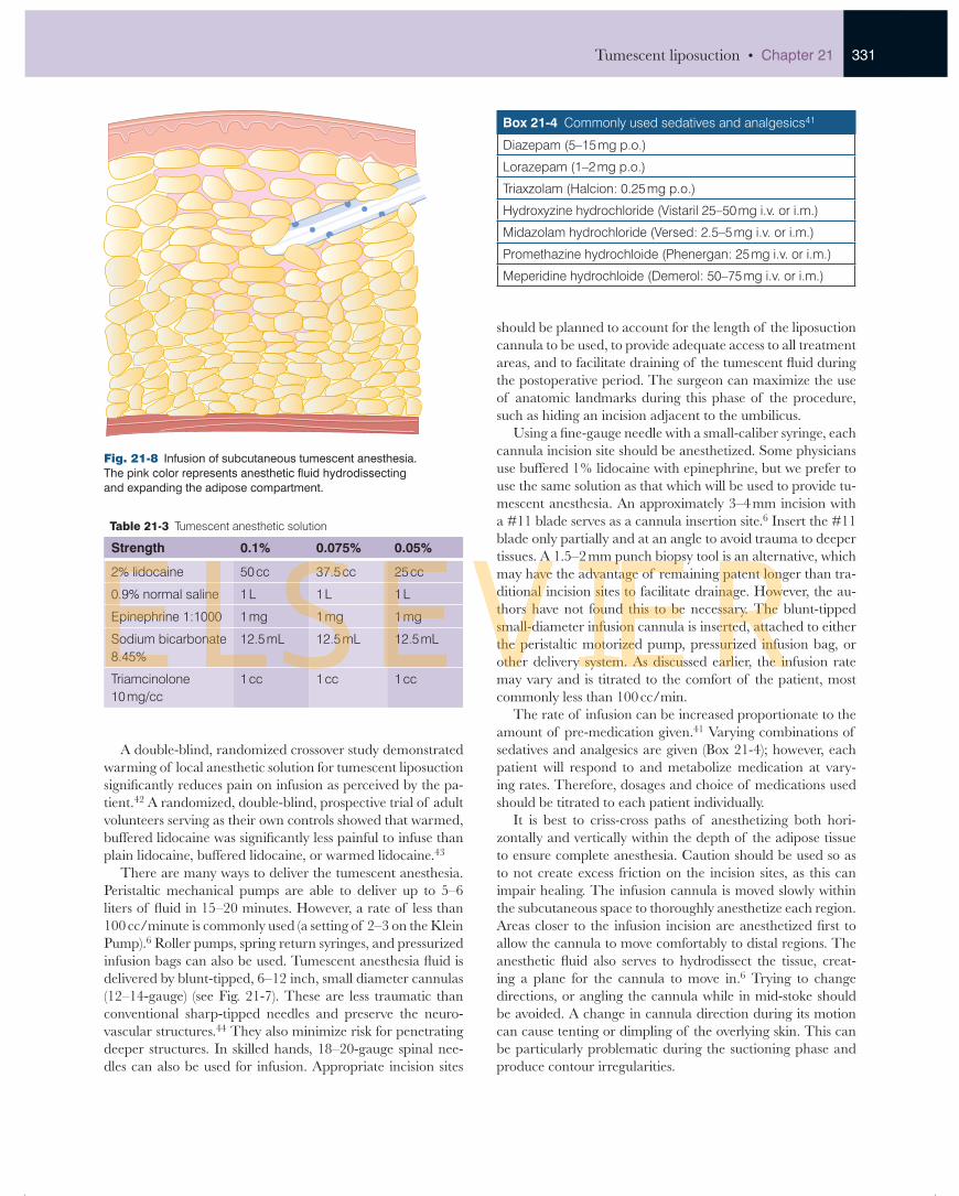
The tumescent technique is a method of delivering large
volumes of dilute lidocaine with epinephrine in buffered
saline via subcutaneous infi ltration to achieve adequate local
anesthesia and assist with hemostasis ( Fig. 21-8 ). 4 Table 21-3
lists the standard preparations.
Epinephrine, without the addition of sodium bicarbonate,
will make the tumescent solution acidic, which may cause
burning with infusion. Also, lidocaine action may be more effi -
cient when it is in solution near its pK (pK of lidocaine = 7.7). 5
Tumescent solution is also buffered because sodium bicarbonate
added to lidocaine in vitro augmented the bacteriocidal activity
of lidocaine. 35 Lidocaine has been shown to be bacteriocidal
for organisms isolated from the skin. 36 Concentrations greater
than 0.5% lidocaine provide a dose-dependent inhibition of
bacterial growth, Gram-negative greater than Gram-positive
organisms. 37 In dilutions of 0.05%, lidocaine is bacteriostatic
for Staphylococcus aureus . 38 In in vitro studies using suspensions
of bacteria (105 cfu/mL), all Gram-positive organisms, includ-
ing Staphylococcus aureus , had signifi cantly lower colony counts in
0.05% lidocaine. 39 When tumescent anesthesia is used, infec-
tion is a rare complication. 40
The higher concentration of lidocaine (0.1%) is used for
more sensitive areas, such as the abdomen, lateral thighs,
knees, inner thighs, periumbilical area, neck, fl anks, and
back 14,41 Some physicians choose to only utilize the 0.05%
lidocaine formula for liposuction to decrease total lidocaine
dosage and enable them to treat larger surface areas. However,
0.05% lidocaine is not as effi cient at producing anesthesia as
the 0.1% concentration. Therefore, an increase in sedation is
often required when using 0.05% lidocaine for local liposuc-
tion anesthesia. Another option is to use the 0.075% solution
which retains much of the anesthetic activity of the 0.1%
solution but with 25% less lidocaine, allowing larger areas to
be treated in one session. The 1:1 000 000 concentration of
epinephrine is more commonly used, but some surgeons have
used concentrations as low as 1:2 000 000 epinephrine with
good results.
A
C
B
Fig. 21-6 Powered cannula varieties. (A) Byron ARC cannula (Byron Medical Tuscon, AZ). (B) MicroAire PAL (MicroAire Surgical Instruments, Charlottesville, VA). (C) VibraSat (LaserPoint AG, Nordkirchen Germany).
Box 21-3 Commonly used infusion and aspiration pumps
Wells Johnson Single or Dual Infusion Pump (Wells Johnson, Tuscon, AZ)
HK Infusion pump (HK Surgical, San Juan Capistrano, CA)
Hercules aspirator (Wells Johnson, Tuscon, AZ)
Reliance aspirator (Bernsco, Hauppauge, NY)
Byron Psi-Tec III (Byron Medical, Tuscon, AZ)
LipoSat (LaserPoint AG, Nordkirchen, Germany)
Titan (Miller Medical, Mesa, AZ)
Infusion cannula
Fig. 21-7 The sprinkler-tip tumescent anesthesia infusion cannula.
331Tumescent liposuction • Chapter 21
A double-blind, randomized crossover study demonstrated
warming of local anesthetic solution for tumescent liposuction
signifi cantly reduces pain on infusion as perceived by the pa-
tient. 42 A randomized, double-blind, prospective trial of adult
volunteers serving as their own controls showed that warmed,
buffered lidocaine was signifi cantly less painful to infuse than
plain lidocaine, buffered lidocaine, or warmed lidocaine. 43
There are many ways to deliver the tumescent anesthesia.
Peristaltic mechanical pumps are able to deliver up to 5 – 6
liters of fl uid in 15 – 20 minutes. However, a rate of less than
100 cc/minute is commonly used (a setting of 2 – 3 on the Klein
Pump). 6 Roller pumps, spring return syringes, and pressurized
infusion bags can also be used. Tumescent anesthesia fl uid is
delivered by blunt-tipped, 6 – 12 inch, small diameter cannulas
(12 – 14-gauge) (see Fig. 21-7 ). These are less traumatic than
conventional sharp-tipped needles and preserve the neuro-
vascular structures. 44 They also minimize risk for penetrating
deeper structures. In skilled hands, 18 – 20-gauge spinal nee-
dles can also be used for infusion. Appropriate incision sites
should be planned to account for the length of the liposuction
cannula to be used, to provide adequate access to all treatment
areas, and to facilitate draining of the tumescent fl uid during
the postoperative period. The surgeon can maximize the use
of anatomic landmarks during this phase of the procedure,
such as hiding an incision adjacent to the umbilicus.
Using a fi ne-gauge needle with a small-caliber syringe, each
cannula incision site should be anesthetized. Some physicians
use buffered 1% lidocaine with epinephrine, but we prefer to
use the same solution as that which will be used to provide tu-
mescent anesthesia. An approximately 3 – 4 mm incision with
a #11 blade serves as a cannula insertion site. 6 Insert the #11
blade only partially and at an angle to avoid trauma to deeper
tissues. A 1.5 – 2 mm punch biopsy tool is an alternative, which
may have the advantage of remaining patent longer than tra-
ditional incision sites to facilitate drainage. However, the au-
thors have not found this to be necessary. The blunt-tipped
small-diameter infusion cannula is inserted, attached to either
the peristaltic motorized pump, pressurized infusion bag, or
other delivery system. As discussed earlier, the infusion rate
may vary and is titrated to the comfort of the patient, most
commonly less than 100 cc/min.
The rate of infusion can be increased proportionate to the
amount of pre-medication given. 41 Varying combinations of
sedatives and analgesics are given ( Box 21-4 ); however, each
patient will respond to and metabolize medication at vary-
ing rates. Therefore, dosages and choice of medications used
should be titrated to each patient individually.
It is best to criss-cross paths of anesthetizing both hori-
zontally and vertically within the depth of the adipose tissue
to ensure complete anesthesia. Caution should be used so as
to not create excess friction on the incision sites, as this can
impair healing. The infusion cannula is moved slowly within
the subcutaneous space to thoroughly anesthetize each region.
Areas closer to the infusion incision are anesthetized fi rst to
allow the cannula to move comfortably to distal regions. The
anesthetic fl uid also serves to hydrodissect the tissue, creat-
ing a plane for the cannula to move in. 6 Trying to change
directions, or angling the cannula while in mid-stoke should
be avoided. A change in cannula direction during its motion
can cause tenting or dimpling of the overlying skin. This can
be particularly problematic during the suctioning phase and
produce contour irregularities.
Fig. 21-8 Infusion of subcutaneous tumescent anesthesia. The pink color represents anesthetic fl uid hydrodissecting and expanding the adipose compartment.
Table 21-3 Tumescent anesthetic solution
Strength 0.1% 0.075% 0.05%
2% lidocaine 50 cc 37.5 cc 25 cc
0.9% normal saline 1 L 1 L 1 L
Epinephrine 1:1000 1 mg 1 mg 1 mg
Sodium bicarbonate 8.45%
12.5 mL 12.5 mL 12.5 mL
Triamcinolone 10 mg/cc
1 cc 1 cc 1 cc
Box 21-4 Commonly used sedatives and analgesics 41
Diazepam (5 – 15 mg p.o.)
Lorazepam (1 – 2 mg p.o.)
Triaxzolam (Halcion: 0.25 mg p.o.)
Hydroxyzine hydrochloride (Vistaril 25 – 50 mg i.v. or i.m.)
Midazolam hydrochloride (Versed: 2.5 – 5 mg i.v. or i.m.)
Promethazine hydrochloide (Phenergan: 25 mg i.v. or i.m.)
Meperidine hydrochloide (Demerol: 50 – 75 mg i.v. or i.m.)

332 Chapter 21 Cosmetic surgery procedures and techniques
The end point for infusion is reached when the tissue
becomes fi rm to hard, and indurated ( Fig. 21-9 ). For both
infusing tumescent anesthesia fl uid and suctioning, one hand
moves the cannula, and the other serves as a ‘smart hand’ to
guide and feel the cannula position. This usually nondomi-
nant hand lies on the skin and palpates, constantly assess-
ing the movement of the cannula, depth in the tissue, and
degree of tissue induration. The end point for infusion can
also be assessed by blanching as a result of vasoconstriction.
The amount of tumescent anesthesia fl uid infi ltrated will
depend on the anatomic location ( Table 21-4 ). The surgeon
must be cautioned that there is no absolute rule as to how
much anesthesia is required to fully treat an area. Factors
that can affect volumes of infi ltration include body weight
and the amount of fat in a particular anatomic area, and
the amounts listed in Table 21-4 represent averages based on
the authors’ experience. Clearly, certain patients will require
more or less local anesthesia depending on individual
anatomic variation.
Infusion patterns for men and women are different. Adipose
tissue in women tends to lie in the mid- to deep subcutaneous
space, and therefore anesthetic solution should be directed to
these locations. Men tend to have fi brous subdermal fat that
requires aggressive suctioning to remove adipose, as well as
mid-subcutaneous space adipose tissue ( Fig. 21-10 ). For this
reason, it is essential to add tumescent anesthetic solution to
the subdermal fat in sensitive areas for men (breasts, abdo-
men, love handles), in addition to anesthetizing the mid- and
deeper subcutaneous compartments.
The ideal time delay between tumescent infusion and lipo-
suction varies from patient to patient, and from location to
location. Approximately 15 minutes is needed to establish
adequate vasoconstriction. 41 A good indicator of the appro-
priate time delay is the visible blanching that occurs in the
tumesced sites. 44 However, a minimum of 30 – 45 minutes is
required to establish the profound anesthesia that is essential
for performing adequate and careful suctioning. The areas an-
esthetized should extend beyond the border of the intended
liposuction sites to prevent tenderness at the periphery, and
allow for feathering.
The concept of liposculpture is evolving as physicians
treat not just one cosmetic unit, but adjacent cosmetic units
(Box 21-5), blending the treatment sites to result in a more
natural symmetry of proportions. Preoperative markings
help the surgeon to delineate areas to be treated, with an
improvement in the overall aesthetic appearance as the goal
of surgery. However, it is the intraoperative technique that ul-
timately determines the fi nal result. The surgeon must pinch,
feel, inspect, move, and contour the subcutaneous tissue in a
manner that will produce an improved skin contour. As lipo-
suction surgeons, we rely on the skin’s remarkable ability to
contract and drape over the underlying soft tissue. It is im-
perative to keep the patient’s unique physical characteristics
and skin type in mind while suctioning. Skin that has poor
elasticity will not re-contour as well as skin with good tone,
and this is part of the art of liposuction/liposculpture. The
surgeon factors in all of these issues to determine just how
much fat to remove, and from which areas, to produce the
fi nal result.
Fig. 21-9 Determining the end point for tumescent anesthesia infi ltration. Prior to infusion (top) the skin is soft, but when infusion is complete (bottom) the skin is fi rm and resists downward pressure.
Table 21-4 Approximate volume of anesthesia used according to body site
Site Volume (liters)
Neck 0.4
Arms 1.0 per side
Upper abdomen 0.75
Lower abdomen 1.0
Hips 0.75 per side
Love handles 1.0 per side
Flanks 0.75 per side
Outer thighs 1.0 per side
Inner thighs 0.75 per side
Knees 0.5 per side
Calves & ankles 1.0 per side

333Tumescent liposuction • Chapter 21
Body position during the procedure must be changed fre-
quently, including that of the physician as well as the patient.
The physician should use all sides of the operative table to ex-
amine and treat the patient, accessing areas from a minimum
of two directions, preferably three. We refer to this method
of suctioning as triangulation . Patient position should also be
changed during surgery if the physician needs to access the
fat. An advantage of tumescent liposuction surgery is that
the patient is awake and therefore able to follow commands.
The patient can be asked to change body position during the
procedure to make it easier for the surgeon to treat an area
of the body. The central premise is that the surgeon must be
certain to treat all marked areas in a manner that will yield
smooth contours. Asking the patient to change position on
the table is one part of the process.
The concept of triangulation is central to obtaining smooth
liposuction results ( Fig. 21-11 ). The surgeon should think
of each unit area of fat as a compartment that needs to be
treated, and linking these areas will produce smooth contours.
Each unit area should be accessed and suctioned from three
directions (triangulated) in order to avoid producing ridges.
When an area is suctioned from one direction it is possible to
leave ridges, as small areas of fat between the cannula tunnels
remain. Suctioning from two directions helps to reduce this
risk, but the third vector dramatically reduces the appearance
and feel of residual fat and ridges.
The nondominant smart hand is one of the most important
elements of liposuction surgery. This hand is used to guide the
cannula, as well as assess cannula position and depth within
the fat, bring fat into the cannula path, stretch or stabilize skin,
and in general serve as the sensory input from the patient back
to the physician ( Fig. 21-12 ). Visual clues are also extremely
helpful for liposuction contouring, but the smart hand is an
invaluable link between the surgeon and patient. A surgeon’s
mastery of the smart hand concept is likely to improve lipo-
suction results signifi cantly.
With the use of tumescent anesthesia blood loss is minimal.
The physician should continuously be monitoring the aspirate
for quantity and quality of adipose ( Fig. 21-13 ). If the amount
of blood increases in the aspirate at any time during the pro-
cedure, active suction of that area should be discontinued.
Klein has reported that approximately 12 cc of blood is lost
for each 1000 cc of fat that is aspirated using the tumescent
technique. 45
Skin
Fascia
Muscle
Skin
Fascia
Fat
Fat
Fat
Fat
Muscle
Men
Women
Fig. 21-10 Relative location of fat in men and women (anterior lower abdomen as example). Note relative size/proportion of fat above and below superfi cial fascia in men and women.
Box 21-5 Liposuction cosmetic units
Neck, submental region, and jowls
Posterior upper arm
Posterior axillary line and upper back
Upper abdomen
Lower abdomen
Hip or love handles
Waistline and mid back
Outer thigh
Inner thigh extending to knee
Anterior thigh
Posterior thigh
Calve and ankle
Breast

334 Chapter 21 Cosmetic surgery procedures and techniques
Adequate tumescent anesthesia should make the procedure
nearly painless. The use of large cannulas initially takes ad-
vantage of the period of maximal anesthesia. Smaller cannu-
las cause less pain as they are advanced through the adipose
tissue and offer more options for fi ne-tuning and removing the
remaining adipose tissue ( Fig. 21-14 ). Changing the angle, di-
rection, diameter of the cannula, altering the patient position,
or applying manual traction with the smart hand may allow
treatment of tender areas, avoiding the need to use further
anesthesia in an area. 6
The structure and function of each body region necessi-
tates variations in the liposuction technique. Local anatomy,
the quality of adipose tissue (soft or fi brous), thickness of the
dermis, and skin elasticity all factor into the approach to a
liposuction cosmetic unit. The following discussions focus on
the unique approach we take for different anatomic regions
( Box 21-6 ).
When adding laser assisted liposuction to the procedure,
the surgeon inserts the fi beroptic cannula and moves it in a to-
and-fro motion very similar to traditional liposuction. The la-
ser part of the procedure is performed under sterile technique
and the same general guidelines for technique and anesthesia
as tumescent liposuction. The goal of treatment is to feel that
the resistance the laser cannula meets diminishes with treat-
ment, which serves as an indicator that the fat to be treated
Fig. 21-11 Triangulation of liposuction cannulas.
Fig. 21-12 Use of the smart hand. The surgeon uses the non-dominant hand (left in this photo) to palpate the skin and give tactile feedback.
Fig. 21-13 Liposuction aspirate demonstrating nearly 1.5 liters of fat without any signifi cant bleeding.

335Tumescent liposuction • Chapter 21
has been adequately heated. The surgeon can also focus on
the subdermal region to theoretically heat that region and en-
hance skin contraction. In some areas (neck, jowls), surgeons
prefer to reduce the power settings on the machine to more
gently heat the fat and reduce risk of nerve injury. This is also
an area of investigation.
Liposuction of the neck and jowls The neck and jowls are both a very diffi cult and very reward-
ing area to treat with liposuction. Traditional methods of
treatment for the aging neck
have primarily included face
and neck lifting procedures.
However, for selected patients,
liposuction can be a defi nitive
treatment. The ideal patient
has good skin tone and elastic-
ity, moderate submental fat,
mild jowl formation, and a
high-set hyoid bone.
There are two basic physi-
cal fi ndings of patients who are candidates for neck liposuc-
tion: aging and obesity. Patients with neck obesity often have
excess adipose tissue in other areas of the body, but many
are interested in treatment of the neck to defi ne their facial
features for appearance enhancement. These patients often
do extremely well with liposuction, in part because many are
young (under age 45) with superb skin elasticity ( Fig. 21-15 ).
Aging can be challenging to treat with neck liposuction, but is
perhaps the most common indication for therapy. These pa-
tients are often older (over age 40), have mild to moderate jowl
formation, fair to good skin tone and elasticity, and mild to
moderate submental fat. Rhytides will often improve follow-
ing careful and aggressive suctioning, and neck contours can
be greatly improved ( Fig. 21-16 ).
Since liposuction is not a skin tightening procedure, patients
must be carefully evaluated. The patient is asked to clench the
teeth, which will tighten the platysma muscle and defi ne fat
location. The surgeon pinches the submental skin between
thumb and forefi nger to assess both quantity and location of
fat. Submental fat can be either pre- or post-platysmal. Pre-
platysmal fat can be suctioned through a small submental inci-
sion, but post- (or retro-) platysmal fat must be excised directly.
Asking the patient to place the tongue up against the hard
palate will also help the surgeon to identify fat location. The
surgeon should also release the skin as part of a snap test to
determine skin elasticity. If the skin feels loose and does not
recoil quickly, then liposuction alone is unlikely to provide
maximal benefi t. For many patients, adjunctive tissue tighten-
ing can enhance results. For those patients seeking to avoid
a facelift, monopolar RF (Thermage, Hayward, CA) can be
useful when performed immediately before neck liposuction.
Skin tightening can be increased by almost twofold when lipo-
suction and RF are performed on the same day.
The clenched teeth test is also useful to evaluate platysma
location and banding. As patients age, some will develop
vertical subcutaneous bands that represent nondecussating
platysma muscle fi bers. When identifi ed preoperatively, the
surgeon can elect to repair the platysma muscle at the time of
liposuction through a submental incision. The surgeon should
also evaluate submandibular gland position. Many patients
Fig. 21-14 Orientation and placement of liposuction cannula tunnels. Smaller cannula diameters are often used to remove super-fi cial fat after deeper fat is suctioned with larger-diameter cannulas.
Box 21-6 Anatomic sites and liposuction aggressiveness 46
Aggressive – 80 – 100% removed
Love handles
Back/fl ank
Male breast
Medial knee
Upper and lower abdomen
Moderate – 50 – 80% removed
Hips
Arms
Outer thighs
Buttock
Inner thighs
Calves/ankles
Neck
Jawline
Light – less than 50% removed
Mid inner thigh
Jowls
Anterior distal thigh and knee
Posterior knee

336 Chapter 21 Cosmetic surgery procedures and techniques
have ptotic submandibular glands which appear as a subcu-
taneous fullness bilaterally along the inferior portion of the
mandibular ramus. This ptotic gland can appear to be jowls,
and it is important to identify this preoperatively. Patients
are told that submandibular gland position is unlikely to im-
prove with liposuction alone, and that may compromise fi nal
results. Platysma repair as well as superfi cial musculoapo-
neurotic system (SMAS) plication can improve gland position
in some cases, but other surgical procedures are available to
defi nitively correct submandibular gland ptosis. They will not
be discussed in this chapter.
The position of the hyoid bone is also an important determi-
nant of postoperative results. The hyoid bone is a central compo-
nent of neck architecture and musculature, and is an important
landmark when evaluating patients preoperatively. Patients
with a relatively low hyoid position will tend to have a less well-
defi ned cervicomental angle, whereas those with a high-set
hyoid are able to obtain greater defi nition to the cervicomental
angle ( Fig. 21-17 ). 47 Although both groups are candidates for
neck liposuction, it is helpful to counsel patients preoperatively
as to the limits of liposuction if they have a low hyoid position.
The authors have found that platysma repair at the time of
A B
Fig. 21-16 Female 51-year-old patient with moderate adipose (A) before and (B) 6 months after neck liposuction.
A B
Fig. 21-15 Female 28-year-old patient (A) before and (B) 6 months after neck liposuction.

337Tumescent liposuction • Chapter 21
liposuction can help to improve cervicomental angle formation in
patients with both low- and high-set hyoid position ( Fig. 21-18 ).
For patients with poor skin tone and/or severe jowl and
rhytide formation, full or partial facelift procedures can be of
signifi cant benefi t.
Liposuction The neck is marked with the patient in the sitting position
( Fig. 21-19 ). The anterior border of the sternocleidomastoid
muscle is defi ned and outlined, and markings continued in-
feriorly across the midline just above the sternal notch. The
submental crease is marked, as is the jawline. Care must be
taken to identify the jowl bilaterally as it will extend slightly
below the mandibular ramus. The superior extents of the jowl
should be marked as well. The extent of submental fat is then
identifi ed and marked.
The neck is anesthetized through a 3 – 4 mm submental
incision using a short 6-inch sprinkler-tip infusion cannula.
Anesthesia is performed with 0.1% lidocaine tumescent so-
lution. Care must be taken to anesthetize beyond the skin
markings to provide a feather zone and margin for error
with cannula motion. The jowls and lateral neck are an-
esthetized with either the infusion cannula or a 20-gauge
spinal needle from a lateral jawline location. Caution must
be use when anesthetizing the jowl, since over-fi lling can
theoretically lead to intraoral airway occlusion. Anesthesia
is allowed to sit for a minimum of 30 – 45 minutes prior to
suctioning.
The patient is asked to hyperextend the neck to provide
optimal access. If the patient has diffi culty with this maneu-
ver, the headrest of the bed should be dropped to facilitate
A B
Highhyoidbone
Lowhyoidbone
Fig. 21-17 Hyoid bone position. Note the change/improvement in cervicomental angle when the hyoid bone is relatively higher in position.
Fig. 21-18 Female 62-year-old patient (A) before and (B) 6 months after neck liposuction with platysma repair.

338 Chapter 21 Cosmetic surgery procedures and techniques
neck hyperextension. The submental region is suctioned fi rst
with either a 12-gauge Klein or 3 mm Accelerator cannula
with the holes oriented away from the dermis. The submental
region should be machine suctioned thoroughly ( Fig. 21-20 ).
However, it is recommended that a small amount of subder-
mal fat be left to prevent skeletonization of the neck. Caution
must be used when suctioning in the region of the marginal
mandibular branch of the facial nerve. It is most vulnerable
to injury below the ramus of the mandible when the neck
is hyperextended, as well as along the anterior third of the
jawline ( Fig. 21-21 ). Suctioning in a superfi cial plane in these
regions is advised. Suctioning of the neck should not extend
beyond the medial border of the sternocleidomastoid muscle
to avoid injury to vascular structures.
The jowls and jawline are suctioned as a unit. Initial de-
bulking should be done through a lateral 3 mm incision placed
along the jawline. A 2 mm, 3-inch spatula-tipped cannula with
5 cc syringe suctioning is used ( Fig. 21-22 ). Caution must be
used on the jowl and medial cheek, as over-aggressive suc-
tioning of the upper jowl can produce an unnatural dimple
appearance. The cannula should also be used to gently un-
dermine the medial jowl to reduce the appearance of labio-
mandibular tethering and rhytides. The inferior jowl, jawline,
and lateral neck are then aggressively suctioned with the 2 mm
spatula-tipped cannula on machine suction. Caution must be
used to stay in the superfi cial fat to avoid nerve injury. The
Fig. 21-19 Preoperative markings for neck liposuction.
Fig. 21-20 Machine suctioning of submental region. Note surgeon’s left hand identifying and protecting the marginal mandibular nerve.
Fig. 21-21 Danger zones (shaded pink boxes) for potential injury to the marginal mandibular branch of the facial nerve (pink line).
Fig. 21-22 The 2 mm, 3 inch spatula-tipped cannula.

339Tumescent liposuction • Chapter 21
cannula orifi ce can be oriented both towards and away from
the dermis. Particular attention should be paid to the medial
fi brous fat which is located just inferior to the ramus of the
mandible along the medial jowl. Failure to adequately suction
this area can produce an unnatural bulge.
After suctioning has been completed, submental fi brous
bands can be released. We have found that this may reduce
the appearance of puckering of the neck postoperatively, but
with the addition of monopolar RF preoperatively, this has
become less useful. Submental fi brous bands can be released
under direct vision if additional surgery is to be performed
(such as platysma repair), or can be eliminated with a closed
neck dissector (Byron, Tucson, AZ) ( Fig. 21-23 ). This instru-
ment has a sharp V-shaped notch at its distal tip which is
gently advanced in the superfi cial fat to catch and cut fi brous
bands ( Fig. 21-24 ).Caution should be used to avoid aggressive
treatment and increased bleeding. The closed neck dissector
should not be used to treat the lateral neck or jawline as this
may increase the risk of permanent marginal mandibular
nerve injury ( Fig. 21-25 ). The marginal mandibular nerve
function should be assessed before, during and after the pro-
cedure by having the patient grimace to show the lower teeth,
pucker, or protrude the lower lip.
Fig. 21-23 The closed neck dissector. Note V-shaped notch at tip.
Fig. 21-24 Superfi cial dissection of fi brous bands of the neck with the closed neck dissector.
Fig. 21-25 Lateral borders of area to be treated with closed neck dissector. Superior edge of dissection is marked in black at approxi-mately 45-degree angles from the submental incision, extending to sternocleidomastoid bilaterally.

340 Chapter 21 Cosmetic surgery procedures and techniques
• Determine hyoid bone placement and presence of submandibular ptosis.
• Add monopolar RF to enhance skin tightening. • Ensure patients wear postoperative garments
appropriately.
PE
AR
LS
Fig. 21-26 Platysma plication. Note initial running suture (pink) from superior to inferior, with continuation of the running suture in an oversewn plication from inferior to superior. Suture is tied on itself at superior end.
Fig. 21-27 Final appearance of platysma plication. Central two layer plication produces majority of tightening. Bilteral oblique plication sutures augment platysma tightening as well as help to suspend and elevate submandibular glands.
Platysma repair Repair of the platysma muscle can be performed through a
submental incision when platysmal bands are present. Numer-
ous techniques can be utilized to eliminate platysmal bands,
but the authors have found this technique to be quick, reliable,
and reproducible. 48,49 An approximately 3 cm elliptical incision
is made with a #15 blade encompassing the 3 mm stab wound
in the submental crease. The skin ellipse is excised, and the
submental region visualized. Hemostasis is obtained with elec-
trocautery. A headlamp is useful for this part of the procedure.
The medial borders of the platysmal bands are identifi ed
with blunt dissection extending from the submental incision
inferior to the level of the thyroid cartilage. If subplatysmal
fat is present, it is removed under direct vision with electrosec-
tion or sharp dissection. The platysma is then plicated in the
midline using a running clear 4-0 nylon, PDS, or comparable
suture. The plication is performed in a superior to inferior
direction, with a buried superior knot. When plication reaches
the level of the thyroid cartilage, the repair is then turned in
a fold-over maneuver from inferior to superior ( Fig. 21-26 ).
The surgeon should be certain to maintain constant tension
on the plication suture to ensure adequate platysmal tighten-
ing. The fold-over is performed by taking bites outside of the
initial plication seam (on either side of the seam) to imbri-
cate the platysma over the initial superior-to-inferior running
suture. The suture is continued superiorly to the initial bur-
ied knot and tied to the free strand of that knot. Additional
plication sutures can be placed bilaterally in the region of the
submandibular glands to both tighten the platysma further as
well as elevate the ptotic submandibular gland ( Fig. 21-27 ).
Hemostasis is obtained and confi rmed, and the submental
wound closed with subcutaneous 5-0 vicryl (polygalactin 910,
Ethicon, Inc., Somerville, NJ) and 6-0 prolene (polypropylene,
Ethicon, Inc., Somerville, NJ) skin sutures.
Postoperative compression A double-layer compression garment is placed postoperatively
to improve skin contraction and reduce postoperative bleed-
ing. Reston foam or French tape can also be applied under the
garments to further improve results. Adequate compression
along the jawline for a minimum of 7 – 10 days is essential to
obtaining adequate tissue adherence and contraction.
• Do not oversuction mid-cheek as this can lead to hollowing.
• Beware of submental skin ridging. • Over-aggressive suctioning of the submentum
can lead to bony prominence of hyoid cartilage.
PIT
FAL
LS

341Tumescent liposuction • Chapter 21
Liposuction of the arms Women with signifi cant lipodystrophy of the proximal arm
often have to wear blouses 2 – 3
sizes larger than they might
otherwise need so that the
arms fi t. For this reason, even
modest improvements result in
signifi cant patient satisfaction
( Fig. 21-28 ). In our experi-
ence, it is a misconception that
liposuction of the upper arms
often results in postoperative
irregularities with poor skin
retraction. 50 – 52 Liposuction of the arms is performed almost
exclusively on women, with the posterior and posterolateral
aspects of the arm involved more often than the anterior and
medial upper arms. On occasion, a localized fat deposit on
the ulnar side of the proximal forearm requires treatment as
well. Conservative but thorough fat extraction is obtainable
without undue trauma due to the soft quality of the fat in the
area. Avoiding trauma to the subdermis with correct cannula
choices, and minimal use of the smart hand will minimize
irregularities. 53 Skin of the upper arm has good potential for
signifi cant skin contraction. Brachioplasty (arm lift) and the
resultant scar can produce such an unattractive and uncor-
rectable result that liposuction, and even a second liposuction
session, are often preferable alternatives. In some patients,
radiofrequency skin tightening can be a useful adjunct to
upper arm fat removal.
The patient is evaluated in a standing position with arms
extended horizontally with the thumb pointing up, or elbows
bent, to maximize the laxity of the posterior and postero-
lateral compartments. As with other sites, skin tone and tex-
ture must be evaluated. If skin tone is poor preoperatively,
the patient may still achieve signifi cant skin contraction if
thorough fat removal is performed, but texture will often
not improve. For some patients, concurrent treatment of the
upper back and anterior/posterior axillary regions is per-
formed as well.
At least two incision sites are needed for infusion of 0.1%
tumescent anesthesia. For infi ltration of the posterior and
posterolateral arm, incision sites are just proximal to the
elbow and at the apex of the posterior axillary line. The soft,
loose tissue of the upper arms can often be infi ltrated more
rapidly than other areas. The amount of total tumescent
anesthesia may vary greatly, with an expected range of 500 –
1000 cc per arm.
Two to three incision sites across the mid-posterior up-
per arm are used for aspiration. For large-volume cases (over
400 cc removed), initial very cautious debulking may be done
with the 3 mm Accelerator cannula. In smaller-volume cases,
we fi nd the 12-gauge Klein cannula is preferable. As previ-
ously mentioned, pinching, lifting, and downward pressure
from the smart hand is not necessary in this area, and may in-
crease the risk for subdermal fi brosis, adhesions, puckering and
indentations. The 3 mm Accelerator (Eliminator) or 12-gauge
Klein cannulas have a relatively nonaggressive tip and, with
their recessed openings placed away from the dermis, are ideal
for fat removal of the arms. Particular attention must be paid
to thoroughly treat the proximal upper arm and fat overlying
the medial epicondyle, as incomplete treatment of these areas
are the most common causes of patient dissatisfaction. An in-
cision site just distal to the fat overlying the medial epicondyle
provides access to that area as well as the more fi brous fat just
proximal to the elbow. This may be the only area of the up-
per arm which may need use of the smart hand to assist with
fat aspiration, due to the fi brous nature of this area. For fi nal
assessment, the patient is instructed to hold her arms straight
up, occasionally exposing residual pockets of fat in the poste-
rolateral compartment.
In the rare case where the anterior upper arm needs treat-
ment, the 3 mm Accelerator (Eliminator) or 12-gauge Klein
A B
Fig. 21-28 Female 34-year-old patient (A) before and (B) 6 months after arm liposuction.

342 Chapter 21 Cosmetic surgery procedures and techniques
cannula is preferred due to the thin dermis in this area.
Incision sites in the anterior axilla and just distal to the area of
fat to be removed are utilized. Postoperative pain of the arms
is minimal compared to other areas, and increases with poorly
fi tting compression garments.
Liposuction of the trunk It is useful to consider trunk anatomy and cosmetic contours
from two views: anterior and lateral. The lateral view will re-
veal contour irregularities of the upper and lower abdomen.
The anterior view will allow the surgeon to view the upper
waist and back, waist, and hips. Patients will often discuss ar-
eas of the trunk that bother them such as the lower abdomen,
but it is the responsibility of the surgeon to identify which
adjacent regions might benefi t from suctioning. Also, the sur-
geon should consider whether any areas, if the patient were to
gain weight, might look unnatural juxtaposed with the treated
area. For these reasons, we have found that this anterior and
lateral classifi cation is useful. Patients interested in lower ab-
dominal liposuction are evaluated for possible upper abdomi-
nal liposuction. Those patients interested in treatment of the
hips or waist are evaluated to determine whether treatment of
the entire lateral waistline unit is indicated.
The abdomen can thus be divided into cosmetic units, many
of which are interrelated. The upper and lower abdomen are
separate cosmetic units, but often treated together in one session
( Fig. 21-29 ). The hip is another cosmetic unit, and the waist/
mid-lower back (below the bra line) is a cosmetic unit as well.
• Choose young skin with good elasticity. • Arms can be a good place for the inexperienced
liposuction surgeon to begin.
PE
AR
LS
A B
Fig. 21-29 Female 39-year-old patient (A) before and (B) 6 months after upper and lower abdominal liposuction.
• Obvious skin laxity can lead to ridging. • Seroma formation may occur if garments are not
properly worn.
PIT
FAL
LS

343Tumescent liposuction • Chapter 21
A B
C D
Fig. 21-30 Female 43-year-old patient (A) before and (B) 6 months after upper and lower abdomen, hips, waist and back liposuction. Female 42-year-old patient (C) before and (D) 6 months after same procedure. Note retraction of pannus in both patients without the need for abdomi-noplasty.
The hip and waist/mid-lower back are considered the lateral
area of the trunk, and these two cosmetic units are often treated
in one session. The upper back (above the bra line) is most often
treated in conjunction with the arms and posterior axilla, and
is not considered part of the abdomen/trunk cosmetic region.
The result is a total of four cosmetic units of the abdomen and
trunk. In some patients, treatment of all four cosmetic units in
one surgical session is indicated. This helps to maximally con-
tour the abdomen/trunk by removing adipose tissue in three
dimensions. In addition, postoperative healing and skin con-
traction along suction vectors provides maximal improvement
of the abdominal pannus and waistline ( Fig. 21-30 ).

344 Chapter 21 Cosmetic surgery procedures and techniques
Upper and lower abdomen Prior to suctioning, the patient must be evaluated for abdomi-
nal hernias and scars. Ventral
hernias including umbilical,
postsurgical, and Spigelian
(lateral rectus sheath) should
be ruled out through clinical
examination. Preoperative
markings should refl ect the
extent of suctioning, areas to
be suctioned, and localized
collections of adipose tissue
( Fig. 21-31 ). The lower abdo-
men, when suctioned alone, is often clearly demarcated and
easily outlined. When the lower and upper abdomen are to be
treated together, the extent of suctioning extends from under
the breasts to the suprapubic region.
Anesthesia is obtained through two incisions placed along the
suprapubic region, as well as from mid-abdominal sites along
the lateral aspect of the area to be treated. Anesthesia is placed
in the mid-subcutaneous space, and allowed to sit for a mini-
mum of 30 minutes prior to suctioning. Tumescent 0.075 – 0.1%
lidocaine anesthesia is used for the upper abdomen, especially
the areas over the costal margin. When possible, 0.1% lidocaine
should be used for the lower abdomen, but this area can be an-
esthetized with 0.075% lidocaine and suctioned effectively. The
cannula will often pass through and under older scars without
diffi culty, but caution should be used with newer scars.
Suctioning is performed with the 3.7 mm swan-neck Keel
Cobra cannula for debulking larger patients. The 3 mm Accelerator
cannula can be used to debulk smaller patients. Triangulation of
areas is essential to produce smooth contours, as near 100% fat
removal is the goal of therapy. All too often surgeons leave too
much fat after suctioning, believing this will help ensure a smooth
result. However, patients are often disappointed if some fat is left.
It is the authors’ experience that removal of 90% or more of ab-
dominal region fat produces smooth results with excellent patient
satisfaction. The 12-gauge Klein cannula is used to feather treat-
ment sites and ensure maximal smooth fat removal.
It is essential to thoroughly suction the periumbilical region,
as well as the deep fat of the upper and lower abdomen. Many
patients will have well-defi ned adipose collections that lie on
the rectus sheath deep to Camper’s and Scarpa’s fascia, both
superior to the umbilicus and inferior/lateral to the umbilicus.
Suctioning of these areas is essential to producing a fl at abdo-
men ( Fig. 21-32 ). It is often necessary to lift the skin with the
smart hand and carefully advance the cannula into a deep
adipose plane to access this fat. Clearly, caution is needed to
prevent sub-rectus suctioning. We have found it helpful to use
short cannula strokes, and avoid any cannula motion lateral
to the rectus sheath when attempting this deep fat maneuver.
By avoiding cannula motion lateral to the rectus, it reduces
the chances of becoming sub-rectus with cannula position. It
is also imperative that cannula position be superfi cial when
crossing the costal margin to prevent injury in that location.
Fig. 21-31 Preoperative markings for abdominal liposuction. Note use of fl ow lines and markings (large ‘X’ to denote areas of adipose concentration) to aid surgeon intraoperatively.
• Whenever possible, include waist and fl anks with abdomen.
• Defi ne extra- and intra-abdominal fat with patient before starting procedure.
• Use smart hand to gently lift the lower layer of adipose to provide thorough deep suctioning.
• Inform patient of possible early start of menses after procedure.
• Warn patients of excessive fl uid in mons or testicular region on postoperative days 0 – 3.
PE
AR
LS
• Incomplete umbilical anesthesia will lead to discomfort.
• Rarely, patients will develop skin mottling. • Beware aggressiveness at midline of rectus sheath. • Failure to remove enough fat can lead to patient
dissatisfaction.
PIT
FAL
LS

345Tumescent liposuction • Chapter 21
Hips The hips are either treated alone or in combination with the
upper back and waistline. Some
women will also have the lateral
thighs treated in combination
with the hips to re-contour the
lateral silhouette ( Fig. 21-33 ).
Correction of the double-bulge
of the hips and lateral thighs
can have a profound impact
on body shape. However, many
women will benefi t from suc-
tioning of the hips alone. These
women typically have an athletic build, and have isolated hip
adipose deposits. Some will have mild outer thigh fullness, but
their athletic shape makes treatment of this area less necessary.
The hip is outlined bilaterally with the patient in the stand-
ing position. The inferior border of the area to be treated is
often easily recognized as a distinct junction between hip and
upper lateral thigh. This pseudo-groove should not be treated,
even when combining hip and lateral thigh liposuction, since
this can produce disfi guring depressions. The anterior and
posterior hip are also usually distinct, and the surgeon can
feel for the boundaries of the hip with the pinch technique.
Typically, the amount of fat one can pinch diminishes sub-
stantially as one moves away from the central area of hip
adipose tissue. The superior hip can have an indistinct border,
and it is for this reason that hip liposuction is often combined
with treatment of the waistline. When treating the hip alone,
it is important to feather suctioning up into the waistline.
Anesthesia is placed with 0.1 or 0.075% tumescent lido-
caine, and allowed to sit for 30 minutes. The right hip is anes-
thetized (and suctioned) with the patient lying on the left side,
and vice versa for the left hip. Initial debulking can be per-
formed with the 12-gauge Klein or 3 mm Accelerator cannu-
las. Final blending is done with the 12-gauge Klein cannula.
Triangulation is essential, and the goal of therapy is creation
of a smooth contour in harmony with the lateral thigh, but-
tock, and waistline. Suffi cient fat is removed to achieve this
result, and can vary signifi cantly. For most patients, 50 – 80%
fat removal from the hip is adequate. Occasionally, near 100%
fat removal is needed to obtain the desired contour.
Mid-lower back and waistline Contouring of the waist can produce beautiful aesthetic results,
and a shapely waistline is one
of the things appreciated most
by patients after their lipo-
suction surgery. To adequately
contour the waistline, the sur-
geon must also treat the back
in the vicinity of the bra line.
For most patients, suctioning
of the hip at the same time is
Skin
Fascia
Muscle
A B
Fig. 21-32 Removal of deep anterior lower abdomen fat. (A) Drawing illustrating location of fat on anterior lower abdomen both above and below superfi cial fascia. (B) Smart hand maneuver to elevate skin and expose deep adipose compartment for suctioning.
• Suction hips along with outer thighs in women with double-bulge ‘violin’ deformity.
PE
AR
LS

346 Chapter 21 Cosmetic surgery procedures and techniques
necessary to achieve optimal results. This allows the surgeon
to maximally contour the lateral abdomen, blending it into the
fl ank and ultimately the back. Volumetric reduction of fat in
three dimensions along the abdomen, fl ank, waistline, and back
is essential for creating substantial improvements in shape and
contour.
The patient is marked in the standing position, with vectors
of suctioning clearly marked ( Fig. 21-34 ). It is important to
suction along intended vectors of skin retraction to promote
skin redraping postoperatively. The surgeon can pinch the
lower back fat to localize it, and this area is marked. Incisions
along the mid-waist, posterior mid-back, and under the lateral
breast are used for anesthesia and suctioning. Additional inci-
sions are placed as needed at the inferior zone of treatment
to promote triangulation and facilitate drainage. Anesthesia
is obtained with 0.075 – 0.1% lidocaine, and allowed to sit for
40 – 45 minutes. It is advisable to allow the tumescent solution
in this region to sit for slightly longer than other areas to pro-
vide maximal anesthesia. The waistline and mid-lower back
can be a particularly sensitive area to treat due to the very
fi brous nature of the fat. Some surgeons prefer to use external
ultrasound in this area (and other areas with fi brous fat) to
make fat removal easier, but the authors have found this to be
cumbersome and unnecessary. 54,55
The goal of waistline and upper back suctioning is near
100% fat removal. Initial debulking is performed with either
the 3.7 mm Keel Cobra or 3 mm Accelerator cannula. The
surgeon must be certain to suction the very deep fat that lies
just superfi cial to the muscular fascia, similar to the technique
for the upper and lower abdomen deep fat. The smart hand
is sometimes used to lift the skin of the waistline to allow the
cannula to access the deeper fat. Final blending and contour-
ing can be done with a 12-gauge Klein cannula, but for many
patients the 3 mm Accelerator is adequate for this task. It must
be emphasized that triangulation and aggressive suctioning
are needed to fully contour the waistline. A few extra minutes
of attention to detail in this region can produce dramatic
improvements in results.
Liposuction of the buttock The buttock is a challenging area to suction, and is almost
exclusively done in women.
Many women are interested in
reducing the overall size of the
buttock, but often have diffi -
culty verbalizing exactly what
it is that bothers them. The
usual concern is size, but the
buttock has so many different
contours, projecting in both
a lateral and posterior direc-
tion, that defi nition of treatment parameters can be diffi cult.
Therefore, it is buttock shape, symmetry and proportion that
• Be aggressive on waistline, especially in males as the adipose tends to suspend from the thick dermis.
• Evaluate hip size to determine if they need to be suctioned along with the waist to prevent step-off deformities.
PE
AR
LS
A B
Fig. 21-33 Female 41-year-old patient (A) before and (B) 6 months after hip and outer thigh liposuction.

347Tumescent liposuction • Chapter 21
should guide surgical judgment, and not absolute size. The
goal is to treat the buttock so that it fi ts in harmony with the
shape and silhouette of the patient. For these reasons, treat-
ment of the buttock is often combined with treatment of the
hips and lateral thighs, considering this to be an extended
cosmetic liposuction unit. Preoperative markings are essential,
and serve as a roadmap and guide for the surgeon.
Caution must be used to avoid the inferomedial buttock
in the vicinity of the sciatic nerve, as well as the medial but-
tock near the gluteal cleft. In addition, suctioning and cannula
motion should never cross the inferior gluteal crease where
the buttock meets the upper posterior thigh. Suctioning of the
inferior buttock/upper thigh crease can disturb the fi brous
junction in that region and lead to an unnatural ptosis of the
buttock. Blending of the buttock with the lower back, hip, and
outer thigh can produce superb contours ( Fig. 21-35 ). Over-
suctioning can produce a fl attened buttock with irregular
contours, which is not desirable.
After appropriate surgical markings have been made,
anesthesia is achieved with 0.075 – 0.1% lidocaine tumescent
anesthesia bilaterally. The patient is often positioned in the
prone position for anesthesia, occasionally rocking gently
onto one hip to allow anesthesia of the contralateral buttock.
Incision sites should be placed in the lateral infragluteal
crease, the upper medial buttock and the upper lateral buttock
to promote triangulation. Anesthesia is placed predominantly
in the mid and deep fat of the buttock.
Anesthesia is allowed to sit for a minimum of 30 minutes
and suctioning begun. Caution must be used to remain in
the mid and deep fat of the buttock, avoiding superfi cial suc-
tioning. Treatment of the superfi cial fat can quickly lead to
contour irregularities in this very technique-sensitive region.
The main theme that should guide the surgeon is to retain the
contour and convexity of the buttock while decreasing size
and improving contours. Mid and deep fat liposuction are es-
sential tools for the physician. Initial gentle debulking can be
done with a 3 mm Accelerator cannula in most patients, while
larger patients can be treated with the more aggressive 3.0 or
3.7 mm Keel Cobra cannula. Following debulking, the mid fat
is contoured with a 12-gauge Klein cannula.
Final results will often depend on the ability of the surgeon
to blend the buttock with the hip, lower back, and lateral thigh.
The challenge of buttock liposuction is to judge the amount
of fat removal correctly, and to precisely contour the junc-
tion of the buttock with the hip and outer thigh. Actual per-
centages for fat removal are less important than the aesthetic
result, and this is judged through intraoperative surgical feel ,
both visual and tactile (with the smart hand). In many cases,
measuring exactly how much fat has been removed from each
side can help the surgeon obtain symmetry.
Liposuction of the legs Localized adipose deposits of the legs are particularly well
suited to liposuction surgery. Although some people (usually
women) have diffusely large legs with abundant adipose tis-
sue, many have well-shaped legs with discrete collections of
fat. It is for these women that liposuction is ideal. Thorough
Fig. 21-34 Preoperative markings for liposuction of the back and waistline. Note use of markings to aide surgeon intraoperatively, and suctioning vectors (three black lines on waistline) to contour the waistline and produce retraction of the pannus and skin. • Suction in the midplane only.
• Avoid over-suctioning with large cannulas. • Measure milliliters of fat removal to ensure
symmetry.
PE
AR
LS
• Excessive removal can lead to a fl at, unattractive contour.
PIT
FAL
LS

348 Chapter 21 Cosmetic surgery procedures and techniques
contouring of these localized fat deposits can dramatically
change the shape and fl ow of leg lines. Clothing fi ts better,
and the patient usually feels much more comfortable with her
shape in general. As opposed to truncal obesity, many women
struggle with the shape of their thighs for years, even when
close to their ideal body weight. Genetics plays a signifi cant
role in determining leg shape.
Women with generalized leg obesity can be improved
with liposuction surgery, but many have underlying bone and
muscle anatomy that will not support the appearance of a thin,
shapely leg. These women must be counseled preoperatively
that although liposuction can alter leg contours and perhaps
thin their legs, it is unlikely that they will convert from some-
one with an obese leg to one with a thin leg. Naturally, there
are exceptions to this rule, but we have found this to be true
in general. Also, there is a subset of women who have soft skin .
This term refers to women who have abundant cellulite in
the setting of a relatively obese leg. The limiting factor for
women with soft skin is that over-aggressive suctioning can
produce rapid and dramatic skin depressions. Caution and, if
anything, under-treatment are the rules for such patients.
It is useful to think of the leg as having discrete cosmetic lipo-
suction units. These include (1) the outer thigh, (2) inner thigh
extending down to and including the knee, (3) anterior thigh,
(4) posterior thigh, and (5) the calves/ankles. With lidocaine
toxicity as the limiting factor, the surgeon must factor in anat-
omy, patient desires, and surgical reality in determining which
cosmetic units to treat. The outer thighs, inner thigh and knee
are commonly treated in one session together. Occasionally the
outer thigh is treated alone in the absence of other leg obesity,
but in many cases the outer thigh and hip are treated together.
Outer thighs The goal of therapy is creating a fl ow of skin that allows the
outer leg to blend naturally
with the hip, buttock, and
trunk ( Fig. 21-36 ). Suction-
ing of the outer thigh must be
approached with caution, as
taking too little fat will yield
a disappointed patient and
taking too much a disfi gured
patient. The outer thigh is
a landmark area, one that is
used as a reference point by
those casually observing a woman’s shape to determine over-
all body type, thinness, and aesthetic contour. When the outer
thigh is large or unnatural in shape, it tends to stand out and
A B
Fig. 21-35 Female 43-year-old patient (A) before and (B) 6 months after liposuction of the abdomen and buttocks.

349Tumescent liposuction • Chapter 21
be noticeable to the eye. Therefore, improvement in outer
thigh contours can have a profound effect on body shape and
patient self-image ( Fig. 21-37 ).
The patient is marked in the standing position ( Fig. 21-38 ).
Anesthesia is obtained with 0.075 or 0.1% lidocaine and
allowed to sit for 30 minutes. Each leg is anesthetized and
treated with the patient lying on the contralateral thigh.
Three to four incisions are used, the most common in the
lateral aspect of the gluteal crease. The other incisions are
placed at 2, 8, and 10 o’clock around the typical oval drawn
to mark the outer thigh.
Initial debulking is performed with the 3 mm Accelerator
cannula in most individuals, but with the 12-gauge Klein in
thin women. The 12-gauge Klein is then used to suction more
superfi cially and to perform fi nal blending and triangulation.
Although relatively superfi cial liposuction is performed, it is
advisable to leave a narrow zone of intact subdermal fat to
retain optimal contours.
Areas in which to be cautious include the upper lateral
thigh, distal lateral thigh, and Gasparotti’s point. 56 The upper
lateral and distal lateral thigh are susceptible to over-suctioning
and ridging or dimpling, particularly around cannula insertion
A B
A B
Fig. 21-36 Female 40-year-old patient (A) before and (B) 6 months after liposuction of the outer thighs and hips.
Fig. 21-37 Female 35-year-old patient (A) before and (B) 3 months after liposuction of the outer thighs.

350 Chapter 21 Cosmetic surgery procedures and techniques
holes. It is important to move the cannula from one inser-
tion hole to the next with regularity to avoid over-suctioning
through one incision and creating a dimple under that incision.
Gasparotti’s point is just posterior to the greater trochanter,
and depressions in this location result from aggressive suc-
tioning of the deep fat ( Fig. 21-39 ). Abduction and internal
rotation of the leg are useful to drop the greater trochanter
out of the surgical fi eld, and thus protect against a Gasparotti
point depression ( Fig. 21-40 ). Numerous devices are available,
including the triangular wedge pillow, that promote this leg
position and aid the surgeon (Wells Johnson, Tuscon, AZ).
The authors prefer to have an assistant abduct the leg.
The endpoint for outer thigh liposuction is subjective.
Contour and fl ow are the most important concerns, and the
smart hand pinch technique is invaluable. The surgeon pinches
other areas of the leg that appear to be well contoured, and
determines how many fi nger-widths (using the index fi nger) of
skin is contained in the pinch ( Fig. 21-41 ). This becomes the set-
point of the leg, and is often 1 to 1.5 fi nger-widths. The goal of
outer thigh suctioning becomes bringing the outer thigh pinch
test to match that 1 to 1.5 fi nger-widths. But the surgeon must
use the pinch test in conjunction with visual and other tactile
clues to determine the optimal liposculpture endpoint.
Inner thighs and knees For many women, the inner thigh region is a diffi cult place to lose
weight and improve contours.
Diet and exercise programs can
have some limited success, but
liposuction is a superb treat-
ment option for this anatomic
region ( Fig. 21-42 ). When eval-
uating the inner thigh and knee,
the surgeon must determine
both the amount of fat removal
to be performed as well as the
extent of surface area to treat.
Specifi cally, a decision must be made to either treat the entire
inner thigh and knee region as one unit, or to treat the upper
inner thigh and/or knee as separate cosmetic units.
The diffi culty with treating the inner thigh or knee as
distinct entities is that the risk of contour irregularities and
step-offs increases. Blending of the knee or inner thigh with
the mid thigh can be challenging, and the amount of fat that
Fig. 21-38 Preoperative markings for liposuction of the outer thigh. Central circle with three ‘X’ marks is the area with the most adipose tissue, and the outer circle denotes the feather zone.
• Abduct leg to prevent over-suctioning at Gasparotti’s point.
• Triangulate and trust the smart hand pinch test.
PE
AR
LS
Greatertrochanter
Femur
Risk zonefor depression
Fig. 21-39 Gasparotti’s point. The area posterior to the greater trochanter is a risk zone for liposuction-induced depressions and soft tissue deformities.
• Soft skin and dimpling may not improve with procedure.
• Beware suctioning under gluteal crease which may lead to a ‘double banana’ roll.
PIT
FAL
LS

351Tumescent liposuction • Chapter 21
A B
Fig. 21-40 Indentation due to over-suctioning at Gasparotti’s point
A B
Fig. 21-41 The pinch technique to evalu-ate liposuction endpoints. (A) Diagram illustrating use of the index fi nger as a measuring device to determine amount of remaining fat. (B) The pinch technique showing very little remaining subcutaneous fat on the lower abdomen (less than one fi nger).

352 Chapter 21 Cosmetic surgery procedures and techniques
can be removed from these regions is limited if the surgeon
must prevent a line of demarcation at the junction with the
mid thigh. Suboptimal cosmetic results can be more frequent
when the mid thigh is not treated.
For these reasons, the authors have developed a preferred
method for treating the entire inner thigh and knee as one
cosmetic unit, extending from the inguinal crease down to the
superomedial calf. Very gentle suctioning of the mid thigh
region allows more thorough fat removal from the upper in-
ner thigh and knee, improves blending and feathering into the
mid thigh, and increases patient satisfaction by contouring
and debulking the entire medial upper leg.
The patient is marked in the standing position with the
knee fully extended, the leg advanced forward (a modifi ed
lunge position), and externally rotated ( Fig. 21-43 ). The fat
pads of the medial knee and upper inner thigh are identifi ed
and delineated, as is the inguinal crease. Markings should in-
clude the proximal medial calf as treatment of this area helps
to defi ne knee contours and improve the fl ow of skin lines.
The surgeon should be certain to mark and identify the pos-
terior knee and posterior upper thigh fat pockets, as failure to
treat these areas will lead to less than optimal postoperative
contours. Adequate contouring of these posterior adipose col-
lections is essential since these fi brous areas will dominate the
postoperative appearance if not removed. Also, reduction of
these compartments allows the remainder of the inner thigh
and knee skin to fall into position after suctioning rather than
being tented by these posterior fi brous adipose collections.
The anterior and posterior borders of the region to be treated
should also be marked, and are loosely used as feathering
guides into the anterior and posterior thigh. Careful feather-
ing is essential to maintain normal thigh contours and fl ow.
Anesthesia is obtained with 0.075 – 0.1% tumescent lido-
caine anesthesia through multiple incision sites. Incision
sites below the knee should be avoided since they often heal
less well than those above the knee. Thorough anesthesia of
the fi brous posterior knee and inner thigh regions is helpful
for improving patient comfort. Anesthesia should be placed
2 – 3 cm beyond the anterior and posterior markings to
allow for feathering. Anesthesia should sit for a minimum of
30 minutes prior to suctioning.
The knee is suctioned fi rst, with the patient in the frog-leg
position and slightly rotated onto the side being treated. The
degree of convexity of the medial femoral condyle and the
tibial plateau may create a pseudolipodystrophy in an area
devoid of fat. Palpation upon physical examination will dif-
ferentiate the depth of the fat pad from the underlying bony
prominences. A 12-gauge Klein cannula is used to treat the
knee, with near complete fat removal as the goal. The sur-
geon should feather this treatment area into the proximal calf
and the mid thigh. It is useful to perform the feather maneu-
vers during the initial phases of suctioning, as this can allow
for more thorough and even fat removal from the knee and
upper inner thigh. Early feathering tends to improve the sur-
geon’s feel during the procedure, often eliminating the need to
‘chase’ a persistent ridge or depression. Treatment of the pos-
terior fi brous knee fat is performed with the 12-gauge Klein
cannula, but in some patients a more aggressive cannula such
as the 12- or 14-gauge Capistrano cannula is needed to debulk
this area.
The proximal inner thigh is initially gently debulked in
the deep fat with the Capistrano cannula. The 12-gauge
Capistrano is used for most patients, but thinner patients can
be debulked with the 14-gauge Capistrano cannula. Caution
A B
Fig. 21-42 Female 28-year-old patient (A) before and (B) 6 months after inner thigh and knee liposuction. Note increased space between thighs and knees. Patient has also had outer thigh liposuction.

353Tumescent liposuction • Chapter 21
is a must when using the Capistrano cannula in this region. Its
benefi t is that it can quickly and thoroughly debulk the upper
inner thigh, including the very fi brous and resistant posterior
inner thigh fat. However, over-zealous or superfi cial use of this
cannula can produce persistent ridges and contour irregulari-
ties. The surgeon should limit the number of cannula strokes
performed from any single incision with the Capistrano can-
nula, so triangulation is essential with this instrument. The up-
per thigh is then fi ne-tuned with the 12-gauge Klein cannula,
with blending and feathering into the mid thigh. Fat removal
from the upper inner thigh should not be 100%, but more in
the 50 – 80% range. This is one area that fl ow and the surgeon’s
aesthetic sense are essential determinants of the end point of
treatment.
The mid thigh is the medial region located between the
upper inner thigh and the knee. It should be viewed as a con-
nector, essentially a bridge between the upper and lower in-
ner thigh region. For this reason, treatment of the mid thigh
helps the surgeon to blend and contour the inner leg. The
overall contour changes of the inner leg come from suction-
ing of the upper inner thigh and knee, but the mid thigh is
the glue that holds the cosmetic unit together. Suctioning of
the mid thigh should be performed gently in the mid fat with
a 12-gauge Klein cannula, taking only what comes very easily.
The endpoint of treatment is when the upper thigh and knee
blend smoothly with the mid thigh, as well as when the entire
inner thigh blends smoothly with the anterior and posterior
thigh. Caution should be exercised in the vicinity of Hunter’s
canal and the femoral artery, since aggressive suctioning in
this area can produce a very unnatural postoperative fullness
(lump) in the area. Final blending with the upper inner thigh,
posterior upper thigh, and knee is performed during the fi nal
stages of mid thigh suctioning. The rule of thumb is that it is
better to remove too little rather than too much fat from the
mid thigh.
It is useful for the surgeon to think of the inner leg as an en-
tire cosmetic unit during the fi nal stages of suctioning. Taking
a literal step back to view the fl ow and contours of this region
can help the surgeon see areas that require further treatment
and blending.
Postoperative compression of the upper inner thigh is espe-
cially important, and the surgeon should be certain to choose
garments that provide adequate support in this area. Some
garments have a cut-out in the groin area to facilitate toilet use,
but this opening can hinder compression of the upper thigh
and should be eliminated as soon as possible postoperatively.
Anterior and posterior thighs The authors infrequently treat the anterior and posterior thighs.
The anterior thigh is occasion-
ally treated as an extension of
the knee, and in most cases in-
volves gentle suctioning of the
distal anterior thigh from two
incisions placed in the medial
and lateral suprapatellar re-
gions. The so-called banana roll
of the upper posterior thigh
is occasionally suctioned very
Fig. 21-43 Preoperative markings for inner thigh and knee lipo-suction. Note the three zones to be treated: upper inner thigh, mid thigh, and knee extending to superior calf.
• Avoid over-suctioning of mid thigh above Hunter’s canal.
• Treat upper inner thigh and knee as one cosmetic unit when possible.
• Feather early to avoid having to chase a ridge.
PE
AR
LS
• Avoid over-suctioning of inner mid thigh as this may create irregular contours.
• Beware soft skin issue with possible accentua-tion of inner thigh skin sagging.
PIT
FAL
LS

354 Chapter 21 Cosmetic surgery procedures and techniques
gently. Over-treatment of this
area can produce a very un-
natural ridging. Gentle mid to
superfi cial suctioning of the
fat can improve contours and
encourage skin tightening with
a good safety margin. The 12-
gauge Klein cannula is used
for both regions.
Calves and ankles Lipodystrophy of the calves and ankles is often not dependent
on body weight, and is usu-
ally present from early ado-
lescence. Fatty deposits of the
calves and ankles often make
patients look much heavier
than they really are, but for-
tunately liposuction can help
in this area. With the use of
tumescent anesthesia, smaller
cannulas, and good postop-
erative compression, excessive
bruising, persistent swelling and irregularities frequently can
be minimized. 57 The challenge of calf and ankle liposuction
is treating a small convex surface so that at the end of the
procedure the patient has an even smaller, natural appearing,
shapely convex lower leg. Patients with a history of deep
venous thrombosis, hypercoagulable state, angina, past myo-
cardial infarction, or large varicose veins are not candidates
for liposuction of the calves and ankles.
The patient is marked in the standing position. It is often
useful to have the patient stand on a stool or low bench. The
anterior leg in the pretibial area is rarely treated due to the
almost universal absence of fat in this region. Tumescent
anesthesia is obtained with 0.1% lidocaine and allowed to
sit for 30 – 45 minutes.
Suctioning is performed with small cannulas, usually a
12-gauge Klein or 3 mm spatula cannula. Incisions are kept to
a minimum (usually 4 – 8 per leg) since lower leg incisions often
do not heal as well as those placed in other areas. Areas to focus
on include the lateral and medial upper calf, as contouring
of this area will often substantially improve leg shape. Further
debulking of the posterior calf can reduce circumference and
provide additional contour improvements. Moderate liposuc-
tion around the ankle region can refi ne distal lower extremity
contours.
The goal of calf and ankle liposuction is gentle but thor-
ough fat removal. The surgeon must use caution so as to avoid
injury to the underside of the dermis, which can produce un-
natural ridging and become quite noticeable in this region.
Therefore it is advisable to treat the calf and ankle as one
cosmetic unit, leaving a small amount of subdermal fat while
enhancing contours through targeted suctioning of the areas
discussed above.
Postoperative compression garments and leg elevation are
essential following calf and ankle liposuction. Patients are
encouraged to ambulate and perform leg exercises to pre-
vent deep venous thrombosis (DVT). Despite the best efforts
of both patient and surgeon, postoperative lower leg edema
is to be expected and often will persist for many months.
Evaluation for DVT should be performed regularly during the
fi rst 2 postoperative weeks, and necessary studies performed
when indicated. Postsurgical swelling resolves slowly and
patient counseling can be invaluable during this time period.
Female breasts Excessive breast tissue can be a physically impairing issue for
some women. Large, pendulous breasts can cause back pain,
shoulder pain (from tight bra straps), and can lead to poor
posture. For women with excessive breast size, or downward
placed nipples (nipple ptosis), mastopexy (cold steel breast re-
duction) is appropriate. Some women, however, have only 1 – 2
cup sizes over what they desire, and for them liposuction of
the breast may be appropriate. It can also be used for patients
who have asymmetrical breasts. Liposuction provides a quick,
virtually scarless alternative to the traditional breast reduction
procedure.
The most appropriate female candidates are those with
good skin tone, an anteriorly oriented nipple complex and
lack of nipple ptosis, and relatively fatty breast tissue with
the absence of a prominent glandular component. Younger
women tend to have more glandular tissue than older women
and are therefore less favorable candidates. The amount
of fat that can be removed via liposuction is less than that
achieved by excision; therefore, patients who expect consid-
erable reduction may not be good candidates. On average,
• Use small cannulas. • Warn patients of prolonged leg edema.
PE
AR
LS
• Avoid over-suctioning on anterior thighs as this may create irregular contours.
PIT
FAL
LS
• The three-dimensional nature of calf liposuction can make smooth fi nal results an elusive goal.
PIT
FAL
LS

355Tumescent liposuction • Chapter 21
breast size can be reduced by 1 – 2 cup sizes with liposuc-
tion. Although liposuction of the breast can give a certain
amount of lift to the breast, it is not predictable and should
not be exaggerated when discussing results with the patient.
However, for some women, the amount of correction of
both nipple and breast ptosis can be signifi cant. Ptosis of
the breast is measured as the shortest distance between the
inframammary crease and the lowest point of the breast
profi le, and from the inframammary crease to the nipple
( Fig. 21-44 ). There is a relative contraindication for patients
who have a family history of breast cancer, and all women
should have pre- and 6-month postoperative mammograms
performed. 58
After photographs have been taken, the patient’s breast size
may be measured via the water displacement method. A 4-liter
beaker is fi lled to the brim and the breast is immersed in the
beaker. The water that is displaced is caught in an underlying
bowl or pan and that water volume is measured. This should
be repeated twice on each breast to obtain precise measure-
ments ( Fig. 21-45 ). Alternatively, a digital hand-held scale
can be used to measure breast weight. Preoperative markings
should be made with the patient in the upright position. It is
important to draw the contours to include the inframammary
crease as a landmark, and to include the anterolateral triangle
of tissue near the axillae if this area is to be treated in the
cosmetic unit ( Fig. 21-46 ).
The breast is anesthetized with 0.1% tumescent anesthesia
through small incisions placed inferolaterally, inferomedially,
and in the anterior axillary line. These same incisions are
used for the aspiration and heal imperceptibly. Twenty-gauge
needles can be used to deliver the anesthesia, or the standard
short sprinkler-tip infusion cannula can be used. It is impor-
tant to thoroughly anesthetize all levels of the breast tissue
(superfi cial, mid, and deep). Typically, the volume of anesthe-
sia required will be from 120 – 150% of the volume of the
breast measured by the water displacement test. On average
this requires 750 – 1250 cc per breast. After anesthesia is com-
plete, the aspiration begins in the mid layer of the breast. It
is important to use the smart hand to steady the breast tissue,
Fig. 21-44 Ptosis of the breasts is measured before breast reduc-tion by liposuction (Courtesy of Habbema and Hanke).
Fig. 21-45 The volume of the breast is measured preoperatively using water displacement (Courtesy of Habbema and Hanke).
Fig. 21-46 Preoperative markings indicate areas to be removed during liposuction. The fat accumulation extends into the posterior axillary line (Courtesy of Habbema and Hanke).

356 Chapter 21 Cosmetic surgery procedures and techniques
and allow for even planes of suctioning. After the mid layer
is suctioned in all four quadrants, it is important to address
both the superfi cial (cautiously) and deep levels. The central
and superfi cial tissue often has a glandular component and
therefore it may be diffi cult to remove suffi cient fat from these
areas. Using a 12- or 14-gauge Capistrano cannula can help
to remove the glandular tissue. A stab incision on the upper
outer perimeter of the areola can be used to access the adipose
which is intertwined with the glandular tissue under the nip-
ple. Approximately 25 – 50% of the volume of breast weight
(based on the preop volume) should be removed. The better
the skin quality and breast position preoperatively, the more
aggressive the surgeon can be intraoperatively. After suction-
ing is complete, it is important to measure the amount of fat
removed from one breast to be consistent with volume reduc-
tion in the other breast.
Postoperatively, absorbent padding and a chest binding
garment are worn for 24 hours. The garment is then worn for
23 hours a day for 1 week. Thereafter, a supportive sports bra
can be worn 23 hours a day for 3 months. The average patient
will achieve a 1 – 2 cup size reduction.
Liposuction for men The male breast and love handles are areas commonly treated
with liposuction. In some male patients, the upper and lower
abdomen will benefi t from suctioning as well. The guid-
ing principle in men is that fat is much more fi brous than in
women, and therefore often requires more aggressive suc-
tioning to remove. In addition, male fat tends to hang off the
underside of the dermis in a more superfi cial location than
female fat. Men also have mid and deep adipose tissue, but
this superfi cial fat is often quite diffi cult to access without
substantial subdermal tissue trauma.
The advantage of treating men is that their skin/dermis
is often quite thick and resilient, responding quite well to
aggressive treatment. The one exception is neck skin, which
does not retract and redrape as well as in the female. The
surgeon is often able to suction with the goal of near 100% fat
removal in men without producing unnatural ridges or sur-
face irregularities. Naturally, extreme care must be exercised
to produce optimal skin contour and fl ow, but male skin and
fat can, for the most part, be treated more aggressively than
in females.
Male breast The male breast responds well to liposuction. A careful history,
screening for breast cancer in
the patient and close relatives
is essential. Some, but not
all, physicians also advocate
mammograms preoperatively.
Screening for fi brous subareo-
lar glandular tissue is also help-
ful, as patients with extensive
glandular tissue will often ben-
efi t from sharp excision under
direct visualization. Marking is performed with the patient in
the standing position. Asking the patient to contract/fl ex the
A B
Fig. 21-47 Female 48-year-old (A) before and (B) 6 months after liposuction of the breasts, demonstrating elevation of the breasts and reduction in ptosis (Courtesy of Habbema and Hanke).

357Tumescent liposuction • Chapter 21
pectoral muscles often helps to delineate the extent of the area
to be treated. The superomedial breast area often requires
minimal suctioning, with most fat removed from the lateral, in-
fra- and subareolar breast regions. Some men will also benefi t
from suctioning axillary and upper back adipose connections
to the breast area.
Anesthesia is obtained with 0.1% tumescent lidocaine an-
esthesia and allowed to sit for 30 – 45 minutes. Incisions should
be placed in concealed areas, such as the inframammary
crease and axillary folds. Initial suctioning is performed with
the 3 mm Accelerator cannula from multiple incision sites and
with careful triangulation. Thorough suctioning under the ar-
eola is important for enhanced contours, and more aggressive
or sharper cannulas are sometimes required (e.g. Pinto can-
nula). In our experience, injury to the nipple complex is rare
with liposuction alone. Final contouring is performed with
the 12-gauge Klein cannula. If glandular tissue persists under
the nipple after healing is complete, it can be removed under
direct visualization during a follow-up procedure, utilizing a
1 – 2-inch curvilinear infra-areolar incision. However, the need
for surgical excision of residual glandular tissue following li-
posuction is uncommon. Direct excision of glandular tissue at
the time of liposuction surgery is avoided since it can induce
necrosis of the nipple complex due to aggressive suctioning
and subdermal trauma to that area.
The optimal amount of breast fat to be removed is some-
times diffi cult to determine. In younger patients, more aggres-
sive treatment is often benefi cial as the skin will redrape well.
Older patients tend to have less skin elasticity, and in these
patients thorough, smooth fat removal is indicated to produce
optimal skin retraction. Some patients will benefi t from selec-
tive suctioning and liposculpting to enhance the defi nition of
chest contours and further defi ne underlying pectoral mus-
cles. The male breast can also have a pseudocapsule, creating
a diffuse mound of tissue that tends to move as the cannula
approaches it. Stabilization of this mound with the smart
hand is essential.
Postoperative compression is important, and obtained with
either a standard male breast liposuction garment or conven-
tional 9 – 12-inch abdominal binders.
Love handles and abdomen Fat removal from the abdomen and love handles should be
thorough for men. Most male patients desire and benefi t from
near 100% fat removal in these
areas. However, adipose tissue
in these areas is quite fi brous,
and aggressive suctioning is
indicated. Careful treatment
of the superfi cial fat compart-
ment is often essential in these
regions, as failure to remove
this tissue can lead to persist-
ent fullness.
The areas to be treated
are marked in the stand-
ing position. The love han-
dles can be treated alone, or
combined with treatment of
the upper and lower abdo-
men. Evaluation for hernias
is essential preoperatively (see
section on liposuction of the
female abdomen). Many men
have intra-abdominal fat that
causes a protuberant abdomen. It is essential to identify this
during the initial consultation and educate patients about the
location of their adipose tissue. Fat that is deep to the rectus
muscle cannot be treated with liposuction. Patients who have
fat superfi cial and deep to the rectus will often be disappointed
with their results if they are not counseled preoperatively as
to the limitations of treatment. The ideal patient has little to
no intra-abdominal fat and well-defi ned adipose collections
superfi cial to the rectus and oblique muscles ( Fig. 21-48 ).
Anesthesia is obtained with 0.075 – 0.1% tumescent lidocaine
anesthesia. Allowing the anesthetic to sit for an additional 15 –
20 minutes (45 – 50 total) is helpful in these very sensitive areas.
Multiple incision sites are used, hiding them in the suprapubic,
periumbilical, and hair-bearing regions when possible. Initial
suctioning can be performed with the 3.0 or 3.7 mm Keel
Cobra cannula in larger individuals, as well as 10 – 12-gauge
Capistrano cannulas. Caution must be used to prevent ridging
when using these aggressive instruments despite the fact that
men tend to have resilient skin. Further suctioning is performed
with the 3 mm Accelerator cannula, and superfi cial fat removed
with either the Accelerator or 12-gauge Klein cannula.
The abdomen is treated with the patient in the fl at supine
position. Caution should be used when crossing the costal mar-
gins. Each love handle is best treated with the patient lying on his
contralateral side. Very aggressive suctioning of the deep fat in
the love handles is essential for contouring. Many men have fi rm,
fi brous fat in the posterior love handle/lower back region, and
this can be a challenge to maximally debulk. Use of aggressive
cannulas such as the 10 – 12-gauge Capistrano can be useful.
Postoperative compression is obtained with 9- or 12-inch
elastic abdominal binders. Male patients are encouraged to
• Stabilize breast tissue to ensure suctioning through pseudocapsulated adipose.
PE
AR
LS
• Over-suctioning can lead to nipple retraction and scarring.
PIT
FAL
LS

358 Chapter 21 Cosmetic surgery procedures and techniques
wear these garments as much as possible to compress and
contour the treated areas. Lycra bicycle shorts may also be
benefi cial for the fi rst 1 – 3 days to prevent fl uid collections in
the scrotal area.
Postoperative clinical considerations
Patients may be greatly distressed by the quantity of drain-
age during the postoperative period unless they have been
adequately prepared by the medical team. Placement and
changing schedules for the various pads and support garments
may be easily confused by the sedated patient, and should be
provided in writing. Any instructions regarding medications
(antibiotics and analgesics), and continued avoidance of cer-
tain products, should also be provided in writing.
The management style of the postoperative liposuction pa-
tient is again unique to each physician. Most patients’ postop-
erative pain is well controlled with acetaminophen, requiring
acetaminophen with codeine or other narcotics in only the
fi rst few days, if at all.
Support garments applied by the medical team in the op-
erating room will facilitate drainage of the tumescent fl uid,
provide signifi cant pain control, improve fi nal outcomes and
contours, and reduce the risk of seroma formation. These gar-
ments should compress all surgical areas; multiple garments
may be needed. A compression level between 17 and 21 mm
of mercury is desired. 59 They should allow the patient to com-
fortably eat, breathe, and use the bathroom. Many companies
offer appropriate garments and even over-the-counter athletic
support braces may be suffi cient, provided all treatment sites
are covered and fi rm compression is obtained. Adequate cir-
culation and perfusion must be ensured before the patient is
discharged. These garments should not be removed by the pa-
tient for the fi rst 24 hours. The patient is instructed to return
to the offi ce on the fi rst postoperative day, where the medi-
cal team assists them with removing the garments for the fi rst
time. This is best done with the patient supine, and they should
be closely monitored for hypotension as the pressure garment
is removed. Wounds are cleaned, and either the original or a
new garment is applied with appropriate bandaging.
Incision sites are not sutured postoperatively, and therefore
tend to drain copious amounts of fl uid. Absorbent pads are ap-
plied over incision sites under the support garment. Additional
pads may be placed over the garment to facilitate changing by
the patient. These may be changed as frequently as necessary
to absorb discharge. Some patients have found sleeping the
fi rst two nights on a plastic mattress cover facilitates clean-up.
Super-absorbent pads are becoming increasingly available,
and may provide some advantages over conventional dress-
ings at the incision sites.
The patient is asked to wear the compression garment for 23 –
24 hours a day for the fi rst 7 postoperative days, removing it to
shower when needed. After the fi rst week, the patient is instructed
to wear the compression garments for 8 – 10 hours a day for the
A B
Fig. 21-48 Male 38-year-old (A) before and (B) 4 months after liposuction of the love handles and abdomen.
• Preoperatively evaluate and discuss presence of intra-abdominal fat.
• Aggressively suction in love handle area. • Warn patient of postoperative fl uid collections
in the groin area.
PE
AR
LS
• Be cautious when suctioning superfi cial fat as over-treatment can lead to contour irregularities and a livedo pattern.
PIT
FAL
LS

359Tumescent liposuction • Chapter 21
following 3 – 4 weeks. Compression is particularly important for
the neck, upper arms, upper inner thighs, and the abdomen.
If preoperative antibiotics were initiated, they are usually
continued for 5 – 7 days postoperatively. The patient is in-
structed not to shower for the fi rst 24 – 48 hours to decrease
the risk of infection. For the same reason, the patient should
not bathe in a tub or sit in a jacuzzi until all incision sites have
healed. Patients should be informed of the signs of possible
infection, such as fever, chills, increased pain or redness and
told to notify the physician immediately if there is concern.
Since the lidocaine plasma peak level may actually occur
after the patient has left the offi ce, it is imperative that the pa-
tient also be aware of the signs of lidocaine toxicity: diffi culty
speaking, ringing in the ears, tremors or tingling around the
mouth, confusion. We prefer to have patients in the company
of another person for 12 – 24 hours after surgery so they are
not left unattended.
The patients are asked to not drink alcohol for 3 days after
surgery, refrain from smoking for as long as possible, and to
avoid strenuous activity for 1 week. They are encouraged to
drink fl uids and have a soft diet for the fi rst 24 hours, after
which they may resume their regular diet.
Edema, ecchymosis, dysesthesia, fatigue, and soreness are
common complaints which improve with time. Wearing the com-
pression garments will also improve these symptoms. For this rea-
son, some patients will choose to wear their garments for many
weeks after the procedure. Areas which become fi rm to the touch
can be gently massaged, twice a day for 10 – 15 minutes until they
resolve. This usually occurs between weeks 2 – 4. Dysesthesia of
the overlying skin tends to resolve over 1 – 3 months, and some
patients may complain of an ‘itchy’ sensation. It is important to
inform the patient that this is a normal phenomenon.
Development of a hematoma, seroma ( Fig. 21-49 ), infection,
or drug reaction is indication to see the patient immediately. Early
intervention is key. Hematomas may require drainage, or even
surgical exploration. Seromas require aspiration, often serially
over several days. The physician must again confi rm that the pa-
tient is being compliant with compression garment use, as poor
compliance is often a cause of hematoma or seroma formation.
Drug reactions may be confused with cellulitis, neither of which
should go untreated in the liposuction patient. Antibiotics should
be promptly changed if a drug reaction is suspected. Incision site
cultures, as wells as urine and blood cultures should be obtained
if systemic infection is suspected. Hospital admission should be
considered depending on the clinical scenario.
Some degree of entry site scars should be expected by the
patient, and followed clinically for improvements over a year. A
double-blind study evaluating the healing of cannula incisions
with and without sutures demonstrated that the sutured sites
healed more slowly and left more visible scars. 60 Therefore,
it is recommended that incision sites are not sutured at the
conclusion of the procedure. In patients with light skin, inci-
sion sites initially turn red by 1 – 2 weeks, and fade completely
by 3 – 6 months, leaving a small porcelain-white scar 2 – 4 mm
in length. Patients with olive or dark skin should be told to
expect some degree of postinfl ammatory hyperpigmentation,
with resolution over 6 – 12 months.
Persistent edema and dysesthesia (usually hypoesthesia) can
be troubling to the patient. Lower leg edema is common, par-
ticularly following calf liposuction. Leg elevation and prop-
erly fi tting compression garments are essential. Acute-onset
edema in the immediate postoperative period necessitates
an evaluation for deep venous thrombosis (DVT). Although
DVT is very uncommon in the postoperative period follow-
ing tumescent liposuction, women on oral contraceptives and
those with a family history of DVT may be particularly at
risk. Edema of the distal arm is occasionally seen after arm
liposuction, and is often due to garment constriction at the
forearm. A change in garments will usually correct this, along
with elevation of the affected limb.
After the initial dressing change on postoperative day 1, pa-
tients are seen again 1 week postoperatively to assess healing
and effectiveness of garments. Modifi cations can be made to
the postoperative plan as indicated, and patients are routinely
seen 1 month postoperatively, and then again at 3 – 6 months.
What the patient may notice most is any degree of asym-
metry, contour imperfections, or dimpling. Preoperative
photographs are essential for comparison during the postop-
erative period. Most patients are not symmetric prior to the
procedure, and this can be pointed out to the patient preop-
eratively. Minor irregularities may improve as the edema im-
proves. More noticeable contour imperfections may be best
served with a ‘touch-up,’ or additional liposuction to a limited
area. Dimpling of the skin can be caused by damage to the
Fig. 21-49 Seroma on postoperative day 3 following neck liposuction.

360 Chapter 21 Cosmetic surgery procedures and techniques
reticular dermis, overaggressive suctioning of the deep fat, or
poor skin tone.
Patients are encouraged to wait a minimum of 3 months
before assessing surgical outcomes. This allows for postopera-
tive edema to resolve, and skin contraction to begin. Some
patients, especially younger patients with taught skin, will be-
gin to see their results on postoperative day 1. Others will see
their results evolve over weeks to months. Touch-up proce-
dures are performed in approximately 5 – 10% of patients, and
surgeons differ in their approach to touch-ups. Some prefer to
intervene early in the postoperative course, while others prefer
to wait until the fi nal result has settled. While it is essential
to tailor decision-making on touch-ups to patient desires, it is
often advisable to wait as long as possible before performing
touch-ups. This is because skin retraction and redraping in the
postoperative period will often improve contours for months
after surgery, and an area that appears to need a touch-up
1 month after surgery may look superb at the 6-month mark
without any intervention.
In the event that surface irregularities do persist postopera-
tively, the surgeon has several options. A central premise to
liposuction surgery is that it is always easier to remove addi-
tional fat than it is to replace fat. Therefore, part of the treat-
ment of postoperative contour irregularities comes during the
initial surgical session in the form of conservative fat removal
in some areas. This is particularly important in challenging
areas such as the inner/outer thighs and upper arms, or any
area where less than 100% fat removal is the goal. Since
liposuction is a contouring procedure, the goal of treatment is
not always to remove 100% of the fat, but rather to leave just
enough fat to produce an aesthetically pleasing contour. If the
surgeon undertreats slightly, then it is possible to go back for
a touch-up procedure and remove additional fat to enhance
results. If the surgeon overtreats, then there is a need to either
replace fat with fat transplantation, or elevate the skin and
improve contours with alternate methods. Clearly, the method
of removing more fat is preferable if a touch-up is to be per-
formed. This point cannot be emphasized enough. Although
all surgeons strive to produce superb results each time a pro-
cedure is performed, it is the cautious surgeon who plans for
worst-case scenario.
When replacement of fat is needed, fat transplantation is
a good option. Fat can be harvested from other areas of the
body and moved to the area to be treated via this relatively
simple technique. The authors utilize a 12-gauge Klein can-
nula to harvest and implant the fat from a 10 cc syringe. It is
important to realize that fat will be less likely to maintain per-
manence in areas of high mobility, such as the outer thighs.
Also, some fat is likely to be absorbed during the healing phase,
so it is advisable to harvest more fat than is needed and freeze
some in the event a fat transplantation touch-up is indicated.
An alternative or complimentary solution to fat trans-
plantation is subdermal undermining. This is done with the
12-gauge Klein cannula, and can be performed alone or in
combination with fat transplantation. The cannula is inserted
in a piston-like motion similar to liposuction, and is directed to
bound-down or fi brous areas that are in need of elevation. By
releasing the tissue in these areas, it is possible to elevate and
improve its contours. The body’s natural response to injury
will cause an infl ammatory response that ultimately produces
a fi brous reaction, which develops into a form of soft tissue
augmentation.
Summary
Liposuction is a challenging surgical procedure that can pro-
duce superb aesthetic results when performed properly. Care-
ful suctioning, use of the smart hand, triangulation, and fl uid
management are all important parts of the liposuction pro-
cedure. Final outcomes depend on both the skill of the sur-
geon and the healing response of the patient. However, it is
the responsibility of the surgeon to have thorough knowledge
and training in the procedure to minimize the possibility that
surgical technique is the contributing factor to less than opti-
mal results. It is the blend of physician skill and artistry that
ultimately determines outcomes, and the surgeon can create
beautiful contours by managing the interplay of skin healing
dynamics, cannula motion and position, thoroughness of fat
removal, and body shape.
REFERENCES
1 . Coldiron B , Coleman W P III , Cox S E , et al. ASDS Guidelines of care for tumescent liposuction . Dermatol Surg 2006 ; 32 : 709 – 716 .
2 . Rischer A , Fischer G . First surgical treatment for molding body’s cellulite with three 5 mm incisions . Bull Int Acad Cosmet Surg 1976 ; 3 : 35 .
3 . Illouz R G . Body contouring by lipolysis: A 5-year experience with over 3000 cases . Plast Reconstr Surg 1983 ; 72 : 591 .
4 . Klein J A . Tumescent technique for liposuction surgery . Am J Cosm Surg 1987 ; 4 : 263 .
5 . Lillis P J . Liposuction surgery under local anesthesia: limited blood loss and minimal lidocaine absorption . J Dermatol Surg Ondol 1988 ; 14 : 1145 .
6 . Narins R S , Coleman W P . Minimizing pain for liposuction anesthesia . Dermatol Surg 1997 ; 23 : 12 ; 1137 – 1140 .
7 . Hanke C W , Bernstein G , Bullock S . Safety of tumescent liposuction in 15 336 patients . Dermatol Surg 1995 ; 21 : 459 – 462 .
8 . Rigel D S , Wheeland R G . Deaths related to liposuction . N Eng J Med May 1999 ; 341 ( 13 ) : 1001 – 1002 .
9 . Housman T S , Lawrence N , Mellen B G , et al. The safety of liposuction: results of a national survey . Dermatol Surg 2002 ; 28 ( 11 ) : 971 – 978 .
10 . Hanke W , Cox S E , Kuznets N , et al. Tumescent liposuction report performance measurement initiative: national survey results . Dermatol Surg 2004 ; 30 ( 7 ) : 967 – 977 .
11 . Coldiron B , Fisher A H , Adelman E , et al. Adverse event reporting: lessons learned from 4 years of Florida offi ce data . Dermatol Surg 2005 ; 31 ( 9 Pt 1 ) : 1079 – 1092 .
12 . Coleman W P III . Liposuction and anesthesia . J Dermatol Surg Oncol 1987 ; 13 : 1295 .
13 . Klein J A . Tumescent technique for regional anesthesia permits lidocaine doses of 35 mg/kg for liposuction . J Dermatol Surg Oncol 1990 ; 16 : 248 – 263 .
14 . Ostad A , Kageyama N , Moy R . Tumescent anesthesia with a lidocaine dose of 55 mg/kg is safe for liposuction . Dermatol Surg 1996 ; 22 : 921 – 927 .

361Tumescent liposuction • Chapter 21
15 . deJong R H . Local Anesthetics . St. Louis, MO : Mosby-Year Book Inc. 1994 .
16 . Covino B G . Clinical pharmacology of local anesthetic agents . In: Cousins M J , Bridenbough P O , eds. Neural Blockade . Philadelphia : Lippincott , 1988 .
17 . Tucker G T , Boas R A . Pharmacokinetic aspects of intravenous regional anesthesia . Anesthesiology 1971 ; 34 : 538 – 542 .
18 . Klein J A , Kassarjdian N . Lidocaine toxicity with tumescent liposuction: a case report of probable drug interactions . Dermatol Surg 1997 ; 23 : 1169 – 1174 .
19 . Nemeroff C B , DeVane L C , Pollock B G . Newer antidepressants and the cytochrome P450 system . Am J Psychiatry 1996 ; 153 : 311 – 320 .
20 . Grazer F M . Complications of the tumescent formula for liposuction . Plast Reconstr Surg 1997 ; 11 ( 7 ) : 1893 – 1896 .
21 . Burk R W , Guzman-Stein G , Vasconez L O . Lidocaine and epinephrine levels in tumescent technique liposuction . Plast Reconstr Surg 1996 ; 97 ( 7 ) : 1379 – 1384 .
22 . Sifton DW ed. Epipen (Dey; Napa CA) Physician’s Desk Reference, 54th edition, 2000:958.
23 . Bouloux P , Perett D , Besser G M , Methodological considerations in the determination of plasma catecholamines by high-performance liquid chromatography with electrochemical detection . Ann Clin Biochem 1985 ; 22 : 194 .
24 . Lewis C M . Lipoplasty in males . Clin Plast Surg 1989 ; 16 : 335 – 339 .
25 . Serdula M K , Collins M E , et al. Weight control practices of US adolescents and adults . Ann Intern Med 1993 ; 119 ( 7 pt2 ) : 667 – 671 .
26 . Technology Assessment Conference Panel . Methods for voluntary weight loss and control: Technology Assessment Conference Statement . Ann Int Med 1992 ; 116 : 942 – 949 .
27 . Hanke C W , Bullock S , Bernstein G . Current status of tumescent liposuction in the United States . Dermatol Surg 1996 ; 22 : 595 – 598 .
28 . DeSouza Pinto E B , deAlmaida A E F , Knudsen A M , et al. A new methodology in abdominoplasty and suction-assisted lipectomy . Aesth Plast Surg 1991 ; 15 : 111 – 121 .
29 . Matarasso A. Evaluation and classifi cation in abdominal contour surgery. In: Vasconez, LL, ed. Abdominoplasty. Oper Tech Plast Surg 1996; 3:7.
30 . Bank D E , Perez M I . Skin retraction after liposuction in patients over the age of 40 . Dermatol Surg 1999 ; 25 : 9 , 673 – 676 .
31 . Glaser D A , Kaminer M S . Body dysmorphic disorder and the liposuction patient . Dermatol Surg 2005 ; 31 : 559 – 561 .
32 . Matarasso A , Matarasso S L . When does your liposuction patient require an abdominoplasty? Dermatol Surg 1997 ; 23 : 1151 – 1160 .
33 . Shippert R D . An ergonomic solution to surgeon fatigue and repetitive stress from lipoplasty . Am J Cosm Surg 2004 ; 21 : 21 – 27 .
34 . Katz B E , Bruck M C , Felsenfeld L , et al. Power liposuction: a report on complications . Dermatol Surg 2003 ; 29 : 925 – 927 .
35 . Thompson K D , Welykyj S , Massa M C . Antibacterial activity of lidocaine in combination with a bicarbonate buffer . J Dermatol Surg Oncol 1993 ; 19 : 216 .
36 . Miller M A , Shell W B . Antimicrobial properties of lidocaine on bacteria isolated from dermal lesions . Arch Dermatol 1985 ; 121 : 1157 .
37 . Parr A M , Zoutman D E , Davidson J S D . Antimicrobial activity of lidocaine against bacteria associated with nosocomial wound infection . Ann Plast Surg 1999 ; 43 : 239 – 245 .
38 . Kiak G A , Koontz F F , Chavez A J . Lidocaine inhibits growth of Staphylococcus aureus in propofol . Anesthesiology 1992 ; 77 : A407 .
39 . Gajraj R J , Hodson M J , Gillespie J A , et al. Antibacterial activity of lidocaine in mixtures with Diprivan . Br J Anesth 1998 ; 81 : 444 .
40 . Klein J A . Antibacterial effects of tumescent lidocaine . Plast Reconstr Surg 1996 ; 104 ( 6 ) : 1934 – 1936 .
41 . Hanke C W , et al. Infusion rates and levels of premedication in tumescent liposuction . Dermatol Surg 1997 ; 23 : 1131 – 1134 .
42 . Kaplan B , Moy R L . Comparison of room temperature and warmed local anesthetic solution for tumescent liposuction: A randomized, double-blind study . Dermatol Surg 1996 ; 22 : 707 – 709 .
43 . Colaric K B , Overton S T , Moore K . Pain reduciton in lidocaine administration through buffering and warming . Am J Emerg Med 1998 ; 16 : 353 – 356 .
44 . Hunstad J P . Addressing diffi cult areas in body contouring with emphasis on combined tumescent and syringe techniques . Clin Plast Surg 1996 ; 23 : 1 , 57 – 80 .
45 . Klein J A . Tumescent technique for local anesthesia improves safety in large volume liposuction . Plast Reconstr Surg 1993 ; 92 : 1085 – 1098 .
46 . Lillis P J . Liposuction: How aggressive should it be? Dermatol Surg 1996 ; 22 : 973 – 976 .
47 . Moreno A , Bell W H , Zhi-Hao Y . Esthetic contour analysis of the submental cervical region . J Oral Maxiofac Surg 1994 ; 52 : 704 – 713 .
48 . Kamer F M , Lefkoff L A . Submental surgery: a graduated approach to the aging neck . Arch Otolaryngol Head Neck Surg 1991 ; 117 : 40 – 46 .
49 . Feldman J J . Corset platysmaplasty . Clin Plast Surg 1992 ; 19 : 369 – 382 .
50 . Grazer, F M . Atlas of suction assisted lipectomy . New York : Churchill Livingstone ; 1992 : 140 .
51 . Illouz Y G , de Villers Y T . Body sculpturing by lipoplasty . Edinburgh : Churchill Livingstone ; 1989 : 281 .
52 . Pitman G H . Liposuction and aesthetic surgery . St. Louis, MO ; Quality Medical Publishing, Inc. ; 1993 : 176 .
53 . Lillis P J . Liposuction of the arms, calves and ankles . Dermatol Surg 1997 ; 23 : 1161 – 1168 .
54 . Cook W R Jr , Cook K K . Manual of tumescent liposculpture and laser cosmetic surgery . Philadelphia : Lippincott Williams & Wilkins ; 1999 .
55 . Cook W R Jr . Utilizing external ultrasonic energy to improve the results of tumescent liposculpture . Derm Surg 1997 ; 23 ( 12 ) : 1207 – 1211 .
56 . Troilius C . Ten year evolution of liposuction . Aesthetic Plas Surg 1996 ; 20 ( 3 ) : 201 – 206 .
57 . Lillis P J . Liposuction of the knees, calves and ankles . Dermatol Surg 1999 ; 17 ( 4 ) : 865 – 879 .
58 . Habbema L , Hanke C W . Female breast reduction by liposuction using tumescent local anesthesia . In: Sattler G , Hanke C W , eds. Procedures in cosmetic dermatology: Liposuction . London : Elsevier ; 2005 : 47 – 54 .
59 . Illouz Y G . History and current concepts of lipoplasty . Clin Plast Surge 1996 ; 23 ( 4 ) : 721 – 730 .
60 . Narins R S , Coleman W P . Liposuction technical tips . Dermatol Surg 1996 ; 22 : 973 – 978 .

362 Chapter 21 Cosmetic surgery procedures and techniques
APPENDIX A
MEDICATIONS WHICH INHIBIT CYTOCHROME P450
APPENDIX B
MEDICATIONS WHICH MAY AFFECT BLEEDING
Generic name (Trade name) Acebutolol (Sectral)
Acetazolamide
Alprazolam (Xanax)
Amiodarone (Cordarone)
Anastrazole (Arimidex)
Atenolol (Tenoretic)
Cannabinoids
Carbamazepine (Tegretol)
Cimetidine (Tagamet)
Chloramphenicol
Clarithromycin (Biaxin)
Ciclosporin (Neoral)
Danazol (Danocrine)
Dexamethasone (Decadron)
Diltiazem (Cardiazam)
Diazepam (Valium)
Erythromycin
Esmolol (Brevibloc)
Fluconazole (Difl ucan)
Fluoxetine (Prozac)
Fluvoxamine (Luvox)
Norfl uoxetine
Flurazepam
Indinivir (Crixivan)
Isoniazid (Rifamate)
Itraconazole (Sporanox)
Ketoconazole (Nizoral)
Labetolol (Normodyne, Trandate)
Methadone
Methylprednisolone (Solu-Medrol,
Depo-Medrol)
Metroprolol (Toprol-XL)
Metronidazole (Flagyl)
Mibefradil (Posicor)
Miconazole (Micatin)
Midazolam (Versed)
Nadolol (Corzide)
Naringenin (grapefruit juice)
Nefazodone (Serzone)
Nelfi navir (Viracept)
Nevirapine (Viramune)
Nicardipine (Cardene)
Nifedipine (Procardia)
Omeprazole (Prilosec)
Paroxetine (Paxil)
Pentoxifylline (Trental)
Pindolol
Propranolol (Inderal)
Propofol (Diprivan)
Quinidine (Quinaglute)
Remacemide
Ritonavir (Norvir)
Saquinavir (Invirase)
Sertinadole
Sertraline (Zoloft)
Stiripentol
Tetracycline (Achromycin, Sumycin)
Terfenadine (Seldane)(not available)
Thyroxine
Timolol (Blocadren, Cosopt, Timolide)
Triazolam (Halcion)
Troglitazon (Rezulin)
Troleandomycin (Tao)
Valproic Acid (Depakote)
Verapamil (Calan)
Zafi rlukast (Accolate)
Zileuton (Zyfl o)
Patient to avoid use of these agents 2 weeks prior to liposuction.
Accutane – Alert MD
Advil
Alka-Seltzer Tablets
Alka-Seltzer Plus Cold Medicine
Anacin Capsules and Tablets
Anacin Maximum Strength Capsules/Tabs
APC Tablets
APC with Codeine, Tabloyd Brand
Arthritis Formula by the makers of
Anacin Tablets
Ascodeen-30
Ascriptin
Aspirin
Modifi ed from Shiffman M. Medication potentially causing lidocaine toxicity. Am J Cosmet Surg 1998:227 – 229. McEvoy GK,
ed. AHFS Drug Information. Bethesda, MD: 2000. Gelman CR, Rumack BH, Hess AJ, eds. Drugdex R. System. Englewood,
CO: Micromedex Inc; 2000.

363Tumescent liposuction • Chapter 21
Aspergum
Aspirin Suppositories
Anarox
Bayer Aspirin
Bayer Children’s Chewable Aspirin
Bayer Children’s Cold Tablets
Bayer Timed-Released Aspirin
BC Powders
Buff-a Comp Tablets
Buffadyne
Bufferin
Bufferin Feldene
Butalbital
Cama Inlay Tablets
Cetased, Improved
Cheracol Capsules
Clinoril
Congespirin
Cope
Coricidin D Decongestant Tablets
Coricidin Medilets Tablets for Children
Darvon
Darvon with Aspirin
Darvon-N with Aspirin
Dristan Decongestant Tablets/Capsules
Duragesic
Ecotrin
Empirin
Emperin with Codeine
Emprazil-C Tablets
Equagesic
Excedrin
Fiorinal with Codeine
Four (4)-Way Cold Tablets
Gemnisyn
Goody’s Headache Powders
Ibuprofen
Indocin
Measurin
Midol
Momentum Muscular Backpain Formula
Monacet with Codeine
Motrin
Naprosyn
Norgesic/Norgesic Forte
Norwich Aspirin
Pabirin Buffered Tablets
Panalgesic/Percodan/Percodan Demi tabs
Persistin
Quiet World Analgesic/Sleeping Aid
Robaxisal Tablets
Salsalate
SK-65 Compound
St. Joseph’s Aspirin for Children
Sine-Aid
Sine-Off Sinus Medicine/Aspirin Formula
Stendin
Stero-Darvon with Aspirin
Sulindac
Supac
Synalgos Capsules
Tolectin
Triamcinilin
Verin
Viromed Tablets
APPENDIX C
LIPOSUCTION CONSENT FORM
Patient Name: ________________________________ Date: _________________
Procedure: ___________________________________
Diagnosis: ___________________________________
Dr. _________________________ and/or the staff has explained the nature of my condition, the nature of the procedure, its
alternative treatments, and the risks/benefi ts to be reasonably expected compared with alternative approaches. This document is
a written confi rmation of this discussion.
By placing my initials next to the following items, I clearly understand and accept the following.
_______ 1. The goal of liposuction surgery, as in any other cosmetic procedure, is improvement, not perfection.
_______ 2. The fi nal results may not be apparent for 6 – 12 months postoperatively.
_______ 3. In order to achieve the best possible result, a ‘touch up’ procedure may be required. There will be a charge for any
‘touch up’ operation performed.
_______ 4. Areas of ‘cottage cheese’ texture, i.e. ‘cellulite,’ are unlikely to improve with the procedure.
_______ 5. Liposuction surgery is a contouring procedure and not performed for purposes of weight reduction or skin
reduction.
_______ 6. Strict adherence to the pre- and postoperative regimen (i.e. wearing garments for 4 weeks and following
instructions) is necessary in order to achieve the best possible results.

364 Chapter 21 Cosmetic surgery procedures and techniques
_______ 7. I have not taken any aspirin or aspirin-containing products, nonsteroidal antiinfl ammatory drugs (i.e. Advil/Motrin
or Vitamin E) for 10 days prior to my surgery.
_______ 8. The surgical fee covers the operation itself and subsequent postoperative offi ce visits. There is no guarantee
that the expected or anticipated results will be achieved.
_______ 9. I authorize the taking of photographs or fi lms during the procedure and their use for teaching and research
purposes.
Although complications following liposuction are infrequent, by placing my initials next to the following, I understand that they
may occur:
_______ 1. Skin irregularities, lumpiness, hardness and dimpling may appear postoperatively. Most of these problems
disappear with time and massage, but localized irregularities may persist permanently. If loose skin is present
in the treated area, it may or may not shrink back to conform to your new contour.
_______ 2. Possible livedo pattern (mottled red and brown coloring) may occur following the procedure.
_______ 3. Infection is rare, but should it occur, treatment with antibiotics and/or surgical drainage may be required.
_______ 4. Numbness or increased sensitivity of the skin over the treated areas may persist for months. It is possible that
localized areas of numbness or increased sensitivity could be permanent.
_______ 5. Objectionable scarring is rare because of the small size of incisions used in liposuction surgery, but scar formation
is possible.
_______ 6. Dizziness may occur during the fi rst week following liposuction surgery, particularly upon rising from a lying
or sitting position. If this occurs, extreme caution must be exercised while walking. Do not attempt to drive a car
if dizziness is present.
_______ 7. Surgical bleeding is very rare, and theoretically could require hospitalization. It is possible that blood clots may
form under the skin and require subsequent surgical drainage.
_______ 8. Although rare, injury to the facial nerve (for neck liposuction only) may occur.
_______ 9. You have arranged for someone to bring you to the offi ce and drive you home. We have cancelled surgeries
in the past if the patient didn’t have a ride.
_______ 10. You have arranged for someone to stay with you for 24 hours after surgery.
_______ 11. In addition to these possible complications, I am aware of the general risks inherent in all surgical procedures
and anesthetic administration.
_______ 12. I am aware that unexpected risks or complications may occur and that no guarantee or promises have been made
to me concerning the results of any procedure or treatment.
My signature certifi es that I have read and discussed the previous material thoroughly with my physician and/or his staff;
and that I understand the goals, limitations and possible complications of liposuction surgery and I wish to proceed with the
operation.
________________________________ _______________
Patient Signature Date
I have explained the above statements to the patient and answered all questions.
________________________________ _______________
Clinical Staff Signature Date
________________________________ _______________
Physician Signature Date