light and energy based therapeutics for genitourinary ......stratified, squamous epithelium (sse),...
TRANSCRIPT

Lasers in Surgery and Medicine
Light and Energy Based Therapeutics for GenitourinarySyndrome of Menopause: Consensus and Controversies
YonaTadir, MD,1�AdrianGaspar, MD,2 AhinoamLev-Sagie, MD,3MacreneAlexiades, MD,PhD,4 RedAlinsod, MD,5
Alex Bader, MD,6 Alberto Calligaro, MD,7 Jorge A. Elias, MD,8 Marco Gambaciani, MD,9 Jorge E. Graviria, MD,10
Cheryl B. Iglesia, MD,11 Ksenija Selih-Martinec, MD,12 Patricia L. Mwesigwa, MD,11 Urska B. Ogrinc, MSc, MD,13
Stefano Salvatore, MD,14 Paolo Scollo, MD,15 Nicola Zerbinati, MD,16 and John Stuart Nelson, MD, PhD1
1Beckman Laser Institute and Medical Clinic, University of California, Irvine, California2Department of Laser Surgery, Mendoza Hospital, Mendoza, Argentina3Department of Obstetrics and Gynecology, Hadassah-Hebrew University Medical Center, Jerusalem, Israel4Department of Dermatology, Yale School of Medicine, New Haven, Connecticut5South Coast Urogynecology, Laguna Beach, California6Reconstruction & Cosmetic Gynecology, London, UK7Department of Public Health, Experimental and Forensic Medicine, University of Pavia, Pavia, Italy8Urogynecology and Aesthetic Gynecology Clinic, Boenos Aires, Argentina9Department of Obstetrics and Gynecology, University Hospital Pisa, Pisa, Italy##Aesthetics and Laser Medical Educational Center, Korpo Laser, Caracas, Venezuela11Departments of Obstetrics and Gynecology, Georgetown University, Washington, District of Columbia12Kalliste Medical Center, Domzale, Slovenia13Juna Clinic, Ljubljana, Slovenia14Department of Obstetrics and Gynecology, San Raffaele University, Milan, Italy15Department of Obstetrics and Gynecology, Cannizzaro Hospital, Catania, Italy16Department of Surgical and Morphological Sciences, University of Insubria, Varese, Italy
Gynecologist and plastic surgeons pioneered the applica-tion of lasers inmedicine and surgery almost 5 decades ago,initially used to treat cervical and vaginal pathologies.Ever since, energy-based devices have been deployed totreat pelvic pathologies and improve fertility. Recenttechnological developments triggered an unprecedentedwave of publications, assessing the efficacy of fractionallaser, and radiofrequency on the vaginal wall in reversingnatural aging processes. Studies have shown that a certaindegree of thermal energy deposited on the vaginal wallstimulates proliferation of the glycogen-enriched epithe-lium, neovascularization, and collagen formation in thelamina propria, and improves natural lubrication andcontrol of urination. This review aimed to review such dataand to guide future research. A unique assembly of expertsfrom around the globe, compiled and edited this manu-script based on a thorough literature review and personalexperience. Lasers Surg. Med.� 2017 Wiley Periodicals, Inc.
Key words: laser; radiofrequency; energy based device;genitourinary syndrome of menopause (GSM); vagina;vulva; rejuvenation; stress urinary incontinence (SUI);lichen sclerosus; vulvodynia
LASERS IN GYNECOLOGY: HISTORIC OVERVIEW
Almost 5 decades ago, gynecologist and plastic surgeonspioneered the integration of lasers for the ablation of
diseased tissue [1]. Energy of the focused CO2 laser beamwas exploited to create incisions by tissue vaporization,while the defocused beam, featuring a lower energydensity, elicited tissue contraction, and was applied totreat various cervical and vaginal pathologies [2]. In the1970’s, various lesions such as genital warts on the uterinecervix, were treated with the CO2 laser which has sincebecome a common treatment approach for genital wartswith micromanipulators connected to colposcopes.
Conflict of Interest Disclosures: All authors have completedand submitted the ICMJE Form for Disclosure of PotentialConflicts of Interest and have disclosed the following: Yona Tadiris a scientific consultant for Alma Lasers. Red Alinsod is aconsultant for Thermi, receive royalty for ThermiVa, CooperSurgical, Lone Star Retractor, and Coloplast. Consultant,Caldera Medical: Stock owner, Monarch Medical Products:Consultant, Royalty for Alinsod Surgical Equipment. Selih-Martinec K. is an invited speaker by Alma Lasers. AlbertoCalligaro, Stefano Salvatore, and Nicola Zerbinati are scientificconsultants for DEKA laser. All other co-authors: nothing todisclose. No commercial source supported the collaboration andpreparation of this manuscript.
�Correspondence to: Yona Tadir, MD, Beckman Laser Instituteand Medical Clinic, University of California, Irvine. 1002 HealthScience Rd., East. Irvine, CA. 92612.E-mail: [email protected]
Accepted 6 January 2017Published online in Wiley Online Library(wileyonlinelibrary.com).DOI 10.1002/lsm.22637
� 2017 Wiley Periodicals, Inc.

Laser Laparoscopy
Natural progression of the potential use of the CO2 laserbrought it to the pelvic surgery arena, where its energy isdelivered via rigid probes [3,4]. Nezhat [5] took thissurgical approach one step further by connecting the laserlaparoscope to a video camera to define this procedure as“videolaseroscopy,” while converting a “single-eye, single-hand” procedure into a minimally invasive team effort,with proper assistance and documentation. Baggish andElBakry [6] introduced the use of flexible hollow fibers todeliver CO2 laser energy for laparoscopic use [7], andrecent progress in the production of flexible hollowwaveguides, generated renewed interest in this surgicalapproach.
Laser Hysteroscopy
Uterine anatomy, involving a narrow cervical canalconnecting the vagina to a small endometrial cavityprotected by thick myometrium, renders this organ anideal target for safe and simple dissection and coagulationof diseased tissue with laser beams [8]. Correction ofcongenital malformations and treatment of uterine bleed-ing, caused by submucosal fibroids or polyps, representpotential indications. A recent publication described theuse of a high-power 980nm diode laser, delivered via adiamond probe, to perform a two-step hysteroscopicmyomectomy [9].
Assisted Reproduction
Developments of laser technologies have enabled the useof laser beams in various procedures performed on singlesperm and oocytes in vitro [10]. Unlike laser beamsdelivered through colposcopes or laparoscopes interactingwith tissue at spot sizes of 200–800mm, in vitro gametemanipulations are performed with microbeams of a spotsize range or 1–5mm. These systems are routinely used inhuman in vitro fertilization procedures, such as pre-embryo genetic diagnosis (PGD), zona pellucida hatching,blastomere biopsy, and more [11].
Photodynamic Therapy (PDT), Photodiagnosis(PD)/Photodynamic Diagnosis (PDD)
PDT is a technique in which light is used in combinationwith photosensitizing agents to achieve a selective effect ontissue. The mechanism of action involves the formation ofsinglet oxygen, which oxidizes biologic molecules andcauses irreversible subcellular damage. PDT has also beenapplied for fluorescence detection (PD/PDD) of dysplasticor cancer cells. The superficial nature of gynecologiclesions may render them an ideal target for PD/PDD andPDT, and studies have shown its potential use in clinicalpractice. While a complete description of PDT lies beyondthe scope of this review, two articles on this topic arepublished in this issue [Hillemanns P. et al., and VicentiniC. et al], details the attempts to translate these conceptsinto clinical tools for minimally invasive, diagnostic, andtherapeutic procedures in gynecology.
Lower Genital Organs
The laser and RF industry has developed a multitude ofdevices employing ablative and non-ablative, fractionaltechnologies, which largely differ in their method ofthermal damage, weigh degrees of efficacy profiles againsteach other [12]. Such “healing effects” popularized thetrend of “skin rejuvenation,” which was later translated toaddress gynecological needs [13]. This publication trig-gered an unprecedented wave of publications, assessingthe efficacy of fractional laser technology on the agingvagina to reverse dryness in the aging vaginal wall, and totreat repeated infections and urinary incontinence, amongothers. Thus far, studies have shown that the beams oflight and RF penetrating the vaginal wall stimulateneovascularization, improve natural lubrication of thevaginal wall and improve collagen synthesis [14,15].
Past, Present, and Future
In a comprehensive review published in this journal [16],Reid and Absten concluded that for certain purposes,lasers provide one of the safest andmost versatile tools thatsurgeons could ask for. Copperman and DeCherney intheir Fertility and Sterility “Editor’s Corner” “Turn, Turn,Turn” [17] paid tribute to “modern pioneers” who usedlasers to trigger changes in gynecologic surgery, whoplayed a major role in the transition from conventional tominimally invasive surgery. Similarly, the ability toreverse natural aging of cells and tissue with energy-based devices may become a “game-changer” in meno-pausal medicine. A steady increase in the number ofcitations is noted in a search through peer-reviewedmedical journals of the last 50 years using the keywords“Laser & Gynecology” (Fig. 1). Recent developments in theuse of energy-based technologies to cure female genitaltract pathologies may increase this trend. This review isaimed at collecting data and guiding future research. Aunique assembly of experts, drawn up based on literaturereview and personal experience, compiled, and edited thismanuscript.
The Vagina: Histology and Tissue Properties
The vaginal wall is composed of four layers [18] (Fig. 2):(i) superficial non-keratinized stratified, squamous epithe-lium; (ii) lamina propria—a dense connective tissuelayer; (iii) muscular layer composed of inner circular andouter longitudinal smooth muscle fibers; and (iv) theadventitia—a loose connective, collagen-, and elasticfiber-rich tissue layer, which supports the vaginal wall.The mucosal epithelium displays estrogen-dependent
behavior and function which naturally reacts to hormonalfluctuations occurring over a woman’s lifetime, as well asduring the menstrual cycle [19]. The estrogenized epithe-lium is rich in glycogen, which is fermented by lactobacilli,lowering the vaginal pH. The lamina propria is primarilycomposed of collagen and elastin fibers and contains adense plexus of small blood vessels, lymphatic vessels, andnerves. It is denser toward the surface and freer toward themuscular layer. In the anterior vaginal wall, the lamina
2 TADIR ET AL.

propria’s papillae are scarce, but in the posterior wall, theyare prominent and deep.Collagen and elastin participate in the control of the
biomechanical properties of thevaginal tissue [18].Collagenfibers are rigid and do not easily distorted while elastinfibers afford tissue elasticity. Therefore, collagen fibers are
the major determinants of vaginal wall strength and itsmechanical resistance. Multiple collagen subtypes co-polymerize to form mixed fibrils, whose size and impacton the biomechanical strength of the tissue is largelydependent on the proportion of collagen subtypeswithin thefiber [20,21]. The main collagen subtypes present in the
Fig. 1. Fifty years’ citations of the key words: “Laser & Gynecology” in peer-review articles (1964–2015).
Fig. 2. Histological sections of the vaginal wall. (A) Normal. (B) Moderately atrophic. (C) Severelyatrophic. (A) Estrogenized vaginal histology. The two upper layers of the vaginal wall are shown:stratified, squamous epithelium (SSE), and the lamina propria (LP). The stratified squamousepithelium is rich in glycogen (larger cells with abundant clear cytoplasm—blue arrow) and isnonkeratinizing. The basal cell layer (black arrow) consists of a single layer of columnar cells. (B)Moderately atrophied vagina: Atrophy is shown by thinner epithelium (E) and loss of maturation(smaller cell size with less cytoplasm) on the surface. (C) Marked vaginal atrophy. (Courtesy:Lev-Sagie A).
LIGHT AND ENERGY BASED THERAPEUTICS FOR GSM 3

vagina are I, which forms large and strong fibers, III, whichforms smaller fibers of lower tensile strength, contributingto the tissue’s elasticity, and V, which forms small fibers oflow tensile strength, which are typically located at the coreof the fibril [18].
The effects of estrogens on urogenital tissues aremediated through estrogen receptors (ERs) a and b, whichare expressed throughout the urogenital tissues, includingthe vagina, vulva, urethra, and bladder trigone [22]. In astudy evaluating the molecular effect of estradiol on thevagina, differentially expressed mRNA transcripts weredetected in vaginal biopsies by microarray analysis beforeversus after estradiol treatment inmenopausalwomen [23].Over 3,000 estradiol-regulated genes, involved in severalsignaling pathways that promote vaginal tissue repair andremodeling were identified, via regulation of cell prolifera-tion, differentiation, apoptosis, pathogen defense, barrierfunction, inflammation, extracellular matrix metabolism,oxidative stress, andneovascularization,were identified.Asa dominant regulator of vaginal physiology, estrogenwithdrawal is associated with significant anatomic andphysiologic changes in the urogenital tissues. The epithe-lium becomes pale, thin, less elastic, and progressivelysmoother as rugal folds decrease. Other changes includereduced collagen content and hyalinization, decreasedelastin, altered appearance and function of smooth musclecells, increaseddensity of connective tissue, and fewer bloodvessels (Fig. 2). Furthermore, blood flow and secretionsdiminish, flexibility, and elasticity of the vaginal vaultdecrease, the tissues become more friable, vaginal florachanges shifting from a lactobacillus-dominant flora to ananaerobic gram-negative rods and gram-positive cocci flora,and pH increases. It is possible that variations in urogenitalsymptoms experienced by menopausal women representinter individual variations in vaginal tissue ER expressionand in genetic regulation, among other factors.
Genitourinary Syndrome of Menopause
Genitourinary syndrome of menopause (GSM), alsoknown as vulvovaginal atrophy (VVA), urogenital atrophy,or atrophic vaginitis, is a condition that results fromdecreased estrogen levels in the urogenital tissues. GSMcan occur at any time in a woman’s life cycle, although it ismost common in postmenopausal women, when it report-edly affects up to 50% of women [24]. GSM is defined as acollection of signs and symptoms involving changes to thevulva, vagina, urethra, and bladder, including, but notlimited to, dryness, burning and irritation, poor lubrica-tion, discomfort or pain, impaired sexual function, andurinary symptoms of urgency, dysuria, and recurrenturinary tract infection [22,24]. The diagnosis of GSM isclinical based upon characteristic symptoms, typical signson physical examination and laboratory tests. Womenmaypresent with some or all of the signs and symptoms, whichare bothersome and are not attributed to another disorder.Classic vaginal findings include a pale, dry vaginalepithelium that is smooth and shiny with loss of mostrugation.
Laboratory evaluations can confirm the diagnosis andare also used to exclude other diagnoses. The vaginal pHgenerally exceeds 5, as opposed to the normal pH of anestrogenized vagina, which ranges from 3.5 to 5.0. Onsalinemicroscopy, the squamous, large superficial cells arereplaced by parabasal or intermediate cells, which aresmaller and rounder, andhave a relatively large nucleus. Avaginal maturation index (VMI) measuring the proportionof parabasal, intermediate, and superficial squamousmature cells can aid in the diagnosis [25]. In general,measuring serum estrogen levels does help make thediagnosis of atrophic vaginitis [26].In most cases, GSM is a chronic condition and long-term
therapy is required to provide symptomatic relief. Symp-tomatic GSM can range in severity from bothersome todebilitating symproms, and the extent of adverseconsequences of urogenital atrophy makes treatmentessential in many women. When left untreated, vaginalsymptoms usually worsen [22]. Choice of therapy dependson the severity of symptoms, the effectiveness, and safetyof therapy for the individual patient, and patientpreference. Treatment goals include symptomatic relief,with reduced systemic exposure to estrogen andminimizedpotential for adverse effects. Recommended first-lineapproaches [22] include nonhormonal vaginal lubricantsand moisturizers, as well as continued sexual activity.Local estrogen therapy is considered effective and welltolerated for the treatment of moderate to severe symp-toms, by re-establishing the pre-menopausal vaginalenvironment, that is, thickened epithelium, increasedsecretions, restoration of vaginal flora, and reduced pH.Overall, therapy decreases vaginal dryness and relievesurogenital symptoms. The potential use of energy-baseddevices to address GSM symptoms is encouraging but callsfor more solid scientific data, as is detailed in this review,and treatment protocol guidance, as discussed in the lastchapter.
LASER AND RADIOFREQUENCY: PHYSICS ANDVAGINAL TISSUE INTERACTIONS
Basic Laser Physics: Ultraviolet to Far-InfraredWavelength Lasers
Lasers, an acronym for “light amplification by stimu-lated emission of radiation,” operate in the ultraviolet(157–400nm), visible (400–800nm), near-infrared(800–3,000 nm), mid-infrared (3,000–30,000 nm), andfar-infrared (>30,000 nm) regions of the electromagneticspectrum [27]. The wavelengths across the electromag-netic spectrum are differentially absorbed by differenttissue chromophores, including hemoglobin, melanin,connective tissue, and water [28]. Selective photother-molysis describes the desirable clinical effect of selec-tively absorbed laser wavelengths by a chromophore inthe target tissue [29]. In practice, to maximize absorptionand enhance treatment efficacy, the appropriate wave-length is determined by the main chromophore withinthe target tissue. For example, melanin and hemoglobinreadily absorb light in the visible and near-infrared
4 TADIR ET AL.

regions. In contrast, in minimally pigmented tissue,wavelengths highly absorbed by water will provideefficient ablation [30]. In addition, the laser pulseduration can be controlled to confine the thermal damageto the target area. This is achieved by administering apulse of light that is less than or equal to the thermalrelaxation time (tr) of the target (time for the tempera-ture of a Gaussian temperature distribution with a widthequal to the target’s diameter to decrease by 50% fromthe temperature immediately after laser exposure) of thetarget chromophore in the tissue [30]. Various fraction-ated beam delivery technologies created microzones oftissue injury, separated by intervening areas of un-treated skin, which hastens recovery and reduces theincidence of adverse events [31,32].
Radiofrequency Wavelengths
Radiofrequency (RF) devices emit within a frequencyrange of 3 kHz to 24GHz the so-called industrial, scientific,and medical user (ISM) bands. Application of RF formedical use generates an electrical field that results is anoscillating electrical current, which, in turn, inducestranslational motion of charged atoms and moleculesand hinders rotation of polar molecules [33]. This molecu-lar motion, largely responsible for the heat capacity,results in a local temperature increase. Water moleculehas a large permanent dipole moment, which is randomlyoriented in the absence of an applied electric field. In thepresence of an electric field, the dipole moments partiallyorient along the direction of the field, yet, due to theviscosity of water, energy is required to rotate the dipolesresulting in energy transfer into the tissue [33]. Theresistance, or impedance, converts electrical current tothermal energy, generating heat relative to the amount ofcurrent and exposure time. Consequently, energy isdispersed to three-dimensional volumes of tissue atcontrolled depths.The configuration of electrodes in RF devices can be
monopolar, bipolar, or tripolar, all of which have been usedfor cutaneous applications. In a monopolar system, which
has been employed for vaginal applications, the electricalcurrent passes through a single electrode in the hand pieceto a grounding pad.
Laser-Tissue Interactions: Ablative Lasers: CarbonDioxide Laser
The CO2 laser emits light at wavelength of a 10,600nm,which is strongly absorbed by tissue water (absorptioncoefficient of 800 cm�1) [29]. The penetration depth isdependent upon the water content, independent ofmelanin and hemoglobin. The CO2 laser has beentraditionally used to ablate tissue, and the uterine cervixis a common target for treatment of various lesions such asgenital warts (Fig. 3). Differences between devices in thedepth of vaporization, crater base carbonization, andthermal coagulation effect are dependent on the amountof energy deposited as a function of time (Fig. 4). At a pulseduration shorter than 1 millisecond, the CO2 laser lightpenetrates approximately 20–30mm into tissue and thezone of residual thermal damage can be confined to a100–150mm—thick layer of tissue, although thermalcoagulation at a depth of up to 1mm has been reported[29]. The vaporization or boiling point of water at oneatmosphere is 1008C; thus, the energy density required toachieve pulsed-laser ablation of skin tissue is approxi-mately 5 J/cm2. During ablation at these parameters, theskin temperature rises to 120–2008C. The beam diameteralso plays an important role in the effect on tissue withsmall diameter beams (100–300mm) achieving highfluences and rapid vaporization. Thus, if the beam is notmoved rapidly across the skin surface, desiccation,charring, and diffusion of heat may occur. Beams of largerdiameters (>2mm) induce non-vaporization heating andincrease the risk of deep thermal damage due to the need toapply low energy densities for longer periods of time beforeachieving visible vaporization. The super-pulsed orscanned CO2 lasers, combining high peak powers withshort pulses and rapid movement across the skin surface,evolved in order to precisely control the depth of ablationand degree of thermal damage.
Fig. 3. (A) Genital warts (condyloma accuminata) on the uterine cervix. (B) The uterine cervixfollowing superficial ablation of the diseased area. Carbonized crater base is visible. (Courtesy:Levavi H, Tadir Y).
LIGHT AND ENERGY BASED THERAPEUTICS FOR GSM 5

Erbium:YAG Laser (Er:YAG)
The Er:YAG laser is another near-infrared ablative laserused to induce tissue resurfacing. The Er:YAG laser emitslight at a wavelength of 2,940nm, which is close to theabsorption peak of water, and yields an absorptioncoefficient that is 16 times higher than that of the CO2
laser. The Er:YAG laser’s penetration depth is limited toapproximately 1–3mm of tissue per J/cm2, versus the20–30mm provided by the CO2 laser [29]. This featureallows formore precise skin ablation,withminimal thermaldamage to the surrounding tissues. The estimated reso-nant-tunneling structure is 10–40mm. Use of the Er:YAGlaser at an energy density of 5 J/cm2 will vaporizes theepidermis after four passes, while an energy density in therange of 8–12J/cm2 requires only two passes. The variable-pulsed Er:YAG laser, with pulse durations ranging from 10to 50 milliseconds, elicits immediate tissue contraction andahealing rate that is intermediatebetween the short-pulsedEr:YAG with pulse durations of 250–350 microseconds andthe CO2 laser [31]. The Er:YAG laser is associated with amilder post-operative discomfort, erythemaand edema, andoverallhealing timesare fasterascomparedto theCO2 laser[31]. In contrast, CO2 laser treatment is bloodless, due to itsability to photocoagulate blood vessels of diameters smallerthan 0.5mm, whereas bleeding increases with successivepasses with the Er:YAG laser.
Fractional Ablative and Non-Ablative Lasers
In fractional laser resurfacing, an array ofmicrobeams oflaser light is delivered to create microscopic columns ofenergy-mediated effects [31]. The microscopic lesions
extend from the epidermal layer into the dermis, or fromthe vaginal epithelium into the lamina propria, to depthsdictated by several parameters, including laser energydensity and spot size. The vast majority of fractionalablative devices use an optical scanner to deliver a verysmall laser spot across the skin. Others employ a“pixelated” technology where an array of laser spots is“stamped” onto the target tissue hough a micro-lens arrayor holographic beam splitter. The wavelength delivered byall the CO2 laser devices is the same and the diameter ofeach fractionatedmicrobeam (termed amicrospot, or pixel)can range from less than 100mm to 1.25mm, dependingupon the power density of the beams differing accordingly.The penetration depth of the microbeams varies from lessthan 50mm to as deep as 1.6mm, depending upon thedevice. Among the fractional Er:YAG laser devices, themicrospot size varies from 50mm to 1.5mm, and the effecton tissue varies accordingly. An additional technologicalfeature of some fractional Er:YAG devices is a thermalcoagulative pulse that may be administered immediatelyfollowing the ablative pulse to provide additionalhemostasis.
Radiofrequency (RF)
In RF-tissue interactions, heat generated in the dermisreaches a thermal dose threshold, above which collagenbegins to denature (�608C) and to fully denature (70–758C)[34]. Partial denaturation of collagen by RF is maximal at678C, and correlates with optimal neocollagenesis andclinical effects in the skin. Temperature at 40–458C induceproduction of collagen by fibroblasts and is effective in skintightening [35]. However, surface temperatures of the skin
Fig. 4. Energy (Joule)¼Power (Watt)�Time (Second). Effect on tissue is different even if the sameenergy is deposited depending on exposure time. Same energy may cause different effect, that is,crater shape, superficial carbonization, and thermal coagulation. Three examples of same energy,90 J, will cause different effect on the tissue. (Courtesy: Tadir Y).
6 TADIR ET AL.

exceeding 458C have been correlated with pain andthermal burns during and after RF treatment [36]. Nothermal burns have been reported in vaginal tissue treatedwith RF up to 478C, however, burns and blisters have beenseen at approximately 558C. Temperature is controlled byusing a dedicated software (Alinsod R. Personalcommunication).Introduction of mobile RF delivery, where energy is
repeatedly delivered within nanoseconds over the samesurface area, enabled the cooling of pain afferents at theepidermal-dermal junction, while allowing efficient heat-ing of dermal collagen [36]. Thus, mobile RF delivery,allows for cooling of cutaneous nerves, avoiding a surfacetemperature exceeding the trigger threshold of 458C,whereas heat accumulates in the target collagen and otherdermal structures, which have a very long tr (225 micro-seconds) [36]. Current monopolar RF devices for vaginaltreatment employ mobile delivery, with target surfacetemperatures at or below 458C.
Histological Effects
When applying application of cutaneous energy-basedtechnologies to vaginal wall indications, histological differ-ences between the skin and vaginal mucosa must beconsidered.
Standard Ablative Skin Histology
Histologic changes observed following ablative skinresurfacing with CO2 and Er:YAG lasers include neocolla-genesis approximately 6 weeks post-treatment [28].Recent reverse-transcriptase polymerase chain reactionand immunohistochemical evaluations from the facial skinof 28 patients following CO2 laser-elicited resurfacingdemonstrated upregulation of procollagens I and III,interleukin 1-b, TNF-alpha, TGF-b 1, metalloproteinases(MMP) [27,28,33].
Fractional Ablative and Non-Ablative Effects andHistology
Histological evaluations following fractional CO2 laserresurfacing have demonstrated the direct correlationbetween energy density and ablative depth of penetra-tion. Increasing the energy density of a fractional CO2
device focused on 135mm diameter microspot, from 10 to75 J/cm2 increased the ablative penetration depth from100mm to 1.6mm [31]. The ensuing wound healingprocess showed granulation tissue at 1–3 days post-treatment, followed by progressive neocollagenesis anddermal remodeling up to 30 days post-treatment. Neo-collagenesis continued for several months thereafter, ashas been observed following standard ablative CO2 laserresurfacing [31].Assessments of the effects of the fractional CO2 ablative
laser on the vaginal mucosa demonstrated an increase insquamous stratified epithelium thickness, with aug-mented levels of glycogen in the epithelial cells and alarge number of glycogen-rich cells being shedded at theepithelial surface [14,15]. Active fibroblasts were detected
in the connective tissue of the lamina propria, as well asincreased extracellularmatrix content, including collagen,and ground substance. In addition, newly formed connec-tive tissue papillae, undulated epithelium, and typicalblood capillaries penetrating inside the papillae wereobserved post-treatment [14,15]. Similarly, fractional Er:YAG laser treatment of vaginal mucosa led to increasedthickness and cellularity of the epithelium and a morecompact lamina propria with a denser arrangement ofconnective tissue. Increases in collagen and elastin contentwere also demonstrated [37].
Unlike the CO2 laser, which causes superficial vaporiza-tion and deeper thermal effects, the Er:YAG laser is a trueablative laser. The Er:YAG laser only produces a thermaleffect at depths of about 5–20mm per impact as opposed tothe 50–125mm-deep incremental thermal effect with eachpass of theCO2 laser. Use of theEr:YAG laserwithminimalablation reportedly resulted in vaginal rejuvenation andimproved control ofurination [38].Attempts touse thesame2,940nm laser in a non-ablative (Smooth) mode resulted insimilar effects. The “smoothmode pulse,” with a duration of250 milliseconds, consisted of a fast sequence of individualsuper-pulse mode (300 microseconds) micropulses withintra-pulse intervals of 50 milliseconds [39]. In this mode,vaginal mucosa temperatures increased to 60–658C, with-out inducing superficial ablation.
RF energy is dispersed in three-dimensional volumes oftissue at controlled depths. The creation of new dermalvolume in response to RF treatment has been extensivelyreported, and has been shown to improve skin laxityand the skin mechanical characteristics of the skin[40]. Both neocollagenesis and elastogenesis are inducedwith improved skin elasticity, which correlates withelastometry [40]. Collagen fibers are composed of a triplehelix of protein chains linked via interchain bonds to formahighly organized structure. When collagen fibers areheated to specific temperatures by any energy baseddevices, they contract due to breakage of intramolecularhydrogen bonds. Contraction causes the triple helixstructure to fold, creating thicker and shorter collagenfibers which are thought to be the mechanism of action ofimmediate tissue tightening seen after skin-resurfacingprocedures. The partially denatured collagen serves as asignal for neocollagenesis [34]. The creation of new elastin,which is relatively unique to RF, may play a role in itseffectiveness in treating vaginal laxity [34,40]. Histologicalstudies of vaginal mucosa will be necessary to evaluate theeffect of monopolar RF on symptomatic relief of GSM andon vulvovaginal prolapse. Cell activation and tissuerejuvenation is discussed in the next section.
The Technology
Beam splitting into multiple microbeams can beachieved by rapid computer controlled scanning, or witha holographic beam splitter, which prints pixelatedmicrobeams on the target tissue and can be used in bothablative and non-ablative mode in order to induce cellactivation (Fig. 5). These changes allowed the conversion of
LIGHT AND ENERGY BASED THERAPEUTICS FOR GSM 7
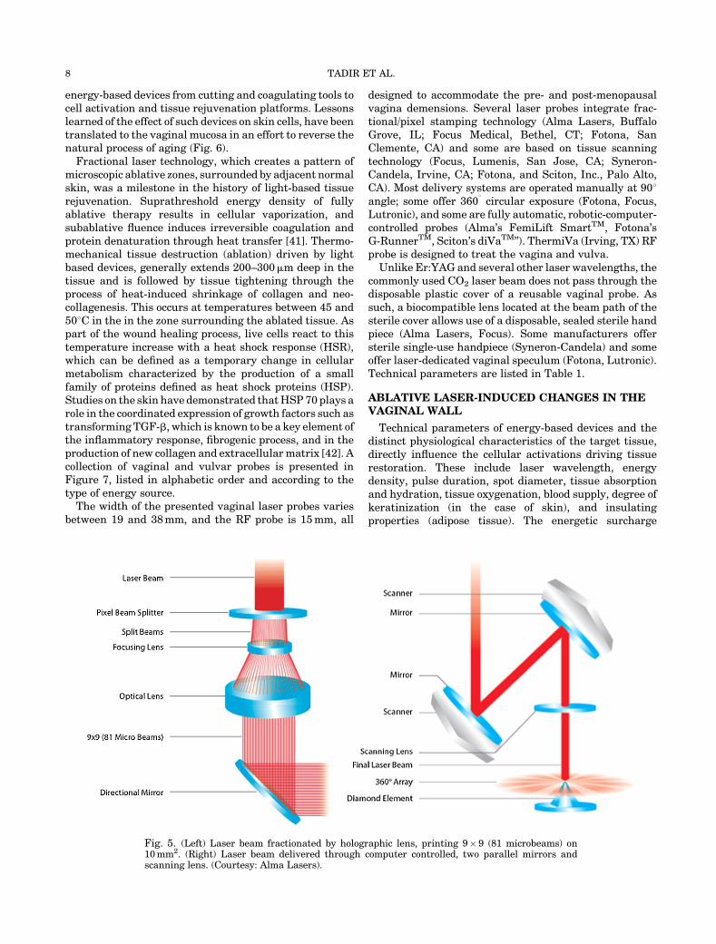
energy-based devices from cutting and coagulating tools tocell activation and tissue rejuvenation platforms. Lessonslearned of the effect of such devices on skin cells, have beentranslated to the vaginal mucosa in an effort to reverse thenatural process of aging (Fig. 6).
Fractional laser technology, which creates a pattern ofmicroscopic ablative zones, surrounded by adjacent normalskin, was a milestone in the history of light-based tissuerejuvenation. Suprathreshold energy density of fullyablative therapy results in cellular vaporization, andsubablative fluence induces irreversible coagulation andprotein denaturation through heat transfer [41]. Thermo-mechanical tissue destruction (ablation) driven by lightbased devices, generally extends 200–300mm deep in thetissue and is followed by tissue tightening through theprocess of heat-induced shrinkage of collagen and neo-collagenesis. This occurs at temperatures between 45 and508C in the in the zone surrounding the ablated tissue. Aspart of the wound healing process, live cells react to thistemperature increase with a heat shock response (HSR),which can be defined as a temporary change in cellularmetabolism characterized by the production of a smallfamily of proteins defined as heat shock proteins (HSP).Studies on the skin have demonstrated thatHSP70 plays arole in the coordinated expression of growth factors such astransforming TGF-b, which is known to be a key element ofthe inflammatory response, fibrogenic process, and in theproduction of new collagen and extracellularmatrix [42]. Acollection of vaginal and vulvar probes is presented inFigure 7, listed in alphabetic order and according to thetype of energy source.
The width of the presented vaginal laser probes variesbetween 19 and 38mm, and the RF probe is 15mm, all
designed to accommodate the pre- and post-menopausalvagina demensions. Several laser probes integrate frac-tional/pixel stamping technology (Alma Lasers, BuffaloGrove, IL; Focus Medical, Bethel, CT; Fotona, SanClemente, CA) and some are based on tissue scanningtechnology (Focus, Lumenis, San Jose, CA; Syneron-Candela, Irvine, CA; Fotona, and Sciton, Inc., Palo Alto,CA). Most delivery systems are operated manually at 908angle; some offer 3608 circular exposure (Fotona, Focus,Lutronic), and some are fully automatic, robotic-computer-controlled probes (Alma’s FemiLift SmartTM, Fotona’sG-RunnerTM, Sciton’s diVaTM”). ThermiVa (Irving, TX) RFprobe is designed to treat the vagina and vulva.Unlike Er:YAG and several other laser wavelengths, the
commonly used CO2 laser beam does not pass through thedisposable plastic cover of a reusable vaginal probe. Assuch, a biocompatible lens located at the beam path of thesterile cover allows use of a disposable, sealed sterile handpiece (Alma Lasers, Focus). Some manufacturers offersterile single-use handpiece (Syneron-Candela) and someoffer laser-dedicated vaginal speculum (Fotona, Lutronic).Technical parameters are listed in Table 1.
ABLATIVE LASER-INDUCED CHANGES IN THEVAGINAL WALL
Technical parameters of energy-based devices and thedistinct physiological characteristics of the target tissue,directly influence the cellular activations driving tissuerestoration. These include laser wavelength, energydensity, pulse duration, spot diameter, tissue absorptionand hydration, tissue oxygenation, blood supply, degree ofkeratinization (in the case of skin), and insulatingproperties (adipose tissue). The energetic surcharge
Fig. 5. (Left) Laser beam fractionated by holographic lens, printing 9�9 (81 microbeams) on10mm2. (Right) Laser beam delivered through computer controlled, two parallel mirrors andscanning lens. (Courtesy: Alma Lasers).
8 TADIR ET AL.

endured by an excessively high increase in tissuetemperature can jeopardize cell survival. In the case ofvaginal mucosa, in which epithelial cells and the groundsubstance of the connective tissue are rich in water, it isimportant to consider that healthy premenopausal mucosais highly hydrated while the atrophic postmenopausalmucosa is characteristically dry [43]. Therefore, a con-trolled power of the source must be used.Based on successful fractional CO2 laser treatment of
aging-related skin changes [44,45], and on preliminaryexperience with the fractional CO2 laser applied for vaginalrejuvenation [14], the irradiation parameters of fractionalCO2 laser were further optimized through an ex vivo studyon the vaginal mucosa as MonaLisa TouchTM (DEKA,Florence, Italy) treatment [14]. A pilot study on 50postmenopausal women suffering from severe symptomsof vaginal atrophy demonstrated the effectiveness of thistreatment [46]. Clinical efficacy, including breast cancersurvivors, for whom estrogen treatment is contraindicated,was confirmed by various authors [47–51]. Histologicallyevident modifications of the postmenopausal atrophicvaginal mucosa following fractional CO2 laser treatmenthas provided a structural basis for understanding themolecular mechanisms responsible for restoration to ahealthy mucosa [14,15,46].
REJUVENATION OF THE VAGINAL MUCOSA
Premenopause
As mentioned earlier, the premenopausal vaginalmucosa is characterized by a squamous stratified non--keratinized epithelium and a lamina propria of connective
tissue, often protruding into the undersurface of theepithelium, with papillae rich in small blood vessels.Considering the absence of glands in the vaginal mucosa,epithelial cell shedding represents a sort of secretoryactivity critical for vaginal health. Starting from thebasal layer, where epithelial cells proliferate in orderto compensate for the loss of the cells shedded at themucosal surface, epithelial cells undergo a process ofdifferentiation. Glycogen begins to be synthesized by thecells of the intermediate layers and is subsequently storedin the superficial cells. When the most superficial cells areshed into the vaginal lumen, their glycogen content isdeposited at the epithelial surface, forming favorableconditions for Lactobacilli activity, which is a key factorin maintaining the low pH of the healthy inner vaginalenvironment.
The connective tissue is rich in blood vessels, whichpenetrate as small capillaries inside the papillae,providing metabolic support (nutrients, oxygen, andother molecules) to the intermediate and superficialcell layers. Collagen fibers are organized in compactbundles of different orientation which, together withelastic fibers, constitute an effective support for themucosal part of the vaginal wall. The ground substance,comprised of glycosaminoglycans, proteoglycans, andmultiadhesive glycoproteins, is a significant componentof the extracellular matrix of the connective tissueand represents the most permeable part of the matrix.Its components are particularly rich in polar groupscapable of linking large quantities of water moleculesand are responsible for the high hydration status(turgidity) of the mucosa. The consequently high
Fig. 6. (A) Fractionalmicro-ablation inducing cell activation and tissue rejuvenation at 45–508C [42].(B) Tissue ablation and thermal effects on adjacent layers (Courtesy: Tadir Y).
LIGHT AND ENERGY BASED THERAPEUTICS FOR GSM 9

permeability of the matrix permits easy diffusion ofwater, ions, nutrients, signaling molecules, and manyother molecular species through the extracellularmatrix.
Postmenopause
The decline and later arrest of ovarian estrogenproduction correlates with significant structural and
functional changes in the vaginal mucosa, resulting inatrophy, characterized by significant thinning of theepithelium with a reduction of epithelial renewaldynamics and the directly related absence of superficialcell desquamation. The epithelium-connective tissueinterface appears smooth due to the reduction and/orabsence of papillae and blood vessels (Fig. 8A). In theconnective tissue, fibroblasts are characterized by a small
TABLE 1. Technical Parameters of Vaginal Probes Listed in Manufacturer’s Alphabetic Order (Courtesy:
Manufacturers)
Brand name
Laser type/ wavelength
(nm) or RF
Pulse duration
(ms)
Maximum
energy/pulse (mJ)
Surface area “lased”/
exposure (mm2)
Alma Lasers, Buffalo Grove, IL. CO2 – 10,600 400 500 (per pixel) 10
Fotona, San Clemente, CA. Er:YAG – 2,940 250 240 J (per pass)
3 J/cm2
80 (cm2), non-ablative,
entire surface
Focus Medical, Bethel, CT. CO2 – 10,600 1–200 60 10
Lumenis, San Jose, CA. CO2 – 10,600 NA 7.5/10/12.5 NA
Lutronic Aesthetics, Burlington, MA. Er:YAG – 2,940 0.2 � max. 1500
(Dual mode)
3.7 J (per pulse) 144 (per pulse)
Sciton, Inc. Palo Alto, CA. Hybrid: 2,940/1,470 150/20 300/100 1.5–2.5
Syneron-Candela, Irvine, CA. CO2 – 10,600 20–1,066 70 10
ThermiVA, Irving, TX. RF 460kHz � Estimated tissue
temp. 47oC
10
NA: not available.
Fig. 7. Energy-based probes for vaginal and vulvar treatment listed in alphabetic order. (Courtesy:manufacturers).
10 TADIR ET AL.
cytoplasm around the nucleus and by frequent projec-tions of very thin processes into the surrounding matrix.In addition, cells exhibit a lower number of organelles,particularly of the rough endoplasmic reticulum andthe Golgi apparatus, both involved in the synthesisand turnover of the molecular components of theextracellular matrix, functions significantly reduced inthe postmenopause.
POSTMENOPAUSAL MUCOSA AFTERFRACTIONAL CO2 LASER TREATMENT
The post-treatment stratified squamous epithelium isthicker and comprised of 20–40 cell layers which providecells for differentiation and superficial shedding. Basallayer cells appear closely packed as in continuouslyrenewing stratified epithelium. The intermediate layercells appear enlarged, with the nucleus surrounded by awide cytoplasm. In the most superficial layer, many widecells, shedding from the epithelial surface into the vaginallumen, are observable (Fig. 8B).The basal surface of the epithelium appears character-
istically indented due to the presence of numerous papillaeof connective tissue, protruding onto the undersurface ofthe epithelium, yielding an uneven appearance to the
epithelial-connective tissue junction. Elongated bloodcapillaries inside the papillae are also clearly observable(Fig. 9). Reliable histochemical methods such as Periodicacid-Schiff (PAS) and Alcian blue staining, enableddetection of large quantities of glycogen in the cytoplasmof epithelial cells of the intermediate and superficial layers,and inside the numerous exfoliating cells of the mostsuperficial layer. The comparison with the atrophicmucosa before treatment is presented as Figure 10.
Epithelial changes induced by the laser treatment, andthe renewed cells differentiation with synthesis, storage,and delivery of glycogen (Fig. 11), are the consequence ofthe delivery of cytokines/growth factors in the connectivetissue stimulating a renewal of its structure and function-ality. This is achieved by a full restoration of the connectivetissue, both fibers (mainly collagen) and the groundsubstance, rich in highly hydrophilic molecules. In thisway, the hydration and permeability of the extracellularmatrix are improved, permitting a high rate of moleculartraffic both inside the connective tissue and betweenthe newly formed vessels and the epithelium, withrestoration of related epithelial functions (proliferation,differentiation with a renewed glycogen synthesis, anddesquamation).
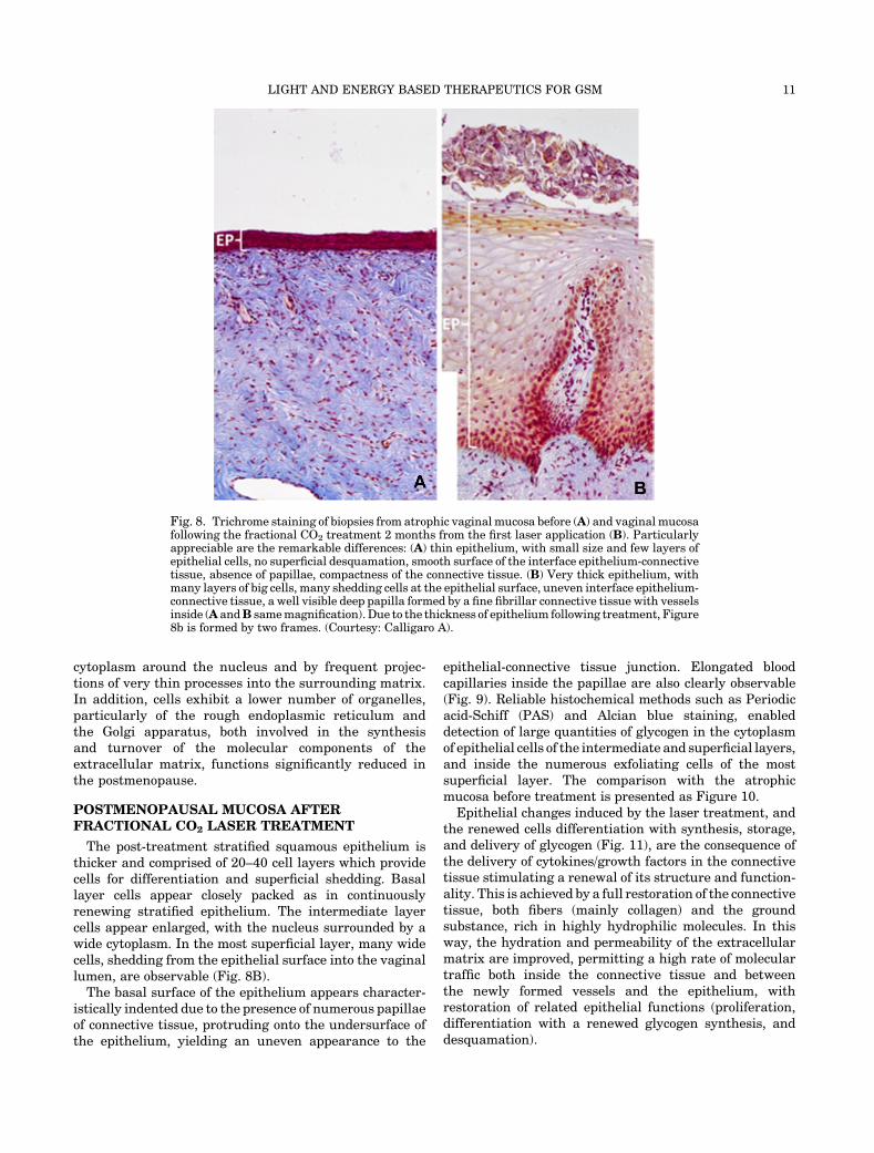
Fig. 8. Trichrome staining of biopsies from atrophic vaginal mucosa before (A) and vaginal mucosafollowing the fractional CO2 treatment 2 months from the first laser application (B). Particularlyappreciable are the remarkable differences: (A) thin epithelium, with small size and few layers ofepithelial cells, no superficial desquamation, smooth surface of the interface epithelium-connectivetissue, absence of papillae, compactness of the connective tissue. (B) Very thick epithelium, withmany layers of big cells, many shedding cells at the epithelial surface, uneven interface epithelium-connective tissue, a well visible deep papilla formed by a fine fibrillar connective tissue with vesselsinside (A andB samemagnification).Due to the thickness of epithelium following treatment, Figure8b is formed by two frames. (Courtesy: Calligaro A).
LIGHT AND ENERGY BASED THERAPEUTICS FOR GSM 11

Comparative microscopic and ultrastructural analysesof biopsies collected before versus after fractionalCO2 lasertreatment, demonstrate the recovery of all the mucosalstructures, suggesting restoration of full physiologicalfunctioning, both in the epithelial and connective tissuecompartments. The observed modifications of the mucosalstructures following fractional CO2 treatment are summa-rized below (Figs. 8–12).
Epithelium
Following the fractional CO2 laser treatment, theepithelial structure is fully restored, due to newly active
proliferative and differentiative processes of epithelialcells, with thickening and desquamation. The lactic acid-producing Lactobacilli vaginalis (bacteria physiologicallyresident in the vagina) feed on glucose derived from theglycogen delivered by shedding epithelial cells, acidify thevaginal transudate fluid at the mucosal surface, andthereby restore the vaginal environment to the acidicpremenopausal state, and prevent the colonization of yeastand other bacterial pathogens [52,53].Connective tissue, lamina propria fibroblasts are
characterized by high organelle content, an extendedrough endoplasmic reticulum, which is the site ofprocollagen synthesis [54], and frequently dilated cister-nae (Fig. 12A and B). A well-developed Golgi apparatuswith flattened stack of membranes and associatedvesicles (Fig. 12C), the site of synthesis of polysacchar-ides and of the glycosylation of the protein components ofthe ground substance, such as glycoproteins, proteogly-cans, and multiadhesive glycoproteins, is also detectable[55]. These structural features provide the machinerynecessary for the synthesis of both the fibrillar and theground substance components of the extracellular ma-trix. In parallel, the high blood vessel content in theconnective tissue supports the renewed activity offibroblasts and capillaries penetrating the newly formedpapillae underneath the epithelium (Fig. 9), and providesmetabolic support for epithelial cell proliferation anddifferentiation.Significant amounts of data relating to the impact of
CO2 laser treatment on human skin, were obtained usingreverse transcriptase real-time polymerase chain reac-tion (PCR) technology and immunohistochemistry[56]. The reported highly significant increase in themRNA levels of procollagen, metalloproteinases (MMPs),
Fig. 10. PAS staining of vaginal mucosa sections from biopsies before (A) and following treatment(B). Besides the evident difference in the epithelial thickness, the PAS signal shows (A) few smallcells containing glycogen (fuchsin-stained) versus (B) many larger cells stained, starting from thelow intermediate cell layer. Also of note in (B) is the superficial desquamation of intensely stainedcells, which deposit glycogen at the surface of mucosal epithelium. (Courtesy: Calligaro A).
Fig. 9. Common feature following treatment. At the center of thefigure, a papilla with a small and long vessel inside is easilyidentifiable due to its erythrocyte content and the thin endothelialprofiles. In the insert, numerous newly formed papillae followingtreatment are recognizable. H&E staining. (Courtesy: Calligaro A).
12 TADIR ET AL.

primary interleukin-1b, tumor necrosis factor-a, andprofibrotic cytokine TGF-b1 following CO2 laser applica-tion to the skin suggests stimulation of tissue renewalmechanisms, which seemingly occurs in the vaginalmucosa as well. Stimulation of connective tissue matrixremodeling by fibroblasts following fractional CO2 laserapplication has been tightly correlated with the activa-tion of heat shock proteins (HSPs). Immunohistochemicalanalysis of HSP in human fractional CO2 laser treatedskin revealed a persistent collagen-associated responsepersisting at least 3 months following treatment [57]. Thecollagen chaperone HSP47 [58,59] localized in the roughendoplasmic reticulum plays an important role in theearly stages of post-treatment collagen biosynthesis [60].In the application of fractional CO2 laser on human skin[61], the local increase in different cytokines has beenhistochemically demonstrated, particularly transforminggrowth factor-b (TGF-b; stimulating matrix proteins,such as collagen), basic fibroblast growth factor (bFGF;
stimulating angiogenetic activity with endothelial cellmigration and proliferation), epidermal growth factor(EGF; stimulating the re-epithelization), platelet-derivedgrowth factor (PDGF; stimulating fibroblasts to produceextracellular matrix components), and vascular endothe-lial growth factor (VEGF; regulating vasculogenesis andangiogenesis) (Fig. 6A). These observations [61] closelyparallel the observed microscopic and ultrastructuralmodifications of the postmenopausal atrophic vaginalmucosa following fractional CO2 laser treatment, that is,regulated stimulation of fibroblast-driven renewal of theextracellular matrix and activation of epithelial celldifferentiation. The expression of these mechanisms isinitiated in the first suprabasal layers of the epithelium(glycogen synthesis) and in the connective tissue with thestimulation of fibroblast activity and the formation ofnew vessels. Other studies on human skin corroboratethese findings relating to increased cytokines/growthfactor expression following fractional CO2 laser applica-tion [62,63].
Deployment of the fractional laser for the treatment ofthe atrophic vaginal mucosa leads to neocollagenesis andto production of ground substance components within theconnective tissue, and glycogen and acidic mucins withinthe epithelium and on the epithelial surface. Theseactivities revert the vaginal mucosa from an atrophicstate to a healthy premenopausal state, resulting inhighly significant symptomatic relief [46].
Sub-ablative (or non-ablative) long pulse modes havebeen evaluated for their ability to induce deep thermaleffects on the surface of the vaginal mucosa. Deepcollagen coagulation by stacking repetitive Er:YAG laserpulses on the same tissue site has been described initiallyby numeric modeling [64]. This mode of laser applicationon the mucosa can generate a thermal change in deeperlayers, with only minor superficial ablation (5mm) on thesurface, but with sufficient caloric effect to thermallyalter the chromophore. Using a large spot size (>5mm),the heating process can be achieved due to the perma-nence of the long-pulse stimulus and the repetition ofexposures. This deepens the heat effect in the laminapropria to a depth of at least 500–1,000mm (depending onthe degree of tissue hydration), inflicting the known“Joule effect” (photothermal and thermochemical effect)on the lamina propria of the vaginal mucosa. In turn, alocal rise in temperature, release of bradykinin andhistamine with relaxation of precapillary arteriolarsphincters, and vasodilation are achieved in an overalleffect called “The Phenomenon of Thermal Reperfusion”(FTR). Deep coagulation of collagen by repetitive Er:YAGlaser pulses has been predicted by theoretical models,demonstrated on animal skin [65,66], and confirmed inthe vaginal wall [67].
These technologies open new perspectives on thepotential use of electromagnetic radiation to re-establishstructural and physiological conditions in other genitouri-nary anatomic sites. Other non-laser energy sources suchas RF and High Intensity Focused Ultrasound (HiFU) arebeing tested for vaginal rejuvenation as well.
Fig. 11. High-resolution, light microscope image of a CO2 lasertreated-section, embedded in epoxy resin for electron microscopy,and stained with toluidine blue. In the lower part of the image,some cells of the lower intermediate layerwith a very clear nucleuscontaining a highly visible nucleolus (as a dark blue spot), showsome violet-stained masses (white arrows). These represent thefirst stores of newly synthesized glycogen inside epithelial cells,resulting from the restored differentiation process. In the upperlayers, glycogen masses are increasingly extended to almostcompletely fill the cytoplasm, until superficial shedding (desqua-mation). (Courtesy: Calligaro A).
LIGHT AND ENERGY BASED THERAPEUTICS FOR GSM 13

Vaginal Tightening focuses on restoring functionality byreversing the deterioration of vaginal wall structuresresulting from natural aging and physical trauma accom-panying childbirth. Lack of strength and support of vaginaltissues correlates with reduced sexual pleasure, as firstdescribed in the 1960s by Masters and Johnson [68].Surgical techniques for vaginal tightening aimed tomodifythe diameter of the vaginal canal by surgically removingextra tissue. In severe cases, the approach ismore invasiveand targets the fascia approximation and muscularplication. Microablative fractional lasers [13], and RF[69–71] devices induce superficial tissue shrinkage as wellas deep stimulation of the collagen layer of the submucosa.Unlike skin complications such as hypertrophic scarring,ectropion formation, and disseminated infection, whichhave all been reported following fractional laser skinresurfacing [72]. No major complications have beenreported following energy-based vaginal rejuvenationprocedures.
Urinary Incontinence (UI)
Involuntary leakage of urine is called urinary inconti-nence, and stress urinary incontinence (SUI) is the most
prevalent type caused by transient increase of intra-abdominal pressure and weakening of support naturallyprovided by the bladder, urethra, and pelvic structures. Inhealthy states, this support is primarily provided bycollagen and elastic connective tissue which degeneratewith age and under various pathological conditions [73].Urethral support can be improved by exploiting theenergy-based tightening effect. Numerous publicationsconfirm enhanced urinary control following treatment ofSUI with an Er:YAG laser [74–78], CO2 laser [79], and RF[80] all of which triggers a photo-thermal effect, as deep as0.5mm inside the vaginal wall, and results in a 30%reduction in tissue volume. Mechanical pull by the deepertissue layers following shrinkage of the upper, photo-thermally processed tissue layers, further contributes tothe tightening effect. At the same time, thermally inducedneo-collagenesis improves thickness, elasticity, and firm-ness of the vaginal wall [75,76].Er:YAG laser treatment for SUI has been performed
with an accessory kit, (Fig. 7B, IncontilaseTM, Fotona),composed of two hand pieces (full spot and fractional orpatterned spot), two adapters or extensions (angular andcircular) to deliver energy within the vaginal canal, and alaser speculum, which facilitates opening of the vaginal
Fig. 12. Electronmicroscopy of fibroblasts in samples collected following fractional CO2 treatment.(A) Connective tissue fibroblasts showing a relatively compacted cell body, with a cytoplasmappearing almost completely filled with rough endoplasmic reticulum (rER). The nucleus iseuchromatic, with a well-presented nucleolus. These features support active synthesis of proteins(collagen and others) to be delivered to the surrounding extracellularmatrix, promoting its renewal.(B) In fibroblasts, close to the rERprofiles (cisternae formed bymembraneswith ribosomes attachedat their surface), dilated cisternae with attached ribosomes are frequently observed as vesiclescontaining a fine, filamentous material, representing the molecular precursors of extracellularfibrillar components. (C) AGolgi apparatus is clearly visible. TwoGolgi complexes, formed by stacksof membranes with associated dilations and vesicles, are recognizable (white arrows). (Courtesy:Calligaro A).
14 TADIR ET AL.
canal and serves as a guide for the handpiece adapter.Using the circular adapter and full spot handpiece, totalenergy of about 650 J is delivered along the entire length ofthe vaginal wall. In the second phase, the patterned spothandpiece is attached to the angular adapter to irradiate(250 J) the anterior, sub-urethral vaginal wall. Thefractional handpiece, delivering energy (�100 J) to theintroitus area, is then deployed to repair connective tissuearound the urethra and to shrink the introitus. All threephases are done in one treatment session. This lasersystem effectively addressed various grades of SUI andoverall urinary control by triggering a rejuvenation effectin various structures of the mucosa, that is, the epitheliumand the lamina propria [76–78], with optimal resultsachieved following three treatment sessions performed at4-week intervals. Statistically significant improvementswere noted in incontinence severity indices (ISI) and in thedegree of incontinence. In the largest published study of175 women treated with this protocol, 77% of the SUIpatients were dry, while only 34% of the patientspresenting with a combination of both, SUI and urgeincontinence (mixedUI), remained dry at 1-year follow-up.At 2 years post-treatment, 82.8% of the patients in thegroup with improved outcome remained dry, while 17.2%of patients developed mild incontinence. Three years afterthe initial treatment, the regimen was repeated in 25% ofpatients to address mild incontinence. No difference intreatment efficacy was noted between pre- versus post-menopausal patients [75].In another study (N¼ 35) in which same protocol is
repeated for the treatment of SUI, urodynamic studies, padtesting, lower urinary tract symptoms (LUTS), and sexualfunction questionnaires were assessed before and aftertreatment. Among 28 women with baseline pad weights>1 g, 11 (39.3%) were objectively cured and 11 (39.3%)improved. Among the 18 women with mild SUI (i.e., padweight 1–10 g) nine (50%) were cured and five (27.8%)improved. Among 32 women with complete questionnairedata at 6 months, seven (21.9%) were subjectively cured,and four (12.5%) improved. The authors concluded that theprocedure for mild SUI was moderate at 6-month follow-up, but was not effective for pad weight>10g. Moreover, itimproved LUTS, QoL, and sexual function [78].A prospective observational study of the efficacy of
fractional CO2 laser in post-menopausal women withmoderate-severe clinical signs (N¼ 53) showed significantimprovement of dyspareunia, dryness, burning, itching,dysuria, frequency, urgency, urgency, and SUI scoresassessed by various standard questionnaires. Participantsreceived intravaginal therapy, once a month for 3consecutive months, with a CO2 laser system (MonaLisaTouchTM, DEKA, Florence, Italy) and the setting wereperformed as previously described [79]. Urinary symptomsimproved significantly, as reflected by the significantreduction in the scores of the relevant urinary and qualityof life questionnaires. Over the course of the study, thenumber of patientswith lower urinary tract symptomswasimproved significantly as reflected by various scores. Allparticipants showed a >5-point improvements in the
King’s Health Questionnaire (KHQ) score, which includesobjective assessments of symptoms and strong psychomet-ric aspects of urinary incontinence. Factors predictive ofideal CO2 laser therapy candidateswere not identified [79].
Non-ablative monopolar transcutaneous temperature-controlled radiofrequency (TTCRF) has been tested for itsefficacy in treating SUI symptoms in a small group women(N¼10/cohort) and compared to sham treatment [80]. Thevaginal sidewalls, as well as the anterior and posteriorwalls, were heated to a temperature of 40–458C bysweeping “half-moon motions” of the RF probe (Fig. 7D,RF). Treatment time was 3–5 minutes per zone. TCRFtreatment was associated with a significant (P< 0.01)improvement of ICIQ-SF and UDI-6 scores. Seven of 10patients (70%) had a negative cough stress test aftertreatment; improvements were maintained for up to 12weeks. The clinical outcomes were supported by thepositive histologically detected vaginal changes seen inwomen suffering from postmenopausal vaginal atrophy.TTCRF was well tolerated with no complications reportedby treated patients. While the cohort size in some of thesestudies was small, and duration of follow-up short theresults are encouraging.
EXTERNAL GENITALS
Anatomy and Histology
The skin of the external genitals is comprised of theepidermis (E), the dermis (D), and the subcutaneous tissue(SG) (Fig. 13). The epidermis consists of a stratifiedsquamous epithelium, containing basal cells (which divideto form keratinocytes), and keratinocytes. Cells divide inthe basal layer andmove up, changing their appearance asthey move from one layer to the next, resulting in thecontinuous replacement of cells in the epidermal layer. Thekeratinocytes synthesize insoluble protein which remainsin the cell and later becomes a major component of theouter layer, called the stratum corneum. The dermis iscomposed of three types of connective tissue: collagen,elastic tissue, and reticular fibers. It is divided into anupper, thin papillary layer, which is composed of thinrandomly arranged collagen fibers, and a deeper reticularlayer, composed of thick collagen fibers arranged parallelto the surface of the skin surface [81].
The vulva exhibits several different types of epithelium[82]. The lateral aspect of the labia majora is covered withdry, keratinized, hair-bearing skin. The medial aspect ofthe labiamajora and the entire labiaminora displaymoist,modified mucous membrane, comprised of a partiallykeratinized epithelium that contains subtle hair follicles,apocrine sweat glands, and sebaceous glands. A variably‘distinct line of demarcation termed the “Hart’s line” isevident at the base of the medial aspect of each labiumminus, separating the modified mucous membranes of thelabia minora from the mucous membrane of the vestibule[82]. This line also demarcates the boundary between theembryonic ectoderm (labia minora) and endoderm (thevestibule). The tissue medial to the Hart’s line is anunkeratinized, non-hair-bearing epithelium with mucous
LIGHT AND ENERGY BASED THERAPEUTICS FOR GSM 15

secreting glands. The hymen separates the vestibule fromthe vagina (Fig. 13).
Lichen Sclerosus (LS) is a chronic, benign, inflammatoryskin disease of unknown etiology which can occur at anysite but has a predilection for the genital area and rarelyinvolves the vagina. In themajority of patients, the diseaseis progressive, causing vulvar scarring, loss of portions orall of the labia minora (resorption), clitoral adhesion andnarrowing of the introitus [83] (Fig. 14A and B).
Vulvar LS can occur at any age, and its true prevalenceis not known, but is estimated at 1 in 59 women in ageneral gynecology practice [84]. The causes for LS areunknown, but several mechanisms have been studied,suggesting multifactorial origin, with involvement of
genetic, hormonal, autoimmune, and inflammatory fac-tors. Symptoms include pruritus, soreness, irritation,and pain. Patients may also complain of dyspareunia,dysuria, and peri-anal involvement although uncommon,active disease may be asymptomatic. The diagnosis ofvulvar LS is based upon the presence of characteristicclinical manifestations, ideally with histological confir-mation [85]. Most frequently, it affects the labia minoraand/or labia majora, clitoris and clitoral hood, and it mayalso involve the perineum and the perianal skin.LS usually requires permanent management to main-
tain remission [85]. If left untreated, it has significantpotential to result in the progressive destruction ofthe vulvar architecture and less commonly, has been
Fig. 13. Normal vulva and histology. Left: Hair bearing vulvar skin A-labia majora, B-labiaminora, C-clitoral hood, D-Hymen, E-vestibule. black arrow-clitoris, red arrow-Hart’s line. Right:Histology of the non-hair bearing vulvar skin. This is hair bearing skin, �40 magnification. Theepidermis (E) does not have estrogen receptors, so does not atrophy like the vaginawhen estrogen iswithdrawn. D-dermis. There are superficial sebaceous glands (Fordyce spots) present (SG). Likehair bearing skin, the labia minora atrophy when estrogen is withdrawn because the dermalfibrioblasts have estrogen receptors. (Courtesy: Lev-Sagie A).
Fig. 14. Vulvar lichen sclerosus—(A) LS can be diagnosed clinically by inspection of the vulva forthe characteristic thin, white, wrinkled skin, and changes in vulvar architecture. Findings in LSinclude hypopigmentation, hemorrhages (black arrow), loss of normal architecture includingdisappearance of labia minora (blue arrow), buried clitoris (red arrow), and narrowing of theintroital opening. Note that the distinction between the labiamajora andminora is lost. The diseaseinvolves the perineal and perianal areas. (B) LS histology �40 magnification—on histologicalexamination, the epidermis (E) is typically thinned (accounting for the older nomenclature “lichensclerosus et atrophicus”), although areas of thickened skin (H-hyperkeratosis) may exist. The upperdermis exhibits homogenization of collagen (black arrow) with a band of lymphocytes below thisregion (blue arrow). (Courtesy: Lev-Sagie A).
16 TADIR ET AL.

associated with vulvar squamous cell carcinoma (SCC)[85]. Topical corticosteroids are the mainstay of therapy,and super-potent topical corticosteroids, such as clobetasolpropionate, have long been considered the standard of carefor vulvar LS. Recommended therapy usually consists ofdaily application of corticosteroid ointment for 6–12weeks,followed by maintenance therapy two to three times perweek, if symptoms improve [85].Medical therapy leads to symptomatic relief in most
women; however, the incidence of long-term sequelae, suchas scarring and SCC, is unclear, although adequatetreatment seems to be associated with a reduced risk ofdevelopment of neoplasia [86].Superficial ablation of LS by means of a CO2 laser has
previously been described [87–89]. The technique requiresuse of general anesthesia and a healing period of severalweeks. The high cost for the laser tools and the need toperform such procedures in surgical facilities has limitedwidespread embracement by physicians and patients.However, advancements in fractional laser technology,
which do not entail use of general anesthesia, and incurminimal superficial ablation alongside thermal cell activa-tion and tissue rejuvenation, have increased the potentialuse of this treatment approach [90]. Several reports of LStreated successfully with fractional CO2 laser are available(Table 2). In a recent prospective study, symptomaticpostmenopausal patients (N¼ 27) diagnosed with LS weretreated with a fractional CO2 laser, three sessions 4–6weeks intervals [91]. Twenty-four patients reportedcessation of itching and pain, and in 26 patients visibleimprovement of skin color, elasticity, and vascularity wasnoticed. No histology is available in this study followingtreatment.
Two additional cases with typical symptoms andhistologically proven LSwho failed long-term conventionaltreatment with clobetasol 0.5% and 2% testosterone creamunderwent successful treatment with fractional CO2 laser.Unlike conventional symptomatic treatment, histologicalevaluations before and after the laser treatment demon-strate curative tissue changes (Fig. 15. Elias J, personal
TABLE 2. Studies Using CO2 Laser for the Treatment of Lichen Sclerosus
Author and year Trial design Primary outcomes Findings Complications
Kartamaa and
Reitamo (1997) [87]
Case series Subjective assessment of
lichen sclerosus (better,
asymptomatic, some
recurrence)
Among male and female
patients (N¼10) with
biopsy proven refractory
LS, 76% were
asymptomatic after CO2
laser treatment with mean
follow up of 32 months.
Three patients needed
repeat treatment
Peterson et al.
(2004) [89]
Case report Subjective symptoms
visual re-epithelization
All patients (N¼ 2) with
refractory anogenital
lichen sclerosus had
resolution of symptoms
and re-epithelization after
2–3 years with CO2 laser
therapy
None
Windahl (2006) [88] Retrospective
cohort
Subjective patient
assessment of
recurrence of symptoms,
any visible penile lesion,
recurrence of meatal
stenosis
In men with histologically
verified penile lichen
sclerosus treated with CO2
laser, 80% (N¼50) had no
local symptoms at mean
follow-up 14 years
Two patients required
further treatment
Lee et al. (2016) [90] Case series Subjective symptom
resolution
In women (N¼5) with biopsy
confirmed severe vulvar
LS recalcitrant to topical
corticosteroid treatment;
four of whom responded
positively to fractional CO2
laser treatment, and one to
ablative CO2 laser therapy
Two patients needed
repeat treatment at 6–8
months. Two patients
reported discomfort
with laser treatment
Baggish (2016) [91] Prospective
(N¼27)
Subjective symptom
resolution
24/27 cessation of itching and
pain. 26/27 visible
improvement of skin color,
elasticity, and vascularity
None
LIGHT AND ENERGY BASED THERAPEUTICS FOR GSM 17

communication). Three treatment sessionswere conductedat 1 month intervals. Three passes were made at eachsession, with the Alma Laser—pixel hand piece set, on lowto medium energy (10–20mJ/pixel). Patients becameasymptomatic shortly after the third treatment session,and the vulva, introitus and clitoral areas appearedhealthy, with elastic closure of the introitus. Tissueappearance was maintained throughout the 6 monthpost-treatment follow-up period. Histological assessmentof samples collected 45 days following the last treatmentsession showed a trophic epithelium with acanthotic areaswithout superficial hyperkeratosis. Prospective trials thatare in progress may determine if this treatment concept isvalid.
Vulvodynia and Provoked Vestibulodynia
Vulvodynia is defined as “pain in the vulva” persistingfor at least 3 months, in the absence of an obviousunderlying cause, and can include various clinical fea-tures. Some patients have continuous, diffuse vulvar pain(generalized, spontaneous vulvodynia), while others expe-rience localized pain, usually provoked by touch.The current nomenclature [92] uses a symptom-based
classification to characterize the pain in regard to itslocation (localized, generalized, mixed), conditions thatprovoke it (contact, spontaneous, or mixed), its temporalpattern (intermittent or constant), and its onset (primaryor secondary).Provoked, localized vestibulodynia (PVD), previously
called “vulvar vestibulitis syndrome,” is the term used todescribe the phenomena of superficial pain confined to thevulvar vestibule, provoked by touch. In some women, paincan be caused by minimal touch (sitting, tight-fittingclothing), whereas in others, it is provoked by vaginalpenetration during sexual intercourse, tampon insertion,or gynecological examination, resulting in dyspareunia orcomplete inability to have intercourse. The pain is oftendescribed as burning or cutting and can radiate throughoutthe entire vestibule or confined to the lower vestibule.Erythema may be evident but is no longer considered adefining criterion.No single causative factor has yet been identified, and
the etiology is considered multifactorial [93]. Clinicalobservations, epidemiological studies, and basic researchsuggest different etiologies, including inflammation, hor-monal factors, pelvic floor muscle dysfunction, peripheraland central neural pain disorders, genetic predisposition,
Fig. 15. Vulvar lichen sclerosus. Histological assessment of Masson’s trichrome-stained tissuesamples before versus after factional CO2 laser treatment. Left, case 1; Right, case 2. Histology: Toprow: Hyperkeratosis (a), dermal atrophy, hydropic degeneration of the basal epithelial cells (b)dermo epidermal clefts (c) homogeneous papillary dermis, afibrillar with frosted glass appearanceand edema (d) and inflammatory infiltrate of polymorphonuclear band and plasma cells (e) andinflammatory infiltrate of polymorphonuclear band and plasma cells. Bottom row: Trophicepithelium without superficial hyperkeratosis (a), persistence in some basal cells hydropicdegeneration (b), persistence of some areas with dermo-epidermal clefts (c), and lamina propriafibrillar with irregular spaces containing translucent material (d). (Courtesy: Elias J, Galich M).
18 TADIR ET AL.
psychosocial, and sexual factors. Current diagnosis isbased upon clinical symptoms while treatment is assessedon a trial and error approach. As a result, many forms oftherapeutic interventions have been used, and outcomesremain largely inconclusive, with response rate rangingbetween 40% and 85% and with many women notresponding to any treatment [94]. Given the high placeboresponse rate of around 50%, only placebo-controlled trialswill provide substantial data about beneficial therapeutictechniques. In a recent study patients (N¼ 70) underwentfractional micro-ablative CO2 laser treatment for vestibu-lar pain plus vestibulodynia (n¼ 37) or GSM (N¼ 33). Astatistically significant improvement of symptoms wasnoted after three sessions of vestibular fractional CO2 lasertreatment. Improvements gradually increased throughoutthe study period andweremaintained through the 4monthfollow-up period [95].
External Genitals
Energy based devices are used to perform surgicalprocedures on the external genitals: some are for medicalindications and some are considered as aesthetic gynecol-ogy. Treatment outcome on the use of low level lasertherapy on episiotomy [Alvarenga A. et al.] and fractionalCO2 laser on cesarean section scars [Karmisholt K. et al]are published in this issue. The field of aestheticgynecology lies beyond the scope of this review and somehighlights are briefly mentioned. (Table 3) [96–98].
Hopes or Hypes
Randomized controlled trials (RCT), peer-reviewedpublications, meta-analyses, and other objective evalua-tions all aim to protect patients from a medical procedurethat has not been adequately tested and proven safe andeffective. This review, drafted by a large group of expertsmay lack some of the above-mentioned “protective tools,”but we hope that it offers relevant collective information todefine directions for future research, and may serve as
“The Guide to the Perplexed” in the arena of gynecologicenergy-based clinical procedures.
A gynecologist’s office serves as a refuge and, occasion-ally, as a confessional when discussing the most privategynecologic, sexual and other intimate concerns. Surgeonsdiscerningly consider behavioral, medical, pharmacologic,and reconstructive pelvic surgical procedures, in additionto potential medical and cosmetic external treatments.Office-based cosmetic procedures offered by plastic, der-matologic, or gynecologic surgeons provide solutions forcommon vulvovaginal disorders; however, it is unclearwhether alternatives are always fully presented to womenseeking cosmetic services. Form and function are bothequally important for pelvic floor health as well asvulvovaginal health. Patients may seek cosmetic gyneco-logical treatments for more than just aesthetic reasons.Functionally, they may struggle with clothing discomfortor dyspareunia [99] in addition to struggles relating tobody image or feelings of inadequacy during intimatemoments. Patients may also have complaints ofvaginal laxity, now defined by the IUGA/ICS as “excessivevaginal looseness,” [100] or have issueswith air trapping orvaginal wind resulting in noises emanating from thevagina during activity [101].
While vaginal laxity refers to loose vaginal tissue, thisbroad and unspecified term is subjective when contrastedwith well-quantified pelvic floor disorders, including pelvicorgan prolapse (cystocele, uterine prolapse, rectocele, andvaginal vault prolapse), SUI and fecal incontinence. Suchdisorders typically result from loss of connective tissuesupport in concert with muscle and nerve damage, mostcommonly occurring after childbirth or related to otherinciting factors such as chronic cough and strainingassociated with constipation. Conversely, vaginal laxityand enlarged labia majora or minora are often a matter ofpersonal preference which can be influenced by the media,social media, and relationships [102–106]. While numer-ous case series have been published on labiaplasty/vaginoplasty outcomes [103–106], no comparative trials
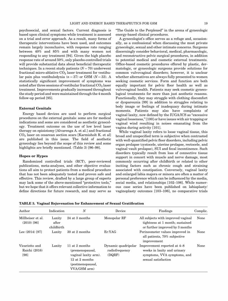
TABLE 3. Vaginal Rejuvenation for Enhancement of Sexual Gratification
Author Indication N Device Findings Complic.
Millheiser et al.
(2010) [96]
Laxity
after
childbirth
24 at 3 months Monopolar RF All subjects with improved vaginal
tightness at 1 month; sustained
or further improved by 3 months
None
Lee (2014) [97] Laxity 30 at 2 months Er:YAG Perineometer values improved in
all patients, 70% subjective
improvement
None
Vicariotto and
Raichi (2016)
[98]
Laxity 11 at 2 months
(premenopausal,
vaginal laxity arm).
12 at 2 months
(postmenopausal,
VVA/GSM arm)
Dynamic quadripolar
radiofrequency
(DQRF)
Improvement reported at 4–8
weeks in laxity and urinary
symptoms, VVA symptoms, and
sexual satisfaction
None
LIGHT AND ENERGY BASED THERAPEUTICS FOR GSM 19

exist on labiaplasty techniques and their clinical efficacy inimproving orgasmic response. Risks related to clitoral andlabial surgery include denervation and scarring. Proce-dures such as re-virgination represent lack of educationandmisinformation perpetuating myths that virginity canbe confirmed by the presence/absence of a hymen.
Data relating to energy-based therapies in cosmetogy-necology primarily focus on treatment of vulvovaginalatrophy/GSM. Unfortunately, the large majority of thetrials are retrospective, single-center studies withshort-term follow-up. Furthermore, outcomes measuresare not standardized and are often only subjective orhistology-based. Energy-based devices are currently beingemployed for treatment of labial hypertrophy, vaginallaxity, and reduced sexual pleasure, but limited publisheddata are available.
The vaginal laser versus estrogen cream therapy(VELVET) trial, comparing the effects of these twotreatment options of vaginal dryness, is currentlyrecruiting GSM patients at the Cleveland Clinic andfive other sites across the United States. The VaginalErbium Laser Academy Study (VELAS) [107] willexamine vaginal erbium laser therapy as a potentiallynon-invasive treatment for GSM and for SUI. Thisinternational study will include 1,500 postmenopausalwomen. The CURLS study, aiming to assess clobetasolversus fractional CO2 laser for treatment of vulvar lichensclerosus, is currently recruiting patients at MedstarHealth Washington Hospital Center in Washington, DC.A trial on the effect of fractional/pixel CO2 laser on GSMsymptoms is in progress at the Cleveland Clinic, Florida,and monitoring of tissue changes with advanced opticaltechniques is in under review at the Beckman LaserInstitute, University of California, Irvine. Other studieswith CO2 laser, Er:YAG laser, and RF are in progress inItaly, Greece, Argentina, Slovenia, Israel, Spain, the UK,UAE, Japan, Korea, Indonesia, and more sites aroundthe globe.
Consensus and Controversies
Data generated from the unprecedented wave ofpublications in peer-review journals on cell activationand vaginal wall rejuvenation following exposure toenergy-based devices justify the quotation: “You can foolall the people some of the time, and some of the people allthe time, but you cannot fool all the people all the time”(Abraham Lincoln).
A consensus has been reached among the authors of thismanuscript on the clinical effectiveness of energy-basedtherapies on GSM-related symptoms. The currentlyavailable data on the effects of fractional laser and RFon the skin, and additional information reported in almost20 peer-reviewed publications on GSM-related symptoms,unequivocally demonstrate the following vaginal changes:thickening of glycogen enriched postmenopausal epithe-lium, neovascularization, and neocollagenesis in thelamina propria, increased lactobacilli counts, reducedpH, vaginal wall tightening, and improved urination
control with minimal risk of short- and long-termcomplications.Controversial issues that remain to be determined: the
preferred energy-based device, be it laser versus RF, CO2
versus Er:YAG laser, tissue scanning versus pixel stamp-ing of the fractional laser technology, ablative versus non-ablative technology, single laser wavelength versus hybridconsecutive beams, the optimal depth of micro-ablation ortemperature of controlled energy deposition. In addition,understanding the physiologic cascade of events during theprocess of tissue rejuvenation, most effective treatmentsession intervals and duration of effects that justify atouch-up session remain to be determined. Studies arenecessary to evaluate the possible curative effect on lichensclerosus, vestibulitis, additive effect of platelet-richplasma/growth factor, and hyaluronic acid when adminis-tered in conjunction with energy deposition. The potentialimpact of the energy-based treatment on estrogen recep-tors levels in the vaginal wall will also need to beaddressed. Add-on therapies such as estrogen primingfor vaginal atrophy (Gaspar A, et al. described elsewhere inthis issue) [67], corticosteroids for lichen sclerosus, orsuperficial tissue cooling [108] for all of the above also needfurther investigation. Computer simulations of thermaltissue remodeling during transvaginal and transurethrallaser treatment of female SUI [Hardy L. et al.] describedelsewhere in this issue, and the potential use of intra-urethral fractional laser treatment of female SUI (GasparA. et al. unpublished data) are tested. The cost-effective-ness of energy-based regimens in comparison to otheravailable treatments, and unwanted long-term effects ofsuch interventions all remain to be defined.
ACKNOWLEDGMENTS
The authors gratefully acknowledge the informationreceived from energy-based device manufacturers aspresented in Table 1, and Figure 7. No commercial sourcesupported the collaboration and preparation of thismanuscript.
REFERENCES
1.Kaplan I, Goldman J, Ger R. The treatment of erosions of theuterine cervix by means of the CO2 laser. Obstet Gynecol1973;41(5):795–796.
2.Bellina JH, Polanyi TG.Management of vaginal adenosis andrelated cervico-vaginal disorders in DES-exposed pregnancyby means of carbon dioxide laser surgery. J Reprod Med1976;16:295–296.
3.Bruhat M, Mage G, Mahnes M. Use of the CO2 laser inlaparoscopy. Proceedings of the 3rd international society forlaser surgery. Ed. Kaplan I. 1979;24:235–238.
4.Tadir Y, Kaplan I, Zukerman Z, Ovadia J. Laparoscopic CO2
laser sterilization. In: Semm K, Mettler L, editors. HumanReproduction. Exerpta Medica, Amsterdam: Elsevier; 1981.551, pp 423–431.
5.Nezhat FR, Nezhat CR, Silfen SL. Videolaseroscopy foroophorectomy. Am J Obstet Gynecol 1991;165:1323–1330.
6.Baggish MS, ElBakry MM. A flexible CO2 laser fiber foroperative laparoscopy. Fertil Steril 1986;46(1):16–20.
7.Cossmann PH, Romano V, Sp€orri S, et al. Plastic hollowwaveguides: Properties and possibilities as a flexible radia-tion delivery system for CO2 laser radiation. Lasers SurgMed1995;16(1):66–75.
20 TADIR ET AL.

8.Tadir Y, Kaplan I, Zukerman Z, Ovadia J. Hysteroscope forCO2 laser application. Lasers Surg Med 1984;4:153–156.
9.Haimovitch S, Lopez-Yarto M, Urresta-Avila J, SaaverdaTascon A, Hernandez JL, Carreras Collado R. Officehysteroscopic laser enucleation of sumcucous myomas with-out mass expraction: A case study. Biomed Res Int 2015;2015:905204.
10.Tadir Y, Wright WH, Vafa O, Ord T, Asch R, Berns MW.Micromanipulation of sperm with a laser generated opticaltrap. Fertil Steril 1989;52:870–875.
11.Harper JC, Wilton L, Traeger-Synodinos J, et al. The ESHREPGD consortium: 10 years of data collection. Hum ReprodUpdate 2012;18(3):234–247.
12.Brightman LA, Brauer JA, Anolik R, Weiss E, Karen J,Chapas A, Hale E, Bernstein L, Geronemus RG. Ablative andfractional ablative lasers. Dermatol Clin 2009;27(4):479–489.
13.Gaspar A, AddamoG, Brandi H. Vaginal fractional CO2 laser:A minimally invasive option for vaginal rejuvenation. Am JCosmetic Surg 2011;28(3):156–162.
14.Salvatore S, Leone Roberti Maggiore U, Athanasiou S, et al.Histological study on the effect of microablative fractionalCO2 laser on atrophic vaginal tissue: An ex vivo study.Menopause 2015;22(8):845–849.
15.Zerbinati N, Serati M, Origoni M, et al. Microscopic andultrastructural modifications of postmenopausal atrophisvaginal mucosa after fractional carbon dioxide laser treat-ment. Lasers Med Sci 2015;30(1):429–436.
16.Reid R, Absten GT. Lasers in gynecology: Why pragmaticsurgeons have not abandoned this valuable technology.Lasers Surg Med 1995;17(4):378–383.
17.Copperman AB, DeCherney AH. Turn, turn, turn. FertilSteril 2006;85(1):12–13.
18.De Landsheere L, Munaut C, Nusgens B, et al. Histology ofthe vaginal wall in women with pelvic organ prolapse: Aliterature review. Int Urogynecol J 2013;24(12):2011–2020.
19.Jannini EA, d’Amati G, Lenzi A. Histology and immunohisto-chemical studies of female genital tissue. In: Goldstein I,Meston CM, Davis S, Traish A, editors. Women’s SexualFunction and Dysfunction: Study, Diagnosis and Treatment.UK: CBC Press, Taylor an Francis Group, 2005. p 784.
20.Wenstrup RJ, Florer JB, Brunskill EW, Bell SM, ChervonevaI, Birk DE. Type V collagen controls the initiation of collagenfibril assembly. J Biol Chem 2004;279(51):53331–53337.
21.Birk DE, Fitch JM, Babiarz JP, Doane KJ, Linsenmayer TF.Collagen fibrillogenesis in vitro: Interaction of types I and Vcollagen regulates fibril diameter. J Cell Sci 1990;95(4):649–657.
22.Menopause society. Management of symptomatic vulvovagi-nal atrophy: 2013 position statement of The North AmericanMenopause Society. Menopause 2013;20(9):888–902 904.
23.CotreauMM, Chennathukuzhi VM, Harris HA, et al. A studyof 17beta-estradiol-regulated genes in the vagina of postmen-opausal women with vaginal atrophy. Maturitas 2007;58(4):366–376.
24.Portman DJ, Gass MLS. Genitourinary syndrome of meno-pause: New terminology for vulvovaginal atrophy from theInternational Society for the Study ofWomen’s Sexual Healthand The North American Menopause Society. Maturitas2014;79(3):349–354, and Menopause 2014;21(10):1063–1068.
25.Willhite LA, O’Connell MB. Urogenital atrophy: Preventionand treatment. Pharmacotherapy 2001;21(4):464–480.
26.Bachmann G. Clinical manifestations and diagnosis ofvaginal atrophy. UpToDate 2014;1–10.
27.AlexiadesMR.Wave science in dermatologic therapy. J DrugsDermatol 2015;14(11):1190.
28.Alexiades-Armenakas MR, Dover JS, Arndt KA. The spec-trum of laser skin resurfacing: Nonablative, fractional, andablative laser resurfacing. J Am Acad Dermatol. 2008;58-(5):719–737, quiz 738–40.
29.Fisher JC. Photons, physiatrics, and physicians: A practi-cal guide to understanding laser light interaction withliving tissue, part 1. J Clin Laser Med Surg 1992;10(6):419–426.
30.Anderson R, Parrish JA. Selective photothermolysis: Precisemicrosurgery by selective absorption of pulsed radiation.Science, New Series 1983;220(4596):524–527.
31.Alexiades-Armenakas MR, Dover JS, Arndt KA. Fractionallaser skin resurfacing. J Drugs Dermatol 2012;11(11):1274–1287.
32.Alexiades-Armenakas M, Sarnoff D, Gotkin R, Sadick N.Multi-center clinical study and review of fractional ablativeCO2 laser resurfacing for the treatment of rhytides, photoag-ing, scars and striae. J Drugs Dermatol 2011;10(4):352–362.
33.Baker-Jarvis J, Kim S. The interaction of radio-frequencyfields with dielectric materials at macroscopic to mesoscopicscales. J Res Inst Stand Technol 2012;117:1–60.
34.Alexiades M, Berube D. Randomized, blinded, 3-arm clinicaltrial assessing optimal temperature and duration for treat-ment with minimally invasive fractional radiofrequency.Dermatol Surg 2015;41(5):623–632.
35.Dunbar SW, Goldberg DJ. Cosmetic dermatology: An update.J Drug Dermatol 2015;14(11):1229–1238.
36.Alexiades-Armenakas M, Dover JS, Arndt KA. Unipolarversus bipolar radiofrequency treatment of rhytides andlaxity using a mobile painless delivery method. Lasers SurgMed 2008;40(7):446–453.
37.Lee MS. Treatment of vaginal relaxation syndrome with anErbium:YAG laser using 908 and 3608 scanning scopes: A pilotstudy & short-term results. Laser Ther 2014;23(2):129–138.
38.Ogrinc UB, Sencar S, Lenasi H. Novel minimally invasivelaser treatment of urinary incontinence in women. LasersSurg Med 2015;47:689–697.
39.Fistonic N, Fistonic I, Gustek SF, et al. Minimally invasive,non-ablative Er:YAG laser treatment of stress urinaryincontinence in women: A pilot study. Laser Med Sci 2016;31:635–643.
40.Alexiades-Armenakas M, Newman J, Willey A, Kilmer S,et al. Prospective multicenter clinical trial of a minimally-invasive temperature-controlled bipolar fractional radiofre-quency system for rhytid and laxity treatment.Dermatol Surg2013;39(2):263–273.
41.SaediN, JalianHR,PetelinA, ZacharyC. Fractionation: Past,present, future. Semin Cutan Med Surg 2012;31(2):105–109.
42.Capon A, Mordon A. Can thermal laser promote skin woundhealing? Am J Clin Dermatol 2003;4(1):1–12.
43.Lev-Sagie A. Vulvar and vaginal atrophy: Physiology, clinicalpresentation, and treatment considerations. Clin ObstetGynecol 2015;58(3):476–491.
44.Berlin AL, Hussain M, Phelps R, Goldberg DJ. A prospectivestudy of fractional scanned nonsequential carbon dioxidelaser resurfacing: A clinical and histopathologic evaluation.Dermatol Surg 2009;35(2):222–228.
45.Lee CJ, Park JH, Ciesielski TE, Thomson JG, Persing JA.Retinoids, 585-nm laser, and carbon dioxide laser: A numericcomparison of neocollagen formation in photoaged hairlessmouse skin. Aesth Plast Surg 2008;32(6):894–901.
46.Salvatore S, Nappi RE, Zerbinati N, et al. A 12-weektreatment with fractional CO2 laser for vulvovaginal atrophy:A pilot study. Climacteric 2014;17(4):363–369.
47.Perino A, Calligaro A, Forlani F, et al. Vulvo-vaginal atrophy:A new treatment modality using thermoablative fractionalCO2 laser. Maturitas 2015;80:296–301.
48.Sokol ER, KarramM.An assessment of the safety and efficacyof a fractional CO2 laser system for the treatment ofvulvovaginal atrophy. Menopause 2016;23(10):1102–1107.
49.Leone Roberti Maggiore U, Parma M, Candiani M, SalvatoreS. Microablative fractional CO2 laser for vulvo vaginalatrophy in women with an history of breast cancer.J Minim Invasive Gynecol 2015;22(6S):S100.
50.Pieralli A, Fallani MG, Becorpi A, et al. Fractional CO2 laserfor vulvovaginal atrophy (VVA) dyspareunia relief in breastcancer survivors. Arch Gynecol Obstet 2016;294(4):841–846.
51.Pagano T, De Rosa P, Vallone R, et al. Fractional micro-ablative CO2 laser for vulvovaginal atrophy in women treatedwith chemotherapy and/or hormonal therapy for breastcancer: A retrospective study. Menopause 2016;23(10):1108–1113.
52.Mac Bride MB, Rhodes DJ, Shuster LT. Vulvovaginalatrophy. Mayo Clin Proc 2010;85(1):87–94.
53.Athanasiou S, Pitsouni E, Antonopoulou S, et al. The effect ofmicroablative fractional CO2 laser on vaginal flora ofpostmenopausal women. Climacteric 2016;19(5):512–518.
LIGHT AND ENERGY BASED THERAPEUTICS FOR GSM 21

54.Stephens DJ. Cell biology: Collagen secretion explained.Nature 2012;482(7386):474–475.
55.AlbertsB, JohnsonA, Lewis J. Transport from theER throughthe Golgi apparatus. Molecular Biology of the Bell, 4thedition. New York: Garland Science; 2002. pp 775–781.
56.Orringer JS, Kang S, Johnson TJ, et al. Connective tissueremodeling induced by carbon dioxide laser resurfacing ofphotodamaged human skin. Arch Dermatol 2004;140(11):1326–1332.
57.Hantash BM, Bedi VP, Kapadia B, et al. In vivo histologicalevaluation of a novel ablative fractional resurfacing device.Lasers Surg Med 2007;39:96–107.
58.Ishida Y, Nagata K. Hsp47 as a collagen-specific molecularchaperone. Methods Enzymol 2011;499:167–182.
59.Tasab M, Batten MR, Bulleid NJ. Hsp47: A molecularchaperone that interacts with and stabilizes correctly-foldedprocollagen. EMBO J 2000;19(10):2204–2211.
60.Dafforn TR, DellaM,Miller AD. Themolecular interactions ofheat shock protein 47 (Hsp47) and their implicationsfor collagen biosynthesis. J Biol Chem 2001;276(52):49310–49319.
61.PrignanoF, Campolmi P, BonanP, et al. Fractional CO2 laser:A novel therapeutic device upon photobiomodulation of tissueremodeling and cytokine pathway of tissue repair. DermatolTher 2009;22(Suppl 1):S8–S15.
62.NowakKC,McCormackM,KochRJ.The effect of superpulsedcarbon dioxide laser energy on keloid and normal dermalfibroblast secretion of growth factors: A serum-free study.Plast Reconstr Surg 2000;105(6):2039–2048.
63.Manolis EN, Kaklamanos IG, Spanakis N, et al. Tissueconcentration of transforming growth factor b1 and basicfibroblast growth factor in skin wounds created with a CO2
laser and scalpel: A comparative experimental study, using ananimal model of skin resurfacing. Wound Repair Regen2007;15:252–257.
64.Reinisch L. Scatter-limited phototherapy: A model for lasertreatment, of skin. Lasers Surg Med 2002;30(5):381–388.
65.Luka€o M, Mqjaron B, Rupnik T. Ablative and thermal effectsof Er:YAG laser on human tissue. In: WaidelichW,WaidelichR, Waldschmidt J, editors. Lasers in Medicine. Berlin:Springer; 1998. pp 566–572.
66.Majaron B, Plesteniak Luka�e M. Thermo-mechanical laserablation of soft biological tissue: Modeling the micro explo-sions. Appl Phys B 1999;69:71–80.
67.Gaspar A, Brandi H, Gomez V, Luque D. Efficacy of Erbium:YAG laser treatment compared to topical estriol treatment forsymptoms of genitourinary syndrome of menopause. LasersSurg Med 2016; DOI: 10.1002/lsm.22569
68.MastersWH, JohnsonVE.Human Sexual Response. Toronto;New York: Bantam Brooks; 1966.
69.Millheiser LS, Pauls RN, Herbst SJ, Chen BH. Radio-frequency treatment of vaginal laxity after vaginal delivery:Nonsurgical vaginal tightening. J Sex Med 2010;7(9):3088–3095.
70.Mulholland RS. Radio frequency energy for non-invasive andminimally invasive skin tightening. Clin Plast Surg2011;38(3):437–448.
71.Alinsod RM. Transcutaneous temperature controlled radio-frequency for orgasmic dysfunction. Lasers Surg Med 2016;48(7):641–645.
72.Metelitsa AI, Alster TS. Fractionated laser skin resurfacingtreatment complications: A review. Dermatol Surg 2010;36(3):299–306.
73.Norton P, Brubaker L. Urinary incontinence in women.Lancet 2006;367:57–67.
74.Fistonic N, Fistonic I, Lukanovic A, Findri-Gustek S, SortaBilajac Turina I, Franic D. First assessment of short termefficacy of Er:YAG laser treatment on stress urinary inconti-nence in women: Prospective cohort study. Climacteric 2015;18(Sup 1):37–42.
75.Ogrinc BU, Sencar S. Novel minimally invasive lasertreatment of urinary incontinence in women. Lasers SurgMed 2015;47:689–697.
76.Fistonic N, Fistonic I, Findri Gustek SS, et al. Minimallyinvasive, non-ablative Er:YAG laser treatment of stress
urinary incontinence in women—A pilot study. J Laser MedSci 2016;31:635–643.
77.Pardo J, Sola V, Morales A. Treatment of female stressurinary incontinence with Erbium YAG laser in non-ablativemode. Eur J Obstet Gynecol Reprod Biol 2016;204:1–4.
78.Tien YW, Hsiao SM, Lee CN, Lin HH. Effects of laserprocedure for female urodynamic stress incontinence on padweight, urodynamics, and sexual function. Int Urogynecol J2016, [Epub ahead of print].
79.Pitsouni E, Grigoriadis T, Tsiveleka A, et al. Microablativefractional CO2-laser therapy and the genitourinary syndromeof menopause: An observational study. Maturitas 2016;94:131–136.
80.Leibaschoff G, Izasa PG, Cardona JL, Miklos JR, Moore RD.Transcutaneous temperature controlled radiofrequency(TTCRF) for the treatment of menopausal vaginal/genitouri-nary symptoms. Surg Technol Int 2016;26(XXIX):149–159.
81.Mirowski GW, Edwards L. Geniatl anatomy. In: Edwards L,editor. Genital Dermatology Atlas. Philadelphia: LippincottWilliams and Wilkins; 2004. pp 1–8.
82.Habif TP. Skin anatomy. In: by Mosby, editor. ClinicalDermatology. St. Louis, MI; 2004.
83.Smith YR, Haefner HK. Vulvar lichen sclerosus: Pathophysi-ology and treatment. Am J Clin Dermatol 2004;5(2):105–125.
84.Goldstein AT, Marinoff SC, Christopher K, Srodon M.Prevalence of vulvar lichen sclerosus in a general gynecologypractice. J Reprod Med 2005;50(7):477.
85.Cooper SM, Arnold SJ. Vulvar lichen sclerosus. In uptodate2016, last updated: Jun 06, 2016. Available at: http://www.uptodate.com/
86.Bradford J, Fischer G. Long-term management of adultvulvar lichen sclerosus: A prospective cohort study of 507women. JAMA Dermatol 2015;151(10):1061–1067.
87.Kartamaa M, Reitamo S. Treatment of lichen sclerosus withcarbon dioxide laser vaporization. Br J Dermatol 1997;136(3):356–359.
88.Windahl T. Is carbon dioxide laser treatment of lichensclerosus effective in the long run? Scand J Urol Nephrol2006;40(3):208–211.
89.Peterson C, Lane J, Ratz J. Successful carbon dioxide lasertherapy for refractory anogenital lichen sclerosus. DermatolSurg 2004;30:1148–1151.
90.Lee A, Lim A, Fischer G. Fractional carbon dioxide laser inrecalcitrant vulval lichen sclerosus. Australas J Dermatol2016;57(1):39–43.
91.Baggish M. Fractional CO2 laser treatment for vaginalatrophy and vulvar lichen sclerosus. J Gynecol Surg2016;32(6):309–317.
92.Bornstein J, Goldstein AT, Stockdale CK, et al. ISSVD,ISSWSH and IPPS consensus terminology and classificationof persistent vulvar pain and vulvodynia. Obstet Gynecol2016;127(4):745–751.
93.Pukall CF, Goldstein AT, Bergeron S, et al. Vulvodynia:Definition, prevalence, impact, and pathophysiological fac-tors. J Sex Med 2016;13(3):291–304.
94.AndrewsJC.Vulvodynia interventions—Systemic reviewandevidence grading. Obstet Gynecol Surv 2011;66(5):299–315.
95.Murina F, Karram M, Salvatore S, Felice R. Fractional CO2
laser treatment of the vestibule for patients with vestibulo-dynia and genitourinary syndrome of menopause: A pilotstudy. J Sex Med 2016;13(12):1915–1917.
96.Millheiser LS, Pauls RN, Herbst SJ, Chen BH. Radio-frequency treatment of vaginal laxity after vaginal delivery:Nonsurgical vaginal tightening. J Sex Med 2010;7(9):3088–3095.
97.Lee MS. Treatment of vaginal relaxation syndrome with anErbium:YAG laser using 908 and 3608 scanning scopes: A pilotstudy & short-term results. Laser Ther 2014;23(2):129–138.
98.Vicariotto F, Raichi M. Technological evolution in the radio-frequency treatment of vaginal laxity and menopausal vulvo-vaginal atrophy and other genitourinary symptoms: Firstexperiences with a novel dynamic quadripolar device.Minerva Ginecol 2016;68(3):225–236.
99.Goodman MP, Otto PJ, Matlock DL, et al. Evaluation of bodyimage and sexual satisfaction in women undergoing female
22 TADIR ET AL.

genital plastic/cosmetic surgery. Aesthet Surg J 2016;36(9):1048–1057.
100.Haylen BT, Maher CF, Barber MD, et al. An InternationalUrogynecological Association (IUGA)/International Conti-nence Society (ICS) joint report on the terminology for femalepelvic organ prolapse (POP). Int Urogynecol J 2016; 27(2):165–194.
101.Miranne J, Marek TM, Mete M, Iglesia CB. Prevalence andresolution of auditory passage of vaginal air in women withpelvic floor disorders. Obstet Gynecol 2015;126(1):136–143.
102.Cartwright R, Cardozo L. FOR: Women should be free to optfor cosmetic genital surgery. BJOG 2014;121(6):767.
103.Iglesia CB. AGAINST: The social vulnerability and culturalview of women as sex objects need to end. BJOG 2014;121(6):768.
104.Creighton S. AGAINST: Labiaplasty is an unnecessarycosmetic procedure. BJOG 2014;121(6):768.
105.Iglesia CB, Yurteri-Kaplan L, Alinsod R. Female genitalcosmetic surgery: A review of techniques and outcomes. IntUrogynecol J 2013;24(12):1997–2009.
106.Sharp G, Tiggemann M, Mattiske J. Predictors of consid-eration of labiaplasty: An extension of the tripartiteinfluence model of beauty ideals. Psychol Women Q 2014;182–193.
107.Gambacciani M, Torelli MG, Martella L, et al. Rationaleand design for the Vaginal Erbium Laser Academy Study(VELAS): An international multicenter observationalstudy on genitourinary syndrome of menopause andstress urinary incontinence. Climacteric 2015; 18(Sup1):43–48.
108.Nelson JS, Milner TE, Anvari B, Tenenbaum BS, SvaasandLO, Kimel S. Dynamic epidermal cooling in conjunction withlaser-induced photothermolysis of port wine stain bloodvessels. Lasers Surg Med 1996;19(2):224–229.
LIGHT AND ENERGY BASED THERAPEUTICS FOR GSM 23