lec. 5 prosthodontics يلامجلا بنيز .د
TRANSCRIPT

1
Prosthodontics Lec. 5 زينب الجماليد.
The face is a significant social stimulus &is regarded as a clue to identity &personality
through various facial expression.
Maxillofacial prosthodontics: is an art and science of anatomic, functional and esthetic
reconstruction by means of non-living substitutes of those regions in the maxilla,
mandible and the face that are missing or defective .
Maxillofacial prosthesis: artificial device use to replace missing facial or oral
structures.
These causes result in defect in:
1. Intra-oral (maxilla &mandible) which will effecting the speech, mastication,
swallowing, &esthetic.
2. Extra-oral (eye, nose, ear, cranial bones) which will effecting the esthetic.
Indications of maxillofacial prosthesis:
1. When plastic surgery is contraindicated.
2. When recurrence of malignancy is expected.
3. When radiotherapy is being instituted, radium appliance and radium protector shield
can be used.
4. Temporary maxillofacial prosthesis can be used when plastic surgery requires various
steps.
Aim of maxillofacial prosthodontist( objectives):
Reconstruction of missing parts in maxilla &mandible &face with nonliving substitutes
(prosthesis) to achieve:
1. Improvement in esthetic or cosmetic appearance of the patient which is of prime
importance for everybody.
2. Restoration of function (as in cleft palate) that include:
a. Speech functions in patient with palatal lost part of the jaw.

2
b. Nutritional function in patient with lost part of the jaw.
c. Avoid escape of food to nasal cavity in children with cleft and overcome feeding
problem.
3. Preservation of residual structure:
a. To protect the adjacent tissue as in radium protective, also to protect wound, stop
bleeding and carry medication after surgery.
b. Protect the teeth as in mouth guard contact sport.
4. Therapeutic or healing effect( such as placement of radium applicator).
5. Psychological therapy: to raise the moral of the patient and help in healing fracture
segments in cases of fracture face.
Essentials of maxillofacial prosthetic appliance:
1. The appliance must be easily seated in place comfortably and securely and securely
as much as possible.
2. The appliance must be durable and easily clean.
3. The material must be inert and biocompatible.
4. The material must be easily adjusted and altered if needed.
Maxillofacial classification:
Patient can be categorized by maxillofacial defects or Causes:
1. Congenital deformations: are typically craniofacial defects that are present from birth.
The most common of these include cleft defects of the palate that may include the
premaxillary alveolus.
2. Traumatic or acquired that are result of trauma, or of disease and its treatment (gun
shot or accidental), these include a soft and or hard palate defect resulting from removal
of a squamous cell carcinoma.
3. Developmental defects are those defects that occur because of some genetic
predisposition that is expressed during growth and development.
Maxillofacial prosthesis are classified according to location into:
Extra-oral prosthesis: includes:-
1. Ocular prosthesis-eye.
2. Nasal prosthesis –nose.
3
3. Auricular prosthesis- ear.
4. Part of the face.
5. Nasal stent-prevent nasal septum collapse.
6. Cranial prosthesis – cranial bone.
7. Radiation stent- direct the radiation beam.
All the above are either fabricated as hard material(acrylic)(1,4,5,6) or soft material
(silicon, RTV)(2,3).
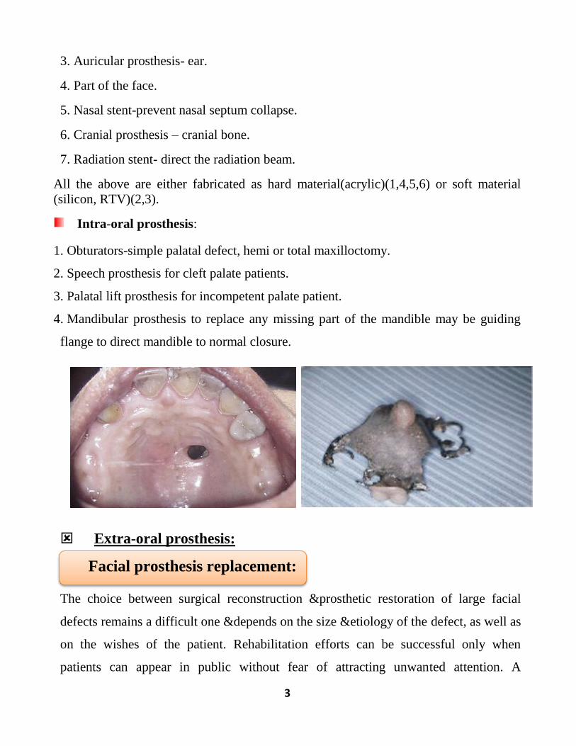
Intra-oral prosthesis:
1. Obturators-simple palatal defect, hemi or total maxilloctomy.
2. Speech prosthesis for cleft palate patients.
3. Palatal lift prosthesis for incompetent palate patient.
4. Mandibular prosthesis to replace any missing part of the mandible may be guiding
flange to direct mandible to normal closure.
Extra-oral prosthesis:
The choice between surgical reconstruction &prosthetic restoration of large facial
defects remains a difficult one &depends on the size &etiology of the defect, as well as
on the wishes of the patient. Rehabilitation efforts can be successful only when
patients can appear in public without fear of attracting unwanted attention. A
Facial prosthesis replacement:

4
replacement Facial prosthesis made from the original mold. A replacement prosthesis
does not require fabrication of a new mold in most of facial prosthesis replacement.
Generally, several prosthesis can be made from the same mold assuming no changes
occur in the tissue bed due to further surgery or age related topographical variations.
Since long time; these prosthesis are retained by an adhesive. These adhesives may
give good result but the duration of their activity &care for the adherence to prosthesis
may complicate the treatment. Facial prosthesis using dental implant &ball
attachments, bars or magnetic abutments may improve the results greatly; although
these attachments may requires additional surgical & technical steps.
1. They provide better retention of the prosthesis, so that the prosthesis is properly
positioned &the patient can wear it more confidently.
2. There is no skin irritation from adhesive & the prosthesis does not need to have
adhesive cleaned off each time it is used.
3. The prosthesis can be made thinner, with feathered edges that blend with the skin,
which offers the patient improved aesthetics.
4. A pre-operative planning meeting with the patient &working team shows not only
different prosthetic options but also e.g. cleaning of the abutments &prosthesis.
Furthermore reports have shown that implants are not uniformly successful, the
failures &complications appear to be site specific &radiation &time dependent.
A removable prosthesis attached to the skin which artificially restores part or all of the
nose. Fabrication of a nasal prosthesis requires creation of an original mold. Additional
Nasal prosthesis(artificial nose)
5
prosthesis usually can be made from the same mold, &assuming no further tissue
changes occur, the same mold can be utilized for extended periods of time.
An artificial ear produced from a previously made mold. A replacement prosthesis
does not require fabrication of a new mold. Generally, several prosthesis can be made
from the same mold, &assuming no further tissue bed changes occur, due to surgery or
age related topographical variations. Unfortunately, the presence of hair &the absence
of anatomic irregularities often result in unfavorable adhesive retention of an auricular
prosthesis. Endosseous implants, may permit positive retention of auricular prosthesis.
A biocompatible, permanently implanted replacement of a portion of the skull bones;
an artificial replacement for a portion of the skull bone.
Loss of eye is emotional and physical problem to the patient. In smaller defects
adhesive retention of the prosthesis may be satisfactory &the limited size of the defect
may prevent implant placement without interference with the prosthesis margins. As
orbital defects increase in size, the need for implant support becomes greater.
Auricular prosthesis (replacement ear):
Cranial prosthesis; skull plate, cranioplasty prosthesis, cranial impact
Orbital prosthesis:

6
Synonymous terminology: radiotherapy prosthesis, carrier prosthesis, radiation
applicator, radium carrier, intracavity carrier, intracavity applicator. A device used to
administer radiation to confined areas by means of capsules, beads or needles of
radiation emitting materials such as radium or cesium. Its function is to hold the
radiation source securely in the same location during the entire period of treatment. It
achieve close approximation &controlled application of radiation to a tumor deemed to
eradication.
It is used to carry skin or mucous membrane graft in vestibule, palate or mouth floor in
approximation to periosteum during initial healing and prevent formation of hematoma
between the graft and the underlying bone and periosteum.
Intra-oral prosthesis:
A maxillofacial prosthesis used to close, cover or maintain the integrity of the oral
&nasal compartments resulting from a congenital, acquired or developmental disease
process. The prosthesis facilitates speech °lutition by replacing those tissues lost
due to the disease process &can, as a result, reduce nasal regurgitation &hypernasal
speech, improve articulation, deglutition & mastication. An obturator prosthesis is
classified as surgical, interim or definitive.
A prosthesis which maintains the right &left maxillary segments of an infant cleft
palate patient in their proper orientation until surgery is performed to repair the cleft. It
closes the oral- nasal cavity defect, thus enhancing sucking &swallowing. Used on an
interim basis, achieves separation of the oral &nasal cavities in infants born with wide
Radiation carrier:
Obturator:
Feeding aid; feeding prosthesis:
Carrier stent:

7
clefts necessitating delayed closure. It is eliminated if surgical closure can be affected
or alternatively, with eruption of the deciduous dentition, a pediatric speech aid may be
made to facilitate closure of the defect.
A removable maxillofacial prosthesis used to restore an required or congenital defect
of the soft palate with a portion extending into the pharynx to separate the oropharynx
&nasopharynx during phonation °lutition, thereby completing pharyngeal
sphincter.
Such a prosthesis consist of:
A palatal component which contacts the teeth to provide stability and anchorage for
retention; a palatal extension, which crosses the residual soft palate.
And a pharyngeal component which fills the palatopharyngeal port during muscular
function, serving to restore the speech valve of the palatopharyngeal region.
A maxillofacial prosthesis used to maintain a functional
position for the jaw, improve speech °lutition following
trauma or/and surgery to the mandible or/and adjacent
structures.
A maxillofacial prosthesis which elevates the soft palate superiorly &aids in
restoration of soft palate functions which may be lost due to an acquired, congenital or
developmental defect.
Speech aid prosthesis:
Mandibular resection prosthesis:
Palatal lift prosthesis:

8
Teamwork for maxillofacial prosthetics :
1. Maxillofacial prosthodontist-play a major role in the treatment planning
&rehabilitation.
2. Skilled general dentist
3. Maxillofacial surgeon.
4. Neurosurgeon-cranail defect.
5. Special nursing team.
6. Radiologist.
7. Chemotherapeutics.
8. Maxillofacial good technician.
9. Speech pathologist.
10. Biocomunication therapy (community).
11. Psychotherapist.
Psychological consideration in maxillofacial patient:
Maxillofacial patients are classified according to the etiology into acquired, congenital
&developmental. Patient with acquired maxillofacial defect are usually resulted from
trauma or cancer, both of them were have normal anatomy &physiology but these are
changed or impaired due to the trauma or cancer. Usually patients with small defect
frequently appear more demanding &have higher expectation than patients with larger
defect. Patients with cancer may face chemotherapy, radiotherapy, recurrence &more
surgical procedure. Patient with congenital defects may understand that they are
different from norm &they may face a knowledge that there may be genetic
predisposition. Those patients usually face multiple surgeries, orthodontics
&prosthodontics procedures over several years in an attempt for correction of the
defect, again in cleft lip &palate patients you may expect variations from simple cleft
lip with minimal loss of function to extensive bilateral cleft lip &palate with sever

9
impairment in function. Patient with developmental defect may display some
emotional response similar to patients with congenital defects.
Requirement of ideal materials used for facial prosthesis:
Prostheses can be made from a variety of materials, such as Poly(methyl)
methacrylate, polydimethylsiloxane, &polyetherurethanes.
Ideal physical &mechanical properties:-
1. High elongation strength.
2. High tear strength.
3. Softness, compatible to the tissue.
4. High edge strength.
5. Translucent.
Ideal processing properties:-
1. Chemically inert after processing.
2. Long working time.
3. Ease of intrinsic &extrinsic coloring with commercially available colorant.
4. No color changes after processing, retain intrinsic &extrinsic colors .
5. Reusable mold.
Ideal biological properties:-
1. Biologically compatible.
2. Cleansable with disinfectant.
3. Color stability.
4. Resistance to the growth of the micro-organisms.
Prosthesis fixation:
These prostheses are retained with adhesives, tissue undercuts, or in some cases extra-
oral osseointegrated implant. Facial &intraoral prostheses can be connected with
magnets. The aesthetic result depends on the amount of tissue removed, type of
reconstruction, morbidity adjunctive treatment, and the physical characteristics of the
tissue base available to support &retain the prosthesis.
So prosthesis fixation may be:

11
1. Anatomic (teeth, alveolar ridge, residual hard palate, undercut)
2. Mechanical.
3. Magnets.
4. Implants.
5. Screw.
Primary factors that affect prosthetic success:
All prostheses must resist a variety of forces that may displace it &generate stress to
the residual structure of the orofacial complex. Prostheses success is often dependent
upon methods of compensation for diminished anatomic capacity for support, retention
&stability of a prosthesis.
In order to achieve a favorable level of retention, remaining teeth &the remaining
soft &hard tissues must be used to the optimal degree. This may be gained by:-
A. It is prudent to extend impressions as much as possible without interfering with
movable tissue.
B. Border molding is performed whenever a prosthesis depends on tissue support
whether that tissue is located within the defect or is part of the remaining structures.
C. In addition, close adaptation to the underlying tissue results in a thin fluid film
between the prostheses &the tissue, the thinner the intervening fluid, the greater the
prosthetic retention.
Frequently, the means of retention is used, &may encompass descriptive adjectives
such as adjacent tissue, teeth, dental/craniofacial implants or a combination of such,
thus appropriate terminology &classification of the prostheses in relation to the
retention means are:-
1. Tissue retained maxillofacial prostheses.
2. Tooth retained maxillofacial prostheses.
3. Implant retained maxillofacial prostheses.
4. Tissue/implant retained maxillofacial prostheses.
Teeth are the greatest asset for providing retention of the obturator prosthesis. The
amount of stress generated by the movement of the obturator may be great. The

11
number, position, &periodontal status of the remaining teeth are the most critical
factors in evaluating the amount of stress that remaining teeth may be able to absorb.
Support is the ability to resist displacement of the prosthesis towards the supporting
structure. Remaining teeth, remaining edentulous areas &the postsurgical defect are
the supporting tissues for prosthesis & prosthesis loads are generated through these
tissues to the underlying supporting bone.
A. Since the tissue has limited capacity for displacement, the greater the surface area of
tissue contact, the less the displacement of the prosthesis towards the tissue.
B. Maximum peripheral extension combined with an accurate adaptation to the
remaining teeth, the residual ridges & the postsurgical site will provide the most
favorable support for prosthesis.
Stability is the ability to resist displacement of the prosthesis by functional forces.
Adhesives enhance retention through optimizing interfacial force. The alteration in the
normal structures results in diminished potential for support &retention. Since the
majority of forces are not directed towards or away from the tissue, but generated at an
angle to the tissue, it is stability that is tested most frequently in function.
An alternative method of prosthetic retention has been developed. Endosseous
implants may be used to address the concerns of diminished support, retention
&stability.
Use of similar implants in extra-oral sites is growing in popularity especially for the
retention of auricular prosthesis and for bone anchored hearing aids.
-The use of endosseous implant support in maxillofacial defects can be complex. As
seen in most maxillofacial prosthetic patients, alterations in normal anatomy reduce
the opportunities for the clinician to place &restore endosseous implants. This
situation occurs when supporting bone is lost due to surgical resection or when tissue
is altered due to therapeutic modalities such as radiation.

12
-prosthetic designs &strategic implant placement must anticipate the functional
demands of the prosthesis while also recognizing the dislodging forces applied to the
prosthesis.
Sequence of treatment for pateints with intra-oral defect(maxilla or
mandible):
1. Pre-operative stage.
2. Post-operative stage.
3. Definitive &follow-up stage.
Pre-operative stage:
1. Close consultation : between the prosthodontics &surgeon (x-ray, study cast) to
outline the defect location &size. The discussion involve the possible preservation of
teeth &supporting structures that may be used for retention &stability of the
prosthesis(obturator). A compromise is necessary &preservation of teeth &structures
should not interfere with the surgical procedure to eliminate the disease.
2. Full dental treatment : for better oral hygiene which helps healing of surgical sight
&successful of future prosthesis:
a. Hopeless teeth.
b. Filling of caries teeth (abutment for prosthesis).
c. Proper periodontal health care.
3. Construction of pre-operative obturator, surgical splint.
A temporary maxillofacial prosthesis inserted during or immediately following
surgical or traumatic loss of a portion or all of one or both maxillary bones and
contiguous alveolar structures (i.e. gingival tissue, teeth). It is secured either by palatal
screw, suture or circumzygomatic wires. Old denture can be used as a surgical
obturator but it might create some problems because the denture mostly not fit as
before surgery therefore relining may help to improve patients’ acceptance and
tolerance. It is mostly used for 10 days more or less depends on treatment plane.
This prosthesis is placed immediately after surgery to:

13
1. Serve as matrix for surgical pack –to be removed
3-10 days.
2. Eliminate the need for naso-gastric tube feeding.
3. Better hygiene for surgical sight –better healing.
4. Improve patient speech.
5. Better psychological status of patient.
6. Reduces the period of hospitalization.
Requirement:
1. Light weight.
2. Strong.
3. Easy to alter( addition, removal to fit the surgical splint).
4. Made from acrylic with or without teeth.
Post-operative stage:
Construction of temporary(interim or transitional) post-operative obturator which is
constructed from post-surgical impression cast which has a false palate and false ridge
and generally no teeth. Every step of prosthesis construction must maximize prosthesis
adaptation to enhance retention and stability to ensure optimum function, esthetic,
occlusion, and correct jaw relations.
The closed bulb extending into the defect area is hollow.
The patient is usually seen every 2weeks because of the rapid soft tissue changes
that occur within the defect during organization and healing of the wound.
Correction of tissue-prosthesis relation can be made by relining.
The temporary obturator will need to function comfortably for as long as 6months.
The timing depending on the size of the defect, the progress of the healing, presence
or absence of teeth.
Used during the healing period of the surgical sight and the tissue conditioning
material .
Has the same advantage as surgical prosthesis.

14
Constructed by obtaining a new post-operative impression or modification of the
surgical prosthesis by adding teeth &clasp.
Definitive stage:
Construction definitive prosthesis(obturator)
Construction after complete healing of surgical sight.
Usually chrome-cobalt.
Periodic follow-up.
Definitive obturator is a maxillofacial prosthesis that replace part or all of the maxilla
and associated teeth lost due to surgery or trauma. It is made when it is deemed that
further tissue changes or recurrence of tumor are likely and more permanent prosthetic
rehabilitation can be achieved, it is intended for long term use.
There are several reasons for constructing a
new definitive obturator:-
1. The periodic addition of interim lining material
increases the bulk &weight of the obturator &this
temporary material may become rough &unhygienic.
2. If teeth are included in the resection, the addition of
anterior denture teeth to the obturator can be of great
psychological benefit to the patient.
3. If retention &stability are inadequate, occlusal contact on the defect side ,may result
in improvement of these aspects.
Approximately 6months after surgery consideration may be given to the construction
of a definitive obturator prostheses, but this period may be extended depending on the
case. It is constructed from the postsurgical maxillary cast. This obturator has a false
palate, false ridge, teeth &closed bulb which is hollow. Changes associated with
healing &remodeling will continue to occur in the border areas of the defect for at least

15
1 year. Dimensional changes are primarily related to the peripheral soft tissues rather
than to bony support areas.
Primary impression: A gauze pack may be placed in the defect undercut area &the
preliminary impression was made in a stock tray using irreversible hydrocolloid
impression material as the tissues were in the healing phase, be careful because in
certain cases alginate may be tear in the defect area during removal. Silicon impression
material can be used. In some cases 2 compatible impression materials can be used in
modified technique. The impression must extend as possible in the defected areas. The
primary cast obtained was used to fabricate a custom tray for the definitive impression.
Any undercuts may interfere with tray construction must be blocked. Relief areas must
be determined also. Proper border molding is done on the non-defect side of the
denture, by following the conventional methods of denture fabrication.
Final impression of the defect area is made in rubber base impression materials.
Sectional trays or double trays technique can be used but this might complicate the
procedure .A master cast is procured out of it and the borders are outlined for the
record bases. The undercuts on the sides of the defect are blocked with wax and also,
the internal part of the cavity is painted with a thin layer of wax before making the
acrylic record bases.

16
Digital impression laser surface scanning was applied to acquired three-dimensional
imaging data of the patient’s facial defect. Transferred to a CAD/CAM interactive
program in computer system for image processing produced a model for fabrication of
the facial prosthesis.
Jaw relation record is made by a conventional method, an attempt must made to
compensate for the loss of facial support on the defect side to improve the esthetics.
Minimal block out should be made because excessive block out result in unstable
record base. Improve esthetic by an attempt to compensate for the loss of facial
support on the defect side. Estimation of the occlusal plane &wax level is difficult in
most of the cases due to the tissue scar &block out procedure. The record jaw relation
was transferred to a semi-adjustable articulator. Teeth are selected &arranged
according to principles. It is advisable to choose cuspless teeth (monoplane occlusion)
may allow for freedom in movement & give a negative overlap especially on the
surgical site.
Verification of jaw relation &esthetic try in: Waxed up dentures are tried and
checked for retention, stability and comfort in the mouth. It must ensure that there is
simultaneous contact of posterior teeth at centric relation position. Adjust the occlusal
plane &teeth alignment, scars &asymmetry of the face due to surgery may require
some modifications &extra-care. The patient approval regarding esthetics must also
obtain.
Flasking: The wax up denture is flasked &dewaxed, finally during the procedure, a
layer of acrylic in dough stage should be packed to the walls of the defect. The center
space is filled with salt &an acrylic lid is placed over which acrylic is packed as for a
conventional denture. The obturator is cured &retrieved. A small perforation is made
in cured bulb of the obturator &salt is flushed out using water in a syringe &the
perforation is seated with autopolymerizing resin.
17
Insertion: all the border must be adjust, use of pressure indicating paste to check for
the pressure areas that must be removed. Jaw relation &occlusion must be checked;
remounting of the prosthesis for occlusal adjustment, premature occlusal contact must
be eliminated to have smooth occlusion. The patient must be instructed that
mastication might be difficult in early time &it is better to use a non-surgical side;
soon the patient will adapt &tolerate the limitations. Adhesives may be given but
concentrate about its used. Give instruction to the patient to maintain good oral
hygiene.
Design of maxillary obturator:
Depend on :1. Size 2. Location (defect). 3. Number &position of the remaining teeth.
General principles in designing of maxillary obturator:
1. Maximum clasping of remaining teeth with multiple occlusion rest (for retention
&stability).
2. Maximum tissue coverage.
3. Maximum use of favorable undercut in the defect region.
4. Flat occlusion to eliminate deflective occlusion.
5. Hallow obturator (bulb) in case of large defect.
Radiotherapy is a common primary or surgical adjuvant treatment for cancer of the
head &neck. While the radiotherapy is effective in destroying residual cancer cells, the
side effects to adjacent tissues are well documented. The bone forming cells,
osteoblasts &osteocytes, within the direct path of irradiation are damaged &killed in
bone, reducing the capacity for new bone synthesis. The periosteum loses cellularity
&vascularity with impairment of osteoid formation. Osteoclast continue resorption
after radiotherapy, patients who have had radiation therapy in or above oral cavity
should be carefully evaluated prior to prosthetic service, since the duration , methods,
&dosage of radiation effect the soft &hard tissues. The use of radiation stent may

18
reduce the side effect. The amount &viscosity of saliva is an important determinant of
prosthodontics success. Compromised salivary function leads to more friction at the
denture mucosal interface &more mucosal irritation. Retention of CD may be
compromised because of diminished salivary quantity &change quality, such as the
increased film thickness of the scanty &more mucinous saliva.
Factors that should be taken in consedration in denture or partial
construction for irradiated patient:
1. Evaluation of psychological status &general health.
2. Tumor is under control.
3. Red, inflamed, edematous mucosa, one year after therapy is contraindicated for
prosthetic treatment(unhealthy denture foundation).
4. Avoidance of pre-prosthetic surgery(tori, bony spicules)due to low body resistance
&slow repair mechanism which may lead to osteradionecrosis.
5. Rubber base is material of choice for impression ,zinc oxide eugenol is contra-
indicated (eugenol is irritant).
6. Good occlusion without disharmony.
7. Wetting of mouth with artificial saliva(due to xerostomia will help in decreasing
friction &improve retention.
8. Continuous follow-up &adjustment.