le journal canadien des sciences neurologiques cerebral
TRANSCRIPT
LE JOURNAL CANADIEN DES SCIENCES NEUROLOGIQUES
Cerebral Edema Associated with Meningioma
Shih-Tseng Lee and Swei Hsueh
ABSTRACT: Cerebral edema associated with meningiomas is a rather common phenomenon. A patient with a small meningioma presented with severe cerebral edema, out of proportion to the size of the tumor. During surgery the meningioma was removed and the adjacent edematous brain tissue biopsied. The histopathoiogical study showed conspicuous plasma cell and lymphocyte infiltration in both the tumor and the cerebral edematous tissue. The pathological findings are presented, and the possible mechanism for cerebral edema associated with meningioma is discussed.
RESUME: Oedeme cerebral associe a un meningiome L'oedeme cerebral associe au meningiome est un phenomene courant. Un patient porteur d'un petit meningiome s'est presente avec un oedeme cerebral severe, dispro-portionne' avec la taille de la tumeur. Le meningiome a ete enleve chirurgicalement et on a procede a une biopsie du tissu cerebral oedematic adjacent a la tumeur. L'etude histopathologique a montre une infiltration importante de la tumeur ainsi que du tissu cerebral oedematie par des plasmocytes et des lymphocytes. Nous presentons les constata-tions anatomo-pathologiques et nous discutons des mecanismes possibles de Toedeme cerebral associe au meningiome.
Can. J. Neurol. Sci. 1989; 16:211-213
Cerebral edema associated with meningioma is a well known clinical and neuroradiological phenomenon. Although extensive clinical and experimental investigation has been performed, the pathogenesis of the associated cerebral edema still remains unclear.
At autopsy, post mortem terminal microcirculation failure with cerebral necrosis makes the interpretation of cerebral edema associated with meningioma more difficult. When a meningioma is removed surgically, the neighboring brain tissue is seldom removed and studied histopathologically.
In reviewing the literature, there was only one report in which the edematous cerebral tissue was examined histopathologically along with the meningioma.1 The edematous brain tissue was biopsied after removal of a meningioma. A histological study of the meningioma with its neighboring edematous cerebral tissue is presented.
CASE REPORT
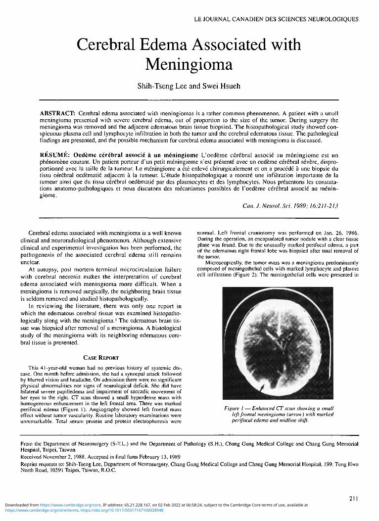
This 41-year-old woman had no previous history of systemic disease. One month before admission, she had a synocpal attack followed by blurred vision and headache. On admission there were no significant physical abnormalities nor signs of neurological deficit. She did have bilateral severe papilledema and impairment of saccadic movement of her eyes to the right. CT scan showed a small hyperdense mass with homogeneous enhancement in the left frontal area. There was marked perifocal edema (Figure 1). Angiography showed left frontal mass effect without tumor vascularity. Routine laboratory examinations were unremarkable. Total serum protein and protein electrophoresis were
From the Department of Neurosurgery (S-T.L.) and the Department of Pathology (S.H.), Chang Gung Medical College and Chang Gung Memorial Hospital, Taipei, Taiwan Received November 2, 1988. Accepted in final form February 13, 1989 Reprint requests to: Shih-Tseng Lee, Department of Neurosurgery, Chang Gung Medical College and Chang Gung Memorial Hospital, 199, Tung Hwa North Road, 10591 Taipei, Taiwan, R.O.C.
normal. Left frontal craniotomy was performed on Jan. 26, 1986. During the operation, an encapsulated tumor nodule with a clear tissue plane was found. Due to the unusually marked perifocal edema, a part of the edematous right frontal lobe was biopsied after total removal of the tumor.
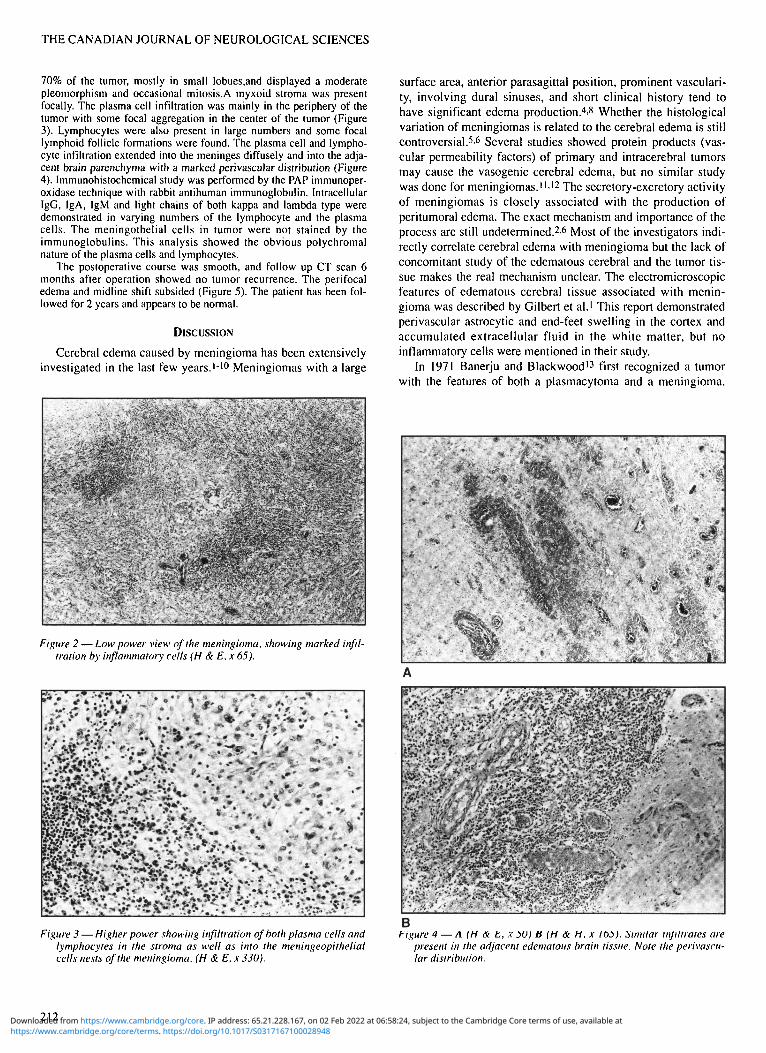
Microscopically, the tumor mass was a meningioma predominantly composed of meningothelial cells with marked lymphocyte and plasma cell infiltration (Figure 2). The meningothelial cells were presented in
Figure 1 — Enhanced CT scan showing a small left frontal meningioma (arrow) with marked perifocal edema and midline shift.
211
https://www.cambridge.org/core/terms. https://doi.org/10.1017/S0317167100028948Downloaded from https://www.cambridge.org/core. IP address: 65.21.228.167, on 02 Feb 2022 at 06:58:24, subject to the Cambridge Core terms of use, available at
THE CANADIAN JOURNAL OF NEUROLOGICAL SCIENCES
70% of the tumor, mostly in small lobues.and displayed a moderate pleomorphism and occasional mitosis.A myxoid stroma was present focally. The plasma cell infiltration was mainly in the periphery of the tumor with some focal aggregation in the center of the tumor (Figure 3). Lymphocytes were also present in large numbers and some focal lymphoid follicle formations were found. The plasma cell and lymphocyte infiltration extended into the meninges diffusely and into the adjacent brain parenchyma with a marked perivascular distribution (Figure 4). Immunohistochemical study was performed by the PAP immunoper-oxidase technique with rabbit antihuman immunoglobulin. Intracellular IgG, IgA, IgM and light chains of both kappa and lambda type were demonstrated in varying numbers of the lymphocyte and the plasma cells. The meningothelial cells in tumor were not stained by the immunoglobulins. This analysis showed the obvious polychromal nature of the plasma cells and lymphocytes.
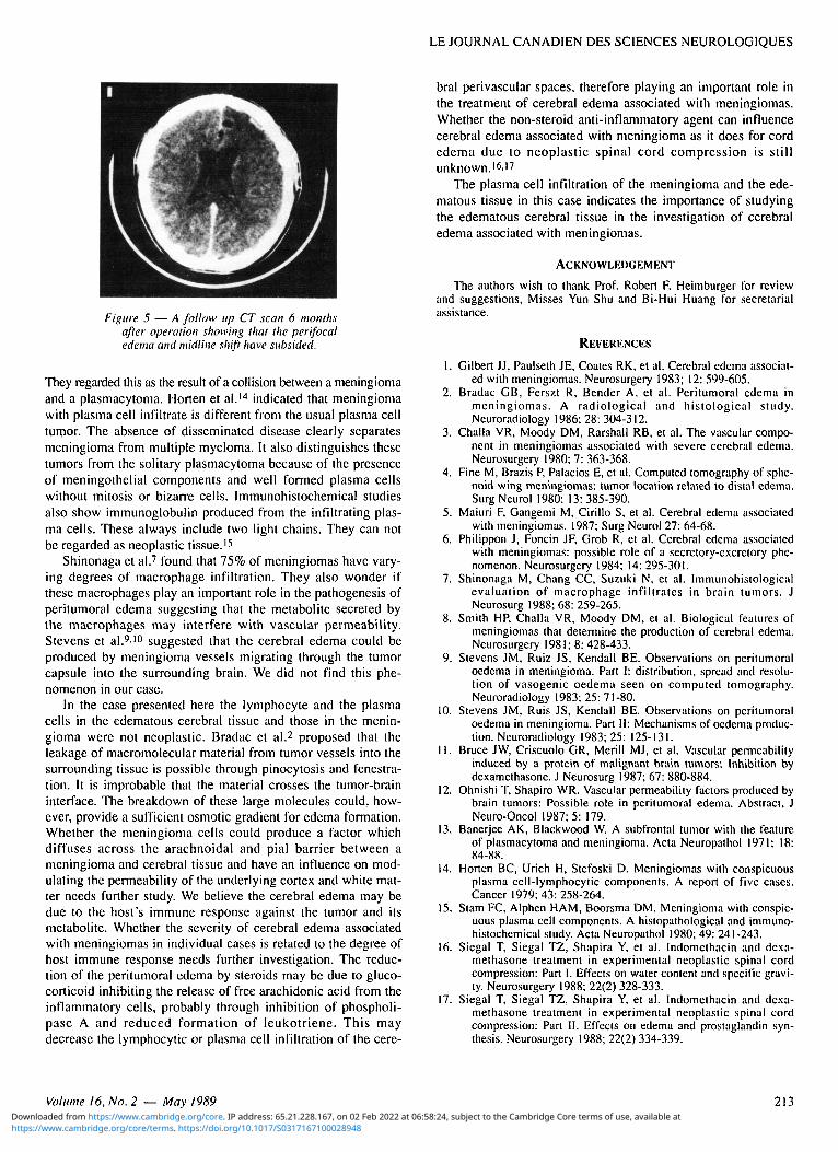
The postoperative course was smooth, and follow up CT scan 6 months after operation showed no tumor recurrence. The perifocal edema and midline shift subsided (Figure 5). The patient has been followed for 2 years and appears to be normal.
DISCUSSION
Cerebral edema caused by meningioma has been extensively investigated in the last few years.1'10 Meningiomas with a large
{ration by inflammatory cells (H & E. x 65).
Figure 3 — Higher power showing infiltration of both plasma cells and lymphocytes in the stroma as well as into the meningeopithelial cells nests of the meningioma. (H & E, x 330).
surface area, anterior parasagittal position, prominent vascularity, involving dural sinuses, and short clinical history tend to have significant edema production.4'8 Whether the histological variation of meningiomas is related to the cerebral edema is still controversial.5'6 Several studies showed protein products (vascular permeability factors) of primary and intracerebral tumors may cause the vasogenic cerebral edema, but no similar study was done for meningiomas."-12 The secretory-excretory activity of meningiomas is closely associated with the production of peritumoral edema. The exact mechanism and importance of the process are still undetermined.26 Most of the investigators indirectly correlate cerebral edema with meningioma but the lack of concomitant study of the edematous cerebral and the tumor tissue makes the real mechanism unclear. The electromicroscopic features of edematous cerebral tissue associated with meningioma was described by Gilbert et al.1 This report demonstrated perivascular astrocytic and end-feet swelling in the cortex and accumulated extracellular fluid in the white matter, but no inflammatory cells were mentioned in their study.
In 1971 Banerju and Blackwood13 first recognized a tumor with the features of both a plasmacytoma and a meningioma.
Figure 4—A(H&E, x50) B (H & H, x 165). Similar infiltrates are present in the adjacent edematous brain tissue. Note the perivascular distribution.
212 https://www.cambridge.org/core/terms. https://doi.org/10.1017/S0317167100028948Downloaded from https://www.cambridge.org/core. IP address: 65.21.228.167, on 02 Feb 2022 at 06:58:24, subject to the Cambridge Core terms of use, available at
LE JOURNAL CANADIEN DES SCIENCES NEUROLOGIQUES
Figure 5 — A follow up CT scan 6 months after operation showing that the perifocal edema and midline shift have subsided.
They regarded this as the result of a collision between a meningioma and a plasmacytoma. Horten et al.14 indicated that meningioma with plasma cell infiltrate is different from the usual plasma cell tumor. The absence of disseminated disease clearly separates meningioma from multiple myeloma. It also distinguishes these tumors from the solitary plasmacytoma because of the presence of meningothelial components and well formed plasma cells without mitosis or bizarre cells. Immunohistochemical studies also show immunoglobulin produced from the infiltrating plasma cells. These always include two light chains. They can not be regarded as neoplastic tissue.15
Shinonaga et al.7 found that 75% of meningiomas have varying degrees of macrophage infiltration. They also wonder if these macrophages play an important role in the pathogenesis of peritumoral edema suggesting that the metabolite secreted by the macrophages may interfere with vascular permeability. Stevens et al.9'10 suggested that the cerebral edema could be produced by meningioma vessels migrating through the tumor capsule into the surrounding brain. We did not find this phenomenon in our case.
In the case presented here the lymphocyte and the plasma cells in the edematous cerebral tissue and those in the meningioma were not neoplastic. Bradac et al.2 proposed that the leakage of macromolecular material from tumor vessels into the surrounding tissue is possible through pinocytosis and fenestration. It is improbable that the material crosses the tumor-brain interface. The breakdown of these large molecules could, however, provide a sufficient osmotic gradient for edema formation. Whether the meningioma cells could produce a factor which diffuses across the arachnoidal and pial barrier between a meningioma and cerebral tissue and have an influence on modulating the permeability of the underlying cortex and white matter needs further study. We believe the cerebral edema may be due to the host's immune response against the tumor and its metabolite. Whether the severity of cerebral edema associated with meningiomas in individual cases is related to the degree of host immune response needs further investigation. The reduction of the peritumoral edema by steroids may be due to glucocorticoid inhibiting the release of free arachidonic acid from the inflammatory cells, probably through inhibition of phospholi-pase A and reduced formation of leukotriene. This may decrease the lymphocytic or plasma cell infiltration of the cere
bral perivascular spaces, therefore playing an important role in the treatment of cerebral edema associated with meningiomas. Whether the non-steroid anti-inflammatory agent can influence cerebral edema associated with meningioma as it does for cord edema due to neoplastic spinal cord compression is still unknown.16'17
The plasma cell infiltration of the meningioma and the edematous tissue in this case indicates the importance of studying the edematous cerebral tissue in the investigation of cerebral edema associated with meningiomas.
ACKNOWLEDGEMENT
The authors wish to thank Prof. Robert F. Heimburger for review and suggestions, Misses Yun Shu and Bi-Hui Huang for secretarial assistance.
REFERENCES
1. Gilbert JJ, Paulseth JE, Coates RK, et al. Cerebral edema associated with meningiomas. Neurosurgery 1983; 12: 599-605.
2. Bradac GB, Ferszt R, Bender A, et al. Peritumoral edema in meningiomas. A radiological and histological study. Neuroradiology 1986; 28: 304-312.
3. Challa VR, Moody DM, Rarshall RB, et al. The vascular component in meningiomas associated with severe cerebral edema. Neurosurgery 1980; 7: 363-368.
4. Fine M, Brazis P, Palacios E, et al. Computed tomography of sphenoid wing meningiomas: tumor location related to distal edema. Surg Neurol 1980; 13:385-390.
5. Maiuri F, Gangemi M, Cirillo S, et al. Cerebral edema associated with meningiomas. 1987; Surg Neurol 27: 64-68.
6. Philippon J, Foncin JF, Grob R, et al. Cerebral edema associated with meningiomas: possible role of a secretory-excretory phenomenon. Neurosurgery 1984; 14: 295-301.
7. Shinonaga M, Chang CC, Suzuki N, et al. Immunohistological evaluation of macrophage infiltrates in brain tumors. J Neurosurg 1988; 68: 259-265.
8. Smith HP, Challa VR, Moody DM, et al. Biological features of meningiomas that determine the production of cerebral edema. Neurosurgery 1981; 8: 428-433.
9. Stevens JM, Ruiz JS, Kendall BE. Observations on peritumoral oedema in meningioma. Part I: distribution, spread and resolution of vasogenic oedema seen on computed tomography. Neuroradiology 1983; 25: 71-80.
10. Stevens JM, Ruis JS, Kendall BE. Observations on peritumoral oedema in meningioma. Part II: Mechanisms of oedema production. Neuroradiology 1983; 25: 125-131.
11. Bruce JW, Criscuolo GR, Merill MJ, et al. Vascular permeability induced by a protein of malignant brain tumors: Inhibition by dexamethasone. J Neurosurg 1987; 67: 880-884.
12. Ohnishi T, Shapiro WR. Vascular permeability factors produced by brain tumors: Possible role in peritumoral edema. Abstract. J Neuro-Oncol 1987; 5: 179.
13. Banerjee AK, Blackwood W. A subfrontal tumor with the feature of plasmacytoma and meningioma. Acta Neuropathol 1971; 18: 84-88.
14. Horten BC, Urich H, Stefoski D. Meningiomas with conspicuous plasma cell-lymphocytic components. A report of five cases. Cancer 1979;43:258-264.
15. Stam FC, Alphen HAM, Boorsma DM. Meningioma with conspicuous plasma cell components. A histopathological and immunohistochemical study. Acta Neuropathol 1980; 49: 241-243.
16. Siegal T, Siegal TZ, Shapira Y, et al. Indomethacin and dexamethasone treatment in experimental neoplastic spinal cord compression: Part I. Effects on water content and specific gravity. Neurosurgery 1988; 22(2) 328-333.
17. Siegal T, Siegal TZ, Shapira Y, et al. Indomethacin and dexamethasone treatment in experimental neoplastic spinal cord compression: Part II. Effects on edema and prostaglandin synthesis. Neurosurgery 1988; 22(2) 334-339.
Volume 16, No. 2 — May 1989 213
https://www.cambridge.org/core/terms. https://doi.org/10.1017/S0317167100028948Downloaded from https://www.cambridge.org/core. IP address: 65.21.228.167, on 02 Feb 2022 at 06:58:24, subject to the Cambridge Core terms of use, available at