lab/x-ray/ecg rounds
DESCRIPTION
Lab/X-ray/ECG Rounds. James Huffman January 15, 2009. 67y.o. Female. Epigastric aching/burning for ~ 4 hours Radiates to LUQ/Left chest, ?back Associate N/V, diaphoresis Onset while walking to car after bingo (she won $50) History of HTN, ++smoking, EtOH abuse. Case: Continued. - PowerPoint PPT PresentationTRANSCRIPT

Lab/X-ray/ECG Rounds
James HuffmanJanuary 15, 2009

67y.o. Female
Epigastric aching/burning for ~ 4 hours
Radiates to LUQ/Left chest, ?back
Associate N/V, diaphoresis
Onset while walking to car after bingo (she won $50)
History of HTN, ++smoking, EtOH abuse
Case: Continued
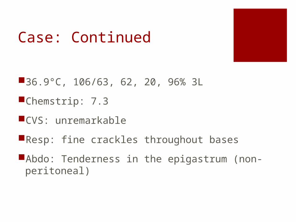
36.9°C, 106/63, 62, 20, 96% 3L
Chemstrip: 7.3
CVS: unremarkable
Resp: fine crackles throughout bases
Abdo: Tenderness in the epigastrum (non-peritoneal)

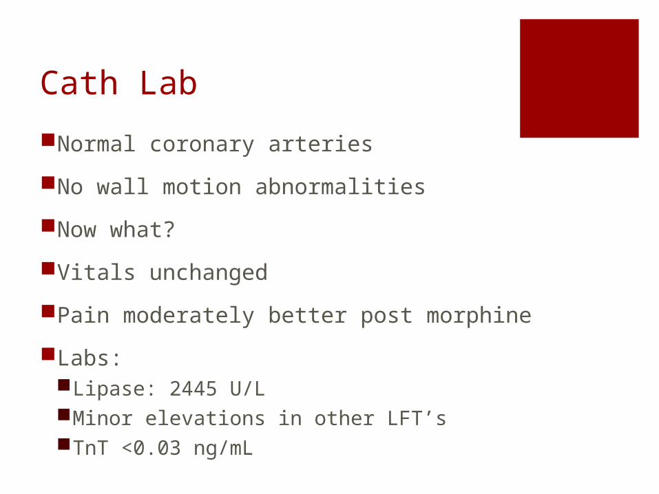
Cath Lab
Normal coronary arteries
No wall motion abnormalities
Now what?
Vitals unchanged
Pain moderately better post morphine
Labs:Lipase: 2445 U/LMinor elevations in other LFT’sTnT <0.03 ng/mL

ECG Manifestations of Gastrointestinal
Disease
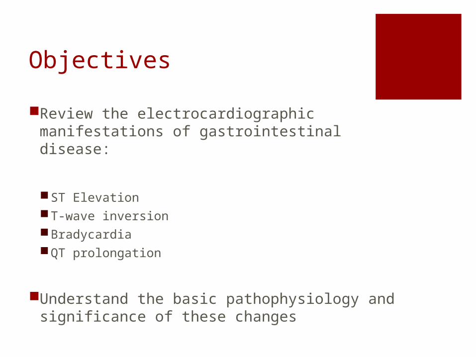
Objectives
Review the electrocardiographic manifestations of gastrointestinal disease:
ST ElevationT-wave inversionBradycardiaQT prolongation
Understand the basic pathophysiology and significance of these changes
ContextChan, T.C. et al. ECG in Emergency Medicine and Acute Care. CH68
ECG is often obtained in initial w/u of abdo pain:Anginal variant (especially women, diabetics)
BUTSeveral GI processes are assoc. with ecg changes:
Pancreatitis, Cholecystitis, PUD, Appendicitis, IBD, Cirrhosis, electrolyte abnormalities
Certain GI processes seem to be assoc. with increased risk for concurrent cardiac ischemia or infarction
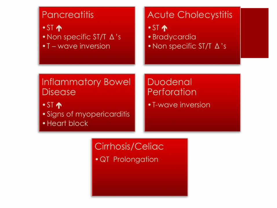
ST Elevation
STE in the setting of abdominal pain should always raise concern for ACS
Two scenarios:1. Certain GI diseases may present with ECG
consistent with pseudoinfarction e.g. acute pancreatitis, cholecystic disease
2. Certain GI diseases and treatments increase the propensity for coronary thrombosis and true ACS
e.g. IBD
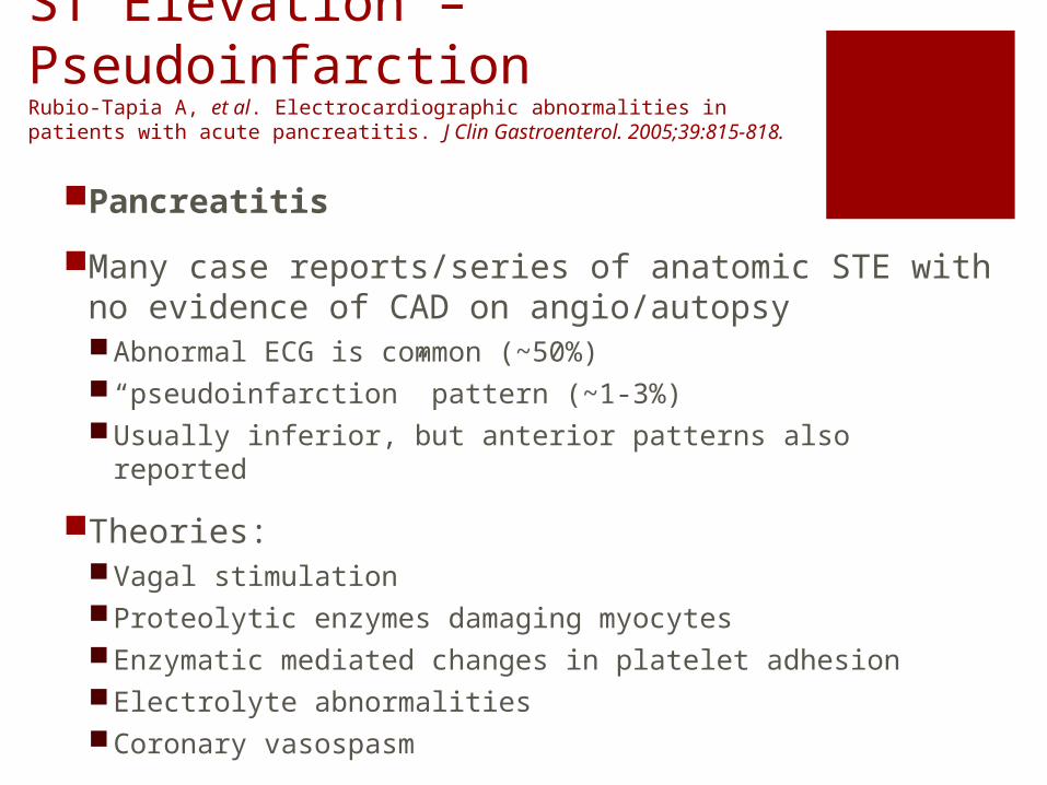
ST Elevation – PseudoinfarctionRubio-Tapia A, et al. Electrocardiographic abnormalities in patients with acute pancreatitis. J Clin Gastroenterol. 2005;39:815-818.
Pancreatitis
Many case reports/series of anatomic STE with no evidence of CAD on angio/autopsyAbnormal ECG is common (~50%)“pseudoinfarction” pattern (~1-3%)Usually inferior, but anterior patterns also reported
Theories:Vagal stimulationProteolytic enzymes damaging myocytesEnzymatic mediated changes in platelet adhesionElectrolyte abnormalitiesCoronary vasospasm

ST ElevationChan, T.C. et al. ECG in Emergency Medicine and Acute Care. CH68.Ryan, E.T. et al. Myocardial infarction mimicked by acute cholecystitis. Ann Int Med 1992; 116:218.
Acute CholecystitisMay present with anterior ischemic patterns on their ECGs that often resolve after GB removal
The cardio-biliary reflex commonly cited as cause:GB distension may lead to vagal response producing intermittent coronary vasospasm
Others:Splenic ruptureDemand ischemia 2° to catecholamine release

ST Elevation – True diseaseEfremidis, M. et al. Acute myocardial infarction in a young patient during an exacerbation of ulcerative colitis. Int J Cardiol 1999; 70:211.
Inflammatory Bowel DiseaseAcute vascular thrombosis is a known complication of both UC and Crohn’s disease
Myopericarditis:Rare but reported complication of both IBD and an adverse drug reaction to mesalamine (5-ASA agent)
T-Wave InversionChan, T.C. et al. ECG in Emergency Medicine and Acute Care. CH68.
Duodenal perforation
Acute pancreatitis
Cholecystitis
ALL occur infrequently
BradycardiaChan, T.C. et al. ECG in Emergency Medicine and Acute Care. CH68.
1. As a result of vagal response to primary GI disorder or pain
2. Specific diseases are associated with bradycardia
Ulcerative Colitis Several cases of 2nd Degree and complete AV
block Jaundice/Bile-acid accumulation
Historically listed in causes of bradycardia Has not borne out in literature/animal studies
QT ProlongationChan, T.C. et al. ECG in Emergency Medicine and Acute Care. CH68.
CirrhosisHistorically thought only to occur in pts with EtOH cirrhosis
Now reported in almost every cirrhotic etiologyGrowing body of evidence that QT-prolongation is associated with a poorer clinical outcome
One case series has shown a significant reduction in QT interval post transplant
Malnutrition/electrolyte disorders
Celiac diseaseOne study found 1/3 of all adult pts had QT prolongation

PEARLS
ST/T wave changes associated with GI disease may represent true ACS or a pseudoischemic pattern
Pts with IBD are at increased risk for thrombotic events, including MI
Biliary-cardiac reflex is a known phenomenon which may explain the ST seen in acute cholecystitis
Cirrhosis and celiac disease can be a cause of QT prolongation