juvenile scleroderma - unesp · juvenile scleroderma ... ana in serum and csf • clinical...
TRANSCRIPT
Juvenile Scleroderma
Carine H Wouters, Pediatric Immune-Inflammatory Diseases, Leuven, Belgium
PReS Latin American Pediatric Rheumatology Course, Sao Paolo, June 2015

Juvenile Scleroderma Agenda
• Definition and pathogenesis
• Localized sclerodermas
• Systemic sclerosis
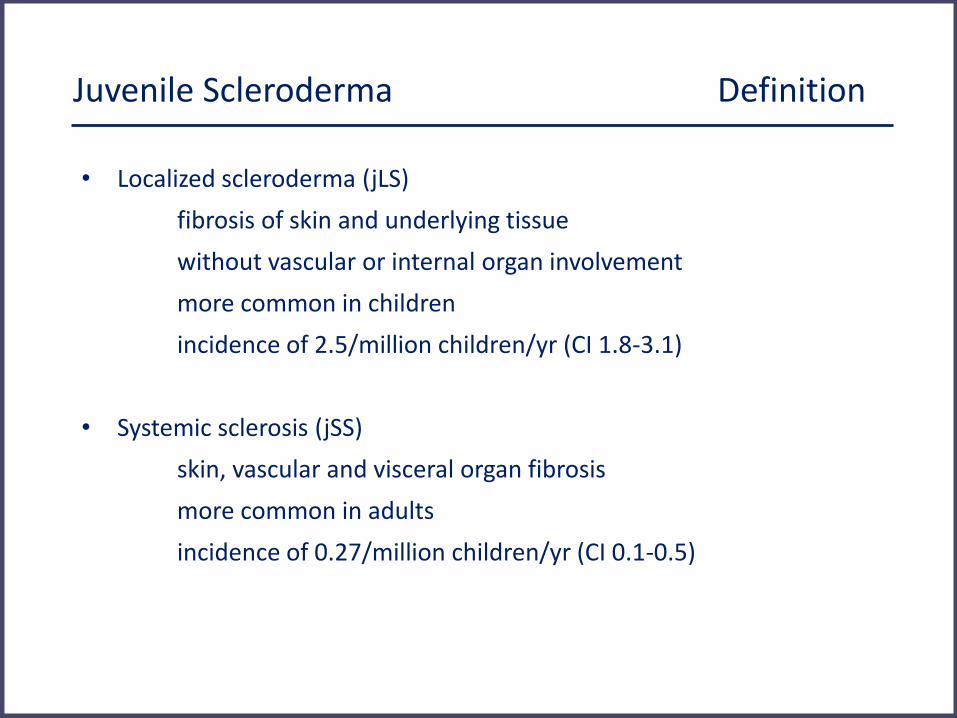
Juvenile Scleroderma Definition
• Localized scleroderma (jLS)
fibrosis of skin and underlying tissue
without vascular or internal organ involvement
more common in children
incidence of 2.5/million children/yr (CI 1.8-3.1)
• Systemic sclerosis (jSS)
skin, vascular and visceral organ fibrosis
more common in adults
incidence of 0.27/million children/yr (CI 0.1-0.5)

“Hard skin” Pathology
• Inflammatory infiltrate
lymphocytes, plasma cells, macrophages, eosinophils and mast cells
hyalinization blood vessel walls, proliferation endothelial cells
• Increase in fibroblasts and collagen < escalating sclerosis
• Entire dermis replaced by compact collagen fibers
• Thinning of epiderm, atrophy dermal appendiges
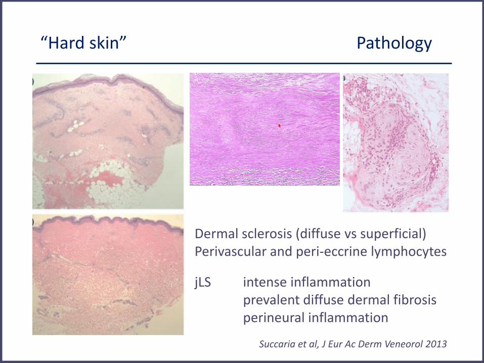
“Hard skin” Pathology
Dermal sclerosis (diffuse vs superficial) Perivascular and peri-eccrine lymphocytes
Succaria et al, J Eur Ac Derm Veneorol 2013
jLS intense inflammation prevalent diffuse dermal fibrosis perineural inflammation
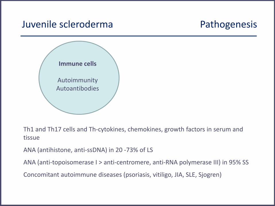
Juvenile scleroderma Pathogenesis
Immune cells
Autoimmunity Autoantibodies
Th1 and Th17 cells and Th-cytokines, chemokines, growth factors in serum and tissue
ANA (antihistone, anti-ssDNA) in 20 -73% of LS
ANA (anti-topoisomerase I > anti-centromere, anti-RNA polymerase III) in 95% SS
Concomitant autoimmune diseases (psoriasis, vitiligo, JIA, SLE, Sjogren)
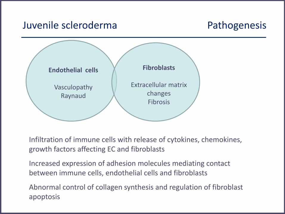
Juvenile scleroderma Pathogenesis
Endothelial cells
Vasculopathy Raynaud
Infiltration of immune cells with release of cytokines, chemokines, growth factors affecting EC and fibroblasts
Increased expression of adhesion molecules mediating contact between immune cells, endothelial cells and fibroblasts
Abnormal control of collagen synthesis and regulation of fibroblast apoptosis
Fibroblasts
Extracellular matrix changes Fibrosis
Localized scleroderma
• Classification and clinical features
• Assessment of activity and damage
• Treatment
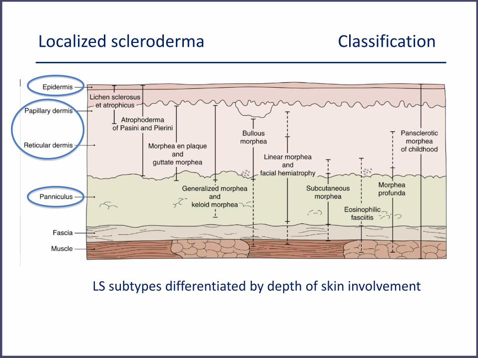
Localized scleroderma Classification
LS subtypes differentiated by depth of skin involvement

Localized scleroderma Classification
Associated: lichen sclerosus et atrophicus, bullous morphea, eosinophilic fasciitis
Padua classification, Laxer, Zulian, Curr Opin Rheumatol 2006
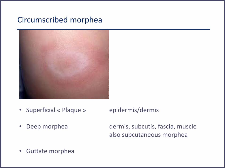
Circumscribed morphea
• Superficial « Plaque » epidermis/dermis • Deep morphea dermis, subcutis, fascia, muscle
also subcutaneous morphea • Guttate morphea
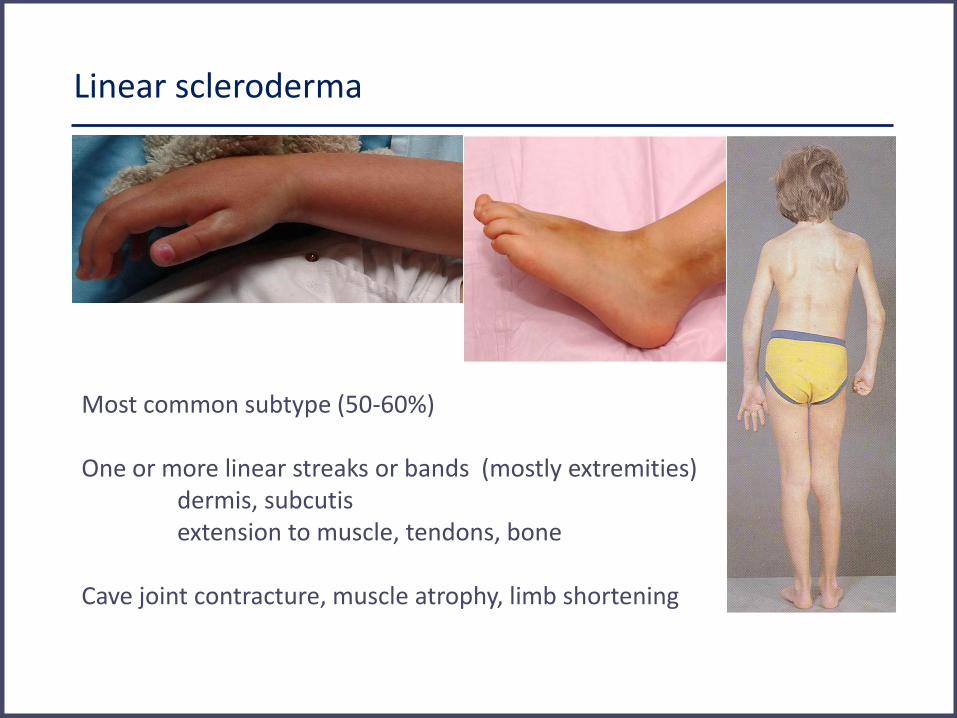
Linear scleroderma
Most common subtype (50-60%) One or more linear streaks or bands (mostly extremities) dermis, subcutis extension to muscle, tendons, bone Cave joint contracture, muscle atrophy, limb shortening

Linear scleroderma en coup de sabre
Linear induration of the skin, generally located at the frontoparietal scalp and/or paramedian forehead often resembling a stroke from a sword
LS en coup de sabre
Hemifacial atrophy of soft tissue of the cheek, progressing to chin and forehead, extending into underlying muscles and bone mild/absent involvement of the superficial skin provoking asymmetry of the face
Parry Romberg

LS en coup de sabre with encephalitis
• Coup de sabre since age 5 yrs
• Hemicranial migraine, complex partial seizures at age 12 yrs MRI T2 hyperintense signals left subcortital white matter SPECT hypoperfusion ANA in serum and CSF
• Clinical remission, MRI stabilisation with MTX therapy
LS with Parry Romberg and autoimmunity
Coeliac disease and bilateral uveitis at 5 yrs ANA+, antigliadin ab+ Depigmentation, loss of eyelashes at right eye, CM lesion cheek at 6 yrs
Progressive hemifacial atrophy at 7 yrs Bilateral wrist synovitis/tenosynovitis with carpal tunnel S at 8 yrs
LSCS and PRS are the same disease entity with LSCS and superficial skin involvement at one end of the spectrum and PFH with involvement of subcutaneous deep tissues, at the other hand. Overlapping cutaneous features may occur with time In both entities, seizures and severe encephalitis mimicking Rasmussen encephalitis can be observed In both entities ocular and dental abnormalities and autoimmune manifestations (arthritis, uveitis, ANA in serum/CSF) may be present
Overlap between linear scleroderma, progressive facial
hemiatrophy and immune-inflammatory encephalitis
De Somer, Eur J Pediatrics 2015; Lehman, J Rheumatol 1992
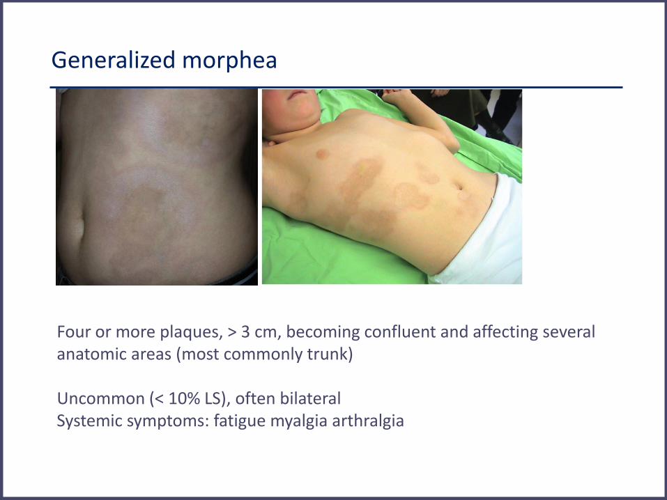
Generalized morphea
Four or more plaques, > 3 cm, becoming confluent and affecting several anatomic areas (most commonly trunk) Uncommon (< 10% LS), often bilateral Systemic symptoms: fatigue myalgia arthralgia
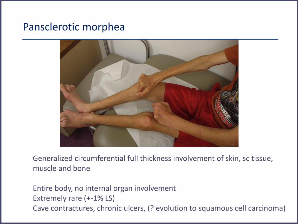
Pansclerotic morphea
Generalized circumferential full thickness involvement of skin, sc tissue, muscle and bone Entire body, no internal organ involvement Extremely rare (+-1% LS) Cave contractures, chronic ulcers, (? evolution to squamous cell carcinoma)
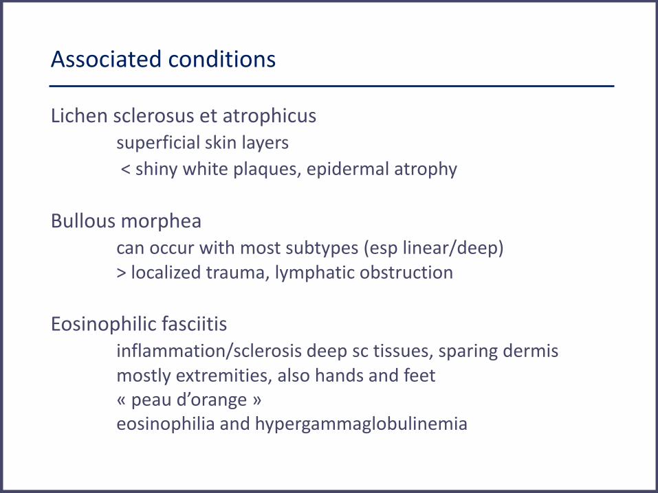
Associated conditions
Lichen sclerosus et atrophicus superficial skin layers
< shiny white plaques, epidermal atrophy Bullous morphea can occur with most subtypes (esp linear/deep) > localized trauma, lymphatic obstruction
Eosinophilic fasciitis inflammation/sclerosis deep sc tissues, sparing dermis mostly extremities, also hands and feet « peau d’orange » eosinophilia and hypergammaglobulinemia

Extracutaneous manifestations LS
Zulian, A&R 2005
In one quarter of LS patients (mostly linear scleroderma) CNS and ocular especially in coup de sabre LS Multiple extracutaneous features in one third

Localized scleroderma Assessment
• Clinical skin scores • Modified Rodnan skin score • Localized scleroderma cutaneous assessment tool • Computerized skin score
• Measurement tools • Durometer (hardness) • Cutometer (elasticity)
• Imaging • Infrared Thermography • Laser Doppler flowmetry • Optical coherence tomography • Ultrasonography • MRI

Localized scleroderma: transition with time Active disease: erythema, skin induration/edema, new/enlarging lesions Disease damage: hypo- and hyperpigmentation, dermal and sc atrophy
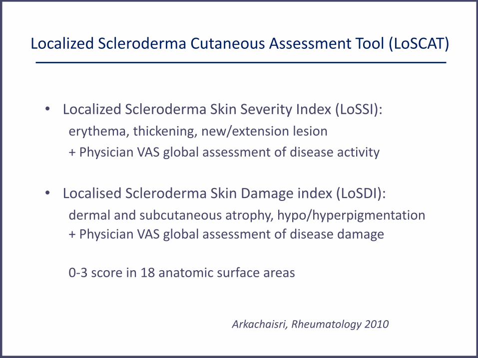
Localized Scleroderma Cutaneous Assessment Tool (LoSCAT)
• Localized Scleroderma Skin Severity Index (LoSSI):
erythema, thickening, new/extension lesion
+ Physician VAS global assessment of disease activity
• Localised Scleroderma Skin Damage index (LoSDI):
dermal and subcutaneous atrophy, hypo/hyperpigmentation
+ Physician VAS global assessment of disease damage
0-3 score in 18 anatomic surface areas
Arkachaisri, Rheumatology 2010
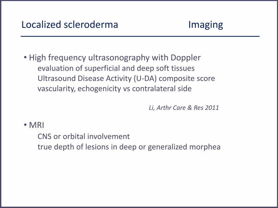
Localized scleroderma Imaging
• High frequency ultrasonography with Doppler evaluation of superficial and deep soft tissues Ultrasound Disease Activity (U-DA) composite score vascularity, echogenicity vs contralateral side Li, Arthr Care & Res 2011
• MRI CNS or orbital involvement true depth of lesions in deep or generalized morphea
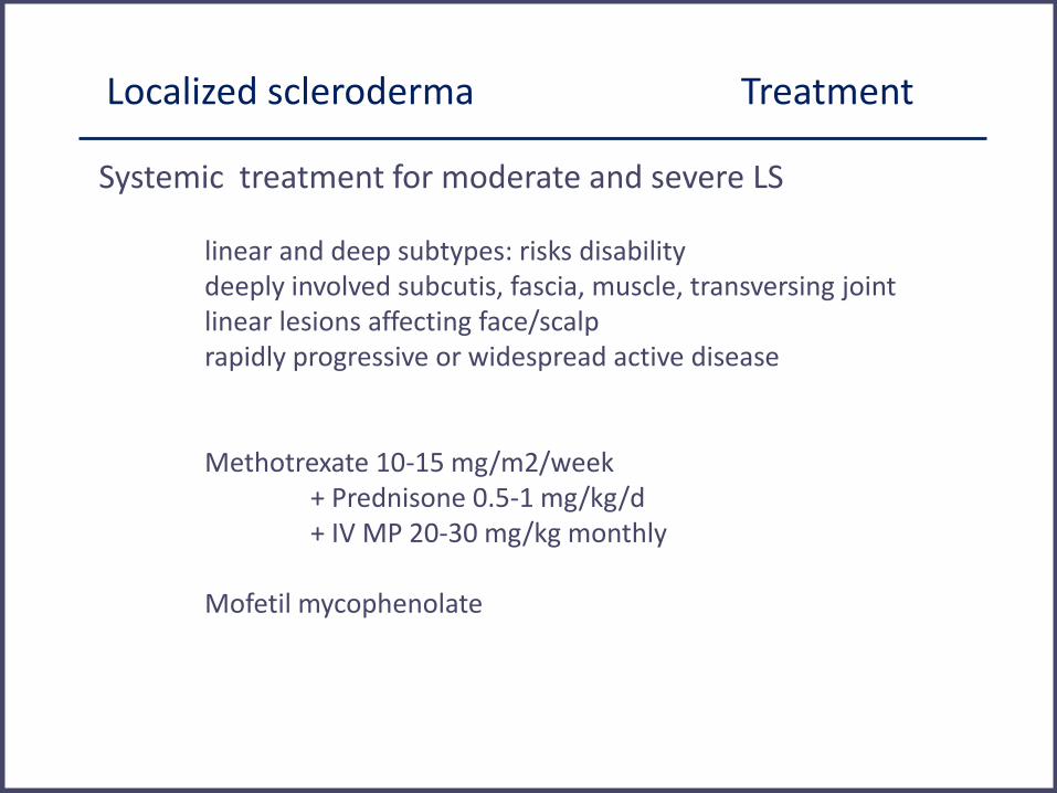
Localized scleroderma Treatment
Systemic treatment for moderate and severe LS linear and deep subtypes: risks disability deeply involved subcutis, fascia, muscle, transversing joint linear lesions affecting face/scalp rapidly progressive or widespread active disease Methotrexate 10-15 mg/m2/week + Prednisone 0.5-1 mg/kg/d + IV MP 20-30 mg/kg monthly Mofetil mycophenolate
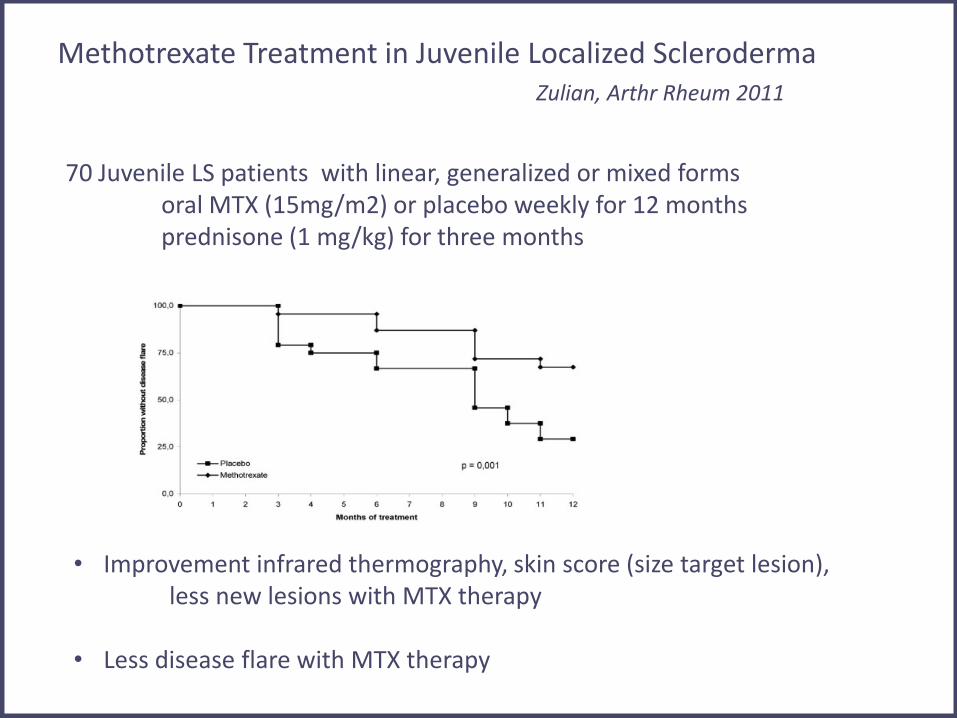
70 Juvenile LS patients with linear, generalized or mixed forms oral MTX (15mg/m2) or placebo weekly for 12 months prednisone (1 mg/kg) for three months
Methotrexate Treatment in Juvenile Localized Scleroderma Zulian, Arthr Rheum 2011
• Improvement infrared thermography, skin score (size target lesion), less new lesions with MTX therapy
• Less disease flare with MTX therapy
Juvenile Systemic Sclerosis
• Classification and clinical features
• Assessment of activity and damage
• Treatment
Juvenile Systemic Sclerosis Classification
• Diffuse cutaneous systemic sclerosis widespread rapidly progressive skin thickening
and early visceral disease
• Limited cutaneous systemic sclerosis skin thickening limited distal extremities
and late visceral disease
includes CREST syndrome
• Overlap scleroderma diffuse or limited SSc with features of another connective tissue disease
eg dermatomhyositis, SLE
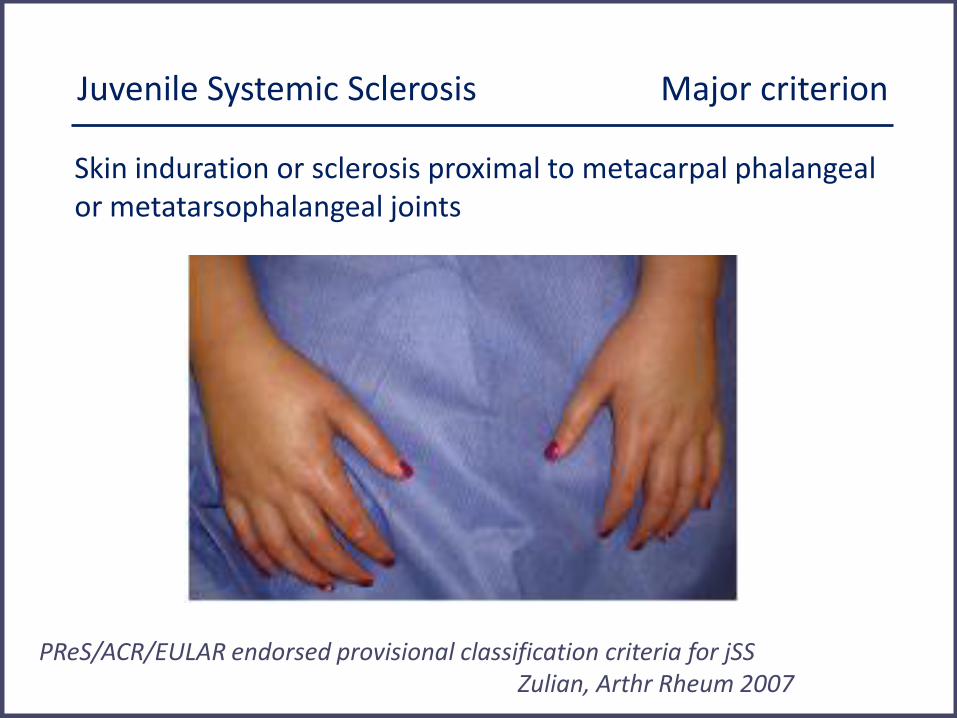
Juvenile Systemic Sclerosis Major criterion
Skin induration or sclerosis proximal to metacarpal phalangeal or metatarsophalangeal joints
PReS/ACR/EULAR endorsed provisional classification criteria for jSS Zulian, Arthr Rheum 2007
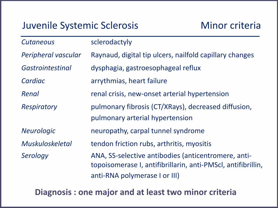
Juvenile Systemic Sclerosis Minor criteria Cutaneous sclerodactyly
Peripheral vascular Raynaud, digital tip ulcers, nailfold capillary changes
Gastrointestinal dysphagia, gastroesophageal reflux
Cardiac arrythmias, heart failure
Renal renal crisis, new-onset arterial hypertension
Respiratory pulmonary fibrosis (CT/XRays), decreased diffusion,
pulmonary arterial hypertension
Neurologic neuropathy, carpal tunnel syndrome
Muskuloskeletal tendon friction rubs, arthritis, myositis
Serology ANA, SS-selective antibodies (anticentromere, anti- topoisomerase I, antifibrillarin, anti-PMScl, antifibrillin,
anti-RNA polymerase I or III)
Diagnosis : one major and at least two minor criteria
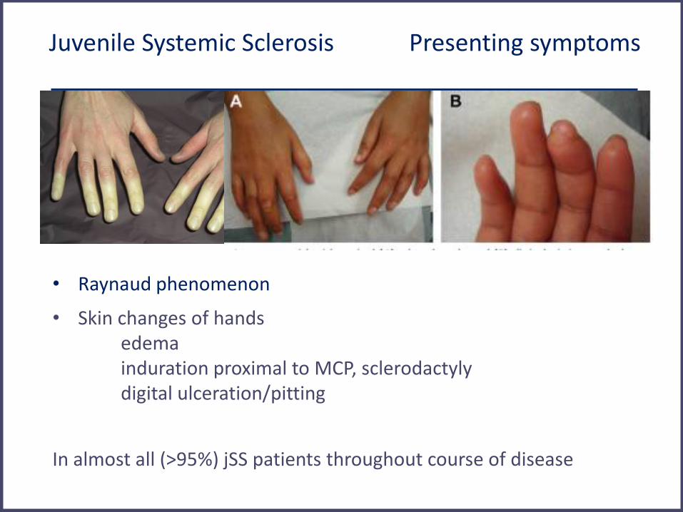
Juvenile Systemic Sclerosis Presenting symptoms
• Raynaud phenomenon
• Skin changes of hands edema induration proximal to MCP, sclerodactyly digital ulceration/pitting
In almost all (>95%) jSS patients throughout course of disease
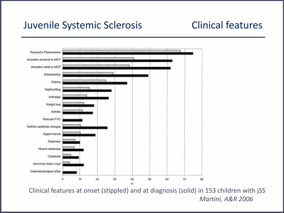
Juvenile Systemic Sclerosis Clinical features
Clinical features at onset (stippled) and at diagnosis (solid) in 153 children with jSS Martini, A&R 2006

Juvenile Systemic Sclerosis Outcome
Martini, Rheumatology 2009
Survival rates (5 to 20 yrs) significantly higher than in adult-onset SS Causes of death: cardiac failure, pulmonary HT, renal insufficiency, respiratory failure Subset with rapid development of internal organ failure and early fatality versus majority of slow insidious course with lower mortality
Petty Table 25-6
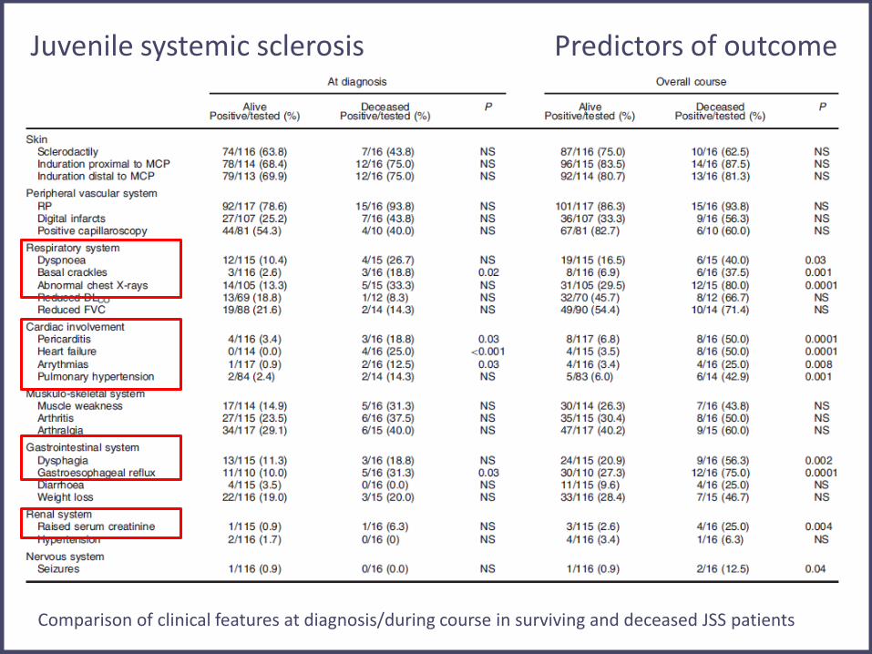
Juvenile systemic sclerosis Predictors of outcome
Comparison of clinical features at diagnosis/during course in surviving and deceased JSS patients
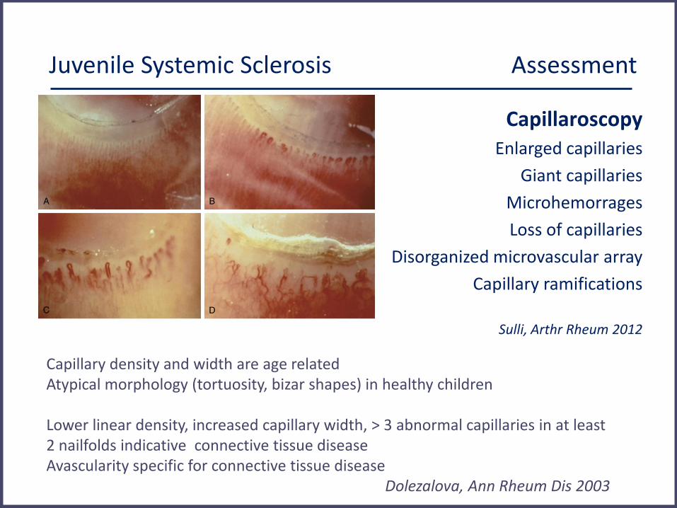
Juvenile Systemic Sclerosis Assessment
Capillaroscopy Enlarged capillaries
Giant capillaries
Microhemorrages
Loss of capillaries
Disorganized microvascular array
Capillary ramifications
Sulli, Arthr Rheum 2012
Capillary density and width are age related Atypical morphology (tortuosity, bizar shapes) in healthy children Lower linear density, increased capillary width, > 3 abnormal capillaries in at least 2 nailfolds indicative connective tissue disease Avascularity specific for connective tissue disease Dolezalova, Ann Rheum Dis 2003
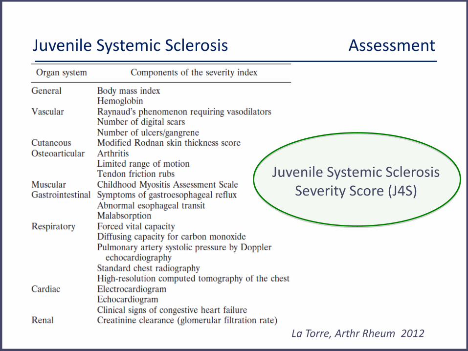
Juvenile Systemic Sclerosis Severity Score (J4S)
La Torre, Arthr Rheum 2012
Juvenile Systemic Sclerosis Assessment
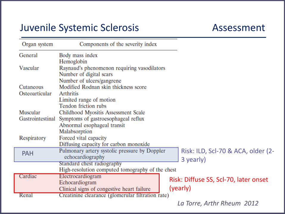
La Torre, Arthr Rheum 2012
Juvenile Systemic Sclerosis Assessment
Risk: Diffuse SS, Scl-70, later onset (yearly)
Risk: ILD, Scl-70 & ACA, older (2-3 yearly)
PAH
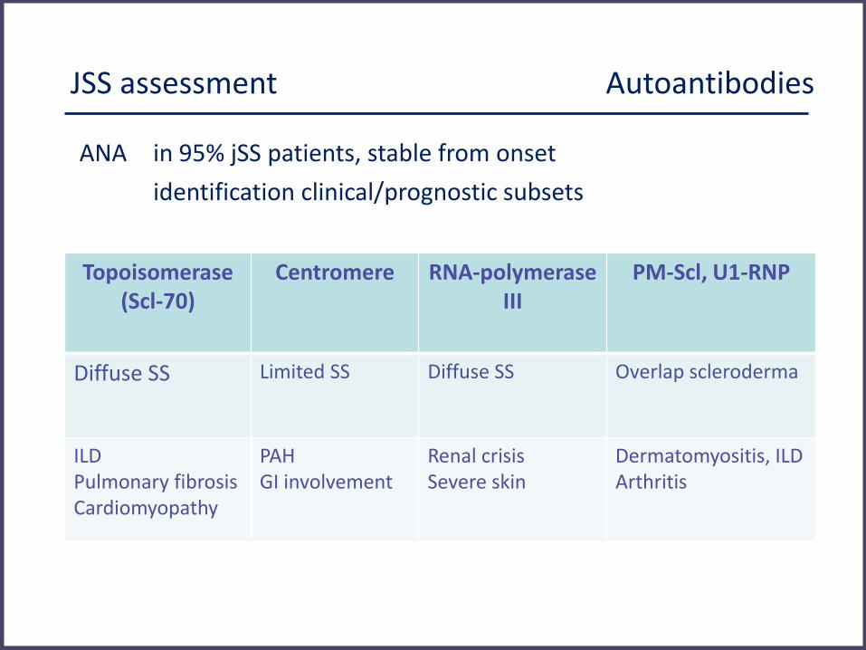
JSS assessment Autoantibodies
ANA in 95% jSS patients, stable from onset
identification clinical/prognostic subsets
Topoisomerase (Scl-70)
Centromere RNA-polymerase III
PM-Scl, U1-RNP
Diffuse SS Limited SS Diffuse SS Overlap scleroderma
ILD Pulmonary fibrosis Cardiomyopathy
PAH GI involvement
Renal crisis Severe skin
Dermatomyositis, ILD Arthritis
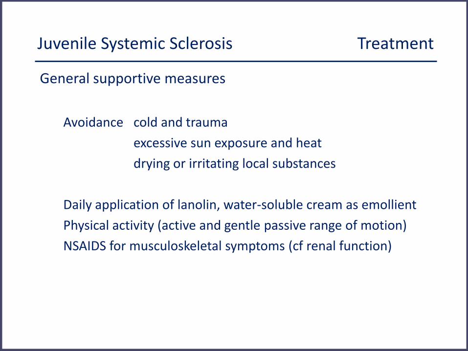
Juvenile Systemic Sclerosis Treatment
General supportive measures
Avoidance cold and trauma
excessive sun exposure and heat
drying or irritating local substances
Daily application of lanolin, water-soluble cream as emollient
Physical activity (active and gentle passive range of motion)
NSAIDS for musculoskeletal symptoms (cf renal function)
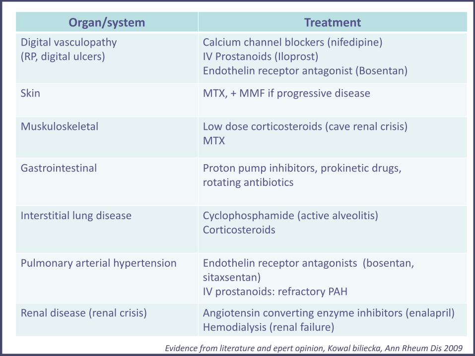
jSS Treatment
Organ/system Treatment
Digital vasculopathy (RP, digital ulcers)
Calcium channel blockers (nifedipine) IV Prostanoids (Iloprost) Endothelin receptor antagonist (Bosentan)
Skin
MTX, + MMF if progressive disease
Muskuloskeletal
Low dose corticosteroids (cave renal crisis) MTX
Gastrointestinal
Proton pump inhibitors, prokinetic drugs, rotating antibiotics
Interstitial lung disease
Cyclophosphamide (active alveolitis) Corticosteroids
Pulmonary arterial hypertension
Endothelin receptor antagonists (bosentan, sitaxsentan) IV prostanoids: refractory PAH
Renal disease (renal crisis)
Angiotensin converting enzyme inhibitors (enalapril) Hemodialysis (renal failure)
Evidence from literature and epert opinion, Kowal biliecka, Ann Rheum Dis 2009