journal of controlled release - medical university of...
TRANSCRIPT

Journal of Controlled Release 168 (2013) 142–150
Contents lists available at SciVerse ScienceDirect
Journal of Controlled Release
j ourna l homepage: www.e lsev ie r .com/ locate / jconre l
NANOMEDICIN
E
Mild hyperthermia triggered doxorubicin release from optimizedstealth thermosensitive liposomes improves intratumoral drug deliveryand efficacy
Li Li a, Timo L.M. ten Hagen a, Martin Hossann b,c, Regine Süss d, Gerard C. van Rhoon e,Alexander M.M. Eggermont a,f, Dieter Haemmerich g, Gerben A. Koning a,⁎a Laboratory Experimental Surgical Oncology, Section Surgical Oncology, Department of Surgery, Erasmus Medical Center, Rotterdam, The Netherlandsb Department of Internal Medicine III, University Hospital Munich, Ludwig-Maximilians University, Munich, Germanyc CCG Tumor Therapy Through Hyperthermia, Helmholtz Zentrum München, German Research Center for Environmental Health, Munich, Germanyd Department of Pharmaceutical Technology and Biopharmacy, Albert-Ludwigs University, Freiburg, Germanye Unit Hyperthermia, Department of Radiotherapy, Erasmus Medical Center — Daniel den Hoed Cancer Center, Rotterdam, The Netherlandsf Institut de Cancérologie Gustave Roussy, Paris, Franceg Medical University of South Carolina, Charleston, USA
⁎ Correspondence to: G.A. Koning, Room Ee151b, LabOncology, Section Surgical Oncology, Department of SRotterdam, PO Box 2040, The Netherlands. Tel.: +31 107
E-mail address: [email protected] (G.A. Konin
0168-3659/$ – see front matter © 2013 Elsevier B.V. Allhttp://dx.doi.org/10.1016/j.jconrel.2013.03.011
a b s t r a c t
a r t i c l e i n f oArticle history:Received 26 December 2012Accepted 15 March 2013Available online 21 March 2013
Keywords:Thermosensitive liposomesHyperthermiaDoxorubicinTriggered drug releaseIntravital microscopyTumor growth control
Liposomemediated anticancer drug delivery has the advantage of reducing cytotoxicity in healthy tissues. However,undesired slowdrug release impedes the therapeutic efficacy of clinically applied PEG-liposomal doxorubicin (Dox).The aim of this study is to combine stealth thermosensitive liposomes (TSL) and local mild hyperthermia (HT) toincrease bioavailable Dox levels in tumors. Doxwas encapsulated in stealth TSL (~80 nm)with optimized PEG con-centration in themembrane, and comparedwith lysolipid-based Dox-LTSL for in vitro stability, release kinetics, andin vivo tumor growth control. In vitro cytotoxicity of Dox-TSL against murine BFS-1 sarcoma and, human BLMmelanoma cell lines and Human Umbilical Vein Endothelial Cells (HUVEC) under normothermia (37 °C) and HT(42 °C) was compared with non-encapsulated Dox. In vitro Dox uptake in nuclei was imaged in BLM and HUVEC.In vivo intravascular Dox release from TSL in BFS-1 tumors under local mild HT in dorsal skin flap window chambermodels was captured by intravital confocal microscopy. Intravascular Dox-TSL release kinetics, penetration depthand interstitial Dox densitywere subjected to quantitative image analysis. Systemic Dox-TSL administration in com-bination with local mild HT on subcutaneous tumor growth control was compared to Dox-LTSL plus local mild HT.Dox-TSLwas stable at 37 °C,while released over 95%Doxwithin 1 min in 90% serumat 42 °C. Dox-TSL demonstrat-ed efficient in vivo intratumoral Dox release under local mild HT, followed by significant Dox uptake by tumor andtumor vascular endothelial cells. Dox-TSL plusmild HT showed improved tumor growth control over Dox-LTSL plusmild HT. Survival after a single treatment of Dox-TSL plusmild HTwas 67%, while survival after Dox-LTSL plus mildHTwas 22%. This combination of Dox-TSL and local mild HT offers promising clinical opportunities to improve lipo-somal Dox delivery to solid tumors.
© 2013 Elsevier B.V. All rights reserved.
1. Introduction
The current available chemotherapeutic drugs used in conventional orcombination chemotherapy are associated with substantial side-effects,but often of limited benefit to cancer patients [1,2]. Doxorubucin (Dox)is an anthracycline antibiotic and widely used in the treatment ofvarious tumors [2]. To reduce side-effects, liposomal chemotherapy isrecommended [3]. For instance, encapsulating Dox within liposomal
oratory Experimental Surgicalurgery, Erasmus MC, 3000CA043963; fax: +31 107044746.g).
rights reserved.
nanoparticles significantly reduces cardiotoxicity, while prolonging thepresence of the drug in the systemic circulation [4–6]. For these reasonslong-circulating or stealth PEG-coated Liposomal Doxorubicin (PLD)was approved for clinical use (Caelyx® in Europe and Doxil® in theUSA) [7]. Despite these promising aspects, PLD did not substantially im-prove efficacy [5,6]. Preclinical evidence suggests that slow and passivedrug release from these stealth liposomes impedes their therapeutic effi-cacy [8]. Thermosensitive liposomes (TSL) combine the features onhigh drug loading efficiency and reduced toxicity due to liposomaldrug encapsulation, with the possibility of triggered drug releasearound a tunablemembrane phase transition temperature (Tm), typically~40 °C [9–11]. Incorporating Dox into stealth thermosensitive liposomes(Dox-TSL) enables a prolonged circulation half-life, and controlled local-ized Dox release from the nanocarrier upon HT [12–14]. However, the

143L. Li et al. / Journal of Controlled Release 168 (2013) 142–150
NANOMEDICIN
E
substantial premature leakage ofDox from lysolipid based TSL (Dox-LTSL)at physiological temperature remains an important issue [15–17]. Recent-ly, we have reported on an optimized stealth TSL formulation formild hy-perthermia (HT) (40–42 °C) induced content release [18]. After adoptingthe optimized stealth TSL formulation, we encapsulated Dox into theseTSL.
Mild HT is routinely used in the clinic and is known to stronglyimprove the outcome of chemotherapy or radiotherapy, without signifi-cant side effects [19,20]. HT can improve the antitumor effect of some che-motherapeutic compounds (i.e. Dox) by several mechanisms. Increasedperfusion, vascular permeability and interstitial microconvection causeincreased drug levels and improved tissue oxygenation [21]. In addition,increased tumor cell sensitivity occurs uponmild HT as well as DNA re-pair inhibition [22]. Although reaching mild hyperthermic tempera-tures (40–43 °C) uniformly within a tumor depends on the location ofthe tumor, and its perfusion, the development in technology and equip-ment nowadays allows for precise heating of a defined tissue volume upto 43 °C, using externalmicrowave or high intensity focused ultrasound[23,24].
The combination of Dox-TSL and local HT has the potential to achievehigh concentrations of bioavailableDox inheated tumors. Lysolipid-basedDox-LTSL was invented by Needham's group, and its temperature sensi-tivity and release mechanisms were reported [10]. Dox-LTSL is currentlyunder thorough study in clinical trials [15,25,26]. The approach is tosystemically administer long-circulating stealthDox-LTSL, in combinationwith local mild HT at the tumor to trigger intravascular Dox release fromthe Dox-LTSL and subsequent diffusion of free Dox throughout the HTtreated tumor tissue [27,28]. Dox concentration reached up to 30 μg/gtumor tissue after Dox-LTSL plus HIFU, over 3-fold increase compared toDox-LTSL without HIFU [27]. An essential requirement for this approachis a desirable TSL formulation, with minimum content leakage at physio-logical temperature in serum andmaximum content release at 41–42 °C.We have previously reported an optimized TSL formulation for contentrelease [18]. In this study, we characterized Dox-TSL for in vitro stabilityand cytotoxicity, in vitro and in vivo Dox release kinetics and cellular up-take undermild HT by fluorescencemicroscopy and confocalmicroscopy.In vivoDox release anddelivery kineticswere quantified.Wepropose thatthe optimized Dox-TSL with prolonged circulation time at physiologicaltemperature and triggered drug release at mild HT further improve thetherapeutic efficacy of the combination approach. For this purpose, wecompared the therapeutic efficacy of Dox-TSL with lysolipid basedDox-LTSL for intravascular Dox release upon local mild HT, on subcutane-ous (s.c.) tumor growth control.
2. Materials and methods
2.1. Lipids and chemical reagents
The phospholipids 1,2-dipalmityol-sn-glycero-3-phosphocholine(DPPC), 1,2-distearoyl-sn-glycero-3-phosphocholine (DSPC), and1,2-distearoyl-sn-glycero-3-phosphoethanolamine-N-PEG2000 (DSPE-PEG2000) were provided by Lipoid (Ludwigshafen, Germany).Monostearoylphosphatidylcholine (MSPC) was purchased from AvantiPolar Lipid Inc. Doxwas purchased fromPharmachemie. Other chemicalreagents were obtained from Sigma Aldrich (The Netherlands) unlessotherwise specified.
2.2. Liposomes
TSL at 80 nm in diameter were composed of DPPC/DSPC/DSPE-PEG2000 in a molar ratio of 80:15:5. We prepared Dox-LTSL at 120 nmin diameter composed of DPPC/MSPC/DSPE-PEG2000 in a molar ratio of85:10:5 for the comparison on the therapeutic efficacy study [29,17]. Li-posomes were prepared by lipid film hydration and extrusion methodat 60 °C (thermobarrel extruder, Northern Lipids, Vancouver, Canada)[18]. Filters with pore sizes between 80 and 200 nm were used for
Dox-TSL, and 100–200 nm for Dox-LTSL. Size and polydispersity index(PDI) were determined by dynamic light scattering using a ZetasizerNano ZS (Malvern Instruments, Worcestershire, UK). Loading of Doxinto TSL and LTSL was performed according to a citrate-basedpH-gradient Dox-loading method [30], with initial Dox/lipid ratio(mole/mole) at 0.15:1 for Dox-TSL and 0.05:1 for Dox-LTSL [17]. Briefly,across membrane 300 mM citrate pH 4 was inside and HN buffer(150 mM NaCl, 20 mM HEPES) pH 7.4 was outside. Mixture of TSL andDox was incubated at 39 °C for 10 min. Free Dox was removed from sus-pension by high speed centrifuge (maximum radial distance 10.70 cm,39,000 rpm, at 4 °C for 1 h). Dox-TSL and Dox-LTSL were stored in Hisbuffer (150 mM NaCl, 20 mM histidine) pH 6.5 at −20 °C, and usedwithin two weeks. Lipid concentration was measured by phosphateassay. Cryo-transmission electron microscopy (Cryo-TEM) images ofDox-TSL were captured by a Leo 912 Omega TEM microscope (CarlZeiss NTS GmbH, Oberkochen, Germany) to visualize the encapsulatedDox inside the TSL [31,32]. Briefly, a thin liquid film placed on amicroperforated copper grid was frozen in liquid ethane and preparedfor imaging in a cooling chamber cooled with liquid nitrogen.
2.3. Cell lines and culture
Murine BFS-1 sarcoma was obtained from Ludwig-MaximiliansUniversity, Munich, Germany, human BLM melanoma was obtainedfrom Nijmegen University, The Netherlands and Human UmbilicalVein Endothelial Cells (HUVECs) were isolated in-house. BFS-1 andBLM were routinely cultured in RPMI-1640 medium with 25 mMHEPES with 2 mM L-glutamine with 10% fetal calf serum (FCS)(Lonza, Belgium), and HUVEC were routinely cultured in human fi-bronectin (Roche) coated flasks (Greiner Bio-One, The Netherlands)in DMEMwith 4.5 g/l glucose with 2 mM L-glutamine, human serum(Lonza, Belgium) (20%) and recombinant human basic fibroblastgrowth factor (Tebu-Bio) and human endothelial-SFM (Invitrogen).
2.4. In vitro Dox encapsulation, stability and release kinetics
Encapsulated Dox was determined after exposure of 20 μl of 5 mM[lipid] suspension of Dox-TSL to 180 μl of detergent (2% Triton X-100 indistilled H2O) at 45 °C for 30 min in a thermal-shaker (EppendorfThermomixer) at 1400 rpm to measure maximal Dox fluorescence byspectrofluorometry (Ex. 482 nm/Em. 594 nm, Ex slit 10 nm/Em slit5 nm) (Hitachi F-4500 Fluorescence Spectrophotometer). After incuba-tion, the samples were diluted in 10 mM Tris/NaCl 0.9% at pH 8.0at 1:50 (v/v) and measured by spectrofluorometry, as positive con-trols (I∞). Samples in the replacement of detergent to 180 μl of pureFCS at 37 °C for 1 h were measured as Dox-TSL leakage (Ileakage).Samples in 180 μl of FCS mixed at room temperature were mea-sured as negative controls (I0). All the samples were adjustedto contain identical amount of FCS. Dox encapsulation efficiencywas determined as Dox/lipid = [Dox]/[lipid] [18]. Stability (%) =100 − (Ileakage − I0) / (I∞ − I0) × 100.
For in vitro temperature-dependent Dox release, 20 μl of 5 mM[lipid] Dox-TSL suspension in HN buffer 7.4 was added to pre-heated180 μl of FCS and incubated for 5 min at desired temperatures (rangingfrom 37 to 45 °C) in a thermal-shaker at 300 rpm. After incubation, thesamples were diluted in 10 mM Tris/NaCl 0.9% at pH 8.0 at 1:50 (v/v)and measured by spectrofluorometry. For in vitro time-dependent Doxrelease, 20 μl of 5 mM [lipid] Dox-TSL suspension in HN buffer 7.4was added to pre-heated 3 ml of FCS and incubated for 1 h at 42 °Cunder stirring, and monitored online by spectrofluorometry. After 1 hincubation, 20 μl of 10% Triton X-100 was added to obtain maximumDox release which was used as positive control. Samples mixed atroom temperature (20 °C) were measured as negative controls.
Dox-TSL encapsulation, stability and release kinetics were com-pared to Dox-LTSL [14], a lysolipid based formulation which is cur-rently in clinical trials.

144 L. Li et al. / Journal of Controlled Release 168 (2013) 142–150
NANOMEDICIN
E
2.5. In vitro Dox-TSL cytotoxicity
BFS-1, BLM and HUVEC were seeded in 96-well flat bottom plates(2000 cell/well for BFS-1 and BLM, 6000 cell/well for HUVEC), and recov-ered overnight. Dox-TSL and Dox were diluted in serum-free medium(at various concentrations) and filtrated through 0.45 μm filters beforeuse. After adding Dox-TSL and Dox to the cells, the 96-well plates wereimmediately placed in a water bath at HT (42 °C) (plates were vacuumsealed to be waterproof) or in an incubator at normothermia (NT)(37 °C) for 1 h. The validity of the experimental setup was confirmedby comparing cell survival at 72 h post-HT to post-NT. The water bathset at 42.5 °C took 10 min to heat medium to 42 °C in the 96-well plates,and an electronic thermometer probe was applied to measure mediumtemperature. After incubation at desired temperatures, the cells werewashed three timeswith serum-freemedium, and incubated in freshme-dium at 37 °C for 72 h. Cytotoxicity was tested using a colorimetric XTTassay for cell viability as described by the manufacturer [33]. IC50s (50%cell inhibition) were calculated.
2.6. In vitro cellular Dox uptake
BLM and HUVEC were seeded in rings (4 cm in diameter) on glasspre-coated with fibronectin (37 °C for 30 min) (50,000 cell/ring forBLM, 25,000 cell/ring for HUVEC), and recovered overnight. HUVECtended to detach under HT conditions, thus low amount of HUVECwas seeded to ease HUVEC attachment during the experiments.Hoechst was added to medium without phenol red (1:100 in rings)and incubated at 37 °C for 30 min, to allow visualization of nuclei.Medium was then refreshed with Dox-TSL suspension (50 μM Dox)in serum-free medium without phenol red. The cells were incubatedat 37 °C (control) or 42 °C (HT) for 1 h (it took 10 min to reach me-dium temperature of 42 °C). Images (40-fold magnification) of brightfield and channels for Hoechst (binding to DNA) and Dox were takenpre-HT, every 10 min during HT up to 1 h under Zeiss microscope100 M (40× oil lens) with a Hamamatsu camera. Cells without theaddition of Dox-TSL were experimented as controls at 42 and 37 °C.
2.7. Animal models and in vivo tumor models
C57BL/6 and NMRI nu/nu mice were purchased from CharlesRiver and housed at 20–22 °C, humidity of 50–60%, and 12 h light–dark cycles. Sterile rodent food and acidified vitamin C-fortifiedwater were given ad libitum. Eight-week old mice at weight of 20–25 g were used. Mice with constitutive vascular endothelial cellexpression of an eNOS-Tag-GFP fusion protein were developed byDr. R. de Crom and R. van Haperen, Department of Cell Biology, Eras-mus MC, Rotterdam, The Netherlands, bred in-house and used forwindow chamber intravital microscopy studies when appropriate.All animal studies were done in accordance to protocols approvedby the committee on Animal Research of Erasmus MC, Rotterdam,The Netherlands.
Murine BFS-1 sarcoma and human BLM melanoma cells wereroutinely cultured as described above. Tumor cells (106) wereinjected subcutaneously in flanks of mice, and bulk tumors of 1 cmin diameter were used for transplantation. A small viable tumorpiece (~1 mm3) of murine BFS-1 sarcoma was removed from thebulk tumor and transplanted in the fascia of a dorsal skin flap placedin a window chamber in mice [18]. For in vivo efficacy study, a tumorpiece (~3 mm3) of human BLM melanoma was transplanted subcu-taneously in the hind leg of NMRI nu/numice. After tumor transplan-tation, mice carrying window chambers were housed individually at30 °C and 60% humidity. Mice were used for experiments, whentumors reached ~5 mm in diameter in dorsal skin flap windowchamber models and s.c. models.
2.8. In vivo Dox release
In vivo Dox release kinetics was studied by intravital fluorescence mi-croscopy. Themicewere anesthetizedwith isofluorane (Nicholas Piramal,London, UK) and placed on a thermal mattress at 37 °C during experi-ments. An external circular resistive heating coil was attached to theglass at the back side of the window chamber to provide homogenouslocal HT [18]. Thermocouples (point-welded thin manganese and con-stantan wires from Thessco®, Amsterdam) were imbedded in windowchambers to monitor tissue temperature online. Dox-TSL (7 mg/kg) wasinjected intravenously through the tail vein. Regions of interest were se-lected by confocalmicroscopy (Zeiss LSM510META). Background imageswere captured before liposome administration. Amplifier offsets for bothGFP andDoxwere adjusted to intensity 0when the laserswere off for ini-tial image calibration. Initial images were taken, and then heating wasstarted within 20 min post-administration. Window chamber tissueswere heated up to 42 °C in ~10 min and remained for 1 h, followed byresting without HT (temperature decreased down to 30 °C in ~15 min).Dox release was monitored online by a helium–neon laser (543 nm),and GFP-expressing endothelial cells were visualized by an argon laser(488 nm). Fluorescent channels (GFP: BP 505–550, Dox: LP 585,Plan-Neofluar 10×/0.3 lens) were recorded in 40 μm thin single imagesand 120 μm thick Z-stack before and after HT. Intermittent images weretaken every 8 s during heating, HT at 42 °C. Images with higher magnifi-cations (Plan-Neofluar 20×/0.5 lens, scan zoom 2.0) were taken at theend of the experiments to capture Dox uptake in nuclei. Images of512 × 512 pixels were analyzed with LSM image software (Zeiss,Germany). Dox intensity was quantified as fluorescence intensity (AU)(area unit) by ImageJ (Wayne Rasband, National Institutes of Health,USA).
2.9. Quantitative image analysis
In vivo Dox release upon local mild HT was quantified by image pro-cessingmethods in softwareMatlab andMeVisLab.Motion compensationwas performed by geometrical transformation between images in eachtime series. Intravascular regions were identified by GFP-tag endotheliallining. Vessel distance maps were then generated by Euclidean dis-tance transfer [34], and average Dox fluorescence intensity calculat-ed depending on distance to closest vessel, up to 27.5 μm [35]. Doxpenetration during HT was visualized by surface plots.
2.10. Therapeutic efficacy and tumor growth control
NMRI nu/numice were transplanted with s.c. human BLMmelanomain their right hind legs. Upon reaching tumor size of 5 mm in diameter,the mice were anesthetized, and the right tumor bearing hind legs werecovered in aquasonic ultrasound gel (Parker Laboratories, Inc.) to ensureefficient heat transfer to the tumor surface. Surrounding tissues andmus-cles were protected by a syringe. The hind legsweremaintained inside ofthe syringes and fixed onto a rack to ensure a steady position in a waterbath during the HT treatment. The mice were placed on a thermal mat-tress during the experiments. Mouse body temperature was measuredwith a rectal probe. Thermocouples were attached to the tumor surfaceat multiple spots to monitor tumor temperature over time. The waterbath temperature was calibrated to 43 °C to reach tumor temperatureat 42 °C. The body temperature of the mice during the whole experi-ments was at 37 °C. When tumor temperature reached 42 °C, Dox-TSLand Dox-LTSL in PBS suspension were administered at 3 mg/kg througha tail vein. The tumor temperature was maintained at 42 °C for 1 h.Mice with Dox-TSL and Dox-LTSL administration under NT, and withPBS administration under HT or NT were used as control groups. Themice were then released back to their cages. Tumor size was measuredon the day of experiments, and every other day after experiments.When tumor size reached 2 cm3 (Height × Width × Depth × 0.4), themice were euthanized, otherwise the mice were euthanized on day 26

Table 1Liposome characterization. ⁎Nonparametric Mann–Whitney test, p value ≤ 0.05. Data are represented as mean ± standard error of the mean (SEM), N = 3.
Size (nm) PDI Dox/lipid (mole/mole) Release (%) at 37 °C in 90% FCS in 1 h Release at 42 °C in 99% FCS (%)
1 min 5 min 1 h
Dox-TSL 86 ± 1 0.03 ± 0.01 0.13 ± 0.01 34 ± 1⁎ 75 ± 14 98 ± 2 99 ± 1Dox-LTSL 127 ± 5 0.04 ± 0.02 0.05 ± 0.01 51 ± 10⁎ 99 ± 1 99 ± 1 99 ± 1
Fig. 1. Cryo-TEM image of Dox-TSL (A). Bar, 200 nm. Temperature-dependent Dox released from Dox-TSL and Dox-LTSL during 5 min incubation in serum at different temperatures (B).Dox-TSL in 50% FCS (□), Dox-TSL in 90% FCS (■), Dox-LTSL in 50% FCS (○), Dox-LTSL in 90% FCS (●). Time-dependent Dox release from Dox-TSL and Dox-LTSL during 1 h incubation at37 °C and 42 °C in 99% FCS (C). Dox-TSL at 37 °C (□), Dox-TSL at 42 °C (■), Dox-LTSL at 37 °C (○), Dox-LTSL at 42 °C (●). N = 3.
145L. Li et al. / Journal of Controlled Release 168 (2013) 142–150
NANOMEDICIN
E
post-treatment. Tumor size related euthanization was used as measurefor survival.
2.11. Statistics
All in vitro data are represented as mean ± standard error of themean (SEM) of experiments in triplicates, and analyzed by pairedMann–Whitney test (p-value ≤ 0.05). In vivo results are analyzed byKruskal–Wallis for multiple groups, followed by pairedMann–Whitneytest (p-value ≤ 0.05).
3. Results
3.1. In vitro characterization of liposomes
In vitro Dox-TSL size, size distribution, stability, temperature-dependent and time-dependent Dox release kineticswere determined(Table 1). Size and homogeneity of the TSL populationwere confirmedby Cryo-TEM (3 independent samples were analyzed) (Fig. 1A) [32].Doxwas present in the liposome interior in a crystallized form as dem-onstrated by the electron dense fibrous appearance inside the TSL.These Dox-TSLs were stable during storage at −20 °C for up to5 weeks with Dox retention in TSL > 80% (data not shown). In thetemperature-dependent release assay, Dox-TSL released their contentwith increasing temperature, starting at 39 °C. Over 80% of Dox wasreleased at the optimal temperature for this formulation at 42 °C in5 min in the presence of 90% FCS (Fig. 1B) [18]. By contrast, LTSL al-ready released significant quantity of their Dox in 5 min at 37 °C in90% of serum and reached maximal release temperature at 40–41 °C.
Table 2IC50 (μM) of Dox-TSL in comparison to Dox in free form. Data are represented as mean ± S
HT
BLM BFS-1 HUVEC
Dox-TSL 0.09 ± 0.03 0.30 ± 0.22 2.50 ± 1.02Dox 0.25 ± 0.02 0.17 ± 0.26 4.01 ± 0.77
At the optimal Dox-TSL release temperature 42 °C, we followed re-lease rate in time. Dox-TSL released virtually all its Dox in 5 min inthe presence of 99.3% FCS (Fig. 1C). In vitro stability characterization re-vealed Dox-TSL to bemore stable at 37 °C in the presence of serum thanLTSL, which released significantly more Dox under these physiologicalconditions (Table 1). Yet Dox-TSL displayed fast Dox release at mild HT.
3.2. In vitro cytotoxicity
The cytotoxicity of Dox-TSL was tested under short-term (1 h) mildHT (42 °C) and NT (37 °C) conditions toward murine BFS-1 sarcoma,humanBLMmelanoma cell lines, andHUVEC. The cytotoxicity of Dox-TSLunder HTwas equivalent to free Dox under HT in bothmurine BFS-1 sar-coma and human BLM melanoma cell lines (Table 2, Fig. 2B,C). Dox-TSLshowed a 60-fold and an 80-fold decrease in IC50 upon HT compared toNT against murine BFS-1 sarcoma and human BLM melanoma cell lines(Table 2). Free Dox under HT caused a 4-fold reduction on IC50 comparedtoNT in both tumor cell lines (Table 2). In viewof possible delivery of Doxto tumor vascular endothelial cellswe also investigated in vitroDox deliv-ery to proliferating endothelial cells (HUVEC). For HUVEC, HT also causeda decrease in the IC50 of Dox-TSL (10-fold reduction compared to NT)(Table 2). However, HUVEC, in contrast to both BLM and BFS-1 tumorcell lines displayed similar Dox chemosensitivity regardless of HT or NT(Fig. 2C and Table 2).
3.3. In vitro Dox uptake in nuclei
Upon exposure of human BLM melanoma cells and HUVEC toDox-TSL under HT (42 °C) and NT (37 °C) for 1 h, cellular uptake of
EM. N = 3.
NT
BLM BFS-1 HUVEC
7.90 ± 0.91 5.31 ± 0.01 26.10 ± 11.400.98 ± 0.09 0.75 ± 0.13 3.12 ± 0.68
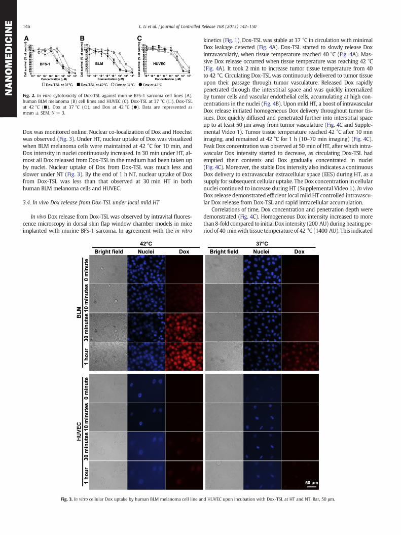
Fig. 2. In vitro cytotoxicity of Dox-TSL against murine BFS-1 sarcoma cell lines (A),human BLM melanoma (B) cell lines and HUVEC (C). Dox-TSL at 37 °C (□), Dox-TSLat 42 °C (■), Dox at 37 °C (○), and Dox at 42 °C (●). Data are represented asmean ± SEM. N = 3.
146 L. Li et al. / Journal of Controlled Release 168 (2013) 142–150
NANOMEDICIN
E
Dox was monitored online. Nuclear co-localization of Dox and Hoechstwas observed (Fig. 3). Under HT, nuclear uptake of Dox was visualizedwhen BLM melanoma cells were maintained at 42 °C for 10 min, andDox intensity in nuclei continuously increased. In 30 min under HT, al-most all Dox released from Dox-TSL in the medium had been taken upby nuclei. Nuclear uptake of Dox from Dox-TSL was much less andslower under NT (Fig. 3). By the end of 1 h NT, nuclear uptake of Doxfrom Dox-TSL was less than that observed at 30 min HT in bothhuman BLM melanoma cells and HUVEC.
3.4. In vivo Dox release from Dox-TSL under local mild HT
In vivo Dox release from Dox-TSL was observed by intravital fluores-cence microscopy in dorsal skin flap window chamber models in miceimplanted with murine BFS-1 sarcoma. In agreement with the in vitro
Fig. 3. In vitro cellular Dox uptake by human BLM melanoma cell line an
kinetics (Fig. 1), Dox-TSL was stable at 37 °C in circulation with minimalDox leakage detected (Fig. 4A). Dox-TSL started to slowly release Doxintravascularly, when tissue temperature reached 40 °C (Fig. 4A). Mas-sive Dox release occurred when tissue temperature was reaching 42 °C(Fig. 4A). It took 2 min to increase tumor tissue temperature from 40to 42 °C. Circulating Dox-TSL was continuously delivered to tumor tissueupon their passage through tumor vasculature. Released Dox rapidlypenetrated through the interstitial space and was quickly internalizedby tumor cells and vascular endothelial cells, accumulating at high con-centrations in the nuclei (Fig. 4B). Upon mild HT, a boost of intravascularDox release initiated homogeneous Dox delivery throughout tumor tis-sues. Dox quickly diffused and penetrated further into interstitial spaceup to at least 50 μm away from tumor vasculature (Fig. 4C and Supple-mental Video 1). Tumor tissue temperature reached 42 °C after 10 minimaging, and remained at 42 °C for 1 h (10–70 min imaging) (Fig. 4C).Peak Dox concentration was observed at 50 min of HT, after which intra-vascular Dox intensity started to decrease, as circulating Dox-TSL hademptied their contents and Dox gradually concentrated in nuclei(Fig. 4C). Moreover, the stable Dox intensity also indicates a continuousDox delivery to extravascular extracellular space (EES) during HT, as asupply for subsequent cellular uptake. TheDox concentration in cellularnuclei continued to increase during HT (Supplemental Video 1). In vivoDox release demonstrated efficient local mild HT controlled intravascu-lar Dox release from Dox-TSL and rapid intracellular accumulation.
Correlations of time, Dox concentration and penetration depth weredemonstrated (Fig. 4C). Homogeneous Dox intensity increased to morethan 8-fold compared to initial Dox intensity (200 AU) during heating pe-riod of 40 minwith tissue temperature of 42 °C (1400 AU). This indicated
d HUVEC upon incubation with Dox-TSL at HT and NT. Bar, 50 μm.

147L. Li et al. / Journal of Controlled Release 168 (2013) 142–150
NANOMEDICIN
E
that liposomes were emptied and Dox intensity became heterogeneousthroughout the tumor tissue as soon as cellular uptake of Dox becamedominant and remained stable afterwards (Supplemental Video 1).
3.5. Tumor growth control
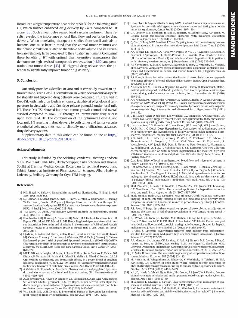
Treatments of systemic Dox-TSL and Dox-LTSL in combination withlocal mild HT at tumors were compared for tumor growth control inhuman BLM melanoma xenografts. When tumor temperature was at42 °C, mouse body temperature remained at 37 °C. Dox-TSL showedoverall significantly better tumor growth control than Dox-LTSL over atime course of 26 days after a single treatment of intravascular releaseof Dox containing TSL in combination with local mild HT (pairedMann–Whitney test, Dox-TSL vs. Dox-LTSL, p b 0.004; Dox-TSL vs. PBS,p b 0.001; Dox-LTSL vs. PBS, p b 0.031) (Fig. 5A,B). The mice after thetreatment of Dox-TSL and mild HT had tumor growth delay of~16 days, while the mice after the treatment of Dox-LTSL and mildHT had tumor growth delay of 8 days. By Day 26, 6 out of 9 mice sur-vived (tumor size b 11 × 15 × 10 mm3) after treatment withDox-TSL and local mild HT; while only 2 out of 9 mice survived(~13 × 16 × 12 mm3) after treatment with Dox-LTSL and localmild HT. In sham group, all of 9 mice had to be euthanized becauseof large tumor size. Intravascular release of Dox from Dox-TSL showedsignificantly improved survival of mice bearing s.c. human BLMmelano-ma up to 26 days after a single treatment (paired Mann–Whitney test,Dox-TSL vs. Dox-LTSL, p b 0.003; Dox-TSL vs. PBS, p b 0.001; Dox-LTSLvs. PBS, p = 0.05) (Fig. 5C).
4. Discussion
Dox-LTSL has been studied in combination with local HT at tumorsites [10] to induce intravascular drug release-mediated control oftumor growth and is currently in clinical trials [15,28]. Lysolipid-basedDox-LTSL is known to release significant quantities of Dox at
Fig. 4. In vivo Dox release from Dox-TSL in murine BFS-1 sarcoma (A), and subsequent cellularmergedGFP endothelial cells (green) and Dox (red) fluorescence. Bar in A, 200 μm. Bar in B, 50the references to color in this figure legend, the reader is referred to the web version of this ar
physiological temperatures in serum and thus has a fast plasma clear-ance upon administration [30,15,36,37]. As early as 1994, Unezaki etal. already proved the benefit of localHT and TSL forDox delivery tomu-rine C26 colon carcinoma [38]. They incorporated 3 mol% of DSPE-PEGwith ameanmolecularweight of 1000 or 5000 in DPPC/DSPC liposomeswith a diameter of 180–200 nm, and suppressed tumor growth withintratumoral Dox two-fold compared to DSPE-PEG lacking TSL andsix-fold compared to freeDox [38]. Inspired by these prior promising re-sults, we aimed to further improve TSL-mediated drug delivery byestablishing a more stable formulation. We omitted the lysolipid andmade use of an optimal DSPE-PEG2000 density to prolong drug retentionat physiological temperature in circulation with peak rate at 42 °C [18].Besides, our Dox-TSL was significantly smaller (80 nm) than LTSL(120 nm) and Dox-PEGTSL (180–200 nm) [38,29,17], to further pro-mote rapid release at their Tm [39]. Our in vitroDox release kinetics illus-trated that the fastest release rate occurred at 42 °C (Fig. 1B).
Dox can relatively rapidly pass cellular membranes followed bystrong binding to DNA in cellular nuclei. Because of the increased perfu-sion, vascular permeability and microconvection in the EES induced byHT, after release, Dox distributed homogeneously into tumor tissue atdistances up to 50 μm away from tumor feeding blood vessels (Fig. 4).In addition, Dox delivery continued to increase during the course ofthe HT treatment, due to new drug-filled Dox-TSL being available in cir-culation and arriving in the heated tumor tissue. Due to the continuoussupply of drug-loaded TSL, Dox delivered by TSL will penetrate deeperinto EES than administration of free Dox, which is rapidly cleared fromcirculation [7]. Importantly, we observed nuclear Dox uptake in vivo intumor cells as well as in tumor vascular endothelial cells (Fig. 4B).Thus, intratumoral Dox release from Dox-TSL confirms a dual targetingof chemotherapy to both intratumoral endothelial cells and adjacenttumor cells as can be achieved by targeted liposomes or using the intra-vascular drug release approach [12,40,41,28,42]. We were able to dem-onstrate that in vitro exposure of HUVEC to Dox in free form andDox-TSL in combination with mild HT causes significant intracellular
uptake by murine BFS-1 sarcoma cells and endothelial cells (B). Images are presented asμm. Quantitative Dox intensity and penetration depth duringHT (C). (For interpretation ofticle.)
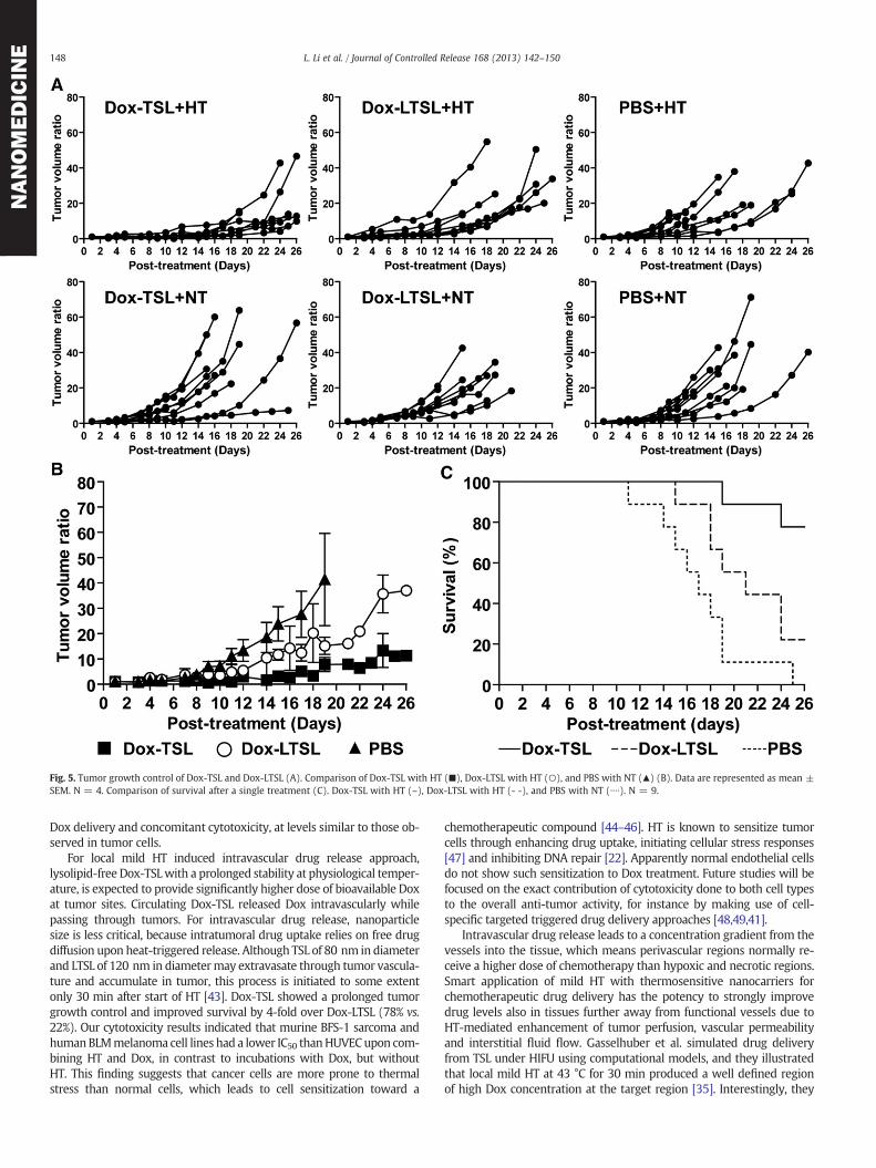
Fig. 5. Tumor growth control of Dox-TSL and Dox-LTSL (A). Comparison of Dox-TSL with HT (■), Dox-LTSL with HT (○), and PBS with NT (▲) (B). Data are represented as mean ±SEM. N = 4. Comparison of survival after a single treatment (C). Dox-TSL with HT (–), Dox-LTSL with HT (- -), and PBS with NT (∙∙∙∙). N = 9.
148 L. Li et al. / Journal of Controlled Release 168 (2013) 142–150
NANOMEDICIN
E
Dox delivery and concomitant cytotoxicity, at levels similar to those ob-served in tumor cells.
For local mild HT induced intravascular drug release approach,lysolipid-free Dox-TSLwith a prolonged stability at physiological temper-ature, is expected to provide significantly higher dose of bioavailable Doxat tumor sites. Circulating Dox-TSL released Dox intravascularly whilepassing through tumors. For intravascular drug release, nanoparticlesize is less critical, because intratumoral drug uptake relies on free drugdiffusion upon heat-triggered release. Although TSL of 80 nm in diameterand LTSL of 120 nm in diametermay extravasate through tumor vascula-ture and accumulate in tumor, this process is initiated to some extentonly 30 min after start of HT [43]. Dox-TSL showed a prolonged tumorgrowth control and improved survival by 4-fold over Dox-LTSL (78% vs.22%). Our cytotoxicity results indicated that murine BFS-1 sarcoma andhumanBLMmelanoma cell lines had a lower IC50 thanHUVECupon com-bining HT and Dox, in contrast to incubations with Dox, but withoutHT. This finding suggests that cancer cells are more prone to thermalstress than normal cells, which leads to cell sensitization toward a
chemotherapeutic compound [44–46]. HT is known to sensitize tumorcells through enhancing drug uptake, initiating cellular stress responses[47] and inhibiting DNA repair [22]. Apparently normal endothelial cellsdo not show such sensitization to Dox treatment. Future studies will befocused on the exact contribution of cytotoxicity done to both cell typesto the overall anti-tumor activity, for instance by making use of cell-specific targeted triggered drug delivery approaches [48,49,41].
Intravascular drug release leads to a concentration gradient from thevessels into the tissue, which means perivascular regions normally re-ceive a higher dose of chemotherapy than hypoxic and necrotic regions.Smart application of mild HT with thermosensitive nanocarriers forchemotherapeutic drug delivery has the potency to strongly improvedrug levels also in tissues further away from functional vessels due toHT-mediated enhancement of tumor perfusion, vascular permeabilityand interstitial fluid flow. Gasselhuber et al. simulated drug deliveryfrom TSL under HIFU using computational models, and they illustratedthat local mild HT at 43 °C for 30 min produced a well defined regionof high Dox concentration at the target region [35]. Interestingly, they
149L. Li et al. / Journal of Controlled Release 168 (2013) 142–150
NANOMEDICIN
E
introduced a high temperature heat pulse at 50 °C for 2 s following mildHT, which further enhanced drug delivery by ~40% compared to HTalone [35]. Such a heat pulse ceased local vascular perfusion. These re-sults revealed the importance of local fluid flow and perfusion for drugdelivery. When translating therapeutic studies from small animals tohumans, one must bear in mind that the animal tumor volumes andtheir blood circulation related to the whole body volume and its circula-tion are relatively large compared to the situation in humans. Combiningthese benefits of HT with optimal thermosensitive nanocarriers thatdemonstrate high levels of nanoparticle extravasation [43,50] and pene-tration into tumor tissues [43], HT triggered drug release bears the po-tential to significantly improve tumor drug delivery.
5. Conclusion
Our study provides a detailed in vitro and in vivo study toward an op-timized nano-sized Dox-TSL formulation, in which several critical aspectsfor stability and triggered drug delivery were combined. This resulted inDox-TSL with high drug loading efficiency, stability at physiological tem-perature in circulation, and fast drug release potential under local mildHT. These Dox-TSL showed improved tumor growth control and overallsurvival compared to Dox-LTSL through an intravascular drug releaseupon local mild HT. The combination of the optimized Dox-TSL andlocalmild HT resulting in drug delivery to both tumor and tumor vascularendothelial cells may thus lead to clinically more efficacious advanceddrug delivery systems.
Supplementary data to this article can be found online at http://dx.doi.org/10.1016/j.jconrel.2013.03.011.
Acknowledgments
This study is funded by the Stichting Vanderes, Stichting Fondsen,SEHK.We thank Halil Ozkal, Debby Schipper, Csilla Scholten and ThomasSoullié at Erasmus Medical Center, Rotterdam for technical support, andSabine Barnert at Institute of Pharmaceutical Sciences, Albert-LudwigsUniversity, Freiburg, Germany for Cryo-TEM imaging.
References
[1] P.K. Singal, N. Iliskovic, Doxorubicin-induced cardiomyopathy, N. Engl. J. Med.339 (1998) 900–905.
[2] D.J. Slamon, B. Leyland-Jones, S. Shak, H. Fuchs, V. Paton, A. Bajamonde, T. Fleming,W. Eiermann, J. Wolter, M. Pegram, J. Baselga, L. Norton, Use of chemotherapy plusamonoclonal antibody against HER2 formetastatic breast cancer that overexpressesHER2, N. Engl. J. Med. 344 (2001) 783–792.
[3] T.M. Allen, P.R. Cullis, Drug delivery systems: entering the mainstream, Science303 (2004) 1818–1822.
[4] D.W. Northfelt, B.J. Dezube, J.A. Thommes, B.J. Miller, M.A. Fischl, A. Friedman-Kien, L.D.Kaplan, C.Du. Mond, R.D. Mamelok, D.H. Henry, Pegylated-liposomal doxorubicin ver-sus doxorubicin, bleomycin, and vincristine in the treatment of AIDS-related Kaposi'ssarcoma: results of a randomized phase III clinical trial, J. Clin. Oncol. 16 (1998)2445–2451.
[5] I. Judson, J.A. Radford, M. Harris, J.Y. Blay, Q. van Hoesel, A. le Cesne, A.T. van Oosterom,M.J. Clemons, C. Kamby, C. Hermans, J. Whittaker, E.D. di Paola, J. Verweij, S. Nielsen,Randomised phase II trial of pegylated liposomal doxorubicin (DOXIL (R)/CAELYX(R)) versus doxorubicin in the treatment of advanced ormetastatic soft tissue sarcoma:a study by the EORTC Soft Tissue and Bone Sarcoma Group, Eur. J. Cancer 37 (2001)870–877.
[6] M.E.R. O'Brien, N. Wigler, M. Inbar, R. Rosso, E. Grischke, A. Santoro, R. Catane, D.G.Kieback, P. Tomczak, S.P. Ackland, F. Orlandi, L. Mellars, L. Alland, C. Tendler, C.B.C.S.Grp, Reduced cardiotoxicity and comparable efficacy in a phase III trial of pegylatedliposomal doxorubicin HCl (CAELYX (TM)/Doxil (R)) versus conventional doxorubicinfor first-line treatment of metastatic breast cancer, Ann. Oncol. 15 (2004) 440–449.
[7] A. Gabizon, H. Shmeeda, Y. Barenholz, Pharmacokinetics of pegylated liposomaldoxorubicin — review of animal and human studies, Clin. Pharmacokinet. 42(2003) 419–436.
[8] A.L.B. Seynhaeve, S. Hoving, D. Schipper, C.E. Vermeulen, G.A. deWiel-Ambagtsheer,S.T. van Tiel, A.M.M. Eggermont, T.L.M. ten Hagen, Tumor necrosis factor alpha me-diates homogeneous distribution of liposomes inmurinemelanoma that contributesto a better tumor response, Cancer Res. 67 (2007) 9455–9462.
[9] W.J. Yatvin MB, W.H. Dennis, R. Blumenthal, Design of liposomes for enhancedlocal release of drugs by hyperthermia, Science 202 (1978) 1290–1293.
[10] D. Needham, G. Anyarambhatla, G. Kong,M.W. Dewhirst, A new temperature-sensitiveliposome for use with mild hyperthermia: characterization and testing in a humantumor xenograft model, Cancer Res. 60 (2000) 1197–1201.
[11] L.H. Lindner, M.E. Eichhorn, H. Eibl, N. Teichert, M. Schmitt-Sody, R.D. Issels, M.Dellian, Novel temperature-sensitive liposomes with prolonged circulationtime, Clin. Cancer Res. 10 (2004) 2168–2178.
[12] Q. Chen, S. Tong, M.W. Dewhirst, F. Yuan, Targeting tumor microvessels using doxoru-bicin encapsulated in a novel thermosensitive liposome, Mol. Cancer Ther. 3 (2004)1311–1317.
[13] A.A. Secord, E.L. Jones, C.A. Hahn, W.P. Petros, D. Yu, L.J. Havrilesky, J.T. Soper, A.Berchuck, I. Spasojevic, D.L. Clarke-Pearson, L.R. Prosnitz, M.W. Dewhirst, PhaseI/II trial of intravenous Doxil (R) and whole abdomen hyperthermia in patientswith refractory ovarian cancer, Int. J. Hyperthermia 21 (2005) 333–347.
[14] P.S. Yarmolenko, Y. Zhao, C. Landon, I. Spasojevic, F. Yuan, D. Needham, B.L. Viglianti,M.W. Dewhirst, Comparative effects of thermosensitive doxorubicin-containing lipo-somes and hyperthermia in human and murine tumours, Int. J. Hyperthermia 26(2010) 485–498.
[15] R.T. Poon, N. Borys, Lyso-thermosensitive liposomal doxorubicin: a novel approachto enhance efficacy of thermal ablation of liver cancer, Expert. Opin. Pharmacother.10 (2009) 333–343.
[16] A. Gasselhuber, M.R. Dreher, A. Negussie, B.J. Wood, F. Rattay, D. Haemmerich, Mathe-matical spatio-temporal model of drug delivery from low temperature sensitive lipo-somes during radiofrequency tumour ablation, Int. J. Hyperthermia 26 (2010)499–513.
[17] A.H. Negussie, P.S. Yarmolenko, A. Partanen, A. Ranjan, G. Jacobs, D.Woods, H. Bryant, D.Thomasson, M.W. Dewhirst, B.J. Wood, M.R. Dreher, Formulation and characterisationof magnetic resonance imageable thermally sensitive liposomes for use withmagneticresonance-guided high intensity focused ultrasound, Int. J. Hyperthermia 27 (2011)140–155.
[18] L. Li, T.L. ten Hagen, D. Schipper, T.M. Wijnberg, G.C. van Rhoon, A.M. Eggermont, L.H.Lindner, G.A. Koning, Triggered content release fromoptimized stealth thermosensitiveliposomes using mild hyperthermia, J. Control. Release 143 (2010) 274–279.
[19] J. van der Zee, D.G. Gonzalez, G.C. van Rhoon, J.D.P. van Dijk, W.L.J. van Putten,A.A.M. Hart, G Dutch Deep Hyperthermia, Comparison of radiotherapy alonewith radiotherapy plus hyperthermia in locally advanced pelvic tumours: a pro-spective, randomised, multicentre trial, Lancet 355 (2000) 1119–1125.
[20] R.D. Issels, L.H. Lindner, J. Verweij, P. Wust, P. Reichardt, B.C. Schem, S.Abdel-Rahman, S. Daugaard, C. Salat, C.M. Wendtner, Z. Vujaskovic, R.Wessalowski, K.W. Jauch, H.R. Durr, F. Ploner, A. Baur-Melnyk, U. Mansmann,W. Hiddemann, J.Y. Blay, P. Hohenberger, E.-S.E. European Org, Neo-adjuvantchemotherapy alone or with regional hyperthermia for localised high-risksoft-tissue sarcoma: a randomised phase 3 multicentre study, Lancet Oncol. 11(2010) 561–570.
[21] C.W. Song, Effect of local hyperthermia on blood flow and microenvironment: areview, Cancer Res. 44 (1984) 4721s–4730s.
[22] P.M. Krawczyk, B. Eppink, J. Essers, J. Stap, H. Rodermond, H. Odijk, A. Zelensky, C.van Bree, L.J. Stalpers, M.R. Buist, T. Soullie, J. Rens, H.J. Verhagen, M.J. O'Connor,N.A. Franken, T.L. Ten Hagen, R. Kanaar, J.A. Aten, Mild hyperthermia inhibits ho-mologous recombination, induces BRCA2 degradation, and sensitizes cancer cellsto poly(ADP-ribose) polymerase-1 inhibition, Proc. Natl. Acad. Sci. U. S. A. 108(2011) 9851–9856.
[23] M.M. Paulides, J.F. Bakker, E. Neufeld, J. Van der Zee, P.P. Jansen, P.C. Levendag,G.C. Van Rhoon, The HYPERcollar: a novel applicator for hyperthermia in thehead and neck, Int. J. Hyperthermia 23 (2007) 567–576.
[24] M. de Smet, E. Heijman, S. Langereis, N.M. Hijnen, H. Grull, Magnetic resonanceimaging of high intensity focused ultrasound mediated drug delivery fromtemperature-sensitive liposomes: an in vivo proof-of-concept study, J. Control.Release 150 (2011) 102–110.
[25] R.T. Poon, N. Borys, Lyso-thermosensitive liposomal doxorubicin: an adjuvant toincrease the cure rate of radiofrequency ablation in liver cancer, Future Oncol. 7(2011) 937–945.
[26] B.J. Wood, R.T. Poon, J.K. Locklin, M.R. Dreher, K.K. Ng, M. Eugeni, G. Seidel, S.Dromi, Z. Neeman, M. Kolf, C.D. Black, R. Prabhakar, S.K. Libutti, Phase I study ofheat-deployed liposomal doxorubicin during radiofrequency ablation for hepaticmalignancies, J. Vasc. Interv. Radiol. 23 (2012) 248–255, (e247).
[27] H. Grull, S. Langereis, Hyperthermia-triggered drug delivery from temperature-sensitive liposomes using MRI-guided high intensity focused ultrasound, J. Control.Release 161 (2012) 317–327.
[28] A.A. Manzoor, L.H. Lindner, C.D. Landon, J.Y. Park, A.J. Simnick, M.R. Dreher, S. Das, G.Hanna, W. Park, A. Chilkoti, G.A. Koning, T.L.M. ten Hagen, D. Needham, M.W.Dewhirst, Overcoming limitations in nanoparticle drug delivery: triggered, intravascu-lar release to improvedrug penetration into tumors, Cancer Res. 72 (2012) 5566–5575.
[29] J.K. Mills, D. Needham, The materials engineering of temperature-sensitive lipo-somes, Methods Enzymol. 387 (2004) 82–113.
[30] M. Hossann, M. Wiggenhorn, A. Schwerdt, K. Wachholz, N. Teichert, H. Eibl,R.D. Issels, L.H. Lindner, In vitro stability and content release properties ofphosphatidylglyceroglycerol containing thermosensitive liposomes, Biochim.Biophys. Acta 1768 (2007) 2491–2499.
[31] X. Li, D.J. Hirsh, D. Cabral-Lilly, A. Zirkel, S.M. Gruner, A.S. Janoff, W.R. Perkins, Doxoru-bicin physical state in solution and inside liposomes loaded via a pH gradient, Biochim.Biophys. Acta 1415 (1998) 23–40.
[32] M. Almgren, K. Edwards, G. Karlsson, Cryo transmission electron microscopy of lipo-somes and related structures, Colloids Surf. A 174 (2000) 3–21.
[33] N.W. Roehm, G.H. Rodgers, S.M. Hatfield, A.L. Glasebrook, An improved colorimetricassay for cell-proliferation and viability utilizing the tetrazolium salt XTT, J. Immunol.Methods 142 (1991) 257–265.

150 L. Li et al. / Journal of Controlled Release 168 (2013) 142–150
NANOMEDICIN
E
[34] M.R. Dreher, W.G. Liu, C.R. Michelich, M.W. Dewhirst, F. Yuan, A. Chilkoti, Tumorvascular permeability, accumulation, and penetration of macromolecular drugcarriers, J. Natl. Cancer Inst. 98 (2006) 335–344.
[35] A. Gasselhuber, M.R. Dreher, A. Partanen, P.S. Yarmolenko, D. Woods, B.J. Wood, D.Haemmerich, Targeted drug delivery by high intensity focused ultrasound mediatedhyperthermia combined with temperature-sensitive liposomes: computationalmodelling and preliminary in vivo validation, Int. J. Hyperthermia 28 (2012) 337–348.
[36] B. Banno, L.M. Ickenstein, G.N. Chiu, M.B. Bally, J. Thewalt, E. Brief, E.K. Wasan, Thefunctional roles of poly(ethylene glycol)-lipid and lysolipid in the drug retentionand release from lysolipid-containing thermosensitive liposomes in vitro and invivo, J. Pharm. Sci. 99 (2010) 2295–2308.
[37] W.T. Al-Jamal, Z.S. Al-Ahmady, K. Kostarelos, Pharmacokinetics & tissue distribu-tion of temperature-sensitive liposomal doxorubicin in tumor-bearing mice trig-gered with mild hyperthermia, Biomaterials 33 (2012) 4608–4617.
[38] S. Unezaki, K. Maruyama, N. Takahashi, M. Koyama, T. Yuda, A. Suginaka, M. Iwatsuru,Enhanced delivery and antitumor-activity of doxorubicin using long-circulatingthermosensitive liposomes containing amphipathic polyethylene-glycol in combina-tion with local hyperthermia, Pharm. Res. 11 (1994) 1180–1185.
[39] M. Hossann, T. Wang, M. Wiggenhorn, R. Schmidt, A. Zengerle, G. Winter, H. Eibl, M.Peller, M. Reiser, R.D. Issels, L.H. Lindner, Size of thermosensitive liposomes influencescontent release, J. Control. Release 147 (2010) 436–443.
[40] A.S. Abu Lila, N.E. Eldin, M. Ichihara, T. Ishida, H. Kiwada, Multiple administration ofPEG-coated liposomal oxaliplatin enhances its therapeutic efficacy: a possible mecha-nism and the potential for clinical application, Int. J. Pharm. 438 (2012) 176–183.
[41] B.M. Dicheva, T.L. Hagen, L. Li, D. Schipper, A.L. Seynhaeve, G.C. Rhoon, A.M. Eggermont,L.H. Lindner, G.A. Koning, Cationic thermosensitive liposomes: a novel dual targetedheat-triggered drug delivery approach for endothelial and tumor cells, Nano Lett.(2012), http://dx.doi.org/10.1021/nl3014154.
[42] J.I. Hare, E.H. Moase, T.M. Allen, Targeting combinations of liposomal drugs toboth tumor vasculature cells and tumor cells for the treatment of HER2-positivebreast cancer, J. Drug Target. 21 (2013) 87–96.
[43] L. Li, T.L.M. ten Hagen, M. Bolkestein, A. Gasselhuber, J. Yatvin, G.C. van Rhoon,A.M.M. Eggermont, D. Haemmerich, G.A. Koning, Improved intratumoral nanoparti-cle extravasation and penetration by mild hyperthermia, J. Control. Release 167(2013) 130–137.
[44] D.A. Averill, C. Su, Sensitization to the cytotoxicity of adriamycin by verapamiland heat in multidrug-resistant Chinese hamster ovary cells, Radiat. Res. 151(1999) 694–702.
[45] M. Hermisson, M. Weller, Hyperthermia enhanced chemosensitivity of humanmalignant glioma cells, Anticancer. Res. 20 (2000) 1819–1823.
[46] G.P. Raaphorst, D.P. Yang, The evaluation of thermal cisplatin sensitization in normaland XP human cells using mild hyperthermia at 40 and 41 degrees C, Anticancer.Res. 25 (2005) 2649–2653.
[47] H.H. Kampinga, Cell biological effects of hyperthermia alone or combined with radia-tion or drugs: a short introduction to newcomers in the field, Int. J. Hyperthermia 22(2006) 191–196.
[48] R.M. Schiffelers, G.A. Koning, T.L. ten Hagen, M.H. Fens, A.J. Schraa, A.P. Janssen, R.J.Kok, G.Molema, G. Storm, Anti-tumor efficacy of tumor vasculature-targeted liposo-mal doxorubicin, J. Control. Release 91 (2003) 115–122.
[49] A.H. Negussie, J.L. Miller, G. Reddy, S.K. Drake, B.J. Wood, M.R. Dreher, Synthesisand in vitro evaluation of cyclic NGR peptide targeted thermally sensitive lipo-some, J. Control. Release 143 (2010) 265–273.
[50] G. Kong, R.D. Braun, M.W. Dewhirst, Hyperthermia enables tumor-specific nano-particle delivery: effect of particle size, Cancer Res. 60 (2000) 4440–4445.