isolation of cellulolytic activities from tribolium
TRANSCRIPT
8/8/2019 Isolation of cellulolytic activities from Tribolium
http://slidepdf.com/reader/full/isolation-of-cellulolytic-activities-from-tribolium 1/6
African Journal of Biotechnology Vol. 8 (23), pp. 6710-6715, 1 December, 2009Available online at http://www.academicjournals.org/AJBISSN 1684–5315 © 2009 Academic Journals
Full Length Research Paper
Isolation of cellulolytic activities fromTribolium
castaneum (red flour beetle)
Fayyaz Ur Rehman1*, Mehwish Aslam1, M. Ilyas Tariq1, Ashraf Shaheen1, Amtul Jamil Sami2,Naima Huma Naveed3 and Aima Iram Batool3
1Department of Chemistry, University of Sargodha, Sargodha, Pakistan.
2Institute of Biochemistry and Biotechnology, University of the Punjab, Lahore, Pakistan.
3Department of Biological Sciences, University of Sargodha, Sargodha, Pakistan.
Accepted 26 May, 2009
Cellulolytic enzymes have immense potential to convert cellulosic biomass into useful products. Tribolium castaneum crude proteins were isolated to screen the cellulolytic activities. The activity wasestablished by substrate-agar plate assay and confirmed by endoglucanase assay. Cellulolytic activitywas further purified and characterized using the different chromatographic techniques andelectrophoresis. Gel filtration chromatography showed the presence of multiple forms of enzymeactivities with different molecular weights. Stability of enzyme activity was investigated at differenttemperatures and pH. Optimum pH for was found 4.8 at 40
oC determined as optimum temperature.
Gradually decreasing Enzyme activity remained half at 60oC. Zymography and SDS-PAGE showed the
presence of multiple forms of endoglucanase activities (Cel I and Cel II) with molecular weight of 55 kDaand 35 kDa.
Key words: Tribolium castaneum , cellulolytic enzymes, endoglucanase, insect cellulase, red flour beetle.
INTRODUCTION
The most common organic compound on earth, cellulose,is about 33% of all plant matter. Cellulose is mainly pro-duced by terrestrial plants and marine algae and used asa food source by many organisms (Teeri, 1997). Cellu-lose production by non-photosynthetic organisms inclu-ding bacteria, marine invertebrates, fungi, slime moldsand amoebae has also been documented (Coughlan,1990; Tomme et al., 1995; Lynd et al., 2002). It has su-preme source of renewable energy in higher animal lifeas well as for lower animals.
Pests are lower animals which obtain food and energy
by degrading plant cellulose into its constituent residuesby the cellulolytic enzymes cellulases. Cellulose hydro-lysis yields cellobiose and glucose monomers which aremost important energy source around the globe(Eveleigh, 1987). In microorganisms, like bacteria andfungi, these enzymes are well studied and characterized(Coughlan, 1990; Nathan et al., 2007). Animals areusually considered dependent on gut microbiota for cellu-
*Corresponding author. E-mail: [email protected].
lose digestion as they are unable to produce indigenouscellulases (Moran et al., 2005; Eckburg et al., 2005Martin and Martin, 1978). Therefore earlier investigationshave been conventionally focused on non-animal cellulase sources such as bacteria and fungi.
Isolation and characterization of cellulase encoding ge-nes from lower animals, in recent years, shows that cellulose hydrolysis in some invertebrate animal taxa involvessynergistic action of flagellates, bacteria, yeasts (Breznakand Brune 1994; König et al., 2002; Varma et al., 1994Cleveland, 1924) and indigenously produced cellulases
Such cellulolytic activities were studied amongst manyinvertebrates like nematodes (Smant et al., 1998; Be´ra-Maillet et al., 2000), arthropods (Watanabe et al., 1997Wei et al., 2006)
and mollusks (Guo et al., 2008
Imjongjirak et al., 2008; Marshall and Grand, 1976; Xu eal., 2000).
Physiological functions of animal cellulases are distincand depend on their source. In bacteria, the enzymes areinvolved in breakdown of fungal cell walls in order toallow them to be used as a food source
and in bioreme
diation. In case of pests especially beetles, cellulase pro-ducing symbiotic protozoan present in intestine (Breznak
8/8/2019 Isolation of cellulolytic activities from Tribolium
http://slidepdf.com/reader/full/isolation-of-cellulolytic-activities-from-tribolium 2/6
and Brune, 1994; Brune, 1998; Dolan, 2001; Dyer, 2002)
hydrolyze cellulose to glucose in synergistic collaborativerelationship with indigenously produced enzymes (Wei et al.,2006; Zverlov et al., 2003). In the present study we hadisolated the cellulolytic activities from a beetle Tribo-lium castaneum (red flour beetle). This beetle cause ex-tensivedamage to the rice and wheat grains (Via, 1999; Weston andRattlingourd, 2000). This beetle uses its amylase andcellulase activities to get energy from the food.
MATERIALS AND METHODS
Insect sampling
T. castaneum (red flour beetle) samples were collected from riceand grain stockpile when temperature was around 40oC and storedat 4oC.
Preparation of crude enzyme sample
Crude enzyme sample was prepared by homogenizing 50 g of in-sect sample in 200 ml 0.1 M Phosphate buffer of pH 7.0. Homo-genate was kept overnight in freezer and centrifuged at 10,000 rpmto discard pellet. 100 ml of supernatant was added to 400 ml of ice-cold acetone and kept overnight at 4oC to get proteins in precipitateform. The mixture was centrifuged at 10,000 rpm for 15 min. Thepellets were air dried and dissolved in 10 ml of 0.1 M phosphatebuffer and 10 ml of Tris-HCl buffer of pH 6.0 and 8.0 respectively.This crude protein sample was stored at 4oC and used as the enzy-me source.
Screening for cellulolytic activity
A modification of substrate-agar plate assay (Teather and Wood,1982; Sami and Akhtar, 1990) was used to screen the cellulase
activity in crude protein extract by observing the cleared zone for-mation around the sample well against a red-stained backgroundon agar plates. 3% agar and 1% CMC were mixed with buffers ofdifferent pH range to prepare agar gel. Agar gel was poured in petriplates to solidify. A sample of 100 l of crude protein extract wasloaded in the hole punched in the plate centre. After overnight incu-bation CMC-agar plates were stained with 0.1% congo red dye for15 min followed by de-staining with 0.5 M sodium chloride solution.
Endoglucanase enzyme assay
Enzyme assay was performed by measuring the amount of redu-cing sugar using modified DNS (dinitrosalicylic acid) method(Nelson, 1944). Concentration of the released glucose was mea-sured from a standard glucose curve. Enzyme activity (U/ml) was
determined considering one I U equal to 1 mol min-1 of glucoseformed in the hydrolysis reaction. Reaction mixture was preparedby mixing 100 µl of the crude enzyme sample with 0.5 ml of carbo-xymethyl cellulose (CMC) solution (2%, w/v) and 0.5 ml 0.1 M so-dium acetate buffer (pH 5.0). Then mixture was incubated for 5 h at50oC with gentle shaking. After incubation, 2 ml of DNS reagent wasadded to reaction mixture and incubated in boiling water bath for 15min. After incubation absorbance was noted at 540 nm.
pH and temperature optima of enzyme activities
To determine the pH profile of cellulase activity, the enzyme assay
Rehman et al. 6711
was carried out using 0.1 M buffers ranging from pH 4-8. Acetatebuffer (pH 4-5), phosphate buffer (pH 5-6) and Tris-HCl buffer (pH7-8) were used. Agar and CMC were mixed with buffers of differenpH and enzyme activity was determined using the DNS method asdescribed in endoglucanase assay. In order to determine theoptimum assay temperature, enzyme assay was performed adifferent temperatures ranges between 4 - 70oC using the buffer o
pH 4.8.
Effect of substrate concentration
Effect of substrate concentration was studied by using different concentrations of CMC ranging from 0.5-5.0 % in the endoglucanaseassay mixture using buffer of pH 4.8 and at temperature 40oC.
Effect of -mercaptoethanol
Endoglucanase activity was studied in the presence of differenconcentrations of -mercaptoethanol in enzyme assay mixture. 100µl of -mercaptoethanol ranging in the concentration of 100-500mM was added to reaction mixture and enzyme activity was deter
mined.
Protein purification
Endoglucanase activity was purified using the chromatographictechniques. The crude enzyme sample was fractionated at 4oCusing the swollen sephadex G-75 suspension, packed in a column(1.6 × 16 cm). The cellulase activity and protein concentration weredetermined by enzyme assay in collected fractions. Active fractionshaving endoglucanase activity were pooled and subjected to ionexchange chromatography for further purification. Active fractionswere loaded into ion exchange column (0.8 x 15 cm) packed withthe DEAE-sephadex gel and equilibrated with Tris-HCl buffer. Sample was loaded and the bound proteins were eluted by a linear gradient of 0-0.5 M NaCl in Tris-HCl buffer (pH 8.5). A total of 50 fractions (2 ml each) were collected. Protein concentration and enzymeactivity were determined in each fraction by dye binding assay(Bradford, 1976) and DNS method. For estimation of protein 2 ml obradford reagent was added to 0.5 ml of each fraction, absorbancewas noted at 595 nm and concentration was determined by usingthe BSA standard curve. Active pooled fractions were stored at 4oC
Native PAGE and zymography
Polyacrylamide gel electrophoresis (PAGE) was performed undenon-denaturing conditions (Sami and Akhtar, 1990). Active fractionsfrom ion exchange chromatography were subjected to electrophoresis. After electrophoresis gel was cut in two strips vertically. Onestrip was placed onto substrate agar plate to locate the position o
endoglucanase activity on gel. Substrate agar gel plate was prepared using the 3% agar and 2% CMC, mixed with buffer of pH 4.8After incubation for 3 h at 40oC, gel strip was removed followed bystaining and detaining of substrate agar plate with 0.1% congo reddye and 0.5 M sodium chloride respectively.
Protein elution from gel
Protein was eluted from gel after locating the position of the bandsof endoglucanase activity. Gel bands were crushed to pieces and 1ml 0.5 M phosphate buffer pH 5.0 was added to elute the enzymeactivity. After overnight elution at 4oC, gel was centrifuged and puri
8/8/2019 Isolation of cellulolytic activities from Tribolium
http://slidepdf.com/reader/full/isolation-of-cellulolytic-activities-from-tribolium 3/6
6712 Afr. J. Biotechnol.
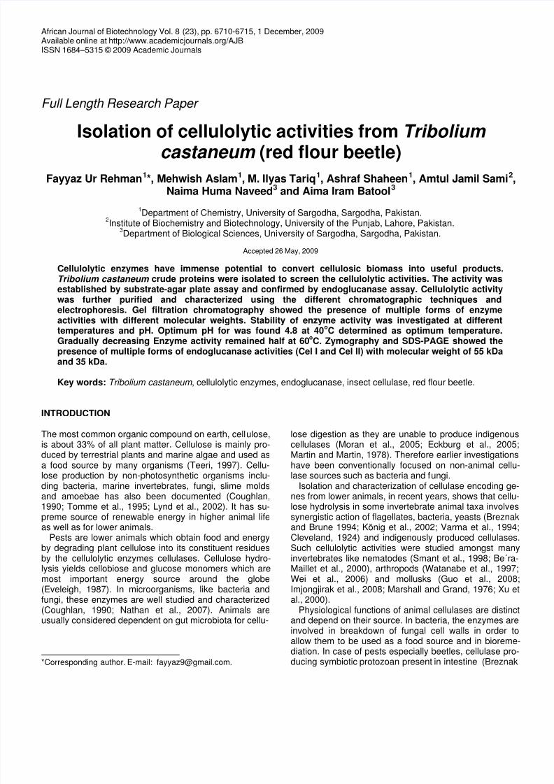
Figure 1. Yellow color area in centre of substrate agar plate shows the hydrolysis of carboxymethyl cellulose by endoglucanase activity.
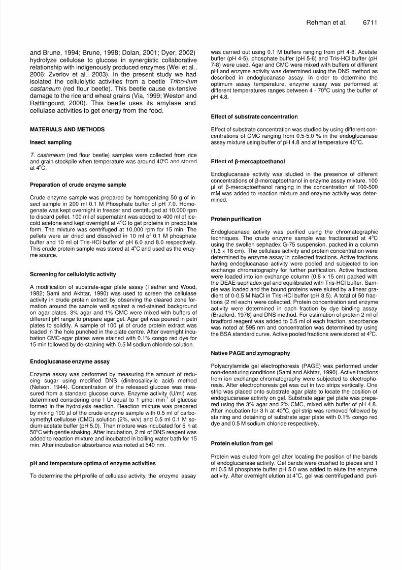
Figure 2. Optimum endoglucanase activity was observed inthe acidic range at pH 4.8 and second peak was observed at6.8 which is pH of insect gut. After pH 6.0 enzyme activity wasdeclined abruptly.
fied enzyme activity was further subjected to SDS-PAGE to calcu-late molecular weight.
Molecular weight determination by SDS- PAGE
SDS-PAGE was performed as described by to determine the mole-cular weight of endoglucanase activities purified by chromatogra-phic techniques and zymography.
RESULTS
Substrate-agar plate assay
Substrate agar plate assay showed the presence of cellu-lase activity by forming the cleared zone around the sam-ple well against a red-stained background on agar plates
Figure 3. Endoglucanase activity was obeservedmaximum at temperature 40oC. Almost half activity wasremained at 60oC.
(Figure 1). The cellulase activity digested the carboxy-methyl cellulose in area that did not retain the congo reddye on destaining.
pH and temperature profile
Endo-glucanase activity peaks were observed both atacidic pH (4.8) and basic pH (7.5). Maximum activity wasobserved at 4.8. Almost no enzyme activity was observedat pH values below 4.4 and above 7.5 (Figure 2). Optimum activity was observed at temperature 50
oC. There
was an increase in enzyme activity with rise in tempera-ture till 40
oC and then there was an abrupt decline in
enzyme activity. Although some enzyme activity was observed at temperature 50
oC but was almost half than the
activity showed at 40oC (Figure 3). At 80
oC almost no en
zyme activity was observed.
8/8/2019 Isolation of cellulolytic activities from Tribolium
http://slidepdf.com/reader/full/isolation-of-cellulolytic-activities-from-tribolium 4/6
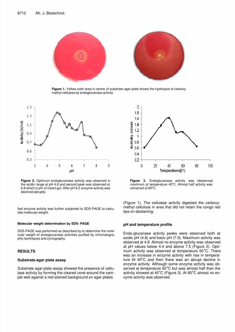
Figure 4. An increasing substrate concentration showed theincrease in enzyme activity. After 2% CMC concentration enzy-
me activity was continuously declined.
Figure 5. Mercaptoethanol gradually increased the enzymeactivity till concentration of 300 mM.
Effect of substrate concentration
Endoglucanase activity was found maximum at 2% of
substrate (CMC) concentration. After the increase in thesubstrate concentration to 2%, slight decline was obser-ved in the enzyme activity (Figure 4). At 5% of substrateconcentration very little endoglucanase activity was ob-served. This may be due to the inhibition of enzyme acti-vity by product formed.
Effect of -mercaptoethanol
-Mercaptoethanol gradually enhanced the endogluca-nase activity. Increase in the concentration of -mercap-
Rehman et al. 6713
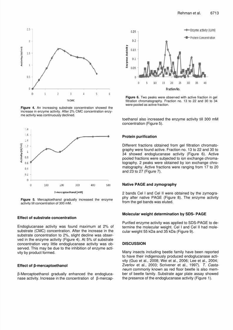
Figure 6. Two peaks were observed with active fraction in gelfiltration chromatography. Fraction no. 13 to 22 and 30 to 34were pooled as active fraction.
toethanol also increased the enzyme activity till 300 mMconcentration (Figure 5).
Protein purification
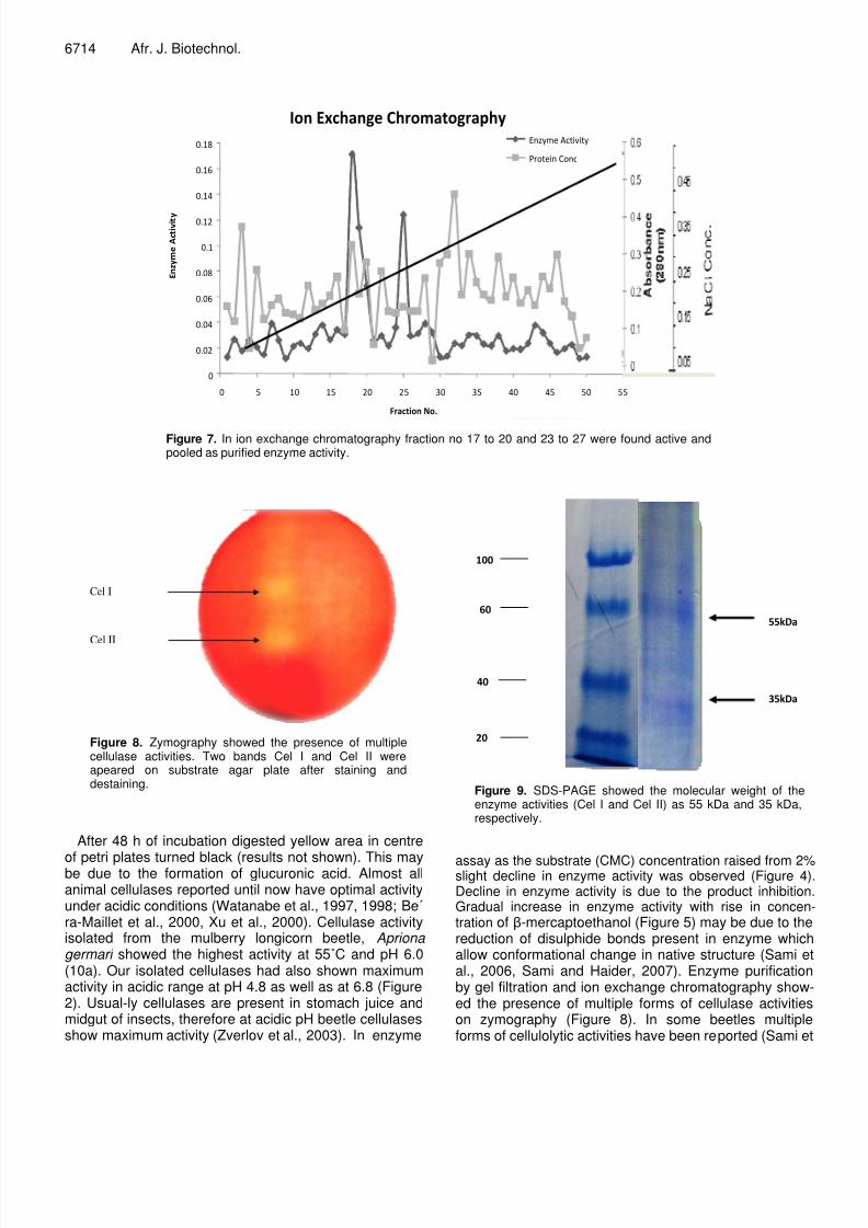
Different fractions obtained from gel filtration chromato-graphy were found active. Fraction no. 13 to 22 and 30 to34 showed endoglucanase activity (Figure 6). Activepooled fractions were subjected to ion exchange chromatography. 2 peaks were obtained by ion exchange chro-matography. Active fractions were ranging from 17 to 20and 23 to 27 (Figure 7).
Native PAGE and zymography
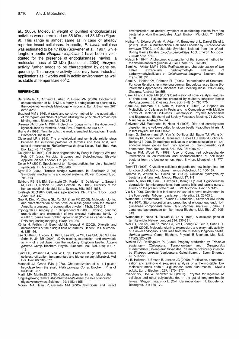
2 bands Cel I and Cel II were obtained by the zymogra-phy after native PAGE (Figure 8). The enzyme activityfrom the gel bands was eluted.
Molecular weight determination by SDS- PAGE
Purified enzyme activity was applied to SDS-PAGE to determine the molecular weight. Cel I and Cel II had mole-
cular weight 55 kDa and 35 kDa (Figure 9).
DISCUSSION
Many insects including beetle family have been reportedto have their indigenously produced endoglucanase acti-vity (Guo et al., 2008; Wei et al., 2006; Lee et al., 2004Zverlov et al., 2003; Scrivener et al., 1997). T. Castaneum commonly known as red floor beetle is also member of beetle family. Substrate agar plate assay showedthe presence of the endoglucanase activity (Figure 1).
8/8/2019 Isolation of cellulolytic activities from Tribolium
http://slidepdf.com/reader/full/isolation-of-cellulolytic-activities-from-tribolium 5/6
6714 Afr. J. Biotechnol.
Figure 7. In ion exchange chromatography fraction no 17 to 20 and 23 to 27 were found active andpooled as purified enzyme activity.
Cel I
Cel II
Figure 8. Zymography showed the presence of multiplecellulase activities. Two bands Cel I and Cel II wereapeared on substrate agar plate after staining anddestaining.
After 48 h of incubation digested yellow area in centre
of petri plates turned black (results not shown). This maybe due to the formation of glucuronic acid. Almost allanimal cellulases reported until now have optimal activityunder acidic conditions (Watanabe et al., 1997, 1998; Be´ra-Maillet et al., 2000, Xu et al., 2000). Cellulase activityisolated from the mulberry longicorn beetle, Apriona germari showed the highest activity at 55˚C and pH 6.0(10a). Our isolated cellulases had also shown maximumactivity in acidic range at pH 4.8 as well as at 6.8 (Figure2). Usual-ly cellulases are present in stomach juice andmidgut of insects, therefore at acidic pH beetle cellulasesshow maximum activity (Zverlov et al., 2003). In enzyme
!
Figure 9. SDS-PAGE showed the molecular weight of theenzyme activities (Cel I and Cel II) as 55 kDa and 35 kDa,respectively.
assay as the substrate (CMC) concentration raised from 2%slight decline in enzyme activity was observed (Figure 4)Decline in enzyme activity is due to the product inhibitionGradual increase in enzyme activity with rise in concentration of -mercaptoethanol (Figure 5) may be due to thereduction of disulphide bonds present in enzyme whichallow conformational change in native structure (Sami eal., 2006, Sami and Haider, 2007). Enzyme purificationby gel filtration and ion exchange chromatography showed the presence of multiple forms of cellulase activitieson zymography (Figure 8). In some beetles multipleforms of cellulolytic activities have been reported (Sami et
8/8/2019 Isolation of cellulolytic activities from Tribolium
http://slidepdf.com/reader/full/isolation-of-cellulolytic-activities-from-tribolium 6/6
6716 Afr. J. Biotechnol.
al., 2005). Molecular weight of purified endoglucanaseactivities was determined as 55 kDa and 35 kDa (Figure9). This range is almost same as in case of alreadyreported insect cellulases. In beetle, P. hilaris cellulasewas estimated to be 47 kDa (Scrivener et al., 1997) whilelonghorn beetle Rhagium inquisitor L have been invest-
tigated for the presence of endoglucanase, having amolecular mass of 32 kDa (Lee et al., 2004). Enzymeactivity further needs to be characterized by gene se-quencing. This enzyme activity also may have industrialapplications as it works well in acidic environment as wellas stable at temperature 60
oC.
REFERENCES
Be´ra-Maillet C, Arthaud L, Abad P, Rosso MN (2000). Biochemicalcharacterization of MI-ENG1, a family 5 endoglucanase secreted bythe root-knot nematode Meloidogyne incognita. Eur. J. Biochem. 267:3255-3263.
Bradford MM (1976). A rapid and sensitive method for the quantitation
of microgram quantities of protein utilizing the principle of protein-dyebinding. Anal. Biochem. 72: 248-254.Breznak JA, Brune A (1994). Role of microorganisms in the digestion of
lignocellulose by termites. Annu. Rev. Entomol. 39: 453-487Brune A (1998). Termite guts: the world's smallest bioreactors. Trends
Biotechnol. 16: 16-21Cleveland LR (1924). The physiological and symbiotic relationships
between the intestinal protozoa of termites and their host, withspecial reference to Reticulitermes flavipes Kollar. Biol. Bull. Mar.Biol. Lab. 46: 117-227.
Coughlan M (1990). Cellulose degradation by Fungi In Fogarty WM andKely CT (eds), Microbial Enzymes and Biotechnology. ElsevierApplied Science, London, UK, pp. 1-36.
Dolan MF (2001). Speciation of termite gut protists: the role of bacterialsymbionts. Int. Microbiol. 4: 203-208.
Dyer BD (2002). Termite hindgut symbionts. In: Seckbach J (ed)Symbiosis: mechanisms and model systems. Kluwer, Dordrecht, pp.
703-713.Eckburg PB, Bik EM, Bernstein CN, Purdom E, Dethlefsen L, Sargent
M, Gill SR, Nelson KE, and Relman DA (2005). Diversity of thehuman intestinal microbial ßora. Science, 308: 1635-1638.
Eveleigh DE (1987). Cellulase: a perspective. Phil. Trans. R. Soc. Lond.A321: 435-447.
Guo R, Ding M, Zhang SL, Xu GJ, Zhao FK (2008). Molecular cloningand characterization of two novel cellulase genes from the molluscAmpullaria crossean. J. comparative physiol. 178(2): 209-215.
Imjongjirak C, Amparyup P, Sittipraneed S (2008). Cloning, genomicorganization and expression of two glycosyl hydrolase family 10(GHF10) genes from golden apple snail (Pomacea canaliculata). J.DNA sequencing mapping. 19(3): 224-236
König H, Fröhlich J, Berchtold M, Wenzel M (2002). Diversity andmicrohabitats of the hindgut flora of termites. Recent Res. Microbiol.6: 125-156.
Lee SJ, Kim SR, Yoon HJ, Kim I, Lee KS, Je YH, Lee SM, Seo SJ, DaeSohn H, Jin BR (2004). cDNA cloning, expression, and enzymaticactivity of a cellulase from the mulberry longicorn beetle, Apriona germari . Comp. Biochem. Physiol. Biochem. Mol. Biol. 139(1): 107-116.
Lynd LR, Weimer PJ, Van WH, Zyl, Pretorius IS (2002). Microbialcellulose utilization: fundamentals and biotechnology. Microbiol. Mol.Boil. Rev. 66: 506-577
Marshall JJ, Grand RJA (1976). Characterization of a -1,4-glucanhydrolase from the snail, Helix pomatia . Comp. Biochem. Physiol.53B: 231-237.
Martin MM, Martin JS (1978). Cellulose digestion in the midgut of thefungus-growing termite Macrotermes natalensis : the role of acquired
digestive enzymes. Science, 199: 1453-1455.Moran NA, Tran P, Gerardo NM (2005). Symbiosis and insect
diversiÞcation: an ancient symbiont of sapfeeding insects from thebacterial phylum Bacteroidetes. Appl. Environ. Microbiol. 71: 88028810.
Nathan A, Ekborg Wendy M, Adam M, Burgoyne Li L, Daniel Distel L(2007). CelAB, a Multifunctional Cellulase Encoded by Teredinibacteturnerae T7902, a Culturable Symbiont Isolated from the WoodBoring Marine Bivalve Lyrodus pedicellatus . Appl. Environ. Microbiol73(23): 7785-7788.
Nelson N (1944). A photometric adaptation of the Somogyi method fothe determination of glucose. J. Biol. Chem. 153: 375-380.
Sami AJ, Akhtar MW (1990). Purification and characterization of twonative extracellular carboxymethyle cellulose ocarboxymethylcellulase of Cellulomonas flavigena . Biochem. SocTrans. 18: 651.
Sami AJ, Haider KM, Rehman FU (2006). Determination of StructureFunction Relationship in Apriona germari Endoglucanases Using Bioinformatics Approaches. Biochem. Soc. Meeting Biosci. 23-27 JulyGlasgow. Abstract No. 558.
Sami AJ and Haider MK (2007) Identification of novel catalytic featuresof endo-beta-1,4-glucanase produced by mulberry longicorn beetleApriona germari . J. Zhejiang Univ. Sci. (B) 8(10): 765-770.
Sami AJ, Rehman FU, Alam M, Haider R (2005). A Repoprt onMultiplicity of Cellulases in Pests and Its Comparison with MultipleForms of Bacterial Cellulases. Biocatalysis: Enzymes Mechanismand Bioprocess, Biochemi-cal Society Focussed Meeting, 21-22 NovManchester. Abstract No. 14.
Scrivener AM, Watanabe H, Noda H (1997). Diet and carbohydratedigestion in the yellow-spotted longicorn beetle Psacothea hilaris. JInsect Physiol. 43: 1039-1052
Smant G, Stokkermans JP, Yan Y, De Boer JM, Baum TJ, Wang XHussey RS, Gommers FJ, Henrissat B, Davis EL, Helder J, Schots ABakker J (1998). Endogenous cellulases in animals: isolation of -1,4endoglucanase genes from two species of plant-parasitic cysnematodes. Proc. Natl. Acad. Sci. USA, 95: 4906-4911.
Teather RM, Wood PJ (1982). Use of Congo red polysaccharideinteractions in enumeration and characterization of cellulolyticbacteria from the bovine rumen. Appl. Environ. Microbiol. 43: 777780.
Teeri TT (1997). Crystalline cellulose degradation: new insight into thefunction of cellobiohydrolases, Trends Biotechnol. 15: 160-167.
Tomme P, Warren AJ, Gilkes NR (1995). Cellulose hydrolysis by
bacteria and fungi. Adv. Microb. Physiol. 37: 1-81Varma A, Kolli BK, Paul J, Saxena S, König H (1994). Lignocellulose
degradation by microorganisms from termite hills and termite guts: asurvey on the present state of art. FEMS Microbiol. Rev. 15: 9-28.
Via S (1999). Cannibalism facilitates the use of a novel environment inthe flour beetle, Tribolium castaneum . Heredity, 82: 267-275.
Watanabe H, Nakamura M, Tokuda G, Yamaoka I, Scrivener AM, NodaH (1997). Site of secretion and properties of endogenous endo-1,4glucanase components from Reticulitermes speratus (Kolbe), aJapanese subterranean termite. Insect Biochem. Mol. Biol. 27: 305313
Watanabe H, Noda H, Tokuda G, Lo N (1998). A cellulase gene otermite origin. Nature (London) 394: 330-331.
Wei YD, Lee KS, Gui ZZ, Yoon HJ, Kim I, Zhang GZ, Guo X, Sohn HDJin BR (2006). Molecular cloning, expression, and enzymatic activityof a novel endogenous cellulase from the mulberry longicorn beetleApriona germari . Comp. Biochem. Physiol. B Biochem. Mol. Biol
145(2): 220-229Weston PA, Rattlingourd PL (2000). Progeny production by Tribolium
castaneum (Coleoptera: Tenebrionidae) and Oryzaephilussurinamensis (Coleoptera: Silvanidae) on maize previously infestedby Sitotroga cerealla (Lepidoptera: Gelechiidae), J. Econ. Entomol93: 533-536.
Xu B, Hellman U, Ersson B, Janson JC (2000). Purification, characterization and amino-acid sequence analysis of a thermostable, lowmolecular mass endo-1, 4-glucanase from blue mussel, Mytiluedulis . Eur. J. Biochem. 267: 4970-4977.
Zverlov VV, Höll W, Schwarz WH (2003). Enzymes for digestion ocellulose and other polysaccharides in the gut of longhorn beetlelarvae, Rhagium inquisitor L. (Col., Cerambycidae). Int. BiodeteriorBiodegrad. 51: 175-179.