computational analysis of an acidic lipase of tribolium ... _27_ pjz-1813-14 23-05-2014.pdf ·...
TRANSCRIPT

Pakistan J. Zool., vol. 46(3), pp. 805-812, 2014. Computational Analysis of an Acidic Lipase of Tribolium castaneum Amtul Jamil Sami,1* Nouraiz Ahmad1 and A.R. Shakoori2 1Institute of Biochemistry and Biotechnology, University of the Punjab, Lahore 2School of Biological Sciences, University of the Punjab, Lahore
Abstract.- Tribolium castaneum is a stored grain pest and generates a battery of enzymes for the utilization of stored grain as food. Genome of Tribolium castaneum reveals that there are 54 genes for the expression of lipase enzyme. Total soluble proteins were isolated from T. castaneum and lipase activity was fractionated using buffers with different pH 4.5, 6.5 and 7.0 in CM sepharose column. At least six different lipases were present in different fractions. Lipolytic activity was visualized on agar-substrate plates, using rhodamine reagent under UV light. Protein sequence for one of the acidic lipase similar to human lysosomal lipase was deduced from NCBI under the accession number XP_972957.2. e. for the identification of novel features of the enzyme. The identified features include catalytic triad, nucleophilic elbow, a flap covering active site of Leu 226-Ile257, Beta 9 loop, Beta 5 loop and oxyanion hole. Four N-glycosylation sites were identified at positions Asn59, Asn230, Asn242 and Asn296. Key words: Tribolium castaneum, acidic lipase, novel features.
INTRODUCTION
Lipase, which is involved in the cleavage or hydrolysis of fats, oils and triglycerides (Hammer and Murphy, 1994; Shimizu and Nakana, 2003) belong to the superfamily of α/ß hydrolases (Nardini and Dijkstra, 1999). Lipases act on substrate at the interface between the aqueous and the lipid phase. The peculiar feature of lipases is the presence of α/ß hydrolase fold in the N-terminal domain which contains a buried active site in the form of a catalytic triad which is similar to the catalytic site of chemotrypsin, trypsin, serine proteases and other esterases (Brzozowski et al., 1991; Brady et al., 1990; Winkler et al., 1990; Schrag et al., 1991; Lowe, 1992; Beer et al., 1996). Hide et al. (1992) reported the structure and evolution of lipse superfamily. Crystal structure of human gastric lipase and model of lysosomal acid lipase, two lipolytic enzymes of medical interest was reported by (Roussel et al., 1999). There are several differences between the acidic lipases, like there is no separate C-terminal domain in acidic lipase which is predominantly present in the neutral lipase and consists of a ß sandwich. Further there are four N-glycosylation sites present in the 3D structure of acidic lipase which may be responsible for providing maximum stability to the _______________________________ * Corresponding author: [email protected],
[email protected] 0030-9923/2014/0003-0805 $ 8.00/0 Copyright 2014 Zoological Society of Pakistan
enzyme to compensate for stability and substrate attachment affinity provided by the C-terminal of neutral lipases. Most lipases from all organisms can be divided into six families defined by sequence relationships within the α/ß hydrolase fold superfamily of proteins (Hide et al., 1992). These are the neutral (PF00151), acid (PF04083), lipase2 (PF01674), lipase3 (PF01764), GDSL (PF00657) and hormone sensitive lipases (PF06350) (Derewenda, 1994; Holmquist, 2004). All six families appear to use the same, two-step reaction mechanism based on a catalytic triad of residues (usually Ser-His-Asp/Glu) that generates a charge relay system and a highly nucleophilic serine. Another small family of lipases with an emerging role in lipid mobilization in insects, the adipose-triglyceride-lipase (ATGL) family (PF01734), also have an N-terminal domain with a predicted alpha/beta hydrolase fold and an active site serine residue (Zimmermann et al., 2004). In Tribolium castaneum, there are 54 genes for lipase enzyme out of which 25 are for neutral lipases, 25 are for acidic lipases, no gene for lipase type 2, 1 gene for lipase type 3, 2 genes for GDSL and 1 gene for hormone sensitive lipase which shows that neutral and acidic lipases, predominate in red flour beetle (Horne et al., 2009). As T. castaneum is a major stored grain pest and relies on lipids and triglycerides for its diet. A number of studies are reported on the effect of different insecticides on T. castanueum from Pakistan (Saleem and Shakoori, 2000a,b). The insect produced a number of hydrolytic enzymes
A. J. SAMI ET AL. 806
which are the focus of attention for several years. The inhibition of its lipase using site specific inhibitors specially from natural sources (Kumar and Prakash, 2009) can be of commercial importance on a large scale. Cygler et al. (1994) had studied the a structural basis for the chiral preferences of lipases. The lipase (TcL) that we are going to discuss here belongs to Tribolium castaneum and is similar to human lysosomal lipase (HLL) and comes under the class of acidic lipase family, along with gastric and lingual lipase (Anderson and Sando, 1991). That is why it is named, similar to lysosomal like lipase in NCBI with accession number XP_972957.2. It is composed of 391 amino acid residues including a signal sequence of 18 amino acids. The HLL is one of the most characterized lipase according to our current study and is widely used to study homology and function of lipase present in other organisms such as insects and bacteria in the past (Langin et al., 1993). Therefore, the various characteristics of the lipase from T. castaneum are determined by its superimposition and sequence alignment with human lysosomal lipase.
MATERIALS AND METHODS Specimens were collected from the local vegetation of district Lahore. The samples were stored in sterilized bottle. All the three insects were crushed separately in TAE buffer pH 8.0 in pestle and mortar. The crude solution was then centrifuged and supernatant was stored in a clean sterilized glass vial at -20ºC. Lipases of T. castaneum were purified on CM-Sepharose using different pH values buffers. This technique was successful for separating acidic, neutral and alkaline lipase. For this purpose, a column (3x10 cm) was properly washed with distilled water. The column was packed with CM-Sepharose gel (soaked at pH 4.5) and was washed with 100 ml, 0.1M phosphate buffer of pH 4.5. Crude sample (2 ml) of T. castaneum was loaded in the column. Proteins were eluted by using buffers of pH 4.5, pH 6.0 and pH of 7.0 buffer (20 ml each). Flow rate was 1ml/min at room temperature. The eluted fractions at different pH values were tested for lipase activity using agar substrate plates. The agar substrate plates were prepared by
homogenizing 1% olive oil in 0.1M Tris- HCl buffer pH 7.1 with 2 g agar, dissolved by heating. Plates were poured with the mixture and after hardening of agar holes were punched. In each hole 20 µl of the eluted samples were loaded and incubated overnight at 50ºC. The plates were stained with rhodamine solution extracted in chloroform. Free fatty acids released by the eluted enzyme fractions from column were visible under UV light. Computational methods Amino acid sequence for a lipase TcL from T. castaneum, showing identity to human lysosomal lipase was selected for the identification of novel catalytic features of the enzyme. The amino acid sequence of lipase from T. castaneum was extracted from NCBI with accession number XP_972957.2 and submitted to Phyre server which is a secondary structure prediction server to build a secondary and tertiary model of enzyme in the form of a protein data bank file. Then the active site and various conserved motifs of the enzyme were determined by sequence alignment with the lysosomal lipase of Homo sapiens (Accession # CAA83495.1) and then with insects using ClustalW. http://www.ebi.ac.uk/Tools/ msa/clustalw2/ Then the protein structure was visualized using Digital Discovery Studio by Accerlys which is a well known suite of software of simulating small molecules and proteins and provides a rich graphical interface. Further the sequence alignments, RMSD, Superimposition of lipase were also calculated and determined using Digital Discovery Studio.
RESULTS AND DISCUSSION Purification of whole body enzyme extract The crude enzyme extract of T. castaneum was subjected to ion exchange chromatography on CM Sepharose column. Alternative fractions were taken for Bradford assay and protein concentration was measured at 545nm (Bradford, 1976). Void volume of 10ml was excluded from the column. After loading sample (1ml) column was washed with 10 ml of buffer pH 4.5 then fractions were collected. Totally 65 fractions were collected using 20ml of each buffer including pH 4.5, pH 6.0 and
ACIDIC LIPASE OF T. CASTANEUM 807
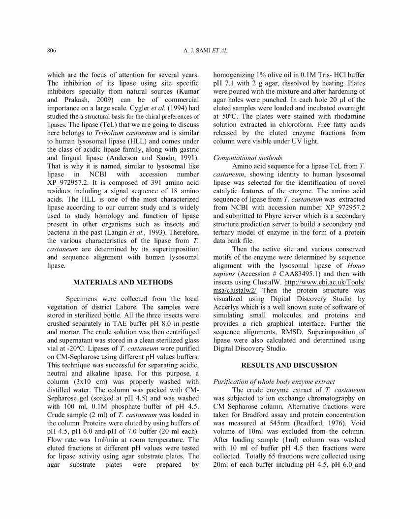
pH 7.0 (Fraction size 1 ml at the flow rate 1ml/min). (Data not shown). The alternate fractions were checked for lipase activity on substrate agar plate. Result are shown in Figure 1. A number of fractions showed lipase enzyme activity eluted at pH 4.5 (1-5, 6, 8, 9-11) and at pH 6.0 (26, 28 30, 38, 39) and at neutral pH (50, 52, 54, 60, 82 and 66). These results showed the functional expression of multiple lipases in T. castaneum. Our results confirm the presence of multiple forms of lipases as, earlier reported by Horne et al. (2009). In Tribolium castaneum, genome there are 54 genes for lipase enzyme including neutral lipases (25 gene), acidic lipases (25 gene) and hormone sensitive lipase. This indicates that neutral and acidic lipases, predominate in red flour beetle.
Fig. 1. 1% oil emulsion agar plates showing florescent zone of lipase activity of eluted fractions from CM-sepharose column of Tribolium enzyme. 20µl sample was added in each well.
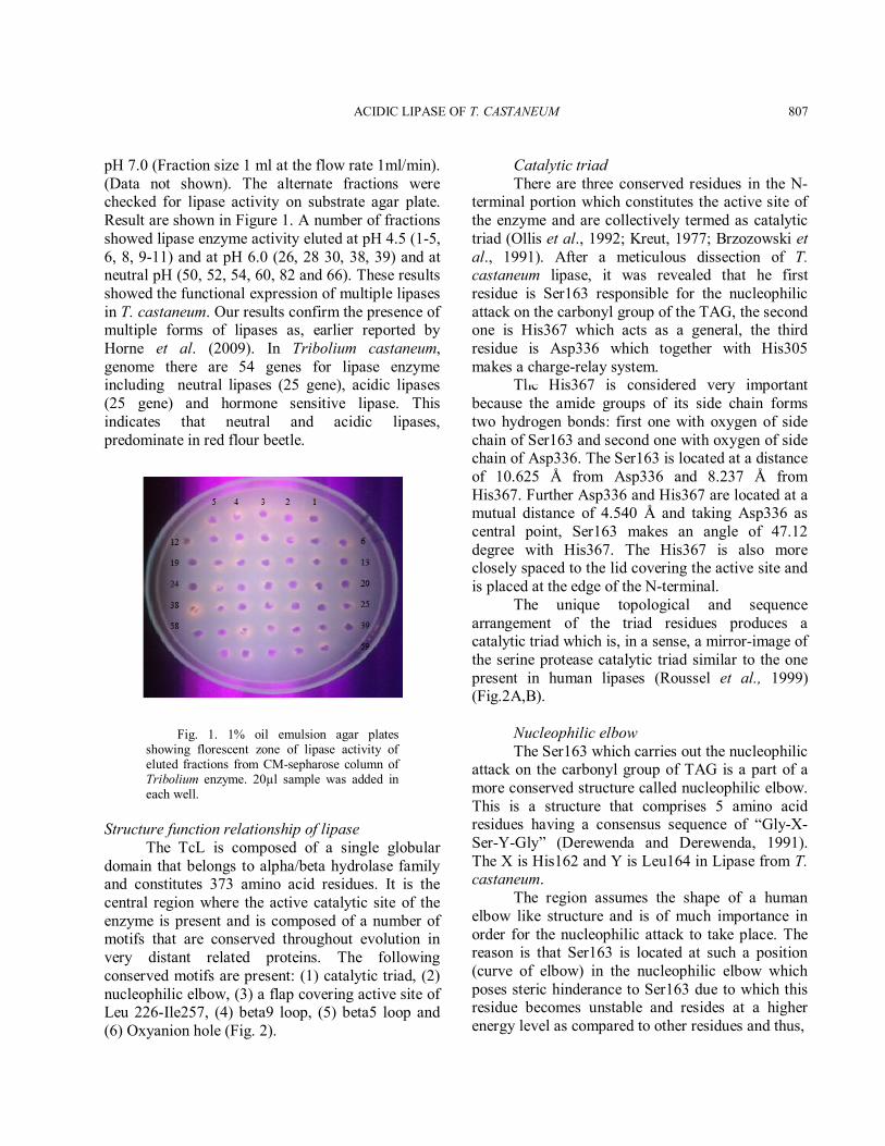
Structure function relationship of lipase The TcL is composed of a single globular domain that belongs to alpha/beta hydrolase family and constitutes 373 amino acid residues. It is the central region where the active catalytic site of the enzyme is present and is composed of a number of motifs that are conserved throughout evolution in very distant related proteins. The following conserved motifs are present: (1) catalytic triad, (2) nucleophilic elbow, (3) a flap covering active site of Leu 226-Ile257, (4) beta9 loop, (5) beta5 loop and (6) Oxyanion hole (Fig. 2).
Catalytic triad There are three conserved residues in the N-terminal portion which constitutes the active site of the enzyme and are collectively termed as catalytic triad (Ollis et al., 1992; Kreut, 1977; Brzozowski et al., 1991). After a meticulous dissection of T. castaneum lipase, it was revealed that he first residue is Ser163 responsible for the nucleophilic attack on the carbonyl group of the TAG, the second one is His367 which acts as a general, the third residue is Asp336 which together with His305 makes a charge-relay system. The His367 is considered very important because the amide groups of its side chain forms two hydrogen bonds: first one with oxygen of side chain of Ser163 and second one with oxygen of side chain of Asp336. The Ser163 is located at a distance of 10.625 Å from Asp336 and 8.237 Å from His367. Further Asp336 and His367 are located at a mutual distance of 4.540 Å and taking Asp336 as central point, Ser163 makes an angle of 47.12 degree with His367. The His367 is also more closely spaced to the lid covering the active site and is placed at the edge of the N-terminal. The unique topological and sequence arrangement of the triad residues produces a catalytic triad which is, in a sense, a mirror-image of the serine protease catalytic triad similar to the one present in human lipases (Roussel et al., 1999) (Fig.2A,B). Nucleophilic elbow The Ser163 which carries out the nucleophilic attack on the carbonyl group of TAG is a part of a more conserved structure called nucleophilic elbow. This is a structure that comprises 5 amino acid residues having a consensus sequence of “Gly-X-Ser-Y-Gly” (Derewenda and Derewenda, 1991). The X is His162 and Y is Leu164 in Lipase from T. castaneum. The region assumes the shape of a human elbow like structure and is of much importance in order for the nucleophilic attack to take place. The reason is that Ser163 is located at such a position (curve of elbow) in the nucleophilic elbow which poses steric hinderance to Ser163 due to which this residue becomes unstable and resides at a higher energy level as compared to other residues and thus,
A. J. SAMI ET AL. 808
A B C
D E F
G H I
J
Fig. 2. A, Alpha/beta hydrolase fold inside the globular domain; B, Catalytic triad of lipase. Green dotted lines in red circles show the H-bonds; C, Position of Ser163 in nucleophilic elbow; D, Relative position of residues involved in catalysis and oxyanion hole formation; E, Protective surface loop from Leu226 to Ile257 covering the catalytic triad; F, Relative position of Beta9 loop from Ile282 to Gly303 to surface loop and catalytic triad; G, Relative position of Beta5 loop from Gly63 to Met85 to surface loop and catalytic triad Oxyanion hole; H, Leu75 and Gln164 involved in oxyanion hole formation; I, Relative position of surface loop, Beta9 loop and Beta5 loop to the catalytic triad providing maximum protection; J, Position of N-Glycosylated residues in the structure of lipase. In all the structural images, the following colour scheme is used except for the space filling model: Ser 163, pink; His 367, purple; Asp 336, yellow; Nucleophilic elbow, orange; Lid, blue; Third loop protecting the active site, brown; Beta5 loop, green; Leu75 and Gln164 of oxyanion hole, Blue.
ACIDIC LIPASE OF T. CASTANEUM 809
in order to reduce its energy and be stabilized, this residue becomes ideally suited to carry out the nucleophilic attack (Ollis et al., 1992) (Fig. 2C). Further Ramachandran plot is also determined for the active site and nucleophilic elbow which shows that Ser163 is present in unfavorable region and is therefore highly unstable in this position. It was noticed that in case of lipase TcL, the turns of nucleophilic elbow on both ends, Gly161 and Gly165 are present. This is also of special importance because it is only Gly that offers no steric hinderance on the turns and curves due to its simplest structure. The nucleophilic elbow also resides at the hydrophobic core of the enzyme due to which the hydrophobicity in that area in increased as compared to other regions which confers maximum stability to the nucleophilic elbow and catalytic Ser163 and practically makes the active site inaccessible to the substrate unless exposed by displacement of the lid. Moreover, the oxygen atom of carboxyl group of His162 also makes a strong H-bond with amide group of Gly165 with a 2.791 Å distance which stabilizes the nucleophilic elbow. Further the Gln164 also assumes such a position that its amide group becomes placed in front of amide group of Leu75 belonging to Beta5 loop with an interatomic distance of 4 Å. Both of them are responsible for the formation of oxyanion hole during the enzymatic activation on binding of the substrate and makes strong H-bonds with the substrate. Flap/lid protecting active site In the case of lipase TcL, the amino acid residues from Leu226 to Ile257 make a protective loop over the surface of the lipase which protects the active site and makes it inaccessible to solvent and renders the core of the enzyme hydrophobic. This loop is called the lid, flap or cap of the enzyme in the N-terminal region. One disulfide bond is present in the loop i.e. between Cys240 and Cys250 which is responsible for the stabilization of the loop. Apart from the disulfide bond, many H-bonds are present inside the loop of the lid conferring additional stability to the curved loop (Figs. 2C,D). This is the lid over the active site that is responsible for much of the hydrophobicity inside the protein’s core and this is the lid that changes its conformation
during the interfacial activation of the enzyme after the binding of the substrate. Winkler et al. (1990) identified a protective surface loop (Fig. 2E) which covers the active site in closed conformation and is stabilized by van der Waal’s interactions with other loops called B5 and B9 loops. Once the lid is displaced, the active site is more exposed to the solvent and Ser163 which is more likely to carry out the nucleophilic attack (Brzozowski et al., 1991; Tilbeurgh et al., 1993; Egloi et al., 1995). Although the lid is also conserved throughout the course of evolution, it shows variability in its length in different species and this variation is seen mostly at the top of the loop which makes a very short alpha helix. It is further elaborated in the section of superimposition of lipase from different sources. Beta 9 loop Winkler et al. (1990) identified a protective surface loop which covers the active site in closed conformation and is stabilized by van der Waal’s interactions with other loops called B5 and B9 loops (Figs. 2E,F). Beta 9 loop is extended from Ile282 to Gly303 and offers further protection to the active site of lipase from another direction by making stable interactions with the surface loop (Winkler et al., 1990). Beta 5 loop For T. castaneum TcL lipase Beta 5 loop extends from Gly63 to Met85 and again offers additional protection to enzyme’s catalytic triad by interacting with beta9 and surface loops (Winkler et al., 1990). However, that loop also plays an important role i.e. the formation of the oxyanion hole (Figs. 2F,G,H). The amide group of Leu75 is placed in close proximity to the amide group of Gln164 and both of them are responsible for the formation of oxyanion hole after the binding of the substrate with the formation of H-bonds (Kraut, 1977). Maximum protection of the catalytic triad by 3 loops According to the current knowledge, the active site is protected by the lid or flap over the enzyme’s surface in the alpha/beta hydrolase family. Here in T. castaneum the lid consists of residues
A. J. SAMI ET AL. 810
from Leu226-Ile257. However, according to space filling model, it is determined that the lid of Leu226-Ile257, Beta9 loop of Ile282-Gly303 and Beta5 loop of Gly63-Met85 and the beta sheets at the bottom, all contribute to the overall protection of the active site residues from every direction and makes the interior of the enzyme more hydrophobic (Fig. 2I). Only after the binding of the substrate, the lid undergoes a conformational change and then in a sequence, the beta5 loop undergoes further change in conformation due to which the inside becomes more exposed to the solvent and becomes ready for the attack and the hydrophobicity is reduced. However the lid/flap is given more importance because beta5 loop undergoes a slight modification in its conformation, beta9 loop remains intact and it is the lid which shows a major shift in conformation after binding of the substrate and covers the top of the active site (Tilbreugh et al., 1993). N-Glycosylated residues In our case, these are present at positions Asn59, Asn230, Asn242 and Asn296 which may be responsible for increase stability of the protein by conferring proper folding. In the acidic lipase, there are four consensus sequences for N-glycosylation of the enzyme which is Asn-X-Ser/Thr (Fig. 2J) (Anderson and Sando, 1991). C-terminal In acidic lipase from Tribolium castaneum there is no separate C-terminal domain which is predominantly present in the neutral lipases. There are several differences between the acidic lipases like there is no separate C-terminal domain in acidic lipase which is predominantly present in the neutral lipase and consists of a beta sandwich. Further there are four N-glycosylation sites present in the 3D structure of acidic lipase which may be responsible for providing maximum stability to the enzyme to compensate for stability and substrate attachment affinity provided by the C-terminal of neutral lipases. In acidic lipase, after the abstraction of proton from His367, there is a local decrease in pKa as compared to the neutral lipases due to which the enzyme acts in the acidic range (Mileda et al., 2000).
Mechanism of action of lipase After lipase has bound to the substrate to form the enzyme-substrate complex, Ser163, in the reaction’s rate determining step, nucleophilically attacks the carbonyl group to form the complex known as the tetrahedral intermediate (covalent catalysis). X-ray and computational studies have shown that Ser163 is ideally positioned to carry out this nucleophilic attack (due to its proximity and orientation effects). The imidazole ring of His367 takes up the liberated proton forming an imidazolium ion (general base catalysis). This process is aided by the polarizing effect of the unsolvated carboxylate ion of Asp336 which is H-bonded to His367. Neutron diffraction studies have demonstrated that Asp336 remains a carboxylate ion rather than abstracting a proton from the imidazolium ion to form an uncharged carboxylic acid group. The tetrahedral intermediate decomposes to the acyl-enzyme intermediate under the driving force of proton donation from N3 of His367 (general acid catalysis). The leaving group of glycerol is released from the enzyme and replaced by the water from the solvent. The acyl-enzyme intermediate (which in the absence of enzyme would be a stable compound) is rapidly deacylated and thereby generating the active enzyme. In this step, water is the attacking nucleophile and Ser163 is the leaving group (Winkler et al., 1990; Tilbreugh et al., 1993; Smith et al., 1992; Cygler et al., 1994). The conformational distortion that occurs with the formation of the tetrahedral intermediate causes the carbonyl oxygen of the substrate to move deeper into the active site so as to occupy the previously unoccupied position, the oxyanion hole. In case of TcL lipase of T. castaneum forms two H-bonds with the enzyme that cannot be formed when the substrate is in its normal regional conformation. One of the H-bond is formed with the amide group of Leu75 in the Beta5 loop and the second H-bond is formed with the amide group of the residue immediately following the catalytic serine which is Gln164 (Kraut, 1977). The existence of that oxyanion hole is based on the premise that convergent evolution has made the active sites of these unrelated enzymes functionally identical.
ACIDIC LIPASE OF T. CASTANEUM 811
ACKNOWLEDGEMENTS Funds for the research work were provided by HEC under National Universities Research Program.
REFERENCES ANDERSON, R. A. AND SANDO, G.N., 1991. Cloning and
expression of cDNA encoding human lysosomal acid lipase/cholesteryl ester hydolase. J. biol. Chem., 266: 2247-22484.
BEER, H.D., WOHLFAHRT, G., MCCARTHY, J.E.G., SCHOMBURG, D. AND SCHMID, R.D., 1996 Analysis of the catalytic mechanism of a fungal lipase using computer-aided design and structural mutants. Protein Eng. 9: 507-517
BRADFORD, M. M., 1976. A rapid and sensitive method for the quantitation of microgram quantities of protein utilizing the principle of protein-dye binding. Analyt. Biochem., 72: 248-254.
BRADY, L., BRZOZOWSKI, A.M., DEREWENDA, Z.S., DODSON, E., DODSON, G., TOLLEY, S., TURKENBURG, J. P., CHRISTIANSEN, L., HUGE-JENSEN, B. AND NORSKOV, L., 1990. A serine protease triad forms the catalytic centre of a triacylglycerol lipase. Nature, 343: 767-770
BRZOZOWSKI, A. M., DEREWENDA, U., DEREWENDA, Z.S., DODSON, G.G., LAWSON, D.M., TURKENBURG, J.P., BJORKLING, F., HUGE-JENSEN, B., PATKAR, S.A. AND THIM, L., 1991. A model for interfacial activation in lipases from the structure of a fungal lipase-inhibitor complex. Nature. 351: 491-494
CYGLER, M., GROCHULSKI, P., KAZLAUSKAS, R.J., SCHRAG, J.D., BOUTHILLIER, F., RUBIN, B., SERREQI, A.N. AND GUPTA, A.K., 1994. A structural basis for the chiral preferences of lipases. J. Am. Chem. Soc., 116: 3180-3186.
DEREWENDA, Z.S., 1994. Structure and function of lipases. Adv. Protein Chem., 45: 1-52.
DEREWENDA, Z.S. AND DEREWENDA, U., 1991. Relationships among serine hydrolases: evidence for a common structural motif in triacylglyceride lipases and esterases. Biochem. Cell Biol., 69: 842-851.
EGLOI, M.P. MARGUET, F., BUONO, G., VERGER, R., CAMBILLAU, C. AND TILBEURGH, H.V., 1995. The 2.46 Aî resolution structure of the pancreatic lipase colipase complex inhibited by a C11 alkyl phosphonate. Biochemistry, 34: 2751-2762.
KUMAR, P. R. AND PRAKASH, V., 2009. Inhibition of rice bran lipase by azadirachtin from Azadirachta indica. J. Sci. Fd. Agric., 89: 1642-1647
HAMMER, M. F. AND MURPHY, J.B., 1994. Lipase activity
and in vivo triacylglycerol utilization during Pinus edulis seed germination. Plant Physiol. Biochem., 32: 861.
HIDE, W. A., CHAN, L. AND LI, W.H., 1992. Structure and evolution of lipse superfamily. J. Lipid Res., 33: 167-178.
HOLMQUIST, M., 2000. Alpha/beta hydrolase fold enzymes: structures, functions and mechanisms. Curr. Protein Peptide Sci., 1: 209-235.
HORNE, I., HORITOS, V. S. AND OAKESHOTT, J.J., 2009. Comparative and Functional genomics of lipases in hematobolous insects. Insect Biochem. Mol. Biol., 39: 547-567.
KRAUT, J., 1977. Serine proteases: structure and mechanism of catalysis. Annu. Rev. Biochem., 46: 331-358
LANGIN, D., LAURELL, H., HOLST, L. S., BELFRAGE, P. AND HOLM, C., 1993. Gene organization and primary structure of human hormone-sensitive lipase: possible significance of a sequence homology with a lipase of Moraxella TA144, an antarctic bacterium. Proc. natl. Acad. Sci. U.S.A., 90: 4897-4901.
LOWE, M.E., 1992. The catalytic site residues and interfacial binding of human pancreatic lipase. J. biol. Chem., 267: 17069-17073.
MILEDA, N., CANAANA, S., DUPUISA, L., ROUSSELB, A., RIVIÈREA, M., CARRIÈREA, F., CAROA, A., CAMBILLAUB, C. AND VERGERA, R., 2000) Digestive lipases: From three-dimensional structure to physiology. Biochimie, 82:973−986
NARDINI, M. AND DIJKSTRA, B.W., 1999. Alpha/beta hydrolase fold enzymes: the family keeps growing. Curr. Opin. Struct. Biol., 9: 732-737.
OLLIS, D.L., CHEAH, E., CYGLER, M., DIJKSTRA, B., FROLOW, F., FRANKEN, S.M., HAREL, M., REMINGTON, S.J., SILMAN, I., SCHRAG, J., SUSSMAN, J.L., VERSCHUEREN, K.H.G. AND GOLDMAN, A., 1992. The K/L hydrolase fold. Protein Eng., 5: 197-211.
ROUSSEL, A., CANAAN, S., EGLOFF, M.P., RIVIERE, M., DUPUIS, L., VERGER, R. AND CAMBILLAU, C., 1999. Crystal structure of human gastric lipase and model of lysosomal acid lipase, two lipolytic enzymes of medical interest. J. biol. Chem., 274: 16995-17002.
SALEEM, M.A. AND SHAKOORI, A.R., 2000a. Synergistic effects of diflubenzuron and permethrin on some macromolecules of Tribolium castaneum larvae. Pakistan J. Zool., 32: 117-122.
SALEEM, M.A. AND SHAKOORI, A.R., 2000b. Effects of mixture of diflubenzuron and cypermethrin on Tribolium castaneum larvae. Pakistan J. Zool., 32: 289-299.
SCHRAG, J. D., LI, Y., WU, S. AND CYGLER, M., 1991. Ser-His-Glu triad forms the catalytic site of the lipase from Geotrichum candidum. Nature, 351: 761-764.
SHIMIZU, S. AND NAKANO, M., 2003. Structural
A. J. SAMI ET AL. 812
characterization of triacylglycerol in several oils containing gamma-linolenic acid. Biosci. Biotechnol. Biochem., 67: 60-67.
SMITH, L.C., FAUSTINELLA, F. AND CLAN, L., 1992. Lipases: Three dimensional structure and mechanism of action. Curr. Opin. Struct. Biol., 2: 490-496.
TILBEURGH, H.V., EGLOI, M.P., MARTINEZ, RUGANI, N., VERGER, R. AND CAMBILLAU, C., 1993. Interfacial activation of the lipaseprocolipase complex by mixed micelles revealed by x-ray crystallography. Nature, 362: 814-820.
WINKLER, F. K., D'ARCY, A. AND HUNZIKER, W., 1990. Structure of human pancreatic lipase. Nature, 343: 771-
774. ZIMMERMAN, R., STRAUSS, J.G., HAEMMERLE, G.,
SCHOISWOHL, G., BIRNER-GRUENBERGER, R., RIEDERER, M., LASS, A., NEUBERGER, G., EISENHABER, F., HERMETTER, A. AND ZECHNER, R., 2004. Fat mobilization in adipose tissue is promoted by adipose triglyceride lipase. Science, 306: 1383-1386.
(Received 14 April 2014, revised 25 May 2014)