isolation identification and characterization … · isolation identification and characterization...
TRANSCRIPT

International Research Journal of Engineering and Technology (IRJET) e-ISSN: 2395 -0056
Volume: 03 Issue: 04 | April-2016 www.irjet.net p-ISSN: 2395-0072
© 2016, IRJET | Impact Factor value: 4.45 | ISO 9001:2008 Certified Journal | Page 2503
ISOLATION IDENTIFICATION AND CHARACTERIZATION OF POTENTIAL
OIL DEGRADING BACTERIA FROM OIL CONTAMINATED SITES
R.Vignesh1, A.Arularasan2, V.Gandhiraj³, R.Charu Deepika 4
1Assistant Professor, Department of Biotechnology, Shri Andal Alagar College of Engineering, Mamandur
² Shri Andal Alagar College of Engineering, Mamandur-603111
---------------------------------------------------------------------***---------------------------------------------------------------------Abstract - The hydrocarbon containing oils like crude oil,
petrol, diesel etc., causes environmental pollution. The oil
contaminated environment rich in microbial community
adapted for utilizing the hydrocarbons for their growth and
metabolic activities. The soil samples were collected near two
oil storage tanks from Chennai harbour. The microorganisms
present in the soil were isolated by enrichment technique
using Bushnell Haas broth with used engine oil as sole carbon
source. The isolated bacteria were identified by biochemical
characterization. The identified bacteria were Streptococcus
spp. (isolate 1) and Pseudomonas spp.1 (isolate 2),
Pseudomonas spp.2 (isolate 3) and Pseudomonas spp.3 (isolate
4) and were allowed to degrade petrol, diesel and engine oil
for twenty days and the amount oil degraded were calculated
by gravimetric analysis. Streptococcus spp. is the potential
bacteria for petrol and engine oil degradation, degraded
89.6% of petrol and 84.6 % of engine oil. Pseudomonas spp.2 is
the potential bacteria for diesel degradation, degraded 97.8%
of diesel.
Key Words: Hydrocarbons, Enrichment culture technique, Oil
degrading bacteria, Gravimetric analysis, Biochemical tests.
1. INTRODUCTION In this modern world the need for petroleum products has highly increased for the contribution of modern lifestyles. Petroleum products are complex mixtures which are mainly derived from crude oil and they are processed in oil refineries [18]. Gasoline and diesel are some of the petroleum products refined with high allocation of energy from the oil refineries [1]. BH media was used as enrichment media for the isolation of diesel oil degrading bacteria [14]. Hydrocarbons found in crude oil are metabolized by a large number of bacterial communities into nontoxic biodegradable products [22]. These are the constituents of simple hydrocarbons to polycyclic aromatic hydrocarbons [16].The release of such petroleum products into the environment shows a great impact on biotic and abiotic communities of that environment if not treated at time. Bacillus cereus was identified as potential oil degrader which was isolated from Lebanese oil polluted soil and marine environment [5]. Acinetobacter calcoaceticus and Alcanivorax dieselolei were identified as potential crude oil degrader from Caspien Sea and Persian Gulf degraded 98% of crude
oil [7]. The removing of some harmful organic compounds in the polluted sites by the biodegradation was done with the help of bacterial species. Bacillus megaterium, Bacillus cereus, Microccus luteus, Staphylococcus aureus, Lactobacillus acidophiles, Neisseria flaviscence and Cornybacterium xerosis were identified as potential hydrocarbon degraders isolated from Hyderabad [17].
2. MATERIALS AND METHODS
2.1 Sterilization
Media and glassware were sterilized in an autoclave at 121ºC with 15 lbs pressure for 20 minutes. 2.2 SAMPLE COLLECTION
Surface soil sample was collected around the two crude oil storage tanks of Chennai harbour and packed in poly bags and stored at 4ºC until the end of the work. 2.3 MEDIA
Bushnell Haas broth, Bushnell Haas agar, Nutrient agar, Nutrient broth, Starch hydrolysis media, Tryptone broth, Carbohydrate utilization media and Methyl red broth. 2.4 ENRICHMENT OF MICROORGANISMS
1g of each soil samples were suspended in 10mL of sterile distilled water separately in two test tubes and vortexed for 1 minute gently. Allow it to settle down and 1mL of suspension was inoculated into 100mL of Bushnell Haas broth with used engine oil as sole carbon source in a 250mL conical flask and placed in shaker for 5-10 days for the enrichment of microbes [12]. After the first enrichment process 1mL from that culture was again transferred into the broth for the second enrichment of microbes.
2.5 SCREENING AND ISOLATION OF OIL DEGRADING
BACTERIA
After the second enrichment of microbes for 5-10 days of incubation, 0.1mL of enriched bacterial culture was spread onto the Bushnell Haas agar plates with used engine oil as sole carbon source [4]. 10mL of used engine oil was added to 1000mL of media with other nutrient supplements. Bushnell Haas media was used as the selective media for screening of oil degrading microbes. The plates were kept at 37 ºC until visible colonies are formed. Selective colonies were taken for
International Research Journal of Engineering and Technology (IRJET) e-ISSN: 2395 -0056
Volume: 03 Issue: 04 | April-2016 www.irjet.net p-ISSN: 2395-0072
© 2016, IRJET | Impact Factor value: 4.45 | ISO 9001:2008 Certified Journal | Page 2504
culture in nutrient agar plates. Pure cultures of bacterial isolates were identified on the basis of their colony morphology by quadrant streaking. 2.6 MEASUREMENT OF BACTERIAL GROWTH
Obtained pure cultures were inoculated into the nutrient broth and growth kinetics was measured by taking OD at 600 nm at regular time interval [20]. 2.7 IDENTIFICATION OF BACTERIAL ISOLATES
2.7.1 Simple staining
Simple staining is the method to identify the shape and structure of bacteria [15]. Basic stains such as crystal violet, safranin and methylene blue are used for staining. Clean and grease free slide was taken and a drop of saline was placed on the slide. The organism to be studied is placed on the saline and made a smear and heat fix the organism [6]. Crystal violet is added to the smear and allow one minute for staining. Wash the dye with running tap water carefully and allowed for air dry and then viewed under microscope.
2.7.1.2 Gram staining
The Gram stain is one of the most important and widely used tools in the identification of unknown bacteria [9]. The Gram stain reaction is dependent on the cell wall structure of the bacteria. The cell wall of Gram-positive bacteria is composed of a thick layer of peptidoglycan that surrounds the plasma or inner membrane. In contrast, a thin layer of peptidoglycan and a second phospholipid bilayer, known as the outer membrane, surround the plasma or inner membrane of Gram-negative bacteria. These characteristically different cell wall structures permit microbiologists to classify bacteria based on the colour of the stain retained by cells treated with the Gram stain. The Gram stain is a differential stain because it divides bacteria into two groups: Gram-positive and Gram-negative, where Gram-positive bacteria stain purple and Gram-negative bacteria stain pink. Sample is heat fixed on to the clean grease free slide. Crystal violet is added in drop to the sample and allows it for one minute and washes the stain in running tap water. And then Gram’s iodine is added which is mordant. Allow it for one minute and washed in tap water. Rinse the sample with ethanol for 30 seconds and gently wash it in water. Add the safranin to the slide and allow it for 1 minute and then wash it off. Allow the slide for air dry and view under microscope.
2.7.1.3 Motility test
The simplest method to examine living micro organism and their motility is by hanging drop method [13]. In this method the organism are observed in a drop that is suspended under a cover glass in a concave slide the hanging drop slides are usually observed in bright field microscope. Cover slides and cover slips are washed with distilled water, dried and wiped
with ethanol. Vaseline placed on four edge of cover of the cover slip. A drop of culture was placed in centre of cover slip. Cavity slide place over cover slip and contact was made between cover slip and slide with help of Vaseline. Curve was such taken that the drop should not touch the inner end of cavity. Slide turned upright and observed under light microscope. 2.7.2 BIOCHEMICAL TESTS
2.7.2.1 Catalase test
The catalase test facilitates the detection of the enzyme catalase in bacteria. It is essential for differentiating catalase-positive Micrococcaceae from catalase negative Streptococcaceae [10]. While it is primarily useful in differentiating between genera, it is also valuable in speciation of certain gram positives such as Aerococcus urinae (positive) from Aerococcus viridians (negative) and gram-negative organisms such as Campylobacter fetus, Campylobacter jejuni, and Campylobacter coli (all positive) from other Campylobacter species [3]. Collect 18-24 hours of culture sample and placed in a clean slide. One drop of hydrogen peroxide solution was placed on the culture organism in the microscopic slide. Observation of air bubbles in the slide represents the positive result.
2.7.2.2 Indole test
The indole test screens for the ability of an organism to degrade the amino acid tryptophan and produce indole. It is used as part of the IMViC (indole, MR-Vp Citrate) procedures, a battery of tests designed to distinguish among members of the family Enterobacteriaceae. Inoculate the tube of tryptone broth with a small amount of a pure culture. Incubate at 37°C for 24 to 48 hours . To test for indole production, add 5 drops of Kovác's reagent directly to the tube. Formation of a pink to red color (“cherry red ring”) in the reagent layer on top of the medium within seconds of adding the reagent shows positive reaction. If the reagent layer will remain yellow or be slightly cloudy shows negative reaction.
2.7.2.3 Methyl red test
This test is to detect the ability of an organism to produce and maintain stable acid end product from glucose fermentation [11]. Some bacteria produce large amount of acids from glucose fermentation that they overcome the buffering action of the system. MR is a pH indicator, which remains red in color at a pH of 4.4 or less. MR broth media was prepared and sterilized. The sterile tubes were taken and the broth was poured. Test organisms were inoculated and the tubes were kept in the incubator for 24 hours. After 24 hours the MR indicator was added to the tubes, and the color change is observed.
International Research Journal of Engineering and Technology (IRJET) e-ISSN: 2395 -0056
Volume: 03 Issue: 04 | April-2016 www.irjet.net p-ISSN: 2395-0072
© 2016, IRJET | Impact Factor value: 4.45 | ISO 9001:2008 Certified Journal | Page 2505
2.7.2.4 Starch hydrolysis test
This test is used for the identification of organism which can hydrolyze the starch. Inoculated the plates of starch agar with the assigned bacteria and incubated at 37 ºC for 24-48 hours. A small amount of Gram’s Iodine dripped on the plate around the inoculated area and a small amount in uninoculated area away from the inoculums. A clear zone was observed around the inoculum. Compared the inoculated area with the uninoculated area, and record the results. 2.7.2.5 Glucose utilization test
This test is used to identify the organism which are utilizing the different carbohydrate components like glucose, fructose, maltose.0.1mL Inoculum of each of the bacteria was transferred into broth of glucose and incubated the broth at 37 ºC for 24-48 hours in a rotary shaker. Results are observed for the broth and compared to the uninoculated controls. 2.8 OIL DEGRADATION STUDIES
For examining the degradation of oil, Bushnell Haas medium (BHM) supplemented with 5g/50mL of petrol, diesel and used engine oil to be used [11]. About 50mL medium was dispensed in 250mL conical flasks. The media was inoculated with 0.1mL of oil degrading bacteria (bacteria obtained by screening of oil degrading bacteria) and incubated at 37°C for 20 days in different flask. 2.8.1GRAVIMETRIC ANALYSIS
Oil degradation was studied by gravimetric analysis. After desired interval of time, the flasks were taken out and bacterial activities were stopped by adding 1% 1N-HCL. For extraction of oil, 50mL of culture broth was mixed with 20mL petroleum ether: acetone (1:1) in a separating funnels and was shaken vigorously to get a single emulsified layer. Acetone was then added to it and shaken gently to break the emulsification, which resulted in three layers [2]. Top layer was a mixture of petroleum ether, oil and acetone; clumping cells make a middle layer and the bottom aqueous layer contains acetone, water and biosurfactant in soluble form. The lower two layers were spread out while top layer containing petroleum ether mixed with oil and acetone was taken in a preweighed clean beaker. The extracted oil was passed through anhydrous sodium sulphate to remove moisture. The petroleum ether and acetone was evaporated on a water bath. The gravimetric estimation of residual oil left after biodegradation was made by weighing the quantity of oil in a tarred beaker. The percentage of degradation was calculated as follows [8];
Weight of Residual oil = Weight of beaker containing extracted oil – Weight of empty beaker.
Amount of crude oil degraded = Weight of oil added – Weight of residual oil % degradation = Amount of oil degraded media x 100
Oil added in media
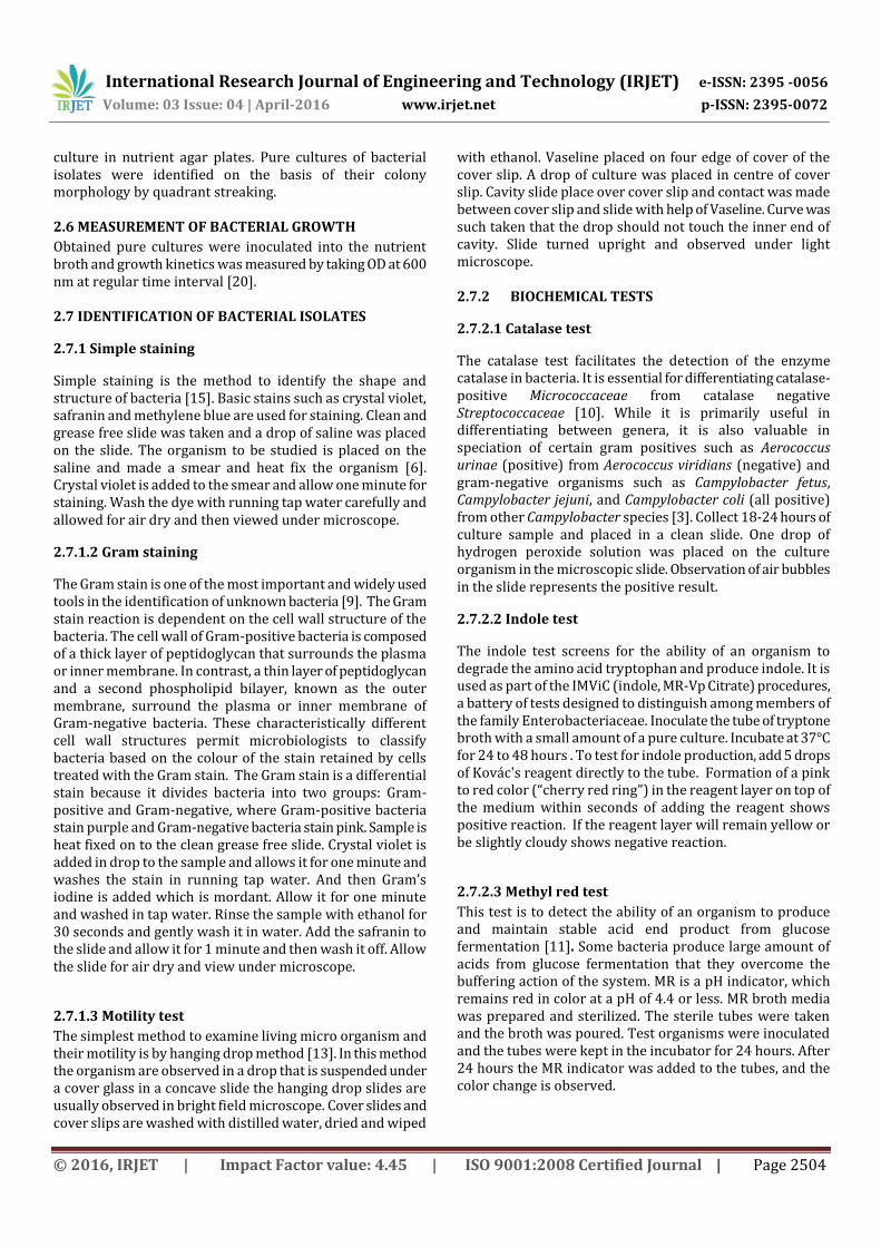
3. RESULTS AND DISCUSSION 3.1 Enrichment of microbes
The microbial cultures were enriched for oil degradation using BH media with engine oil as sole carbon source and it was confirmed by comparing the flasks before and after enrichment of microbes.
Fig-1: Enrichment in BH broth A- Before, B-After
3.2 SCREENING OF OIL DEGRADING BACTERIA
Enriched culture of two soil samples were spread onto the BH agar plates which is the selective media for oil degrading microbes. The oil degrading bacteria were grown on the media which is compared to the control plate.
A-Control B-Tank 1 C-Tank 2
Fig-2: Screened bacteria
3.3 ISOLATION OF OIL DEGRADING BACTERIA The oil degrading bacteria grown in BH agar of selected colonies are randomly taken for culture in Nutrient agar by streaking method. From the streaking four different isolates are chosen for degradation studies by the morphological characteristics and are quadrant streaked to obtain pure culture of the isolates.
International Research Journal of Engineering and Technology (IRJET) e-ISSN: 2395 -0056
Volume: 03 Issue: 04 | April-2016 www.irjet.net p-ISSN: 2395-0072
© 2016, IRJET | Impact Factor value: 4.45 | ISO 9001:2008 Certified Journal | Page 2506
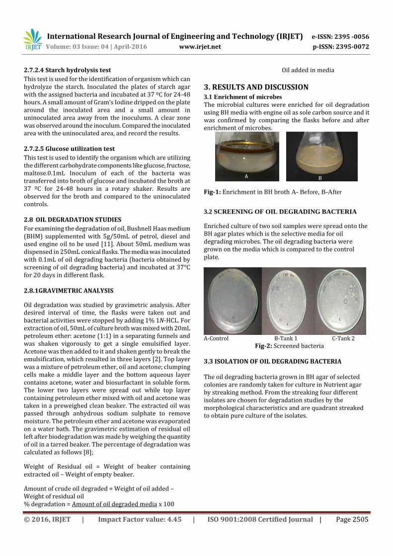
Fig-3: Isolated bacteria
(A-Isolate 1, B-Isolate 2, C-Isolate 3, D-Isolate 4)
3.4 IDENTIFICATION OF BACTERIA
The isolated bacteria were identified by Simple staining, Gram staining, Motility test and also by some biochemical tests. The results of biochemical test were compared to the Bergeys manual. 3.4.1 PRELIMINARY TEST
3.4.1.1 Simple staining Isolate 1 was observed as spherical shape, Isolate 2 and 3 and 4 as rod shape. 3.4.1.2 Gram staining Isolate 1 was observed as gram positive, Isolate 2, 3 and 4 was gram negative. 3.4.1.3 Motility test
Isolate 1 was non motile, Isolate 2, 3 and 4 are observed as motile.
Fig-4: Gram’s staining
A-Isolate 1, B-Isolate 2, C-Isolate 3, D-Isolate 4
TABLE 1: PRELIMINARY TEST
TEST ISOLATE
1 ISOLATE
2 ISOLATE
3 ISOLATE
4
SIMPLE STAINING
Cocci Rod Rod Rod
GRAM’S STAINING
+ - - -
MOTILITY Non
motile Motile Motile Motile
3.4.2 BIOCHEMICAL CHARACTERIZATION
3.4.2.1 Catalase test
Isolate 1 and 2 are negative for the catalase test as there was no formation of bubbles when reacts with hydrogen peroxide solution and Isolate 3 and 4 are positive for the catalase test. 3.4.2.2 Indole test
Isolate 1 and 2 are indole negative, Isolate 3 and 4 are indole positive and are confirmed by formation of cherry red ring. 3.4.2.3 Methyl red test
Isolate 1 and 2 are MR positive, Isolate 3 and 4 are MR negative and it was confirmed by observation of color change. 3.4.2.4 Starch hydrolysis test
All four isolates are positive for the starch hydrolysis and it was confirmed by clear zone formation. 3.4.2.5 Glucose utilization test
Isolate 1 was negative for carbohydrate (glucose) utilization, it was confirmed by no growth in the media and other three isolates are positive for carbohydrate utilization as it was confirmed by growth in the media.
From the above tests and compared with Bergeys manual Isolate 1 was identified as might be Streptococcus spp. , Isolate 2, 3 and 4 as Pseudomonas spp.
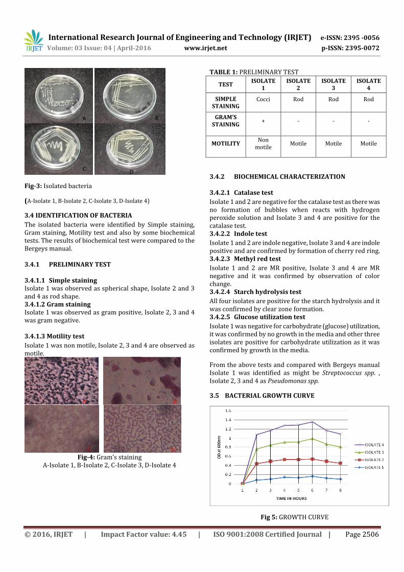
3.5 BACTERIAL GROWTH CURVE
Fig 5: GROWTH CURVE

International Research Journal of Engineering and Technology (IRJET) e-ISSN: 2395 -0056
Volume: 03 Issue: 04 | April-2016 www.irjet.net p-ISSN: 2395-0072
© 2016, IRJET | Impact Factor value: 4.45 | ISO 9001:2008 Certified Journal | Page 2507
All four Isolates reached stationary phase in four hours.
3.6 OIL DEGRADATION Four isolated bacteria were allowed to undergo oil degradation of petrol, diesel and used engine oil for 20 days.
(A) (B)
(A) (B)
(A) (B)
Fig-6: DEGRADATION OF OIL (A)-Before, (B)-After
3.6.1 GRAVIMETRIC ANALYSIS Gravimetric analysis was done to extract the oil degraded by bacterial culture using petroleum ether and acetone in the 1:1 ratio of 20ml.
Fig-7: GRAVIMETRIC ANALYSIS
Three layers were formed during gravimetric analysis; top layer contains remaining oil mixed with acetone and petroleum ether. It was collected in a preweighed beaker and placed in water bath for the evaporation of acetone and petroleum ether. After that it was weighed to measure the amount of oil in the beaker.
4. CONCLUSIONS From this study it was concluded that oil contaminated soil is the best source for the isolation of oil degrading bacteria and may be used for the isolation of other microorganisms. The isolated bacteria can be used for the degradation of different hydrocarbon containing oils and it can also be used for the remediation of contaminated sites. The degrading efficiency of Streptococcus spp. was 89.6% for petrol, 97% for diesel and 84.6 % for engine oil. The degrading efficiency of Pseudomonas spp.1 was 87.8% for petrol, 97% for diesel and 71.4% for engine oil. The degrading efficiency of Pseudomonas spp.2 was 87.2% for petrol, 97.8% for diesel and 67.8% for engine oil. The degrading efficiency of Pseudomonas spp.3 was 85.6% for petrol, 94% for diesel and 70.6% for engine oil. Streptococcus spp. is the potential bacteria for petrol and engine oil degradation among the four isolates. Pseudomonas spp.2 is the potential bacteria for diesel degradation. The future prospects are checking for the degradation of other organic compounds like chemical fertilizers with the isolated bacteria and also for the production of biosurfactants using the isolated bacterial strains.
REFERENCES [1] Anupama Mittal and Padma Singh (2009) ‘Isolation of
hydrocarbon degrading bacterial from soil contaminated with crude oil spills’, Indian journal of experimental biology Vol.47, pp.760-765.
[2] Bharti, P. and Irfan, M. (2011) ‘Pseudomonas aerugenosa is present in crude oil contaminated soil of Barmer region, India’, Journal of bioremediation and biodegradation Vol.2(5), pp.1-2.
[3] Burger, J. (1997) ‘Oil spills’, Rutges university press, Piscataway, NJ, USA, pp.280.
[4] Cerniglia, C E. (1984) ‘Microbial metabolism of Polycyclic aromatic hydrocarbons’, Adv Appl Microbiol Vol.30, pp.31-76.
[5] Darin Maliji, Zakia Olama and Hanafy Holail (2013) ‘Environmental studies on the microbial degradation of oil hydrocarbon and its application in Lebanese oil polluted coastal and marine ecosystem’, International journal of current microbiology and applied sciences Vol.2, No.6, pp.1-18.
[6] Dig Ke Qiang and Luo Yong (2002) ‘Bioremediation of soil contaminated with petroleum forced aeration’, Pedosphere Vol.12(2), pp.145-150.
[7] Geetha, S J. Sanket J Joshi and Shailesh Kathrotiya (2013) ‘Isolation and characterization of hydrocarbon degrading bacterial isolates from oil contaminated sites’, APCBEE Procedia Vol.5, pp.237-241.
[8] Guru, G S. Gohel, H R. Panchal, M R. Ghosh, S K. and Braganza, V B. (2013) ‘Isolation and enrichment of microorganism for degradation of crude oil’, International journal of engineering science and innovative technology Vol.4, pp.144-147.

International Research Journal of Engineering and Technology (IRJET) e-ISSN: 2395 -0056
Volume: 03 Issue: 04 | April-2016 www.irjet.net p-ISSN: 2395-0072
© 2016, IRJET | Impact Factor value: 4.45 | ISO 9001:2008 Certified Journal | Page 2508
[9] Huang, T L. and Ren, L. (2000) ‘Simulating and modelling of the runoff pollution of petroleum pollutants in loers plateau, China’, Environmental science Vol.20(4), pp.345-348.
[10] Jahir Alam Khan and Syed Hasan Abbs Rizvi (2011) ‘Isolation and characterization of microorganisms from oil contaminated sites’, Advances in applied science research Vol.2(3), pp.455-460.
[11] Jyothi, K. Surendara Babu, K. Nancy Clara, and K. Amita Kashyap (2012) ‘Identification and characterization of hydrocarbon degrading by molecular characterization’, Helix Vol.2, pp.105-111.
[12] Latha and Kalaivani (2012) ‘Bacterial degradation of crude oil by gravimetric analysis’, Advances in applied science research Vol.3(5), pp.2789-2795.
[13] Mandri, T. and Lin, J. (2007) ‘Isolation and characterization of engine oil degrading indigenous microorganism in Kwazulu, South Africa’, Journal of bioremediation Vol.6(1), pp.23-27.
[14] Meenakshisundaram, M. and Bharathiraja, C. (2014) ‘Isolation and molecular identification of hydrocarbon degrading bacteria from oil contaminated soil from Tamilnadu’, Indian journal of applied research Vol.4, pp.39-42.
[15] Mulligan, C N. (2005) ‘Environment pollution’, Env App for bioscience Vol.33, pp.183-198.
[16] Nalinee Kumari, Abhishek Vashishtha, Pooja Saini and
Ekta Menghani (2013) ‘Isolation identification and characterization of oil degrading bacterial isolate from the contaminated sites of Barmu, Rajasthan’, International journal of biotechnology and bioengineering research Vol.4, pp.429-436.
[17] Panda, S K. Kav, R N. and Panda, C R. (2013) ‘Isolation and identification of petroleum hydrocarbon degrading microorganism from oil contaminated environment’, International journal of environment science Vol.3(5), pp.1314-1321.
[18] Rafik M. Hesnawi and Farid S. Mogadami (2013) ‘Bioremediation of Libyan crude oil contaminated soil under mesophilic and thermophilic condition’, APCBEE Procedia Vol.5, pp.82-87.
[19] Singh, C. and Lin, J. (2008) ‘Isolation and characterization of diesel oil degrading indigenous microorganisms in Kwazulu-Natal, South Africa’, African journal of biotechnology Vol.7(12), pp.1927-1932.
[20] Teli Nikhil, Verma Deepa, Gavankar Rohan and Bhalerao Satish (2013) ‘Isolation characterization and identification of diesel oil degrading bacteria from garage soil and comparison of their bioremediation potential’, International research journal of environmental sciences Vol.2(2), pp.48-52.
[21] Van Hamme, J D. Singh, A. and Ward, O P. (2003) ‘Recent advances in petroleum microbiology’, Microbiol Mol Rev Vol.67, pp.649.
[22] Wilson, S C. and Jones, K C. (1993) ‘Bioremediation of soil contaminated with polynuclear aromatic
hydrocarbons’, Environmental pollution Vol.81, pp.229-249.