isolation and comparison of galactose-binding … journal of biological chemistry vol. 249, no. 3,...
TRANSCRIPT
THE JOURNAL OF BIOLOGICAL CHEMISTRY Vol. 249, No. 3, Issue of February 10, pp. 803-310, 1974
Printed in U.S.A.
Isolation and Comparison of Galactose-binding Lectins from
Abrus precatorius and Ricinus communis*
(Received for publication, July 11, 1973)
SJUR OLSNES, ERLING SALTVEDT, AND ALEXANDER PIHL
From Nor& Hydro’s Institute for Cancer Research, Department of Biochemistry, Montebello, Oslo 3, Noru,ay
SUMMARY
The agglutinins present in the seeds of Abrus precatorius and Ricinus communis have been separated from the toxins, abrin and ricin, and extensively purified. The procedure involves ion exchange chromatography on DEAE- and CM- cellulose columns, affinity chromatography on Sepharose 4B columns, and sucrose gradient centrifugation.
The purified agglutinins which possessed almost no toxic activity were found by analytical ultracentrifugation to have molecular weights of 134,000 (abrus agglutinin) and 120,000 (ricinus agglutinin). After treatment with sodium dodecyl sulfate and /3-mercaptoethanol the agglutinins were split into four peptide chains, as revealed by polyacrylamide gel electrophoresis.
The binding of the agglutinins and the toxins to human erythrocytes was inhibited by galactose or galactose-contain- ing carbohydrates, like lactose. Equilibrium dialysis ex- periments indicate that the agglutinins possess two binding sites for lactose, while the nonagglutinating toxins, abrin and ricin, have only one binding site.
The agglutinins as well as the toxins were found to consist of polypeptide chains of nearly the same size (30,000 to 35,000). Abrin and ricin consist of only two polypeptide chains, one of which is involved in the binding to cells. The agglutinins consist of four polypeptide chains, only two of which appear to be involved in the binding to cell surfaces. The data suggest that, within one kind of seed, the polypep- tide chains involved in the binding of the agglutinins and the toxins to cell surfaces are identical or very similar.
Although numerous agglutinins or lectins’ are known, only comparatively few have been isolated in pure form and their structure studied (for review see Refs. l-3). It appears that most of the lectins exert their agglutinating activity by way of binding to carbohydrate residues on the cell surface (2). The recent finding that malignant cells are often more easily ag- glutinated by lectins than the corresponding normal cells (2-4) and the possibility that this selective agglutination might be
* This work was supported by The Norwegian Cancer Society. r The term lectin is used also for proteins that bind to cells with-
out the formation of cell aggregates (see “Discussion”). Thus, the term includes the toxins abrin and ricin.
used in cancer therapy has stimulated work on the structure and properties of lectins.
Lectins were first discovered in extracts from the seeds of Abrus precatorius L. and Ricinus communis L. (5-7)) and such extracts were widely used in early immunological research (8- 10). The high toxicity of the preparations used was originally attributed to their hemagglutinating activity (5, 6). More re- cent work has indicated that the toxic and the agglutinating properties of such extracts belong to separate proteins (11-16). Thus, the highly purified toxins abrin and ricin, which are present in the seeds of A. precatorius and R. communis, respectively, have very low agglutinating activity and, as we have earlier suggested (15, 16)) the toxicity of purified agglutinin preparations from these seeds may be due to contamination by traces of residual toxins.
In previous papers we have isolated and studied the properties of the toxins abrin and ricin (15, 16). In the present paper we have purified two nontoxic agglutinating lectins from A. preca- torius and R. communis. These lectins have been characterized with respect to molecular weight, subunit structure, and sugar- binding specificity, and their properties have been compared with those of abrin and ricin.
EXPERIMENTAL PROCEDURE
Materials-Semen jequiriti (the seeds of A. precatorius) was obtained from Norsk Medisinaldepot, Oslo. Castor beans (the seeds of R. communis) were kindly provided by Deutsche Rizinus- Oelfabrik Boley & Co., Krefeld-Uerdingen, West Germany. DE52 cellulose and CM52 cellulose (Whatman) were obtained from E. & R. Balstone Ltd., Springfield, Maidstone, Kent, England. Sepharose 4B was obtained from Pharmacia, Fine Biochemicals, Uppsala, Sweden. [I-r4C]Lactose (20 Ci per mole) was obtained from The Radiochemical Centre, Amersham, England.
Preparation of Crude Lectins-The procedures used for the ex- traction of lectins were the same as those previously used for the extraction of abrin and ricin (15, 16)) except that the defatted castor beans were extracted with 0.14 M NaCl, rather than with distilled water. The solubilized material was dialyzed against distilled water and then, in the case of the abrus extract, against 10 mM Tris-HCl (pH 7.7) for 24 hours. In the case of the ricinus extract, the dialysis was carried out against 5 mM sodium phos- phate (pH 6.5). After dialysis the extracts were centrifuged at 8,000 x g for 20 min, the pellets were discarded, and the super- natants were centrifuged once more in the same way. The final supernatants are referred to below as the crude lectins.
803
by guest on March 12, 2019
http://ww
w.jbc.org/
Dow
nloaded from

804
Isolation of Abrin and Ricin-Abrin and ricin were isolated from the crude lectin extracts as described earlier (15, 16).
Preparation of Antisera to Abrin and R&n-Due to the high toxicity of the intact toxins, immunization of rabbits was initiated with formaldehyde-treated toxins. The immunization was car- ried out as described earlier (15, 16). Complement was inacti- vated by incubating the sera at 56” for 30 min.
Polyacylamide Gel Electrophoresis-A solution of protein was treated with sodium dodecyl sulfate and submitted to polyacryl- amide gel electrophoresis as described earlier (17, 18). In some cases the proteins were treated with 1% /3-mercaptoethanol at room temperature for 1 hour before the electrophoresis.
Analytical Ultracentrzjugation-The centrifugation was carried out in a Beckman-Spinco model E analytical ultracentrifuge at 20” with rotor speed of 10,000 rpm, as described by Olsen et al. (19). The samples were dissolved in 0.1 M NaCl and 10 mM Tris-HCl (pH 7.7). The partial specific volume was assumed to be 0.73 ml per g and the starting concentration was 0.5 to 1.5 mg per ml.
Hemagglutination Tests-Increasing amounts of protein were mixed with 1.6 X lo7 thoroughly washed human erythrocytes, blood group B-, in 1 ml of 10 mM Tris-HCl (pH 7.7) in 0.15 M NaCl. In experiments with agglutinins the mixtures were incu- bated at room temperature for 9 hours and then the sedimented erythrocytes were resuspended. One drop of the solution was examined under the microscope. One unit of hemagglutinating activity was defined as the amount of lectin per ml needed to give clearly visible aggregation of human erythrocytes under these conditions.
In experiments with abrin and ricin, which in low concentra- tions do not spontaneously agglutinate cells, agglutination was studied by adding, after 1 hour, 5 ~1 of specific antisera against the toxins and examination after 8 more hours of incubation.
Toxicity Test-Various protein fractions were injected intra- peritoneally into mice weighing 25 f 3 g and LDbo was meas- ured as described earlier (15, 16).
Equilibrium Dialysis-The different lectins were concentrated by precipitation in the presence of 80% saturated ammonium sulfate, dissolved in a small volume, and dialyzed against 50 mM sodium phosphate (pH 7.1) and 0.02% sodium azide. The con- centrated lectins contained 5 to 15 mg of protein per ml as meas- ured by the absorbance at 280 nm. Equilibrium dialysis (20) was carried out with 200.~1 samples which were dialyzed against 50 mM sodium phosphate (pH 7.1), containing varying amounts of unlabeled a-lactose and 17 nCi of [14C]lactose per ml. The dialysis was carried out at room temperature for 18 to 20 hours, then 25+1 samples were removed from the solution inside and outside of the dialysis bags and radioactivity as well as absorb- ance at 280 nm was measured in each sample. Control experi- ments showed that equilibrium was reached after 6 to 10 hours. Protein concentrations were determined using the following ex- tinction coefficients (E:5m) for the different protein fractions: abrin, 15.9; abrus agglutinin, 14.6; ricin, 11.8; ricinus agglutinin, 11.7; and Fraction Q, 11.2. The extinction coefficients were measured by weighing the lyophilized and desiccated proteins.
RESULTS
Isolation of Abrus Agglutinin-Crude abrus lectin, extracted as described under “Experimental Procedure,” was purified first by chromatography on a DEAE-cellulose column. The protein was applied to the column in the presence of 30 mM NaCl in 10 IIiM Tris-HCl (pH 7.7) under which conditions only a minor part of the material was not adsorbed. The adsorbed proteins were
20
E
B 5 1.5
fw *
i 0.5
I It I I I 50 100 IS0
Fraction number
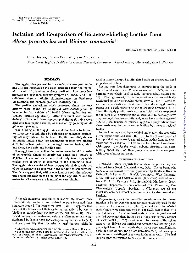
FIG. 1. DEAE-cellulose chromatography of proteins extracted from semen jequiriti. Crude lectin (600 ml) (see “Experimental Procedure”), was applied to a column, 2.6 X 40 cm, of DE52 cellulose equilibrated with 30 mM NaCl in 10 mM Tris-HCl (pH 7.7) and eluted with a 1600-ml linear gradient from 30 to 180 mM NaCl in the same buffer. Fractions (14 ml) were collected and the absorbance was measured.
TABLE I
Toxicity and hemagglutinating activity of different abrus fractions
Purification step Fraction number
DEAE-cellulose chro matography (Fig. 1)
Sepharose 4B chro- matography (Fig.
2)
49 (1) 86 (II)
108 (III) 119 (IV) 149 (V)
12 (VI) 25 (VII)
0 100
CM-cellulose chro- matography (Fig.
3)
96 (VIII) 110 (IX) 120 (X)
100 100
10
Sucrose gradient 12 (XI) 100 centrifugation 16 (XII) 100 (Fig. 4) 20 (XIII) 60
Hemagglu- tinating activity
Toxicity
units/fig LD~o/PK of @3lein protein
0 1.6 0.1 6.6
60 1.1 40 0.9
3 0.3
<O.l 1.1
0.1 1.0
10.0
<0.005 0.005 0.20
eluted with a linear NaCl gradient in the same buffer. As shown in Fig. 1 the proteins were eluted as four peaks, two of which were not separated. In agreement with previous findings (15) the second peak (II) was found to represent the toxic pro- teinabrin (Table I), whereas most of the hemagglutinating activity was present in the double Peaks III and IV.
In the further purification advantage was taken of the fact that abrus agglutinin has a high affinity to Sepharose 4B (15, 21). The material in Peaks ZZZ and ZV was pooled and applied to a Sepharose 4B column from which it was eluted with galac- tose as shown in Fig. 2. A number of contaminating proteins did not bind to the column and were removed by this procedure. However, since abrin also binds to a Sepharose 4B column and is
by guest on March 12, 2019
http://ww
w.jbc.org/
Dow
nloaded from
10 20 30
Fraction number
0.4 5 :: ‘= c
E x 0.3 c & cl2
=x s * 0.2 ” . 1 ! 0.1
0.1
20 40 60 60 100 120 140
Fraction number
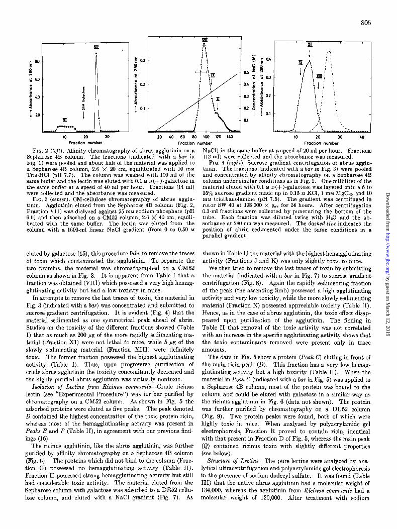
FIG. 2 (left). Affinity chromatography of abrus agglutinin on a Sepharose 4B column. The fractions (indicated with a bar in Fig. 1) were pooled and about half of the material was applied to a Sepharose 4B column, 2.6 X 20 cm, equilibrated with 10 mM Tris-HCl (pH 7.7). The column was washed with 100 ml of the same buffer and the lectin was eluted with 0.1 M D(+)-galactose in the same buffer at a speed of 40 ml per hour. Fractions (14 ml) were collected and the absorbance was measured.
FIG. 3 (center). CM-cellulose chromatography of abrus agglu- tinin. Agglutinin eluted from the Sepharose 4B column (Fig. 2, Fraction VII) was dialyzed against 25 mM sodium phosphate (pH 6.0) and then adsorbed on a CM52 column, 2.6 X 40 cm, equili- brated with the same buffer. The lectin was eluted from the column with a 1660-ml linear NaCl gradient (from 0 to 0.50 M
eluted by galactose (15), this procedure fails to remove the traces of toxin which contaminated the agglutinin. To separate the two proteins, the material was chromatographed on a CM52 column as shown in Fig. 3. It is apparent from Table I that a fraction was obtained (VIII) which possessed a very high hemag- glutinating activity but had a low toxicity in mice.
In attempts te remove the last traces of toxin, the material in Fig. 3 (indicated with a bar) was concentrated and submitted to sucrose gradient centrifugation. It is evident (Fig. 4) that the material sedimented as one symmetrical peak ahead of abrin. Studies on the toxicity of the different fractions showed (Table I) that as much as 200 pg of the more rapidly sedimenting ma- terial (Fraction XI) were not lethal to mice, while 5 pg of the slowly sedimenting material (Fraction XIII) were definitely toxic. The former fraction possessed the highest agglutinating activity (Table I). Thus, upon progressive purification of crude abrus agglutinin the toxicity concomitantly decreased and the highly purified abrus agglutinin was virtually nontoxic.
Isolation of Lectins from Ricinus communis-Crude ricinus lectin (see “Experimental Procedure”) was further purified by chromatography on a CM52 column. As shown in Fig. 5 the adsorbed proteins were eluted as five peaks. The peak denoted D contained the highest concentration of the toxic protein ricin, whereas most of the hemagglutinating activity was present in Peaks E and F (Table II), in agreement with our previous find- ings (16).
The ricinus agglutinin, like the abrus agglutinin, was further purified by affinity chromatography on a Sepharose 4B column (Fig. 6). The proteins which did not bind to the column (Frac- tion G) possessed no hemagglutinating activity (Table II). Fraction H possessed strong hemagglutinating activity but still had considerable toxic activity. The material eluted from the Sepharose column with galactose was adsorbed to a DE52 cellu- lose column, and eluted with a NaCl gradient (Fig. 7). As
Y
IO 20 30 40
Fraction number
NaCl) in the same buffer at a speed of 20 ml per hour. Fractions (12 ml) were collected and the absorbance was measured.
FIG. 4 (right). Sucrose gradient centrifugation of abrus agglu- tinin. The fractions (indicated with a bar in Fig. 3) were pooled and concentrated by affinit,y chromatography on a Sepharose 4B column under similar conditions as in Fig. 2. One milliliter of the material eluted with 0.1 M n(+)-galactose was layered onto a 5 to 15% sucrose gradient made up in 0.15 M KCl, 1 mM MgC12, and 10 mM triethanolamine (pH 7.5). The gradient was centrifuged in rotor SW 40 at 198,000 X g,,” for 24 hours. After centrifugation 0.3-ml fractions were collected by puncturing the bottom of the tube. Each fraction was diluted twice with Hz0 and the ab- sorbance at 280 nm was measured. The dashed line indicates the position of abrin sedimented under the same conditions in a parallel gradient.
shown in Table II the material with the highest hemagglutinating activity (Fractions J and K) was only slightly toxic to mice.
We then tried to remove the last traces of toxin by submitting the material (indicated with a bar in Fig. 7) to sucrose gradient centrifugation (Fig. 8). Again the rapidly sedimenting fraction of the peak (the ascending limb) possessed a high agglutinating activity and very low toxicity, while the more slowly sedimenting material (Fraction N) possessed appreciable toxicity (Table II). Hence, as in the case of abrus agglutinin, the toxic effect disap- peared upon purification of the agglutinin. The finding in Table II that removal of the toxic activity was not correlated with an increase in the specific agglutinating activity shows that the toxic contaminants removed were present only in trace amounts.
The data in Fig. 5 show a protein (Peak C) eluting in front of the main ricin peak (D). This fraction has a very low hemag- glutinating activity but a high toxicity (Table II). When the material in Peak C (indicated with a bar in Fig. 5) was applied to a Sepharose 4B column, most of the protein was bound to the column and could be eluted with galactose in a similar way as the ricinus agglutinin in Fig. 6 (data not shown). The protein was further purified by chromatography on a DE52 column (Fig. 9). Two protein peaks were found, both of which were highly toxic in mice. When analyzed by polyacrylamide gel electrophoresis, Fraction R proved to contain ricin, identical with that present in Fraction D of Fig. 5, whereas the main peak (Q) contained ricinus toxin with slightly different properties (see below).
Structure of Lectins-The pure lectins were analyzed by ana- lytical ultracentrifugation and polyacrylamide gel electrophoresis in the presence of sodium dodecyl sulfate. It was found (Table III) that the native abrus agglutinin had a molecular weight of 134,000, whereas the agglutinin from Ricinus communis had a molecular weight of 120,000. After treatment with sodium
by guest on March 12, 2019
http://ww
w.jbc.org/
Dow
nloaded from

806
40 60 120
Fraction number
D
r n
.
IO 20 30 40
Fraction number
FIG. 5 (left). CM-cellulose chromatography of proteins ex- tracted from castor beans. Crude lectin (600 ml), prepared as described under “Experimental Procedure,” was applied to a column of CM52 (Whatman), 2.6 X 40 cm, equilibrated with 5 mM sodium phosphate (pH 6.5), and eluted at a speed of 80 ml per hour with a 1600-ml linear NaCl gradient (from 10 to 100 mM) in the same buffer. Fractions (14 ml) were collected and the ab- sorbance was measured.
FIG. 6 (center). Affinity chromatography of ricinus agglutinin on a Sepharose 4B column. The fractions from Peak E (Fig. 5) (indicated with a bar) were pooled and applied to a column of Sepharose 4B, 2.6 X 20 cm, equilibrated with 10 mM Tris-HCl
Purification step
CM52 cellulose chro- matography (Fig. 5)
Sepharose 4B chroma- matography (Fig. 6)
DE52 cellulose chro- matography (Fig. 7)
Sucrose gradient cen- trifugation (Fig. 8)
DE52 cellulose chro- matography (Fig. 9)
--
.-
Fraction number
40 (A) 75 03)
103 (C) 109 (D) 125 (E) 133 (F)
11 G) 29 (HI
29 (1) 48 (J) 62 W)
13 CL) 16 CM) 20 WI
48 (Q) 56 (R)
--
-
TABLE II
Toxicity in mice and hemagglutinating activity of different ricinus fractions
Hemagglu- tinating activity
units/pg of )rotcin
0.1 0.5 1 3
100 100
0 100
10 100 100
100 100
30
0.1 1.0
Toxicity
~- LDso/ps 4
protein
0.3 1.2 3.3 5.0 1.6 0.3
<O.l 1.4
3.3 0.1 0.1
<0.005 0.007 0.10
8.0 6.6
20 10 60 60 Fraction number
(pH 7.7). The column was washed with 100 ml of the same buffer and the lectin was eluted with 0.1 M n(S)-galactose in 10 mM Tris-HCl (pH 7.7) at a speed of 40 ml per hour. Fractions (16 ml) were collected and the absorbance was measured.
FIG. 7 (right). DE52 cellulose chromatography of ricinus agglu- tinin. Fractions 28 and 29 (Fig. 6) were pooled, dialyzed against 10 mM Tris-HCl (pH 8.5), and about half of the material was applied to a DE52 column, 1.6 X 15 cm, equilibrated with the same buffer. The lectin was eluted from the column with a 500-ml linear N&l gradient (from 0 to 0.1 M NaCl) in the same buffer at a speed of 20 ml per hour. Fractions (5 ml) were col- lected and the absorbance was measured.
FIG. 8 (left). Sucrose gradient centrifugation of ricinus agglu- tinin. The material in the fractions indicated with a bar in Fig. 7 was concentrated and analyzed by sucrose gradient centrifugation as described for abrus lectin in Fig. 4. The dashed line indicates the position of ricin sedimented under the same conditions in a parallel gradient.
FIG. 9 (right). DE52 cellulose chromatography of ricin. The material in Peak C (indicated with a bar in Fig. 5) was pooled and applied to a Sepharose 4B column as described in Fig. 6 for ricinus agglutinin. The material eluted with 0.1 M galactose was dialyzed against 10 mM Tris-HCl (pH 8.5) and applied to a DE52 column, 1.6 X 15 cm, equilibrated with the same buffer. The lectin was eluted with a 500-ml linear NaCl gradient (from 0 to 50 mM NaCl) in the same buffer at a speed of 20 ml per hour. Fractions (8 ml) were collected and the absorbance was measured.
by guest on March 12, 2019
http://ww
w.jbc.org/
Dow
nloaded from
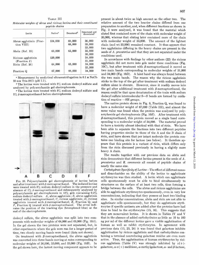
TABLE III
Molecular weights of abrus and ricinus lectins and their constituent eptide chains P -
I Lectin
Abrus agglutinin (Frac- tion VIII)
Abrin (Ref. 15)
Ricinus agglutinin (Fraction J)
Ricin (Ref. 16)
Fraction Q
Native”
____-
134,000
63,000
120,000
64,000
67,000
_-
-
Denatureda
69,000 68,000
65,000
65,000
66,000
I
-
knatured and dissociatedC
36,000 35,000 33,000 35,000 30,000 34,000 31,000 34,000 32,000 34,000
a Measurement by analytical ultracentrifugation in 0.1 M NaClt 10 my Tris-HCl (pH 7.7).
b The lectins were treated with 5% sodium dodecyl sulfate and analyzed by polyacrylamide gel electrophoresis.
c The lectins were treated with 5yo sodium dodecyl sulfate and 1% fl-mercaptoethanol before electrophoresis.
A B C D E F FIG. 10. Polyacrylamide gel electrophoresis of lectins before
and after treatment with@-mercaptoethanol. The isolated lectins were treated with 59& sodium dodecyl sulfate in the presence and absence of 1% p-mercaptoethanol and subsequently analyzed by polyacrylamide gel electrophoresis in 1370 gels containing ‘0.1% sodium dodecyl sulfate. A, abrus agglutinin; B, abrus agglutinin treated with p-mercaptoethanol; C, ricinus agglutinin; D, ricinus agglutinin treated with fi-mercaptoethanol; E, Fraction Q; and F, Fraction Q treated with @-mercaptoethanol. The arrow indi- cates the position of the tracking dye (bromphenol blue) at the end of the electrophoresis.
dodecyl sulfate, the abrus agglutinin was split into two com- ponents with molecular weights of 68,000 and 69,000 (Fig. 10A). In the gel shown the two proteins were not separated-but, in other experiments where the gels were run for a longer period of time, two closely moving bands were found (data not shown).
On treatment with P-mercaptoethanol, the abrus agglutinin was converted into three bands moving at rates corresponding to molecular weights of 36,000, 35,000, and 33,000 (Fig. 10B). In the gel shown here, the fastest moving component appears to be
807
present in about twice as high amount as the other two. The relative amount of the two heavier chains differed from one preparation to another, and, when different fractions as shown in Fig. 3 were analyzed, it was found that the material which eluted first contained more of the chain with molecular weight of 36,000, whereas that eluting later contained more of the chain with molecular weight of 35,000. The amount of the lightest chain (mol wt 33,000) remained constant. It thus appears that two agglutinins differing in the heavy chains are present in the seeds of A. precatorius and that they are not separated under the conditions used.
In accordance with findings by other authors (22) the ricinus agglutinin did not move into gels under these conditions (Fig. lOC), but after treatment with P-mercaptoethanol it moved as two main bands corresponding to molecular weights of 31,000 and 34,000 (Fig. 1OD). A faint band was always found between the two main bands. The reason why the ricinus agglutinin sticks to the top of the gel after treatment with sodium dodecyl sulfate alone is obscure. However, since it readily moves into the gel after additional treatment with fi-mercaptoethanol, the reason could be that upon denaturation of the toxin with sodium dodecyl sulfate intermolecular S-S bonds are formed by oxida- tion of reactive -SH groups.
The native protein shown in Fig. 9, Fraction Q, was found to have a molecular weight of 67,000 (Table III), and almost the same value was found when the protein was analyzed by poly- acrylamide gel electrophoresis (Fig. 10E). After treatment with &mercaptoethanol, this protein moved as a single band corre- sponding to a molecular weight of 34,000. The material proved to have a toxicity almost identical with that of ricin. We have been able to separate the fractions into two different peptides having properties similar to those of the A and the B chain of ricin, and have shown that per intact molecule the protein con- tains one binding site for lactose (see below). It therefore ap- pears that this protein is a variant of ricin, which differs only from the ricin discussed previously in having a slightly more heavy A chain.
The results together with our previous data on abrin and ricin demonstrate that different lectins present in the seeds of A. precatorius and R. communis all consist of peptide chains of nearly the same size.
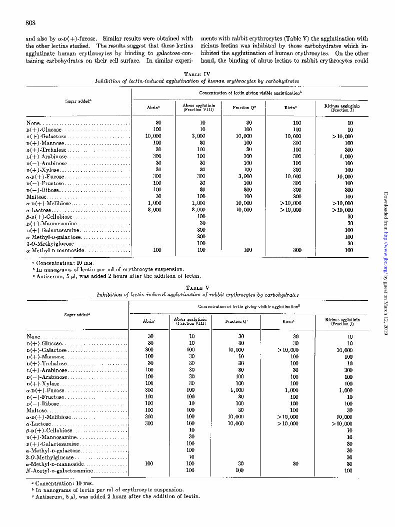
Carbohydrate Specijkity of Lectins-The effect of different mono- and disaccharides on the ability of the lectins to agglutinate erythrocytes was then studied. A lectin which can agglutinate cells spontaneously must be able to bind simultaneously to structures on the surface of at least two cells, thus forming a bridge between the cells. The abrus and ricinus agglutinins are able to agglutinate erythrocytes spontaneously, even in very low concentrations, indicating that they possess at least two binding sites. In similar concentrations, abrin and ricin are not able to agglutinate cells spontaneously, but they do agglutinate eryth- rocytes if specific antisera are added after the proteins have had time to bind to the erythrocytes (15, 16). This suggests that they are monovalent lectins. It is shown in Tables IV and V that in the absence of added carbohydrates as little as 10 to 100 ng per ml of the different lectins gave a visible agglutination of human as well as rabbit erythrocytes. In agreement with previous data (13, 23, 24) it was found that galactose inhibits agglutination by ricinus agglutinin and that carbohydrates con- taining a terminal nonreducing galactose residue are even more active. Thus, the agglutination of human erythrocytes by rici- nus agglutinin (Table IV) was strongly inhibited by D(+)-
galactose, a-~( +)-melibiose, a-methylgalactose, (Y- and P-lactose,
by guest on March 12, 2019
http://ww
w.jbc.org/
Dow
nloaded from
808
and also by WD( +)-fucose. Similar results were obtained with ments with rabbit erythrocytes (Table V) the agglutination with the other lectins studied. Theresults suggest that these lectins ricinus lectins was inhibited by those carbohydrates which in- agglutinate human erythrocytes by binding to galactose-con- hibited the agglutination of human erythrocytes. On the other taining carbohydrates on their cell surface. In similar experi- hand, the binding of abrus lectins to rabbit erythrocytes could
TABLE IV lnhibition of lectin-induced agglutination of human erythrocytes by carbohydrates
Sugar addeda
None .................................. D(+)-Glucose. ......................... D(+)-Galactose. ....................... D(+)-Mannose. ........................ D(+)-Trehalose ........................ I,(+)-Arabinose ........................ D(-)-Arabinose ........................ D(+)-Xylose ........................... a-D(+)-Fucose. ........................ D(-)-Fructose ......................... D(-)-Ribose. .......................... Maltose ................................ a-D(+)-Melibiose ....................... a-Lactose .............................. p-n(+)-Cellobiose ...................... D(+)-Mannosamine. ................... D(+)-Galactosamine ................... U-Methyl-D-galactose. .................. 3-O-Methylglucose . .................... a-Methyl-n-mannoside .................
0 Concentration: 10 mM.
AbrinC
30 100
10,ooo 100
30 300
30 30
300 100 100 30
l,ocJo 3,ooo
100
Concentration of lectin giving visible agglutination*
Abrus agglutinin (Fraction VIII)
10 10
3,ooo 30
100 100 30 30
300 30 30
100
l,ooo 3,ooo
100 30
300 300 100 100
Fraction Q” RicinC
30 100
10,ooo 100
30 300 100 100
3,ooo 100 300 100
10,cOo 10,cOo
100
100 100
10,oocl 300 100 300 100 300
10,ooo 300 300 300
>lO,ooo >lO,ooo
300
* In nanograms of lectin per ml of erythrocyte suspension. c Antiserum, 5 ~1, w&s added 2 hours after the addition of lectin.
TABLE V Inhibition of lectin-induced agglutination of rabbit erythrocytes by carbohydrates
Sugar added”
None ................................ I)(+)-Glucose. ....................... D(+)-Galactose ...................... D(+)-Mannose. ...................... D(+)-Trehalose. .................... L(+)-Arabinose. ..................... D(-)-Arabinose ..................... D(+)-Xylose ......................... a-D(+)-Fucose ..................... D(-)-Fructose ...................... D(-)-Ribose. ........................ Maltose .............................. a-D(+)-Melibiose ..................... a-Lactose ............................ B-D(+)-Cellobiose. ................... D(+)-Mannosamine. ................. D(+)-Calactosamine. ................ a-Methyl-D-galactose ................. 3GMethylglucose. .................. or-Methyl-D-mannoside. ............... iv-Acetyl-D-galactosamine. ...........
a Concentration: 10 mM.
- I
Ricks agglutinin (Fraction J)
10 10
>lO,ooo 100 300
l,OOCJ 100 100
10,ooo 100 300 100
>lO,ooo > 10,ooo
30 30
100 100
30 100
AbrinC
30 30
300 100 30
100 100 100 300 100 100 100 300 300
100
Concentration of lectin giving visible agglutination*
Abrus agglutinin (Fraction VIII)
10 10
100 30 30 30 30 30
100 100
10 100 100 100
10 30
100 100
10 100 100
Fraction Q” RicinC
30 30
10,ooo 10 30 30
100 100
l,fJc@ 30
100 30
10,ooo 10,ooo
30 100
30 30
>lO,ooo 100 100
30 100 100
l,ooO 100 100 100
>lO,ooo >lO,ooo
30
Ricinus agglutinin (Fraction J)
10 10
10,ooo 100
10 300 100 100
1mJ 10
100 30
10,ooo > 10,ooo
10 10 30 30 30 30
100
* In nanograms of lectin per ml of erythrocyte suspension. c Antiserum, 5 ~1, was added 2 hours after the addition of lectin.
by guest on March 12, 2019
http://ww
w.jbc.org/
Dow
nloaded from
not be inhibited by any of the carbohydrates tested. It is there- fore clear that abrus lectins differ somewhat from the ricinus lectins in their binding specificity.
Blood Group Specijkity of Lectins-The ability of the dif- ferent lectins to agglutinate human red blood cells from dif- ferent blood groups was then tested. It is clear from Table VI that the abrus lectins agglutinate erythrocytes of blood groups B and 0 somewhat better than cells from blood group A, in accordance with earlier findings (14). With ricinus lectins no difference was found. On the other hand, when the incubation was carried out in the presence of a low concentration (1 mM) of a-lactose, the amount of both abrus and ricinus lectins nec- essary to agglutinate erythrocytes from blood groups 0 and B was lower than that required for the agglutination of erythro- cytes from blood group A.
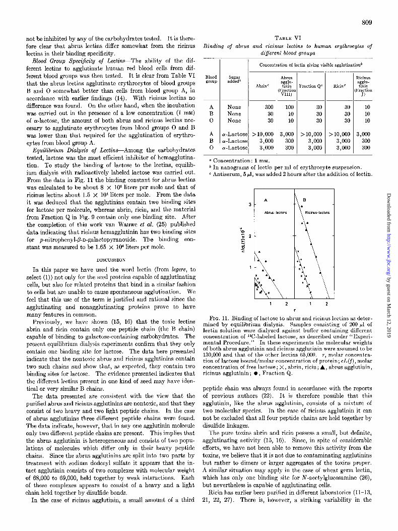
Equilibrium Dialysis of Lectins-Among the carbohydrates tested, lactose was the most efficient inhibitor of hemagglutina- tion. To study the binding of lactose to the lectins, equilib- ium dialysis with radioactively labeled lactose was carried out. From the data in Fig. 11 the binding constant for abrus lectins was calculated to be about 8 x 1Oa liters per mole and that of ricinus lectins about 1.5 x lo4 liters per mole. From the data it was deduced that the agglutinins contain two binding sites for lactose per molecule, whereas abrin, ricin, and the material from Fraction Q in Fig. 9 contain only one binding site. After the completion of this work van Wauwe et al. (25) published data indicating that ricinus hemagglutinin has two binding sites for p-nitrophenyl-P-n-galactopyranoside. The binding con- stant was measured to be 1.65 x lo4 liters per mole.
DISCUSSION
In this paper we have used the word lectin (from Zegere, to select (1)) not only for the seed proteins capable of agglutinating cells, but also for related proteins that bind in a similar fashion to cells but are unable to cause spontaneous agglutination. We feel that this use of the term is justified and rational since the agglutinating and nonagglutinating proteins prove to have many features in common.
Previously, we have shown (15, 16) that the toxic lectins abrin and ricin contain only one peptide chain (the B chain) capable of binding to galactose-containing carbohydrates. The present equilibrium dialysis experiments confirm that they only contain one binding site for lactose. The data here presented indicate that the nontoxic abrus and ricinus agglutinins contain two such chains and show that, as expected, they contain two binding sites for lactose. The evidence presented indicates that the different lectins present in one kind of seed may have iden- tical or very similar B chains.
The data presented are consistent with the view that the purified abrus and ricinus agglutinins are nontoxic, and that they consist of two heavy and two light peptide chains. In the case of abrus agglutinins three different peptide chains were found. The data indicate, however, that in any one agglutinin molecule only two different peptide chains are present. This implies that the abrus agglutinin is heterogeneous and consists of two popu- lations of molecules which differ only in their heavy peptide chains. Since the abrus agglutinins are split into two parts by treatment with sodium dodecyl sulfate it appears that the in- tact agglutinin consists of two complexes with molecular weight of 68,000 to 69,000, held together by weak interactions. Each of these complexes appears to consist of a heavy and a light chain held together by disulfide bonds.
In the case of ricinus agglutinin, a small amount of a third
809
TABLE VI
Binding of abrus and ricinus lectins to human erythrocytes of different blood groups
Blood group
- Concentration of lectin giving visible agglutinationb
sugar addeda
Abrin’
None 300 None 30 None 30
a-Lactose > 10,000 a-Lactose 3,000 a-Lactose 3,000
Abrus apt;-
Fraction VIII)
100 10 10
3,000 300 300
-
1
Fraction Q” RicinC
30 30 30
>10,000 3,000 3,000
30 30 30
> 10,000 3,000 3,000
Ricinus a&-
Fraction J)
10 10 10
3,000 300 300
a Concentration: 1 mM. b In nanograms of lectin per ml of erythrocyte suspension. c Antiserum, 5 ~1, was added 2 hours after the addition of lectin.
FIG. 11. Binding of lactose to abrus and ricinus lectins as deter- mined by equilibrium dialysis. Samples consisting of 200 ~1 of lectin solution were dialyzed against buffer containing different concentration of ‘%-labeled lactose, as described under “Experi- mental Procedure.” In these experiments the molecular weights of both abrus agglutinin and ricinus agglutinin were assumed to be 130,000 and that of the other lectins 65,000. T, molar concentra- tion of lactose bound/molar concentration of protein; CL(f), molar concentration of free lactose; X, abrin, ricin; A, abrus agglutinin, ricinus agglutinin; l , Fraction Q.
peptide chain was always found in accordance with the reports of previous authors (22). It is therefore possible that this agglutinin, like the abrus agglutinin, consists of a mixture of two molecular species. In the case of ricinus agglutinin it can not be excluded that all four peptide chains are held together by disulfide linkages.
The pure toxins abrin and ricin possess a small, but definite, agglutinating activity (15, 16). Since, in spite of considerable efforts, we have not been able to remove this activity from the toxins, we believe that it is not due to contaminating agglutinins but rather to dimers or larger aggregates of the toxins proper. A similar situation may apply in the case of wheat germ lectin, which has only one binding site for N-acetylglucosamine (26), but nevertheless is capable of agglutinating cells.
Ricin has earlier been purified in different laboratories (11-13, 21, 22, 27). There is, however, a striking variability in the
by guest on March 12, 2019
http://ww
w.jbc.org/
Dow
nloaded from

810
hemagglutinating activity of the different ricin preparations. Thus, Gtirtler and Horstmann (22) reported that ricin had a hemagglutinating activity of 1.7 units per mg, whereas the agglutinin had 2.4 units per mg. Nicolson and Blaustein (13) found that ricin had about 10% of the hemagglutinating ac- tivity of the ricinus agglutinin. On the other hand, Wald- Schmidt-Leitz and Keller (12), as well as Tomita et al. (21), found, in accordance with our own data, that ricin has only 1 to 3% of the hemagglutinating activity of ricinus agglutinin. One reason for the discrepancies could be that different condi- tions of isolation and storage may influence the extent of dimer formation of the toxin in solution. Contamination with ag- glutinin in some ricin preparations may be another explanation. Conversely, the high toxicity reported for some ricinus agglutinin preparations (13, 21, 27) is probably due to contamination with toxin.
Many agglutinins stimulate DNA synthesis and blastoid transformation in lymphocytes (2). In the case of abrus and ricinus agglutinins, such studies have been hampered by the fact that previous agglutinin preparations contained sufficient amounts of abrin and ricin to kill the lymphocytes. However, preliminary experiments with our highly purified agglutinins indicate that they indeed initiate DNA synthesis in lympho- cytes.
Acknowledgments-The expert technical assistance of Sidsel Brakstad and Synnove Eilertsen is gratefully acknowledged. We are indebted to A. M. Pappenheimer, Jr., for stimulating discussions and to Terje Christensen for carrying out the ana- lytical ultracentrifugations.
REFERENCES
1. BOYD, W. C. (1970) Ann. N. Y. Acad. Sci. 169, 168 2. SHARON. N.. AND LIS. H. (1972) Science 177.949
Biochem. J. 129, 847 27. ONOZAKI, K., TOMITA, M., SAKURAI, Y. AND UKITA, T. (1972)
, , , . , Biochem. Biophys. Res. Commun. 46,783
3. 4. 5. 6. 7. 8.
9.
10.
11.
12.
13.
14.
BURGER, M. (1973) Fed. Proc. 32,91 NICOLSON, G. L. (1972) Nature New Biol. 239,193 STILLMARK, H. (1339) Arb. Pharmak. Inst. Dorpat 3,59 HELLIN, H. (1891) Ph.D. thesis, Universitiit zu Dorpat ELFSTRAND, M. (1897) Ph.D. thesis, Upsala University EHRLICH, P. (1957) in Collected Papers (Dale, H., ed) Vol. 2,
pp. 21-44, Pergamon Press, London KOBERT, R. (1906) Lehrbuch der Intozikationen, Vol. 2, pp. 695-
707, Enke, Stuttgart LANDSTEINER, K., AND VON JAGIC, N. (1904) Muench. Med.
Wochenschr. 61,1185 ISHIGURO. M., TAKAHASHI, T., FUNATSU, G., HAYASHI, K.,
AND FUNATSU, M. (1964) J. Biochem. 66,587 WALDSCHMIDT-LEITZ, E., AND KELLER, L. (1969) Hoppe-
Seyler’s Z. Physiol. Chem. 360, 503 NICOLSON, G. L., AND BLAUSTEIN, J. (1972) Biochim. Biophys.
Acta 266, 543 KAHN, A. H., GUL, B., AND RAHMAN, M. A. (1966) J. Immunol.
96, 554 15. OLSNES, S., AND PIHL, A. (1973) Eur. J. Biochem. 36,179 16. OLSNES, S., AND PIHL, A. (1973) Biochemistry 12, 3121 17. WEBER, K., AND OSBORN, M. (1969) J. Biol. Chem. 244,4406 18. OLSNES, S. (1971) Eur. J. Biochem. 23, 557 19. OLSEN, B. R., AASLAND TORGNER, I., CHRISTENSEN, T. B.,
AND KVAMME, E. (1973) J. Mol. Biol. 74,239 20. So, L. L., AND GOLDSTEIN. I. J. (1968) Biochim. Biovhus. Acta
i66, 398 - ”
21. TOMITA, M., KUROKAWA, T., ONOZAKI, K., ICHIKI, N., OSAWA, T., AND UKITA. T. (1972) Ezverientia 23.84
22. G~R&LER, L. G.,‘AND.Ho&TM~NN, H. J. (1973) Biochim. Bio- phys. Acta 296, 682
23. PARDOE, G. I., BIRD, G. W. G., AND UHLENBRUCK, G. (1969) Z. Immunitaetsforsch. 137, 442
24. DRYSDALE, It. G., HERRICK, P. It., AND FRANKS, D. (1968) Trot Sang. 16, 194
25. VAN WAUWE, J. P., LOONTIENS, F. G., AND DE BRUYNE, C. K. (1973) Biochim. Biophys. Acta 313,99
26. LEVINE, I)., KAPLAN, M. J., AND GREENAWAY, P. J. (1972)
by guest on March 12, 2019
http://ww
w.jbc.org/
Dow
nloaded from

Sjur Olsnes, Erling Saltvedt and Alexander PihlRicinus communis
andAbrus precatoriusIsolation and Comparison of Galactose-binding Lectins from
1974, 249:803-810.J. Biol. Chem.
http://www.jbc.org/content/249/3/803Access the most updated version of this article at
Alerts:
When a correction for this article is posted•
When this article is cited•
to choose from all of JBC's e-mail alertsClick here
http://www.jbc.org/content/249/3/803.full.html#ref-list-1
This article cites 0 references, 0 of which can be accessed free at
by guest on March 12, 2019
http://ww
w.jbc.org/
Dow
nloaded from