comparison of intraoperative procedures for isolation of ... · enzymatic intraoperative isolation...
TRANSCRIPT
University of Groningen
Comparison of intraoperative procedures for isolation of clinical grade stromal vascularfraction for regenerative purposesvan Dongen, Joris A; Tuin, A Jorien; Spiekman, Maroesjka; Jansma, Johan; van der Lei,Berend; Harmsen, Martin CPublished in:Journal of tissue engineering and regenerative medicine
DOI:10.1002/term.2407
IMPORTANT NOTE: You are advised to consult the publisher's version (publisher's PDF) if you wish to cite fromit. Please check the document version below.
Publication date:2017
Link to publication in University of Groningen/UMCG research database
Citation for published version (APA):van Dongen, J. A., Tuin, A. J., Spiekman, M., Jansma, J., van der Lei, B., & Harmsen, M. C. (2017).Comparison of intraoperative procedures for isolation of clinical grade stromal vascular fraction forregenerative purposes: a systematic review. Journal of tissue engineering and regenerative medicine.https://doi.org/10.1002/term.2407
CopyrightOther than for strictly personal use, it is not permitted to download or to forward/distribute the text or part of it without the consent of theauthor(s) and/or copyright holder(s), unless the work is under an open content license (like Creative Commons).
Take-down policyIf you believe that this document breaches copyright please contact us providing details, and we will remove access to the work immediatelyand investigate your claim.
Downloaded from the University of Groningen/UMCG research database (Pure): http://www.rug.nl/research/portal. For technical reasons thenumber of authors shown on this cover page is limited to 10 maximum.
Download date: 24-12-2019
This article has been accepted for publication and undergone full peer review but has not been through the copyediting, typesetting, pagination and proofreading process which may lead to differences between this version and the Version of Record. Please cite this article as doi: 10.1002/term.2407
This article is protected by copyright. All rights reserved.
Comparison of intraoperative procedures for isolation of clinical grade stromal
vascular fraction for regenerative purposes: a systematic review
Joris A. van Dongen, B.Sc1, 2, #, A. Jorien Tuin, MD3, #, Maroesjka Spiekman, B.Sc1,
Johan Jansma, MD, DMD, PhD3, Berend van der Lei, MD, PhD2, 4, Martin C.
Harmsen, PhD1,*
1 Department of Pathology & Medical Biology, University of Groningen and University Medical Center Groningen, Groningen, the Netherlands 2 Department of Plastic Surgery, University of Groningen and University Medical Center Groningen, Groningen, the Netherlands 3 Department of Oral & Maxillofacial Surgery, University Medical Center Groningen, University of Groningen, Groningen, the Netherlands 4 Bergman Clinics, locations Heerenveen, Zwolle and Groningen, the Netherlands # Authors contributed equally *Corresponding author
Prof. dr. Martin C. Harmsen, PhD Department of Pathology and Medical Biology University of Groningen University Medical Center Groningen Hanzeplein 1 9713 GZ Groningen Phone: +31.50.361.4776
Fax +31.50.361.9911 Email: [email protected]
This article is protected by copyright. All rights reserved.
Running title
Intraoperative procedures for stromal vascular fraction isolation
Conflict of interest
The authors have no conflict of interest to disclose in relation to the content of this
work.
Funding
This study was funded by the University Medical Center Groningen
Abstract
Introduction
Intraoperative application of stromal vascular fraction (SVF) of adipose tissue,
requires a fast and efficient isolation procedure of adipose tissue. This review was
performed to systematically assess and compare procedures currently used for the
intraoperative isolation of cellular SVF (cSVF) and tissue SVF (tSVF) which still
contains the extracellular matrix.
Methods
Pubmed, EMBASE and The Cochrane Central Register of controlled trials databases
were searched for studies that compare procedures for intraoperative isolation of
SVF (searched 28th of September, 2016). Outcomes of interest were cell yield,
viability of cells, composition of SVF, duration, cost and procedure characteristics.
Procedures were subdivided in procedures resulting in a cSVF or tSVF.
Results
Thirteen out of 3038 studies were included, evaluating eighteen intraoperative
isolation procedures, were considered eligible. In general, cSVF and tSVF
intraoperative isolation procedures had comparable cell yield, cell viability and SVF
composition compared to a non-intraoperative (i.e. culture lab-based collagenase
protocol) control group within the same studies. The majority of intraoperative
isolation procedures are less time consuming than non-intraoperative control groups,
however.
This article is protected by copyright. All rights reserved.
Conclusion
Intraoperative isolation procedures are less time-consuming than non-intraoperative
control group with similar cell yield, viability of cells and composition of SVF and
therefore more suitable for use in the clinic. Nevertheless, none of the intraoperative
isolation procedures could be designated as preferred procedure to isolate SVF.
Keywords: Lipografting, Stromal vascular fraction, Adipose derived stem/stromal
cells, Non-enzymatic isolation, Enzymatic isolation, Collagenase
1. Introduction
Adipose tissue seems to be an outstanding source for regenerative therapies, since
it is an easy accessible source for adipose-derived stem or stromal cells (ASCs).
Adipose tissue can easily be harvested with liposuction, a low risk procedure that
can be performed under local anesthesia. Several clinical trials have been published
using ASCs for soft tissue reconstruction (Tanikawa et al. 2013), cardiac repair
(Perin et al. 2014), pulmonary repair (Tzouvelekis et al. 2013) and cartilage repair
(Jo et al. 2014). All these trials show promising results for future use of ASCs in
tissue repair and regeneration.
To harvest ASCs, adipose tissue or lipoaspirate is subjected to enzymatic
dissociation followed by several centrifugation steps (Bourin et al. 2013), which is a
relative long-lasting procedure that cannot be performed during surgery. The cell
population obtained by this enzymatic digestion and centrifugation is the stromal
vascular fraction (SVF), containing ASCs, endothelial cells, supra-adventitial cells,
lymphocytes and pericytes (Eto et al. 2009, Bourin et al. 2013). ASCs in vivo are
characterized as CD31min/CD45min/CD34pos/CD90pos/CD105low cells
(Yoshimura et al. 2006). After isolation, the SVF can either be used directly in clinical
procedures or can be cultured to increase the number of cells before using them in
the clinic (Gir et al. , Suga et al. 2007). In case of cell culturing, only ASCs and their
precursor cells (supra-adventitial cells and pericytes) are able to adhere and survive
(Zuk et al. 2001, Zimmerlin et al. 2010). Upon passaging in vitro, the phenotype of
ASCs starts to deviate from their in vivo phenotype (Spiekman et al., 2016): in this
process CD34 surface expression is lost, while CD105 expression is up-regulated to
mention a few (Yoshimura et al. 2006, Corselli et al. 2012). Alternatively,
This article is protected by copyright. All rights reserved.
administration of the enzymatically prepared vascular stromal fraction of adipose
tissue might have a therapeutic capacity that is similar to cultured ASCs. Although,
no formal scientific evidence exists, the consensus is, that the therapeutic benefit of
SVF predominantly relies on the abundantly present ASCs.
The current protocol to isolate and culture ASCs from adipose tissue involves
enzymatic digestion with collagenase. This is a laborious and time consuming
protocol and requires a specialized culture lab (Good Manufacturing Practice
facilities (cGMP)), which is not available in most peripheral hospitals (Gimble et al.
2010). Therefore, intraoperative procedures for SVF isolation are warranted, in
particular systems that do not employ enzymatic treatment, such as mechanical
dissociation.
At present, several (commercial) procedures are available for intraoperative
isolation of SVF (Aronowitz et al. 2015, Oberbauer et al. 2015). These intraoperative
isolation procedures differ in various aspects: isolation of a single cell SVF (cellular
SVF (cSVF)) resulting in a pellet with hardly any volume or isolation of SVF cells
containing intact cell-cell communications (tissue SVF (tSVF). Most of the enzymatic
intraoperative isolation procedures result in a cSVF, because of the loss of cell-cell
communications and extracellular matrix. In most of the non-enzymatic intraoperative
isolation procedures the cell-cell communications remain intact, resulting in an end
product with more volume (tSVF). Different studies assessed the cell yield and
phenotype of the isolated cSVF or tSVF of the various intraoperative isolation
procedures compared to other intraoperative (commercial) procedures or to the gold
standard for SVF isolation (non-intraoperative culture lab-based collagenase
protocols which require cGMP facilities for clinical use, referred to as ‘non-
intraoperative isolation protocol’). Recently, new intraoperative isolation procedures
are introduced and tested. It is not clear yet if intraoperative isolation procedures
generate a similar quality and quantity of SVF as non-intraoperative isolation
protocols. Next to this, the distinction between end products of intraoperative
isolation procedures, e.g. cSVF and tSVF have never been studied. Therefore, a
systematic review was performed to assess the efficacy of intraoperative isolation
procedures of human SVF based on number of cells, cell viability and composition of
SVF. In addition, duration and costs of the intraoperative isolation procedures were
compared.
This article is protected by copyright. All rights reserved.
2. Material & Methods
2.1. Protocol and registration
This study was performed using the PRISMA protocol (Moher et al. 2009). The
search strategy for this systematic review was based on a Population, Intervention,
Comparison, and Outcome (PICO) framework (Schardt et al. 2007). The study was
not registered.
2.2. Eligibility criteria
Studies were included when at least two different types of intraoperative isolation
procedures or one intraoperative isolation procedure with a non-intraoperative
isolation protocol were assessed using human adipose tissue to isolate SVF. Studies
need to use the adipose fraction of lipoaspirate. Studies only evaluating
centrifugation forces, sonication or red blood cell (RBC) lysis buffer were excluded.
Studies focusing on processing methods of adipose tissue for the use in fat grafting
were excluded as well as case reports, case series and reviews. Searches were not
limited to date, language or publication status (Table 1).
2.3. Information sources and search
Pubmed, EMBASE (OvidSP) and The Cochrane Central Register of controlled trials
databases were searched (searched 28th September, 2016). The search was
restricted to human studies. The search terms (Table 2) were based on three
components: (P) adipose stromal cell, adipose stem cell, stromal vascular fraction,
autologous progenitor cell, or regenerative cell in combination with (I) cell separation,
isolation, dissociation, digestion, emulsification, isolation system, cell concentrator
and finally connected with (C) enzymatic, non-enzymatic, or mechanical.
2.4. Study selection and data collection process
Two authors (JAD, AJT) selected studies independently based on the eligibility
criteria. Inconsistencies were discussed during a consensus meeting. In case of
disagreement, the senior author (MCH) gave a binding verdict.
This article is protected by copyright. All rights reserved.
2.5. Data items
Search term was partly based on a Population, Intervention, Comparison, Outcome
(PICO) framework. Outcomes of interest were not included in the search term. For
this review the outcomes of interest were cell yield, viability of the nucleated cells,
composition of the SVF and duration, cost and characteristics of the intraoperative
isolation procedures. Effect sizes were calculated on cell yield and viability in studies
with a comparison of intraoperative isolation procedures versus regular non-
intraoperative isolation protocols. Differences in harvesting procedure were not taken
into account.
2.6. Risk of bias in individual studies
It is known that the quality of ASCs depends on age and harvest location of the
donor (Engels et al. 2013, Dos-Anjos Vilaboa et al. 2014, Di Taranto et al. 2015,
Maredziak et al. 2016). The inclusion of young healthy patients may positively affect
the results. Therefore, detailed information about demographics are described in this
review.
2.7. Summary measurements
Effect sizes were calculated of the outcome variables cell yield and percentage of
viable nucleated cells from cSVF between enzymatic intraoperative isolation
procedures and non-intraoperative isolation protocols (gold standard). The following
effect size formula was used: effect size = (difference in mean outcomes between
enzymatic intraoperative isolation procedures and gold standard) / (standard
deviation of the gold standard). Studies which presented results in mean and
standard deviation were analyzed. Intraoperative isolation procedures focusing on
tSVF instead of cSVF were not taken into account in the effect size of cell yield,
because of different start volumes of lipoaspirate and end volumes of tSVF.
2.8. Synthesis of results
In some studies, derivate numbers of graphs are used when the actual number of
outcomes was not given. Cell types within the SVF can be distinguished based on
CD marker expression or immuno-staining. To compare SVF compositions between
different studies and to compare intraoperative procedures with their control (i.e.
non-intraoperative protocols or other intraoperative procedures) in the same study,
only CD marker expression was used. Studies evaluating a single CD marker
expression to analyze different cell types were seen as insufficient distinctive and
were excluded. Cells were divided into two major groups: CD45min (adipose tissue-
This article is protected by copyright. All rights reserved.
derived) and CD45pos (blood derived) cells to analyze the expression of stromal
cells, pericytes, vascular endothelial cells/endothelial progenitor cells, endothelial
cells, lymphocytes, leukocytes and hematopoietic stem cells. All other cells are
placed in the category: other cell types. The CD34pos/CD146pos population is
excluded from analysis because of the inability to discriminate between progenitor
pericytes and progenitor endothelial cells (Bianchi et al. 2013).
2.9. Risk of bias across studies
Included studies could present different outcome variables related to SVF analysis.
There is a risk that studies did not present a full SVF characterization and thereby
bias their results. In order to provide an overview of the used outcome variables per
study, a Modified IFATS/ISCT Index Score was used (see 2.10). The risk of
publication bias of positive results might be expected in those articles were the
authors have benefits in the investigated products. Disclosure agreements were
reviewed for each study.
2.10. Modified IFATS/ISCT Index Score for the measurement of adipose tissue-
derived stromal vascular fraction
Studies were assessed based on the reported outcome variables. The assessment
of quality was evaluated based on the position statement of the International
Federation of Adipose Therapeutics and Science (IFATS) and the International
Society of Cellular Therapy (ISCT) (Bourin et al. 2013). The IFATS and ISCTS
proposed guidelines to develop reproducible standardized endpoints and methods to
characterize ASCs and SVF cells. For each of the following characterization
methods a grade was given by the authors (JAD, AJT) to an article if the
characterization was carried out: viability of nucleated cells, flow cytometry of SVF
cells, flow cytometry of ASCs (CD13, CD29, CD31, CD34, CD44, CD45, CD73,
CD90, CD105, CD235a), proliferation and frequency (CFU-F) and functional assays
(adipogenic, osteogenic and chondrogenic differentiation assays) of ASCs. The
maximum score in case of a full characterization was 5.
This article is protected by copyright. All rights reserved.
3. Results
3.1. Included studies
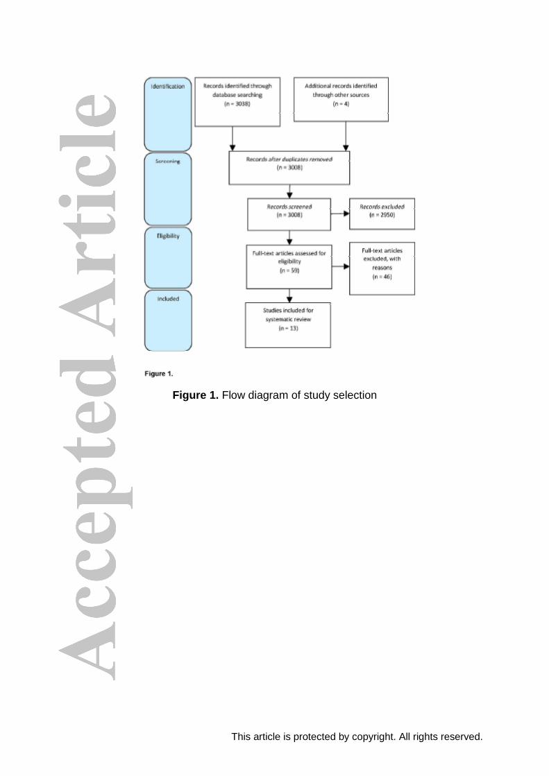
A total of 3038 studies were identified after database searching. 2955 articles were
excluded after abstract screening. 59 full text studies were assessed on eligibility
criteria. Fourteen studies were excluded based on the use of a non-intraoperative
protocol for isolation as experimental method (Yoshimura et al. 2006, Jiang et al.
2007, Pilgaard et al. 2008, Vykoukal et al. 2008, Al Battah et al. 2011, Fink et al.
2011, Condé-Green and Lamblet 2012, Okura et al. 2012, Carvalho et al. 2013,
Escobedo-Lucea et al. 2013, Siciliano et al. 2013, Chen et al. 2014, Doi et al. 2014,
Seaman et al. 2015). Seven studies described isolation protocols in general but gave
no results (Hicok and Hedrick , Bernacki et al. 2008, Dubois et al. 2008, Yu et al.
2011, Zachar et al. 2011, Buehrer and Cheatham 2013, Zhu et al. 2013). Seven
studies were excluded based on the lack of a control group (i.e. non-intraoperative
isolation protocols or other intraoperative isolation procedures) (Zuk et al. 2001,
Zeng et al. 2013, Dos-Anjos Vilaboa et al. 2014, Inoue et al. 2014, Sadighi et al.
2014, Van Pham et al. 2014, Raposio et al. 2016). Four studies were excluded
based on their study design (Kim 2014, Marincola 2014, Aronowitz and Hakakian
2015, Bertheuil and Chaput 2015). Three studies were excluded based on the use of
culture methods to isolate ASCs, because culture methods are incompatible with
intraoperative applications (Wu et al. 2012, Busser et al. 2014, Priya et al. 2014).
Four studies used only centrifugation, centrifugation or RBC lysis buffer as isolation
protocol and were thereby excluded (Baptista et al. 2009, Markarian et al. 2014,
Raposio et al. 2014, Amirkhani et al. 2016). Three studies used the blood saline
fraction of lipoaspirate and were thereby excluded (Francis et al. 2010, Shah et al.
2013, Cicione et al. 2016). Four studies did not describe an outcome of interest
(Reshak et al. 2013, Fraser et al. 2014, Yi et al. 2014, Aronowitz et al. 2015). Four
additional studies were identified through other sources (Fig. 1). Thus, thirteen
studies with eighteen intraoperative isolation procedures remained for analysis.
3.2. Study characteristics
In total, 93 subjects were enrolled in the thirteen studies. Nine studies reported
gender of which 95% was female (n=58). Nine studies reported the mean age or age
variance of the subjects and ten other studies described the use of infiltration (Table
1, supplemental content). No meta-analysis could be performed because the metrics
and outcomes were too diverse.
This article is protected by copyright. All rights reserved.
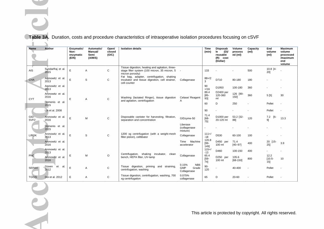
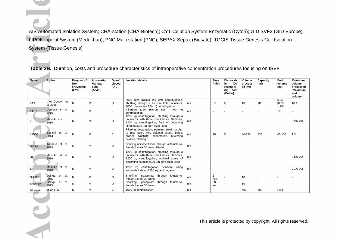
3. 3. Characteristics of the intraoperative isolation procedures
All intraoperative isolation procedures are divided into two categories: enzymatic and
non-enzymatic procedures resulting in cSVF and tSVF respectively (Table 3A and
table 3B). Eight of the eighteen intraoperative isolation procedures were based on
enzymatic digestion and ten isolation procedures were based on non-enzymatic
procedures. Two non-enzymatic procedures, the Residual tissue of emulsified fat
procedure and the Fractionation of adipose tissue procedure, are named differently,
but are almost identical. One intraoperative isolation procedure, the Filtrated fluid of
emulsified fat, is a combined procedure of two other intraoperative isolation
procedures i.e. the Fractionation of adipose tissue procedure and the Nanofat
procedure (Tonnard et al. 2013, Mashiko et al. 2016, van Dongen et al. 2016).
3.3.1 Start volume versus end product
The Automated isolation system, GID SVF2, Lipokit system and Multi station are
enzymatic intraoperative isolation procedure that resulted in large average amounts
of SVF (7.2 ml – 20 ml), suggesting inefficient enzymatic digestions (SundarRaj et al.
2015, Aronowitz et al. 2016). The non-enzymatic intraoperative isolation procedures
resulted in larger end volumes than only a pellet. Prior the Lipogems procedure, 130
ml of adipose tissue can be obtained to mechanical dissociate to 100 ml of
lipoaspirate. Hence, this a reduction of the volume of 1.3 times, suggesting an
inefficient mechanical dissociation to our opinion (Bianchi et al. 2013). In contrast,
the Fractionation of adipose tissue procedure resulted in a 10.4-fold volume
reduction (van Dongen et al. 2016). For all other intraoperative isolation procedures,
no data is mentioned about the end volume of the lipoaspirate (Table 3A and table
3B).
3.3.2 Duration and costs
Duration of the intraoperative isolation procedures varied from 5 seconds to 133
minutes (n=12). Isolation with the Automated isolation system was the longest
intraoperative isolation procedure (SundarRaj et al. 2015). Shuffling lipoaspirate 5 or
30 times through a luer-to-luer lock syringe will take 5 or 30 seconds respectively
and were therefore the fastest procedures (Osinga et al. 2015). In general, the tested
non-enzymatic procedures take less time than the enzymatic procedures (Table 3A
and table 3B).
This article is protected by copyright. All rights reserved.
The costs of only enzymatic procedures Celution system (2013: $1950 and
2016: $2400), CHA-station ($710), Multi station (2013: $460 and 2016: $250), Lipokit
system (2013: $530 and 2016: $450) and GID SVF2 ($1000) are mentioned, the
enzymatic Celution system being the most expensive (Aronowitz and Ellenhorn
2013, Aronowitz et al. 2016). No data of non-enzymatic intraoperative procedures
were available (Table 3A and table 3B).
3.4. Cell yield
Thirteen studies evaluated the cell yield of eighteen different intraoperative isolation
procedures (Millan , Lin et al. 2008, Guven et al. 2012, Aronowitz and Ellenhorn
2013, Bianchi et al. 2013, Doi et al. 2013, Tonnard et al. 2013, Domenis et al. 2015,
Osinga et al. 2015, SundarRaj et al. 2015, Aronowitz et al. 2016, Mashiko et al.
2016, van Dongen et al. 2016) (Table 2A and table 2B, supplemental content). The
reported cell yield after those different procedures varied between 0.19 – 11.7 x 10⁵
cells per ml in enzymatic intraoperative isolation procedures and between 1.8 – 22.6
x 105 cells per ml in non-enzymatic intraoperative isolation procedures. Non-
enzymatic intraoperative procedures yielded higher number of cells since the cell
yield was based on 1ml of end volume, whereas the enzymatic intraoperative
isolation cell yield was based on the obtained pellet per 1 ml start volume of
lipoaspirate. Of the enzymatic intraoperative isolation procedures, the Celution
system, Multi station and Lipokit system were evaluated by more than one group of
authors (Lin et al. 2008, Aronowitz and Ellenhorn 2013, Domenis et al. 2015,
Aronowitz et al. 2016). Interestingly, obvious different yields were seen using the
same procedure in different studies (Lin et al. 2008, Aronowitz and Ellenhorn 2013,
Domenis et al. 2015, Aronowitz et al. 2016). Reproducibility is thereby questioned in
our opinion. The cell yield using the enzymatic Celution system was significantly
higher as compared to the Lipokit system (p=0.004), the Multi station (p=0.049) and
CHA-station (p<0.001) (Aronowitz and Ellenhorn 2013). In contrast, Domenis et al.
did not find a statistical difference between the enzymatic Celution system and
Lipokit system. Moreover, Aronowitz et al. again compared the enzymatic Celution
system with the Lipokit system and Multi station. This time, Multi station and the
Lipokit system resulted in significant more cells as compared to the Celution system
(p<0.05) (Aronowitz et al. 2016).
This article is protected by copyright. All rights reserved.
In the non-enzymatic intraoperative isolation procedures, the Squeezed fat,
Residual fluid of emulsified fat and Fractionation of fat procedures resulted in the
relative highest cell yields per ml harvested lipoaspirate (Mashiko et al. 2016, van
Dongen et al. 2016). Non-enzymatic intraoperative isolation procedures such as
shuffling (5 times and 30 times), the Nanofat procedure and Fastem did not mention
the begin and end volumes, so the relative yield by isolation cannot be calculated
(Tonnard et al. 2013, Domenis et al. 2015, Osinga et al. 2015). Osinga et al,
reported that most of the adipocytes remain intact after shuffling 5 or even 30 times
(Osinga et al. 2015). Consequently, to our opinion, the effect of shuffling only cannot
be stated as an isolation procedure. We deem it possible that the lipoaspirate after
both two procedures did not differ from the initial lipoaspirate obtained at the start of
the procedure. However, the benefit might be at a different level, because shuffling
does improve the injectability of lipoaspirates as shown by Tonnard et al. (Tonnard et
al. 2013).
More interesting than comparing intraoperative isolation procedures evaluated
in different studies might be the comparison between an intraoperative isolation
procedure and a non-intraoperative isolation protocol (gold standard) starting from
the same lipoaspirate. Six studies reported the results of such comparisons (Table
4A) (Millan , Lin et al. 2008, Guven et al. 2012, Doi et al. 2013, Domenis et al. 2015,
SundarRaj et al. 2015). The Automated isolation system and Tissue genesis cell
isolation system resulted in the same cell yield as the non-intraoperative isolation
protocol control (effect size, respectively, 0.07 and 0.00) (Doi et al. 2013, SundarRaj
et al. 2015). Sepax isolated a higher cell yield compared to a non-intraoperative
isolation protocol (effect size 1.11) (Table 4A) (Guven et al. 2012). Lower cell yield
was seen after using the Lipokit system compared to the non-intraoperative isolation
protocol control (effect size -0.52) (Domenis et al. 2015). Interestingly, the highest
positive as well as the most negative effect sizes were seen with the enzymatic
Celution system related to regular isolation with a non-intraoperative isolation
protocol (Lin et al. 2008, Domenis et al. 2015).
This article is protected by copyright. All rights reserved.
3.5. Viability of nucleated cells
Eight studies described viabilities from 39% to 98% of nucleated cells in the SVF. No
big differences in viability were seen between enzymatic and non-enzymatic
intraoperative isolation procedures. The Filtrated fluid of emulsified fat procedure
showed the lowest viability (Mashiko et al. 2016), while the Automated isolation
system showed the highest viability of nucleated cells of 98% after isolation (Table
2A and table 2B, supplemental content) (SundarRaj et al. 2015). Three enzymatic
and three non-enzymatic intraoperative isolation procedures were compared to a
non-intraoperative isolation protocol regarding the viability of nucleated cells (Table
4B) (Lin et al. 2008, Doi et al. 2013, SundarRaj et al. 2015). The viability of five
intraoperative isolation procedures was comparable to their non-intraoperative
isolation protocol controls; the effect sizes were close to zero in many studies (Table
4B). Only the Filtrated fluid of emulsified fat procedure showed an effect size of -45.4
(Mashiko et al. 2016). In general, viability did not differ between non-intraoperative
isolation protocols and the individual intraoperative isolation procedures tested.
3.6. Composition of stromal vascular fractions
The SVF compositions is reported in nine studies evaluating six enzymatic
procedures and three non-enzymatic procedures. The stromal cell population is
larger in the SVF isolated by the enzymatic Celution system, Sepax and Tissue
genesis cell isolation system and the non-enzymatic Residual of emulsified fat and
Squeezed fat procedures compared to other intraoperative isolation procedures
(Guven et al. 2012, Aronowitz and Ellenhorn 2013, Doi et al. 2013, Mashiko et al.
2016) (Table 5, supplemental content). The percentage of stromal cell population of
the SVF isolated by the enzymatic Celution system only differs with 25.2% between
two studies (Aronowitz and Ellenhorn 2013, Domenis et al. 2015) and 32.8%
between two other studies, both evaluated by Aronowitz et al. (Aronowitz and
Ellenhorn 2013, Aronowitz et al. 2016). In general, non-enzymatic procedures
yielded same amounts of CD31min/CD34pos stromal cells.
The stromal cell population, including pericytes, ASCs and supra-adventitial
cells, are the most important cell types in regenerative therapies because of their
paracrine effect and multi-lineage differentiation capacity (Zuk et al. 2001, Pawitan
2014).
This article is protected by copyright. All rights reserved.
Pericytes defined using other CD markers than to define the stromal cell population
are placed separately in the table. The enzymatic Celution system evaluated by Lin
et al. resulted in the lowest percentage of pericytes in the SVF (0.8%), but used
more than three CD markers to detect pericytes (Lin et al. 2008). SundarRaj et al.
resulted in a higher percentage (2.0%) of pericytes in SVF obtained by the
Automated isolation system, but used only two CD markers to determine the pericyte
population and other cell types (SundarRaj et al. 2015). The use of multiple CD
markers results in a more specific population than the use of less CD markers and
so a lower percentage of that specific cell type e.g. pericytes (Bianchi et al. 2013).
Bianchi et al. used CD34min/CD146pos/CD90pos to detect the pericyte-like
population in the SVF and isolated the highest percentage of pericytes using the
non-enzymatic Lipogems procedure as compared to other intraoperative isolation
procedures (Bianchi et al. 2013). However, Bianchi et al. mostly used other
combinations of CD markers in comparison to other studies (Bianchi et al. 2013).
This renders their SVF composition incomparable with SVF compositions obtained
by other intraoperative isolation procedures.
The enzymatic procedures: Automated isolation system, Tissue genesis cell
isolation system and Sepax isolated more endothelial progenitor cells in comparison
to other intraoperative isolation procedures (Guven et al. 2012, Doi et al. 2013,
SundarRaj et al. 2015). Nonetheless, more endothelial progenitor cells were not
corresponding to less stromal cells or pericytes. In all differently obtained SVF, the
origin of large numbers of cells remains unidentified. This is partly because not every
study identified both adipose tissue-derived and blood-derived cell types, but
probably not every subpopulation of all cell types is already known as well.
When donor variability is neutralized by the use of the same lipoaspirate,
intraoperative isolation procedures resulted in different SVF compositions. Lipogems
isolated significantly more pericytes and stromal cells than the non-intraoperative
isolation protocol control (p<0.05) (Bianchi et al. 2013) (Fig. 2). The enzymatic
Celution system resulted in significantly more endothelial progenitor cells in
comparison with the CHA-system, Lipokit system and Multi station, which is not
necessarily preferred (p=0.003) (Aronowitz and Ellenhorn 2013). All other
intraoperative isolation procedures compared with non-intraoperative isolation
protocols showed no significant differences.
This article is protected by copyright. All rights reserved.
3.7. Modified IFATS/ISCT Index Score for the measurement of adipose tissue-
derived stromal vascular fraction
Modified IFATS/ISCT index scores ranged from 1 to 4.6 out of 5. Güven et al. scored
4.6 and presented the most complete characterization of the SVF and ASCs (Guven
et al. 2012) (Table 5). Tonnard et al. scored 2 points, but had only used CD34 as a
marker to identify a subpopulation in the SVF (Tonnard et al. 2013). Two studies
used other methods than flow cytometry to determine the composition of SVF
(Osinga et al. 2015, van Dongen et al. 2016). No studies were excluded based on a
low number of outcomes of interest measured by the modified IFATS/ISCT Index
Score, because five out of thirteen studies scored less than half of the possible
points given. This high number of low scores given to studies underlines the need for
standardization.
3.8 Disclosure agreements of included articles
A disclosure agreement of support by the manufacturer was provided in five of the
thirteen studies (Lin et al. 2008, Guven et al. 2012, Aronowitz and Ellenhorn 2013,
Bianchi et al. 2013, Doi et al. 2013) (Table 6, supplemental content). The company,
which was mostly involved in the studies, was Cytori, the manufacturer of the
enzymatic Celution system.
4. Discussion
Grafting of lipoaspirates and of SVF in particular, is a rapidly evolving treatment
modality for scars and other skin defects, arthritis, neuropathy, diabetic ulcers to
mention a few. Many of these, initially small scale, single center studies, are on the
verge of expansion to multicenter placebo-controlled double-blind randomized
clinical trials. An important prerequisite is the use of an efficient and standardized
intraoperative isolation procedure of SVF. This systematic review shows that none of
these procedures supersedes other procedures in terms of cell yield, viability and
SVF composition while being time and cost efficient too when analyzed using the
same lipoaspirate. However, three intraoperative isolation procedures (shuffling 5
times, shuffling 30 times and Lipogems) showed only a minimal reduction of the
volume of lipoaspirate, implicating that most of the adipocytes still are intact.
Consequently, these three procedures are methods of processing rather than
isolation procedures (Bianchi et al. 2013, Osinga et al. 2015). Moreover, there is a
wide variation in cell yield, viability of cells and composition of SVF when all
This article is protected by copyright. All rights reserved.
intraoperative isolation procedures are compared together. Study characteristics
showed small and varied sample sizes regarding the number, sex and age of the
donors. It is known that the cell yield and viability of SVF differ among donors,
depending on age, harvest location and co-morbidities, such as obesity, of the
donors (Engels et al. 2013, Dos-Anjos Vilaboa et al. 2014, Di Taranto et al. 2015,
Maredziak et al. 2016, Pachon-Pena et al. 2016). This interdonor variability is a
possible explanation for the variations found between several studies. To avoid
variation bias, isolation procedures should be investigated using identical
lipoaspirates in the same study. There are, however, differences between non-
enzymatic and enzymatic isolated SVFs on a different level. Non-enzymatic isolation
procedures resulted in larger volumes (tSVF) than the resulting pellets (cSVF) after
enzymatic intraoperative isolation procedures. Because the final products of both
types of isolation procedures are different, the clinical purpose of the use of SVF is
an important factor which isolation procedure suits best. In some cases, such as the
intra-articular injection of SVF in temporomandibular joints requires very small
volumes, whereas the end volume of SVF enriched lipofilling is less relevant.
Isolation procedures of SVF of adipose tissue are based on reduction of large
volume containing tissue or cells, such as ECM and/or adipocytes to concentrate the
stromal vascular fraction. Non-enzymatic isolation of SVF results in a smaller volume
of adipose tissue containing intact ECM and cell-cell communications between SVF
cells (tSVF), because the shear forces are too low to disrupt cell to cell and cell to
ECM adhesions (Lin et al. 2008, Corselli et al. 2012). Therefore, the tissue structure
of lipoaspirate is still intact in the tSVF. Enzymatic procedures, however, likely result
in a single cell cSVF, because enzymes likely disrupt all cell-cell interactions and
ECM (Fig. 3) (Aronowitz et al. 2015). This is may not happen in the Automated
isolation system, GID SVF2, Lipokit system and Multi station, possibly due to
insufficient enzymatic digestion (SundarRaj et al. 2015, Aronowitz et al. 2016).
Clinical use of tSVF has several advantages over the use of cSVF in different
clinical applications of regenerative medicine. It is well known that single cells
migrate within 24 hours after application (Parvizi and Harmsen 2015). The ECM,
containing a microvasculature structure, might function as a natural scaffold for cells
like ASCs and most likely also augments rapid vascularization and reperfusion. This
will probably increase cell retention rates after injection and enhance clinical effects.
In case of early scar formation, wound healing, or organ fibrosis, tSVF might
This article is protected by copyright. All rights reserved.
therefore be more an appropriate therapy, which implicates that non-enzymatic
procedures are more suitable as compared to enzymatic isolation procedures. In
case of excessive pre-existing scar formation, the ECM in the SVF might not be
appropriate and therefore the application of a cSVF or ASCs might be more eligible.
ASCs could remodel excessive scar formation by immunomodulation or instruction of
resident cells.
Characterization of subpopulations in the SVF depends upon selection of
appropriate markers. Selection of an insufficient number of markers will give a
disfigured image of the actual SVF composition (Fig. 3). SVF of adipose tissue can
be divided into two major subpopulations based on the expression of CD45, which is
a hematopoietic cell marker: adipose derived (CD45min) and blood derived
(CD45pos) (Yoshimura et al. 2006). Adipose derived cell populations can be divided
into endothelial cells (CD31pos) and stromal cells (CD31min) (Yoshimura et al.
2006). Three important subpopulations of the stromal cell population
(CD45min/CD31min) are supra-adventitial cells: CD34pos/CD146min, pericytes:
CD34pos/min/CD146pos and ASCs: CD34pos/CD90pos/CD105low (Yoshimura et
al. 2006, Zimmerlin et al. 2010, Corselli et al. 2012, Corselli et al. 2013). Supra-
adventitial cells and pericytes are both identified as precursor cells of ASCs,
although there remains some controversy about this item (Lin et al. 2008, Traktuev
et al. 2008, Zimmerlin et al. 2010, Corselli et al. 2012). Ideally, to discriminate
between those three cell types within the CD45min/CD31min subpopulation, CD146
and/or CD90 markers should be used additionally. However, in most studies two CD
markers or inappropriate combinations of CD markers have been used to determine
cell types; only Lin et al. used all the aforementioned combinations (Lin et al. 2008).
Because Lin et al. focus mainly on blood derived cells and not on the stromal cell
population or pericytes, this did not affect their results. Doi et al. ascribed
CD31min/CD34min/CD45min to the pericyte population, so therefore the CD34pos
pericytes will be missed (Doi et al. 2013). SundarRaj et al. and Güven et al. used
CD34pos/CD31min to determine the number of ASCs (Guven et al. 2012, SundarRaj
et al. 2015), while pericytes and supra-adventitial cells also express CD34.
Therefore, the number of ASCs contains pericytes and supra-adventitial cells as well
(Yoshimura et al. 2006, Zimmerlin et al. 2010). To cover pericytes, supra-adventitial
cells and ASCs, Domenis et al., Aronowitz et al. and Mashiko et al. used
CD34pos/CD31min/CD45min to determine the stromal cell population (Aronowitz
This article is protected by copyright. All rights reserved.
and Ellenhorn 2013, Domenis et al. 2015, Aronowitz et al. 2016, Mashiko et al.
2016). CD34pos is frequently used as a marker to describe cells with stem cell
characteristics in both hematopoietic and non-hematopoietic stem cells (Suga et al.
2009). The differences in use of CD marker expression to determine pericytes and
the stromal cell population might be a possible explanation for the large variations
found in SVF between different studies. No solid conclusions could be made about
which isolation procedure generates the most stromal cells or pericytes.
Unfortunately, a limited number of commercially available intraoperative SVF
isolation procedures not yet have reached scientific validation at an acceptable level.
The American Society for Aesthetic Plastic Surgery (ASAPS) and the American
Society of Plastic Surgeons (ASPS) published a position statement in 2012 on fat
grafting and stem cells (Eaves et al. 2012). All specialized equipment for the use of
stem cell extraction should be fully verified regarding efficacy and safety before use
in clinical settings. In 2013, the IFATS and ICTS proposed guidelines with
standardized endpoints and methods to verify and compare SVF isolation
procedures (Bourin et al. 2013). None of the included studies fully verified their
isolation procedure according to these IFATS and ICTS guidelines. Moreover,
viability was measured in different ways among studies (e.g. directly on obtained
SVF or after an extra non-intraoperative isolation protocol) and lipoaspirate was
processed differently prior to isolation (e.g. centrifugation or decantation). For those
reasons, we propose new adjusted IFATS and ICTS guidelines to validate
intraoperative isolation procedures (Fig. 3). All intraoperative isolation procedures
should be validated using centrifuged adipose tissue to determine the actual volume
of lipoaspirate prior to isolation. It is known that increased centrifugal forces have a
harmful effect on the viability of fat grafts (Xie et al. 2010, Tuin et al. 2016). However,
the use of centrifuged adipose tissue is necessary to determine the actual cell yield
after an isolation procedure. Furthermore, cell viability of tSVF should be determined
directly on tSVF, instead of using an extra non-intraoperative isolation protocol which
possibly results in more cell damage. However, the proposed adjusted standardized
endpoints and methods by IFATS and ICTS are time-consuming and expensive
since it requires cultured ASCs. In order to quickly verify isolation procedures
intraoperatively during clinical trials, the end product of non-enzymatic intraoperative
isolation procedures should be centrifuged to separate the oily fraction from the tSVF
and pellet fraction based on density. For enzymatic intraoperative isolation
This article is protected by copyright. All rights reserved.
procedures, microscopy can be used to visualize single cells. In this way, isolation
procedures can be quickly evaluated during clinical trials.
A large number of SVF isolation procedures without applying a full verification
according to the IFATS and ICTS guidelines is available (Oberbauer et al. 2015).
Oberbauer et al. presented a narrative overview of enzymatic and non-enzymatic
intraoperative SVF isolation procedures (Oberbauer et al. 2015). In twenty-one out of
thirty (both enzymatic as well as non-enzymatic) intraoperative isolation procedures
reported in their study, there was a lack of verification data. In two studies
intraoperative isolation procedures without scientific evidence e.g. viability of SVF,
flow cytometry of SVF cells and ASCs, were used to treat patients. One study used
SVF obtained by ultrasonic cavitation to treat patients with migraine and tension
headache (Bright et al. 2014). Another study used SVF in combination with platelet
rich plasma for meniscus repair (Pak et al. 2014). Hence, it cannot be guaranteed
that the isolation procedures indeed isolate SVF, which is clinical safe for use. It
seems that the use of most SVF isolation procedures with its concomitant clinical
application is far ahead of a sound scientific base upon which these procedures
should be used.
Moreover, the clinical safety of isolated SVF or ASCs is not clear yet,
especially regarding clinical use in patients with any kind of malignancy. It is
demonstrated, in vitro, that ASCs influence growth, progression and metastasis of
cancer cell lines through e.g. promoting angiogenesis and differentiation of ASCs
into carcinoma-associated fibroblasts (Freese et al. 2015). Zimmerlin et al. showed
in vitro that ASCs influence growth of active malign cell lines, but this is not seen in
latent cancer cell lines (Zimmerlin et al. 2011). Clinical data suggest that the use of
isolated SVF or ASCs is safe in patients without an oncological history (Charvet et al.
2015). In vitro studies often use higher concentrations of ASCs as compared to
clinical studies and this might be the cause of differences found between in vitro and
in vivo studies (Charvet et al. 2015). However, to test clinical safety it is important to
reach scientific validation of the commercially available procedures at an acceptable
level. In this review it become clear that the reproducibility of the procedures as well
as characterization of the SVF had shortcomings. If this is reached, further scientific
research with proper controls with regard to the clinical effect and safety of SVF or
ASCs are definitely wanted.
This article is protected by copyright. All rights reserved.
5. Conclusion
There is no evidence thus far that any intraoperative isolation procedure could be
designated as preferred procedure for isolating SVF. However, three isolation
procedures are rather processing techniques than isolation procedures. Enzymatic
and non-enzymatic procedures had comparable results as it comes to cell yield,
viability, and SVF composition. Non-enzymatic isolation procedures end products
resulted had greater volumes (tSVF) than the pellets (cSVF) of the enzymatic
isolation procedures. The results of intraoperative isolation procedures are
comparable with those of the gold standard, the collagenase based non-
intraoperative isolation protocol. Since intraoperative isolation procedures are less
time-consuming, but as efficient as the non-intraoperative isolation protocol, the use
of intraoperative isolation procedures seems to be more suitable for clinical
purposes. However, only small sample sizes have been used to validate the isolation
procedures. To test clinical safety, it is important to reach scientific validation of the
commercially available procedures at an acceptable level. Regarding to this review,
this level is not yet reached by many procedures.
6. Acknowledgement
We thank prof. dr. A. Vissink (Department of Oral & Maxillofacial Surgery, University
of Groningen and University Medical Center Groningen) for his contribution during
the preparation of this manuscript.
This article is protected by copyright. All rights reserved.
References
Al Battah, F., J. De Kock, E. Ramboer, A. Heymans, T. Vanhaecke, V. Rogiers and S. Snykers (2011). "Evaluation of the multipotent character of human adipose tissue-derived stem cells isolated by Ficoll gradient centrifugation and red blood cell lysis treatment." Toxicol In Vitro 25(6): 1224-1230. Amirkhani, M. A., R. Mohseni, M. Soleimani, A. Shoae-Hassani and M. A. Nilforoushzadeh (2016). "A rapid sonication based method for preparation of stromal vascular fraction and mesenchymal stem cells from fat tissue." Bioimpacts 6(2): 99-104. Aronowitz, J. A. and J. D. Ellenhorn (2013). "Adipose stromal vascular fraction isolation: a head-to-head comparison of four commercial cell separation systems." Plast Reconstr Surg 132(6): 932e-939e. Aronowitz, J. A. and C. S. Hakakian (2015). "A novel and effective strategy for the isolation of adipose-derived stem cells: minimally manipulated adipose-derived stem cells for more rapid and safe stem cell therapy." Plast Reconstr Surg 135(2): 454e. Aronowitz, J. A., R. A. Lockhart and C. S. Hakakian (2015). "Mechanical versus enzymatic isolation of stromal vascular fraction cells from adipose tissue." Springerplus 4: 713. Aronowitz, J. A., R. A. Lockhart, C. S. Hakakian and Z. E. Birnbaum (2016). "Adipose Stromal Vascular Fraction Isolation: A Head-to-Head Comparison of 4 Cell Separation Systems #2." Ann Plast Surg 77(3): 354-362. Aronowitz, J. A., R. A. Lockhart, C. S. Hakakian and K. C. Hicok (2015). "Clinical Safety of Stromal Vascular Fraction Separation at the Point of Care." Ann Plast Surg 75(6): 666-671. Baptista, L. S., R. J. do Amaral, R. B. Carias, M. Aniceto, C. Claudio-da-Silva and R. Borojevic (2009). "An alternative method for the isolation of mesenchymal stromal cells derived from lipoaspirate samples." Cytotherapy 11(6): 706-715. Bernacki, S. H., M. E. Wall and E. G. Loboa (2008). "Isolation of human mesenchymal stem cells from bone and adipose tissue." Methods Cell Biol 86: 257-278. Bertheuil, N. and B. Chaput (2015). "A novel and effective strategy for the isolation of adipose-derived stem cells: minimally manipulated adipose-derived stem cells for more rapid and safe stem cell therapy." Plast Reconstr Surg 135(2): 454e-455e. Bianchi, F., M. Maioli, E. Leonardi, E. Olivi, G. Pasquinelli, S. Valente, A. J. Mendez, C. Ricordi, M. Raffaini, C. Tremolada and C. Ventura (2013). "A new nonenzymatic method and device to obtain a fat tissue derivative highly enriched in pericyte-like elements by mild mechanical forces from human lipoaspirates." Cell Transplant 22(11): 2063-2077. Bourin, P., B. A. Bunnell, L. Casteilla, M. Dominici, A. J. Katz, K. L. March, H. Redl, J. P. Rubin, K. Yoshimura and J. M. Gimble (2013). "Stromal cells from the adipose tissue-derived stromal vascular fraction and culture expanded adipose tissue-derived stromal/stem cells: a joint statement of the International Federation for Adipose Therapeutics and Science (IFATS) and the International Society for Cellular Therapy (ISCT)." Cytotherapy 15(6): 641-648. Bright, R., M. Bright, P. Bright, S. Hayne and W. D. Thomas (2014). "Migraine and tension-type headache treated with stromal vascular fraction: a case series." J Med Case Rep 8: 237. Buehrer, B. M. and B. Cheatham (2013). "Isolation and characterization of human adipose-derived stem cells for use in tissue engineering." Methods Mol Biol 1001: 1-11.
This article is protected by copyright. All rights reserved.
Busser, H., C. De Bruyn, F. Urbain, M. Najar, K. Pieters, G. Raicevic, N. Meuleman, D. Bron and L. Lagneaux (2014). "Isolation of adipose-derived stromal cells without enzymatic treatment: expansion, phenotypical, and functional characterization." Stem Cells Dev 23(19): 2390-2400. Carvalho, P. P., J. M. Gimble, I. R. Dias, M. E. Gomes and R. L. Reis (2013). "Xenofree enzymatic products for the isolation of human adipose-derived stromal/stem cells." Tissue Eng Part C Methods 19(6): 473-478. Charvet, H. J., H. Orbay, M. S. Wong and D. E. Sahar (2015). "The Oncologic Safety of Breast Fat Grafting and Contradictions Between Basic Science and Clinical Studies: A Systematic Review of the Recent Literature." Ann Plast Surg 75(4): 471-479. Chen, S. Y., M. Mahabole, E. Horesh, S. Wester, J. L. Goldberg and S. C. Tseng (2014). "Isolation and characterization of mesenchymal progenitor cells from human orbital adipose tissue." Invest Ophthalmol Vis Sci 55(8): 4842-4852. Cicione, C., G. Di Taranto, M. Barba, M. A. Isgro, A. D'Alessio, D. Cervelli, F. V. Sciarretta, S. Pelo, F. Michetti and W. Lattanzi (2016). "In Vitro Validation of a Closed Device Enabling the Purification of the Fluid Portion of Liposuction Aspirates." Plast Reconstr Surg 137(4): 1157-1167. Condé-Green, A. and H. Lamblet (2012). "Immediate cell-supplemented lipotransfer (iCSL)." European Journal of Plastic Surgery 35(5): 373-378. Corselli, M., C. W. Chen, B. Sun, S. Yap, J. P. Rubin and B. Peault (2012). "The tunica adventitia of human arteries and veins as a source of mesenchymal stem cells." Stem Cells Dev 21(8): 1299-1308. Corselli, M., M. Crisan, I. R. Murray, C. C. West, J. Scholes, F. Codrea, N. Khan and B. Peault (2013). "Identification of perivascular mesenchymal stromal/stem cells by flow cytometry." Cytometry A 83(8): 714-720. Di Taranto, G., C. Cicione, G. Visconti, M. A. Isgro, M. Barba, E. Di Stasio, E. Stigliano, C. Bernardini, F. Michetti, M. Salgarello and W. Lattanzi (2015). "Qualitative and quantitative differences of adipose-derived stromal cells from superficial and deep subcutaneous lipoaspirates: a matter of fat." Cytotherapy 17(8): 1076-1089. Doi, K., S. Kuno, A. Kobayashi, T. Hamabuchi, H. Kato, K. Kinoshita, H. Eto, N. Aoi and K. Yoshimura (2014). "Enrichment isolation of adipose-derived stem/stromal cells from the liquid portion of liposuction aspirates with the use of an adherent column." Cytotherapy 16(3): 381-391. Doi, K., S. Tanaka, H. Iida, H. Eto, H. Kato, N. Aoi, S. Kuno, T. Hirohi and K. Yoshimura (2013). "Stromal vascular fraction isolated from lipo-aspirates using an automated processing system: bench and bed analysis." J Tissue Eng Regen Med 7(11): 864-870. Domenis, R., L. Lazzaro, S. Calabrese, D. Mangoni, A. Gallelli, E. Bourkoula, I. Manini, N. Bergamin, B. Toffoletto, C. A. Beltrami, A. P. Beltrami, D. Cesselli and P. C. Parodi (2015). "Adipose tissue derived stem cells: in vitro and in vivo analysis of a standard and three commercially available cell-assisted lipotransfer techniques." Stem Cell Res Ther 6: 2. Dos-Anjos Vilaboa, S., M. Navarro-Palou and R. Llull (2014). "Age influence on stromal vascular fraction cell yield obtained from human lipoaspirates." Cytotherapy 16(8): 1092-1097. Dubois, S. G., E. Z. Floyd, S. Zvonic, G. Kilroy, X. Wu, S. Carling, Y. D. Halvorsen, E. Ravussin and J. M. Gimble (2008). "Isolation of human adipose-derived stem cells from biopsies and liposuction specimens." Methods Mol Biol 449: 69-79.
This article is protected by copyright. All rights reserved.
Eaves, F. F., 3rd, P. C. Haeck and R. J. Rohrich (2012). "ASAPS/ASPS Position statement on stem cells and fat grafting." Plast Reconstr Surg 129(1): 285-287. Engels, P. E., M. Tremp, P. J. Kingham, P. G. di Summa, R. D. Largo, D. J. Schaefer and D. F. Kalbermatten (2013). "Harvest site influences the growth properties of adipose derived stem cells." Cytotechnology 65(3): 437-445. Escobedo-Lucea, C., C. Bellver, C. Gandia, A. Sanz-Garcia, F. J. Esteban, V. Mirabet, G. Forte, I. Moreno, M. Lezameta, A. Ayuso-Sacido and J. M. Garcia-Verdugo (2013). "A xenogeneic-free protocol for isolation and expansion of human adipose stem cells for clinical uses." PLoS One 8(7): e67870. Eto, H., H. Suga, D. Matsumoto, K. Inoue, N. Aoi, H. Kato, J. Araki and K. Yoshimura (2009). "Characterization of structure and cellular components of aspirated and excised adipose tissue." Plast Reconstr Surg 124(4): 1087-1097. Fink, T., J. G. Rasmussen, P. Lund, L. Pilgaard, K. Soballe and V. Zachar (2011). "Isolation and expansion of adipose-derived stem cells for tissue engineering." Front Biosci (Elite Ed) 3: 256-263. Francis, M. P., P. C. Sachs, L. W. Elmore and S. E. Holt (2010). "Isolating adipose-derived mesenchymal stem cells from lipoaspirate blood and saline fraction." Organogenesis 6(1): 11-14. Fraser, J. K., K. C. Hicok, R. Shanahan, M. Zhu, S. Miller and D. M. Arm (2014). "The Celution® system: Automated processing of adipose-derived regenerative cells in a functionally closed system." Advances in Wound Care 3(1): 38-45. Freese, K. E., L. Kokai, R. P. Edwards, B. J. Philips, M. A. Sheikh, J. Kelley, J. Comerci, K. G. Marra, J. P. Rubin and F. Linkov (2015). "Adipose-derived stems cells and their role in human cancer development, growth, progression, and metastasis: a systematic review." Cancer Res 75(7): 1161-1168. Gimble, J. M., F. Guilak and B. A. Bunnell (2010). "Clinical and preclinical translation of cell-based therapies using adipose tissue-derived cells." Stem Cell Res Ther 1(2): 19. Gir, P., S. A. Oni G Fau - Brown, A. Brown Sa Fau - Mojallal, R. J. Mojallal A Fau - Rohrich and R. J. Rohrich "Human adipose stem cells: current clinical applications." (1529-4242 (Electronic)). Guven, S., M. Karagianni, M. Schwalbe, S. Schreiner, J. Farhadi, S. Bula, K. Bieback, I. Martin and A. Scherberich (2012). "Validation of an automated procedure to isolate human adipose tissue-derived cells by using the Sepax(R) technology." Tissue Eng Part C Methods 18(8): 575-582. Hicok, K. C. and M. H. Hedrick "Automated isolation and processing of adipose-derived stem and regenerative cells." (1940-6029 (Electronic)). Inoue, K. I., H. Nomura, R. Sohma, K. Akimoto, N. Kobayashi, T. Kamai, O. Yamas, I. Taguchi, H. Asato, T. Inoue, O. Mitsumori, H. Iwaguro and K. I. Yoshida (2014). "Feasibility of exploiting Celution™ system in autologous cell therapy in Dokkyo medical university hospital: Safety and reproducibility." Dokkyo Journal of Medical Sciences 41(1): 7-12. Jiang, Y., L. Q. Han, Z. Y. Zhu, L. P. Xiao and Z. Liu (2007). "Isolating culture and osteogenic potential of human adipose derived-stromal cells." Journal of Clinical Rehabilitative Tissue Engineering Research 11(37): 7381-7384. Jo, C. H., Y. G. Lee, W. H. Shin, H. Kim, J. W. Chai, E. C. Jeong, J. E. Kim, H. Shim, J. S. Shin, I. S. Shin, J. C. Ra, S. Oh and K. S. Yoon (2014). "Intra-articular injection of mesenchymal stem cells for the treatment of osteoarthritis of the knee: a proof-of-concept clinical trial." Stem Cells 32(5): 1254-1266.
This article is protected by copyright. All rights reserved.
Kim, S. K. (2014). "Adipose stromal vascular fraction isolation: a head-to-head comparison of four commercial cell separation systems." Plast Reconstr Surg 133(6): 889e. Lin, G., M. Garcia, H. Ning, L. Banie, Y. L. Guo, T. F. Lue and C. S. Lin (2008). "Defining stem and progenitor cells within adipose tissue." Stem Cells Dev 17(6): 1053-1063. Lin, K., Y. Matsubara, Y. Masuda, K. Togashi, T. Ohno, T. Tamura, Y. Toyoshima, K. Sugimachi, M. Toyoda, H. Marc and A. Douglas (2008). "Characterization of adipose tissue-derived cells isolated with the Celution system." Cytotherapy 10(4): 417-426. Maredziak, M., K. Marycz, K. A. Tomaszewski, K. Kornicka and B. M. Henry (2016). "The Influence of Aging on the Regenerative Potential of Human Adipose Derived Mesenchymal Stem Cells." Stem Cells Int 2016: 2152435. Marincola, F. M. (2014). "Expression of concern: Clinical use of dieletrophoresis separation for live adipose derived stem cells." J Transl Med 12: 297. Markarian, C. F., G. Z. Frey, M. D. Silveira, E. M. Chem, A. R. Milani, P. B. Ely, A. P. Horn, N. B. Nardi and M. Camassola (2014). "Isolation of adipose-derived stem cells: a comparison among different methods." Biotechnol Lett 36(4): 693-702. Mashiko, T., S. H. Wu, J. Feng, K. Kanayama, K. Kinoshita, A. Sunaga, M. Narushima and K. Yoshimura (2016). "Mechanical micronization of lipoaspirates: squeeze and emulsification techniques." Plast Reconstr Surg. Millan, A. "Comparison Between Collagenase Adipose Digestion and StromaCell Mechanical Dissociation for Mesenchymal Stem Cell Separation " McNar Scholar Journal 15. Moher, D., A. Liberati, J. Tetzlaff and D. G. Altman (2009). "Preferred reporting items for systematic reviews and meta-analyses: the PRISMA Statement." Open Med 3(3): e123-130. Oberbauer, E., C. Steffenhagen, C. Wurzer, C. Gabriel, H. Redl and S. Wolbank (2015). "Enzymatic and non-enzymatic isolation systems for adipose tissue-derived cells: current state of the art." Cell Regen (Lond) 4: 7. Okura, H., A. Saga, M. Soeda, A. Ichinose and A. Matsuyama (2012). "Adipose tissue-derived multi-lineage progenitor cells as a promising tool for in situ stem cell therapy." Current Tissue Engineering 1(1): 54-62. Osinga, R., N. R. Menzi, L. A. Tchang, D. Caviezel, D. F. Kalbermatten, I. Martin, D. J. Schaefer, A. Scherberich and R. D. Largo (2015). "Effects of intersyringe processing on adipose tissue and its cellular components: implications in autologous fat grafting." Plast Reconstr Surg 135(6): 1618-1628. Pachon-Pena, G., C. Serena, M. Ejarque, J. Petriz, X. Duran, W. Oliva-Olivera, R. Simo, F. J. Tinahones, S. Fernandez-Veledo and J. Vendrell (2016). "Obesity Determines the Immunophenotypic Profile and Functional Characteristics of Human Mesenchymal Stem Cells From Adipose Tissue." Stem Cells Transl Med 5(4): 464-475. Pak, J., J. H. Lee and S. H. Lee (2014). "Regenerative repair of damaged meniscus with autologous adipose tissue-derived stem cells." Biomed Res Int 2014: 436029. Parvizi, M. and M. C. Harmsen (2015). "Therapeutic Prospect of Adipose-Derived Stromal Cells for the Treatment of Abdominal Aortic Aneurysm." Stem Cells Dev 24(13): 1493-1505. Pawitan, J. A. (2014). "Prospect of stem cell conditioned medium in regenerative medicine." Biomed Res Int 2014: 965849. Perin, E. C., R. Sanz-Ruiz, P. L. Sanchez, J. Lasso, R. Perez-Cano, J. C. Alonso-Farto, E. Perez-David, M. E. Fernandez-Santos, P. W. Serruys, H. J. Duckers, J.
This article is protected by copyright. All rights reserved.
Kastrup, S. Chamuleau, Y. Zheng, G. V. Silva, J. T. Willerson and F. Fernandez-Aviles (2014). "Adipose-derived regenerative cells in patients with ischemic cardiomyopathy: The PRECISE Trial." Am Heart J 168(1): 88-95.e82. Pilgaard, L., P. Lund, J. G. Rasmussen, T. Fink and V. Zachar (2008). "Comparative analysis of highly defined proteases for the isolation of adipose tissue-derived stem cells." Regen Med 3(5): 705-715. Priya, N., S. Sarcar, A. S. Majumdar and S. SundarRaj (2014). "Explant culture: a simple, reproducible, efficient and economic technique for isolation of mesenchymal stromal cells from human adipose tissue and lipoaspirate." J Tissue Eng Regen Med 8(9): 706-716. Raposio, E., G. Caruana, S. Bonomini and G. Libondi (2014). "A novel and effective strategy for the isolation of adipose-derived stem cells: minimally manipulated adipose-derived stem cells for more rapid and safe stem cell therapy." Plast Reconstr Surg 133(6): 1406-1409. Raposio, E., G. Caruana, M. Petrella, S. Bonomini and M. P. Grieco (2016). "A Standardized Method of Isolating Adipose-Derived Stem Cells for Clinical Applications." Ann Plast Surg 76(1): 124-126. Reshak, A. H., M. M. Shahimin and F. Buang (2013). "Comparative study on human and bovine AT-SC isolation methods." Prog Biophys Mol Biol 113(2): 295-298. Sadighi, S., A. Khoshzban, A. H. Tavakoli, R. K. Semnani, Z. Sobhani and N. D. Majidabad (2014). "Isolation, amplification and identification of mesenchymal stem cells derived from human adipose tissue." Tehran University Medical Journal 72(1): 27-32. Schardt, C., M. B. Adams, T. Owens, S. Keitz and P. Fontelo (2007). "Utilization of the PICO framework to improve searching PubMed for clinical questions." BMC Med Inform Decis Mak 7: 16. Seaman, S. A., S. C. Tannan, Y. Cao, S. M. Peirce and K. Y. Lin (2015). "Differential Effects of Processing Time and Duration of Collagenase Digestion on Human and Murine Fat Grafts." Plast Reconstr Surg 136(2): 189e-199e. Shah, F. S., X. Wu, M. Dietrich, J. Rood and J. M. Gimble (2013). "A non-enzymatic method for isolating human adipose tissue-derived stromal stem cells." Cytotherapy 15(8): 979-985. Siciliano, C., M. Ibrahim, G. Scafetta, C. Napoletano, G. Mangino, L. Pierelli, G. Frati and E. De Falco (2013). "Optimization of the isolation and expansion method of human mediastinal–adipose tissue derived mesenchymal stem cells with virally inactivated GMP-grade platelet lysate." Cytotechnology 67(1): 165-174. Spiekman, M., J.A. van Dongen, J. Willemsen, D. Hoppe, B. van der Lei, M.C. Harmsen. 2016. "The power of fat - emerging concepts for fibrotic scar treatment." Journal of Tissue Engineering and Regenerative Medicine. In press Suga, H., D. Matsumoto, H. Eto, K. Inoue, N. Aoi, H. Kato, J. Araki and K. Yoshimura (2009). "Functional implications of CD34 expression in human adipose-derived stem/progenitor cells." Stem Cells Dev 18(8): 1201-1210. Suga, H., T. Shigeura, D. Matsumoto, K. Inoue, H. Kato, N. Aoi, S. Murase, K. Sato, K. Gonda, I. Koshima and K. Yoshimura (2007). "Rapid expansion of human adipose-derived stromal cells preserving multipotency." Cytotherapy 9(8): 738-745. SundarRaj, S., A. Deshmukh, N. Priya, V. S. Krishnan, M. Cherat and A. S. Majumdar (2015). "Development of a System and Method for Automated Isolation of Stromal Vascular Fraction from Adipose Tissue Lipoaspirate." Stem Cells International 2015.
This article is protected by copyright. All rights reserved.
Tanikawa, D. Y., M. Aguena, D. F. Bueno, M. R. Passos-Bueno and N. Alonso (2013). "Fat grafts supplemented with adipose-derived stromal cells in the rehabilitation of patients with craniofacial microsomia." Plast Reconstr Surg 132(1): 141-152. Tonnard, P., A. Verpaele, G. Peeters, M. Hamdi, M. Cornelissen and H. Declercq (2013). "Nanofat grafting: basic research and clinical applications." Plast Reconstr Surg 132(4): 1017-1026. Traktuev, D. O., S. Merfeld-Clauss, J. Li, M. Kolonin, W. Arap, R. Pasqualini, B. H. Johnstone and K. L. March (2008). "A population of multipotent CD34-positive adipose stromal cells share pericyte and mesenchymal surface markers, reside in a periendothelial location, and stabilize endothelial networks." Circ Res 102(1): 77-85. Tuin, A. J., P. N. Domerchie, R. H. Schepers, J. C. Willemsen, P. U. Dijkstra, F. K. Spijkervet, A. Vissink and J. Jansma (2016). "What is the current optimal fat grafting processing technique? A systematic review." J Craniomaxillofac Surg 44(1): 45-55. Tzouvelekis, A., V. Paspaliaris, G. Koliakos, P. Ntolios, E. Bouros, A. Oikonomou, A. Zissimopoulos, N. Boussios, B. Dardzinski, D. Gritzalis, A. Antoniadis, M. Froudarakis, G. Kolios and D. Bouros (2013). "A prospective, non-randomized, no placebo-controlled, phase Ib clinical trial to study the safety of the adipose derived stromal cells-stromal vascular fraction in idiopathic pulmonary fibrosis." J Transl Med 11: 171. van Dongen, J. A., H. P. Stevens, M. Parvizi, B. van der Lei and M. C. Harmsen (2016). "The fractionation of adipose tissue procedure to obtain stromal vascular fractions for regenerative purposes." Wound Repair Regen. Van Pham, P., N. B. Vu, N. L. C. Phan, D. M. Le, N. C. Truong, N. H. Truong, K. H. T. Bui and N. K. Phan (2014). "Good manufacturing practice-compliant isolation and culture of human adipose derived stem cells." Biomedical Research and Therapy 1(4). Vykoukal, J., D. M. Vykoukal, S. Freyberg, E. U. Alt and P. R. Gascoyne (2008). "Enrichment of putative stem cells from adipose tissue using dielectrophoretic field-flow fractionation." Lab Chip 8(8): 1386-1393. Wu, C. H., F. K. Lee, S. Suresh Kumar, Q. D. Ling, Y. Chang, Y. Chang, H. C. Wang, H. Chen, D. C. Chen, S. T. Hsu and A. Higuchi (2012). "The isolation and differentiation of human adipose-derived stem cells using membrane filtration." Biomaterials 33(33): 8228-8239. Xie, Y., D. Zheng, Q. Li, Y. Chen, H. Lei and L. L. Pu (2010). "The effect of centrifugation on viability of fat grafts: an evaluation with the glucose transport test." J Plast Reconstr Aesthet Surg 63(3): 482-487. Yi, T., W. K. Kim, J. S. Choi, S. Y. Song, J. Han, J. H. Kim, W. S. Kim, S. G. Park, H. J. Lee, Y. K. Cho, S. J. Hwang, S. U. Song and J. H. Sung (2014). "Isolation of adipose-derived stem cells by using a subfractionation culturing method." Expert Opin Biol Ther 14(11): 1551-1560. Yoshimura, K., T. Shigeura, D. Matsumoto, T. Sato, Y. Takaki, E. Aiba-Kojima, K. Sato, K. Inoue, T. Nagase, I. Koshima and K. Gonda (2006). "Characterization of freshly isolated and cultured cells derived from the fatty and fluid portions of liposuction aspirates." J Cell Physiol 208(1): 64-76. Yu, G., Z. E. Floyd, X. Wu, Y. D. Halvorsen and J. M. Gimble (2011). "Isolation of human adipose-derived stem cells from lipoaspirates." Methods Mol Biol 702: 17-27. Zachar, V., J. G. Rasmussen and T. Fink (2011). "Isolation and growth of adipose tissue-derived stem cells." Methods Mol Biol 698: 37-49.
This article is protected by copyright. All rights reserved.
Zeng, G., K. Lai, J. Li, Y. Zou, H. Huang, J. Liang, X. Tang, J. Wei and P. Zhang (2013). "A rapid and efficient method for primary culture of human adipose-derived stem cells." Organogenesis 9(4): 287-295. Zhu, M., S. Heydarkhan-Hagvall, M. Hedrick, P. Benhaim and P. Zuk (2013). "Manual isolation of adipose-derived stem cells from human lipoaspirates." J Vis Exp(79): e50585. Zimmerlin, L., A. D. Donnenberg, J. P. Rubin, P. Basse, R. J. Landreneau and V. S. Donnenberg (2011). "Regenerative therapy and cancer: in vitro and in vivo studies of the interaction between adipose-derived stem cells and breast cancer cells from clinical isolates." Tissue Eng Part A 17(1-2): 93-106. Zimmerlin, L., V. S. Donnenberg, M. E. Pfeifer, E. M. Meyer, B. Peault, J. P. Rubin and A. D. Donnenberg (2010). "Stromal vascular progenitors in adult human adipose tissue." Cytometry A 77(1): 22-30. Zuk, P. A., M. Zhu, H. Mizuno, J. Huang, J. W. Futrell, A. J. Katz, P. Benhaim, H. P. Lorenz and M. H. Hedrick (2001). "Multilineage cells from human adipose tissue: implications for cell-based therapies." Tissue Eng 7(2): 211-228.
This article is protected by copyright. All rights reserved.
Figure 1. Flow diagram of study selection
This article is protected by copyright. All rights reserved.
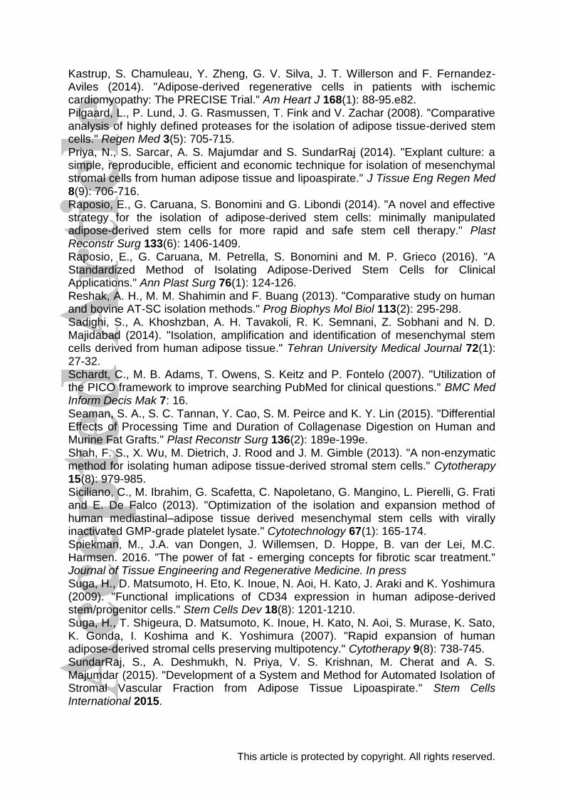
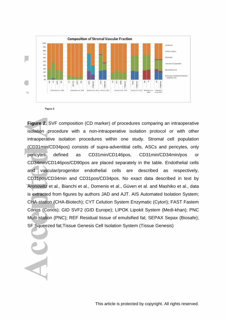
Figure 2. SVF composition (CD marker) of procedures comparing an intraoperative
isolation procedure with a non-intraoperative isolation protocol or with other
intraoperative isolation procedures within one study. Stromal cell population
(CD31min/CD34pos) consists of supra-adventitial cells, ASCs and pericytes, only
pericytes defined as CD31min/CD146pos, CD31min/CD34min/pos or
CD34min/CD146pos/CD90pos are placed separately in the table. Endothelial cells
and vascular/progenitor endothelial cells are described as respectively,
CD31pos/CD34min and CD31pos/CD34pos. No exact data described in text by
Aronowitz et al., Bianchi et al., Domenis et al., Güven et al. and Mashiko et al., data
is extracted from figures by authors JAD and AJT. AIS Automated Isolation System;
CHA-station (CHA-Biotech); CYT Celution System Enzymatic (Cytori); FAST Fastem
Corios (Corios); GID SVF2 (GID Europe); LIPOK Lipokit System (Medi-khan); PNC
Multi station (PNC); REF Residual tissue of emulsified fat; SEPAX Sepax (Biosafe);
SF Squeezed fat;Tissue Genesis Cell Isolation System (Tissue Genesis)
This article is protected by copyright. All rights reserved.
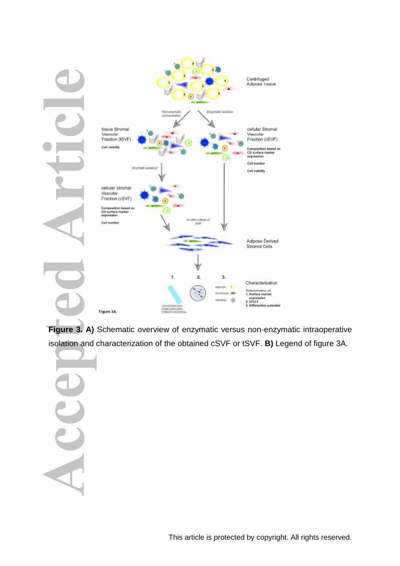
Figure 3. A) Schematic overview of enzymatic versus non-enzymatic intraoperative
isolation and characterization of the obtained cSVF or tSVF. B) Legend of figure 3A.
This article is protected by copyright. All rights reserved.
Table 1. Inclusion and exclusion criteria
Inclusion criteria Exclusion criteria
Clinical trials Case reports
Comparative studies Case series
Full text available Reviews
All languages Letters to editor
Human studies Non-comparative studies
No full text available
≥2 different types of SVF isolation procedures
Processing methods for fat grafting Protocols using centrifugation or RBC lysis buffer only
1 SVF isolation procedure compared with control group
Intraoperative procedures
Mesenchymal cells derived from other source than adipose tissue Blood saline fraction used instead of adipose fraction of the lipoaspirate
Laboratory based enzyme protocols as experimental group No outcome of interest: SVF composition (CD markers), cell yield, viability of SVF
This article is protected by copyright. All rights reserved.
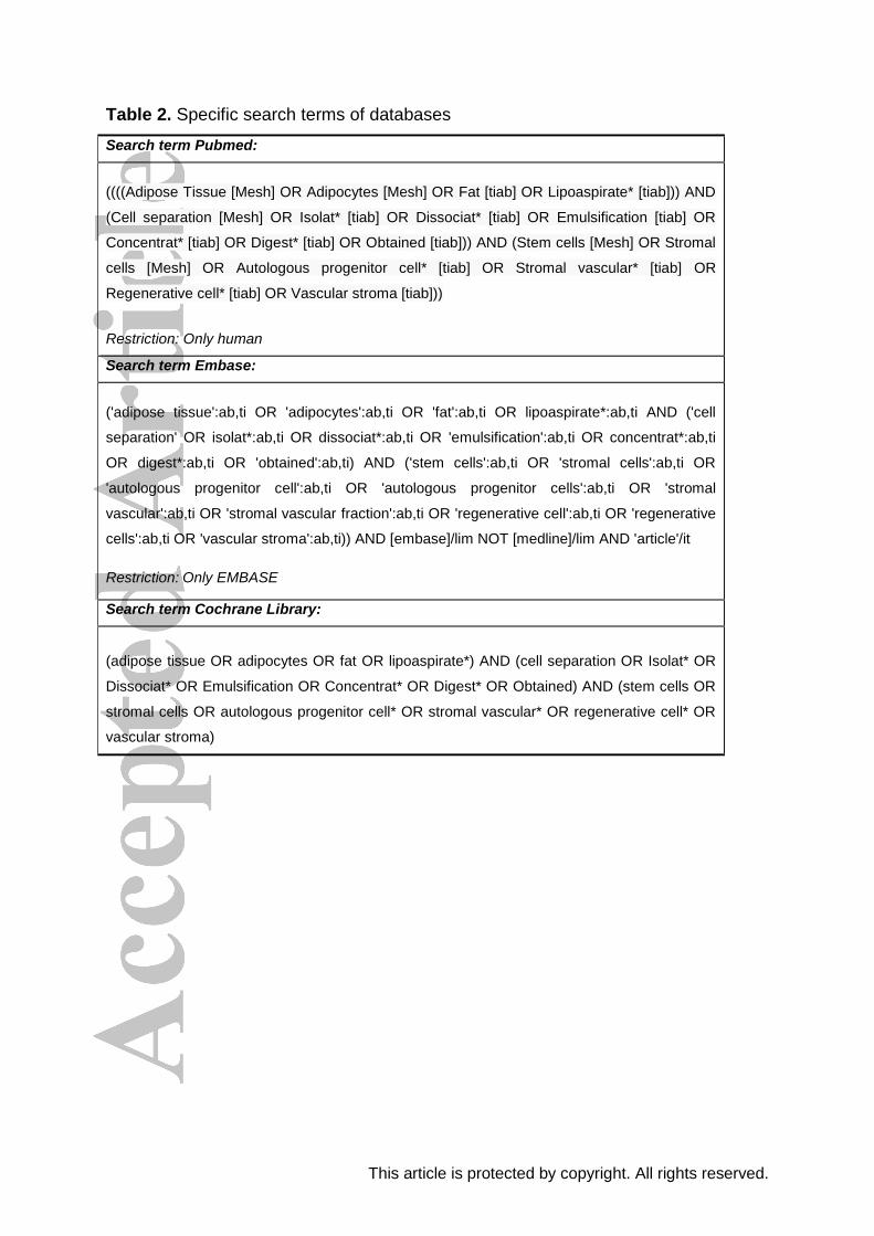
Table 2. Specific search terms of databases
Search term Pubmed:
((((Adipose Tissue [Mesh] OR Adipocytes [Mesh] OR Fat [tiab] OR Lipoaspirate* [tiab])) AND
(Cell separation [Mesh] OR Isolat* [tiab] OR Dissociat* [tiab] OR Emulsification [tiab] OR
Concentrat* [tiab] OR Digest* [tiab] OR Obtained [tiab])) AND (Stem cells [Mesh] OR Stromal
cells [Mesh] OR Autologous progenitor cell* [tiab] OR Stromal vascular* [tiab] OR
Regenerative cell* [tiab] OR Vascular stroma [tiab]))
Restriction: Only human
Search term Embase:
('adipose tissue':ab,ti OR 'adipocytes':ab,ti OR 'fat':ab,ti OR lipoaspirate*:ab,ti AND ('cell
separation' OR isolat*:ab,ti OR dissociat*:ab,ti OR 'emulsification':ab,ti OR concentrat*:ab,ti
OR digest*:ab,ti OR 'obtained':ab,ti) AND ('stem cells':ab,ti OR 'stromal cells':ab,ti OR
'autologous progenitor cell':ab,ti OR 'autologous progenitor cells':ab,ti OR 'stromal
vascular':ab,ti OR 'stromal vascular fraction':ab,ti OR 'regenerative cell':ab,ti OR 'regenerative
cells':ab,ti OR 'vascular stroma':ab,ti)) AND [embase]/lim NOT [medline]/lim AND 'article'/it
Restriction: Only EMBASE
Search term Cochrane Library:
(adipose tissue OR adipocytes OR fat OR lipoaspirate*) AND (cell separation OR Isolat* OR
Dissociat* OR Emulsification OR Concentrat* OR Digest* OR Obtained) AND (stem cells OR
stromal cells OR autologous progenitor cell* OR stromal vascular* OR regenerative cell* OR
vascular stroma)
This article is protected by copyright. All rights reserved.
Table 3A. Duration, costs and procedure characteristics of intraoperative isolation procedures focusing on cSVF
Name Author Enzymatic/ Non-enzymatic (E/N)
Automatic/ Manual/ Semi (A/M/S)
Open/ closed (O/C)
Isolation details Time (min)
Disposable (D)/ reusable (R) cost (Dollar)
Volume processed (ml)
Capacity (ml)
End volume (ml)
Maximum volume processed /maximum end volume
AIS SundarRaj et al. 2015
E A C Tissue digestion, heating and agitation, three-stage filter system (100 micron, 35 micron, 5 micron porosity)
- 133 - - 500 10.8 [4-20]
-
CHA Aronowitz et al. 2013
E S C Fat bag, adapter, centrifugation, shaking incubator and tissue digestion, cell strainer, cell counter
Collagenase 88+/23
D710 80-180 180 - -
CYT
Aronowitz et al. 2013
E A C Washing (lactated Ringer), tissue digestion and agitation, centrifugation
Celase/ Reagent A
90 +/16
D1950 100-180 360 - -
Aronowitz et al. 2016
89.4 [85-93]
D2400 per 120-360 ml
126 [90-150]
360 5 [5] 30
Domenis et al. 2015
60 D 250 - Pellet -
Lin et al. 2008 90 - - - Pellet -
GID-SVF2
Aronowitz et al. 2016
E M C Disposable canister for harvesting, filtration, separation and concentration
GIDzyme-50 71.4 [68-75]
D1000 per 20-120 ml
53.2 [32-88]
120 7.2 [6-9]
13.3
LIPOK
Domenis et al. 2015
E S C 1200 xg centrifugation (with a weight-mesh filter piston), celltibator
Liberase (collagenase mixture)
- - - - - -
Aronowitz et al. 2013
Collagenase 111+/-18
D530 60-100 100 - -
Aronowitz et al. 2016
Time Machine accelerator
120.8 [99-149]
D450 per 100 ml
71.4 [40–97]
400 20 [15-25]
3.9
PNC
Aronowitz et al. 2013
E M O Centrifugation, shaking incubator, clean bench, HEPA filter, UV-lamp
Collagenase
115+/-13
D460 100-150 400 - -
Aronowitz et al. 2016
65.4 [59-74]
D250 per 100 ml
105.6 [68-150]
800 12.2 [10.5-15]
10
SEPAX Güven et al. 2012
E A C Tissue digestion, priming and straining, centrifugation, washing
0.15% NB6 GMP Grade Collagenase
90-120
- 40-400 - Pellet -
TGCIS Doi et al. 2012 E A C Tissue digestion, centrifugation, washing, 700 xg centrifugation
0.075% collagenase
65 D 20-60 - Pellet -
This article is protected by copyright. All rights reserved.
AIS Automated Isolation System; CHA-station (CHA-Biotech); CYT Celution System Enzymatic (Cytori); GID SVF2 (GID Europe);
LIPOK Lipokit System (Medi-khan); PNC Multi station (PNC); SEPAX Sepax (Biosafe); TGCIS Tissue Genesis Cell Isolation
System (Tissue Genesis)
Table 3B. Duration, costs and procedure characteristics of intraoperative concentration procedures focusing on tSVF
Name Author Enzymatic/ Non-enzymatic (E/N)
Automatic/ Manual/ Semi (A/M/S)
Open/ closed (O/C)
Isolation details Time (min)
Disposable (D)/ reusable (R) cost (Dollar)
Volume processed (ml)
Capacity (ml)
End volume (ml)
Maximum volume processed /maximum end volume
FAT Van Dongen et al. 2016
N M O 3000 rpm (radius 9.5 cm) centrifugation, shuffling through a 1.4 mm hole connector, 3000 rpm (radius 9.5 cm) centrifugation
n/a 8-10 R 10 10 0.96 [0.75 - 1.75]
10.4
FAST Domenis et al. 2015
N M - Filterbag (120 micron filter), 400 xg centrifugation
n/a - - - - 10 -
FEF Mashiko et al. 2016
N M O
1200 xg centrifugation, shuffling through a connector with three small holes 30 times, 1200 xg centrifugation, fluid of decanting
filtration (500-m pore size) used
n/a - - - - - 9.9+/-2.0
LIPOG Bianchi et al. 2013
N M C
Filtering, decantation, stainless steel marbles to mix layers (oil, adipose tissue, blood, saline), washing, decantation, reversing devices, filtering
n/a 20 D 40-130 130 60-100 1.3
NANO Tonnard et al. 2013
N M O Shufling adipose tissue through a female-to-female luerlok 30 times, filtering
n/a - - - - - -
REF Mashiko et al. 2016
N M O
1200 xg centrifugation, shuffling through a connector with three small holes 30 times, 1200 xg centrifugation, residual tissue of
decanting filtration (500-m pore size) used
n/a - - - - - 2.5+/-0.2
SF Mashiko et al. 2016
N M O 1200 xg centrifugation, squeeze using automated slicer, 1200 xg centrifugation
n/a - - - - - 2.1+/-0.2
SHUF5 Osinga et al. 2015
N M O Shuffling lipoaspirate through female-to-female luerlok 30 times
n/a 5 sec.
- 10 - - -
SHUF30 Osinga et al. 2015
N M O Shuffling lipoaspirate through female-to-female luerlok 30 times
n/a 30 sec.
- 10 - - -
STCELL Milan et al. N M C 1000 xg centrifugation n/a - - 400 500 Pellet -
This article is protected by copyright. All rights reserved.
FAT Fractionation of Adipose Tissue procedure; FAST Fastem Corios (Corios); FEF Filtrated fluid of emulsified fat; LIPOG
Lipogems (Lipogems); NANO Nanofat procedure; REF Residual tissue of emulsified fat; SF Squeezed fat; SHUF5 Shuffling 5
times; SHUF30 Shuffling 30 times
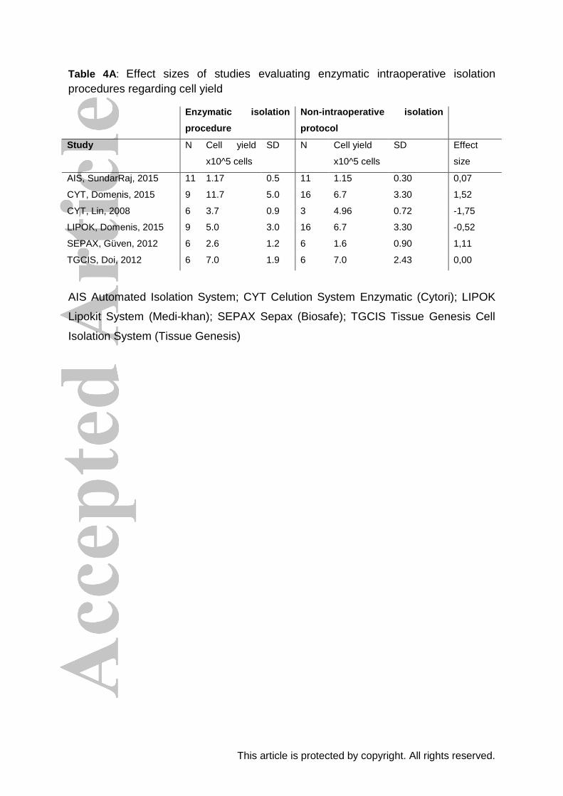
This article is protected by copyright. All rights reserved.
Table 4A: Effect sizes of studies evaluating enzymatic intraoperative isolation
procedures regarding cell yield
Enzymatic isolation
procedure
Non-intraoperative isolation
protocol
Study N Cell yield
x10^5 cells
SD N Cell yield
x10^5 cells
SD Effect
size
AIS, SundarRaj, 2015 11 1.17 0.5 11 1.15 0.30 0,07
CYT, Domenis, 2015 9 11.7 5.0 16 6.7 3.30 1,52
CYT, Lin, 2008 6 3.7 0.9 3 4.96 0.72 -1,75
LIPOK, Domenis, 2015 9 5.0 3.0 16 6.7 3.30 -0,52
SEPAX, Güven, 2012 6 2.6 1.2 6 1.6 0.90 1,11
TGCIS, Doi, 2012 6 7.0 1.9 6 7.0 2.43 0,00
AIS Automated Isolation System; CYT Celution System Enzymatic (Cytori); LIPOK
Lipokit System (Medi-khan); SEPAX Sepax (Biosafe); TGCIS Tissue Genesis Cell
Isolation System (Tissue Genesis)
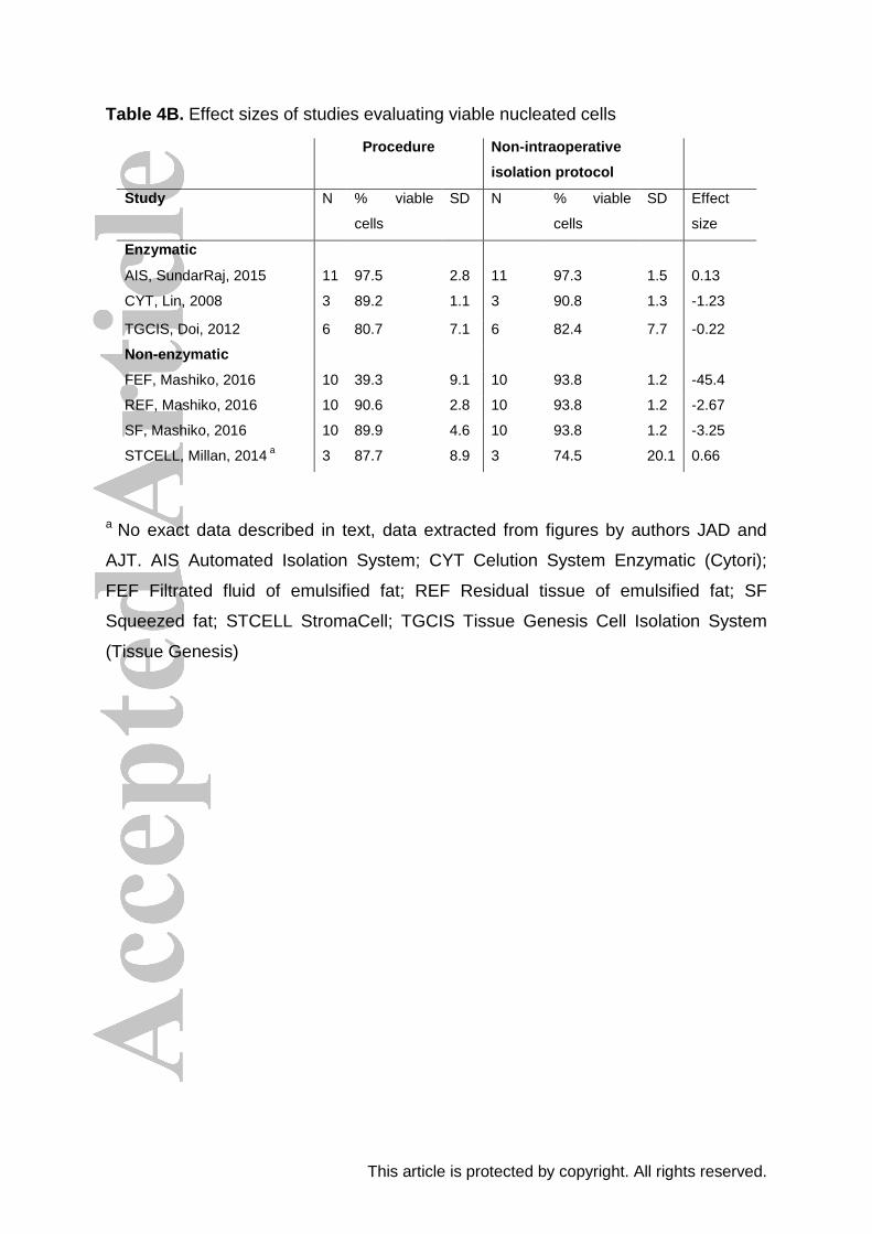
This article is protected by copyright. All rights reserved.
Table 4B. Effect sizes of studies evaluating viable nucleated cells
a No exact data described in text, data extracted from figures by authors JAD and
AJT. AIS Automated Isolation System; CYT Celution System Enzymatic (Cytori);
FEF Filtrated fluid of emulsified fat; REF Residual tissue of emulsified fat; SF
Squeezed fat; STCELL StromaCell; TGCIS Tissue Genesis Cell Isolation System
(Tissue Genesis)
Procedure Non-intraoperative
isolation protocol
Study N % viable
cells
SD N % viable
cells
SD Effect
size
Enzymatic
AIS, SundarRaj, 2015 11 97.5 2.8 11 97.3 1.5 0.13
CYT, Lin, 2008 3 89.2 1.1 3 90.8 1.3 -1.23
TGCIS, Doi, 2012 6 80.7 7.1 6 82.4 7.7 -0.22
Non-enzymatic
FEF, Mashiko, 2016 10 39.3 9.1 10 93.8 1.2 -45.4
REF, Mashiko, 2016 10 90.6 2.8 10 93.8 1.2 -2.67
SF, Mashiko, 2016 10 89.9 4.6 10 93.8 1.2 -3.25
STCELL, Millan, 2014 a 3 87.7 8.9 3 74.5 20.1 0.66
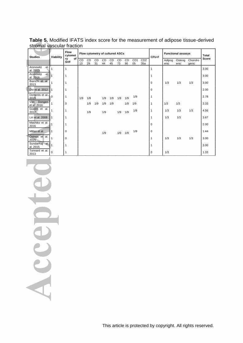
This article is protected by copyright. All rights reserved.
Table 5. Modified IFATS index score for the measurement of adipose tissue-derived stromal vascular fraction
Studies Viability
Flow cytometry of SVF
Flow cytometry of cultured ASCs
CFU-F
Functional assasys Total Score
CD13
CD29
CD31
CD44
CD45
CD73
CD90
CD105
CD235a
Adipogenic
Osteogenic
Chondrogenic
Aronowitz et al. 2013
1 1
1
3.00
Aronowitz et al. 2016
1 1 1 3.00
Bianchi et al. 2013
1 1
0 1/3 1/3 1/3 3.00
Doi et al. 2012 1 1
0
2.00
Domenis et al. 2015
0 1 1/9
1/9
1/9
1/9
1/9
1/9
1/9
1
2.78
Van Dongen et al. 2016
1 0 1/9 1/9 1/9 1/9 1/9 1/9 1 1/3 1/3 3.33
Güven et al. 2012
1 1
1/9
1/9
1/9
1/9
1/9
1 1/3 1/3 1/3 4.56
Lin et al. 2008 1 1
1 1/3 1/3
3.67
Mashiko et al. 2016
1 1 0 2.00
Millan et al. 1 0
1/9
1/9
1/9
1/9
0
1.44
Osinga et al. 2015
1 0
1 1/3 1/3 1/3 3.00
SundarRaj et al. 2015
1 1
1
3.00
Tonnard et al. 2013
0 1
0 1/3
1.33