introduction to the microscope the first light microscopes around 1590 zaccharias and hans janssen...
TRANSCRIPT

Introduction to the Microscope
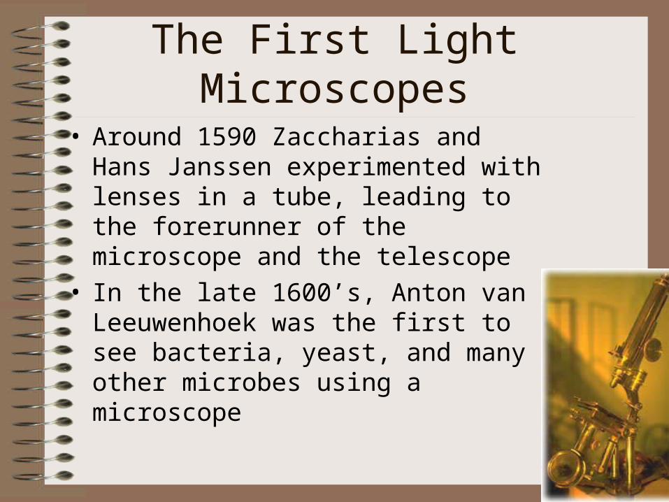
The First Light Microscopes
• Around 1590 Zaccharias and Hans Janssen experimented with lenses in a tube, leading to the forerunner of the microscope and the telescope
• In the late 1600’s, Anton van Leeuwenhoek was the first to see bacteria, yeast, and many other microbes using a microscope
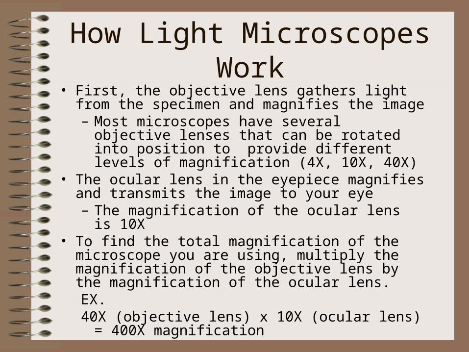
How Light Microscopes Work• First, the objective lens gathers light from the
specimen and magnifies the image– Most microscopes have several objective lenses that
can be rotated into position to provide different levels of magnification (4X, 10X, 40X)
• The ocular lens in the eyepiece magnifies and transmits the image to your eye– The magnification of the ocular lens is 10X
• To find the total magnification of the microscope you are using, multiply the magnification of the objective lens by the magnification of the ocular lens. EX. 40X (objective lens) x 10X (ocular lens) = 400X
magnification
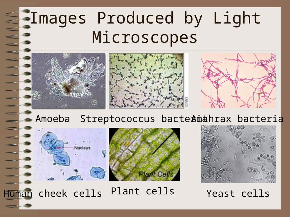
Images Produced by Light Microscopes
Amoeba Streptococcus bacteria Anthrax bacteria
Human cheek cells Plant cells Yeast cells
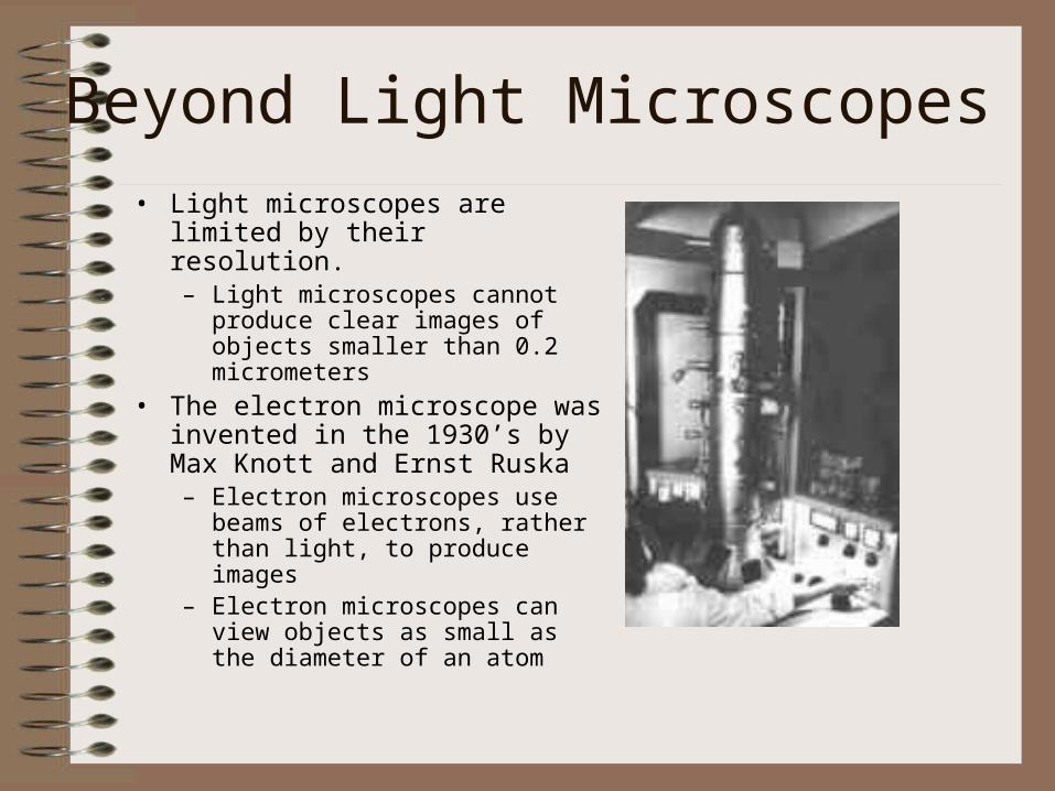
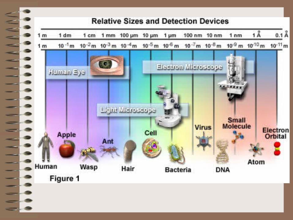
Beyond Light Microscopes
• Light microscopes are limited by their resolution.– Light microscopes cannot produce
clear images of objects smaller than 0.2 micrometers
• The electron microscope was invented in the 1930’s by Max Knott and Ernst Ruska– Electron microscopes use beams of
electrons, rather than light, to produce images
– Electron microscopes can view objects as small as the diameter of an atom
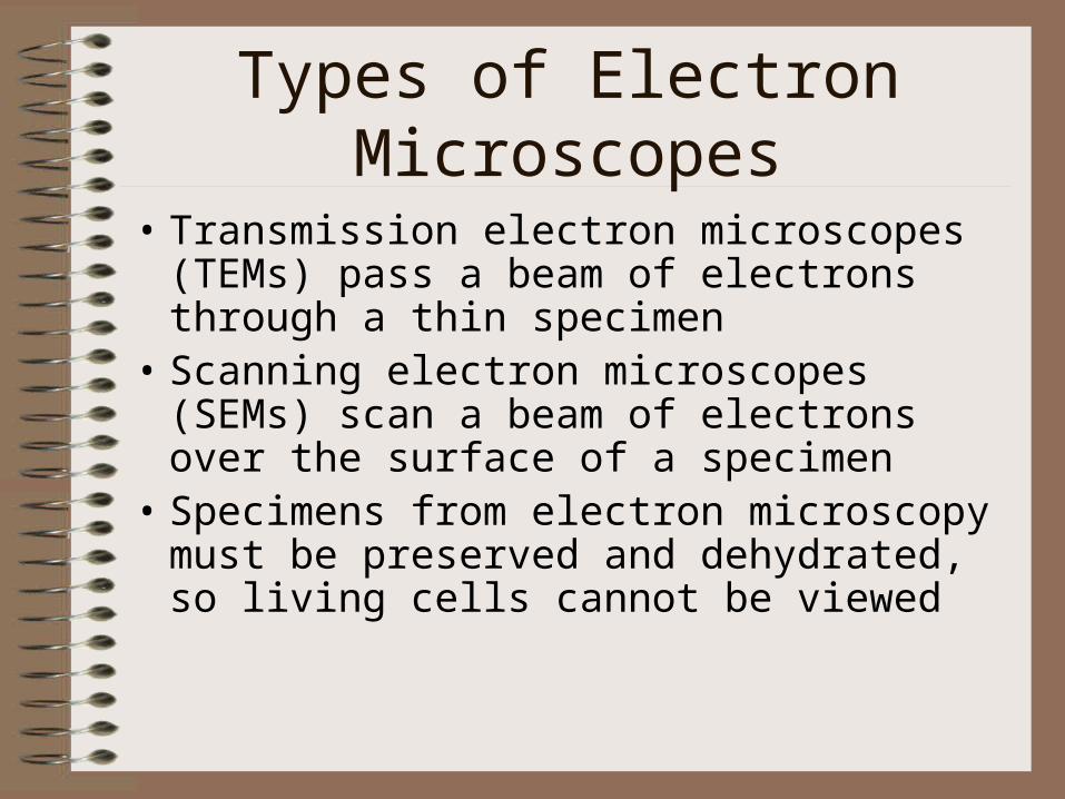
Types of Electron Microscopes
• Transmission electron microscopes (TEMs) pass a beam of electrons through a thin specimen
• Scanning electron microscopes (SEMs) scan a beam of electrons over the surface of a specimen
• Specimens from electron microscopy must be preserved and dehydrated, so living cells cannot be viewed

Images Produced by Electron Microscopes
Cyanobacteria (TEM) Lactobacillus
(SEM)Campylobacter
(SEM)Deinococcus
(SEM)
House ant Avian influenza virus
Human eyelash Yeast

• Always carry with 2 hands
• Only use lens paper for cleaning
• Do not force knobs
• Always store covered
• Keep objects clear of desk and cords

Eyepiece
Body Tube
Revolving NosepieceArm
Objective Lens
StageStage Clips
Coarse Focus
Fine Focus
Base
Diaphragm
Light
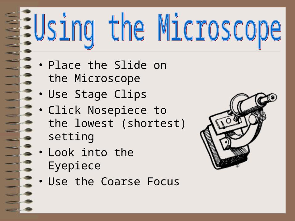
• Place the Slide on the Microscope
• Use Stage Clips • Click Nosepiece to the lowest
(shortest) setting• Look into the Eyepiece• Use the Coarse Focus
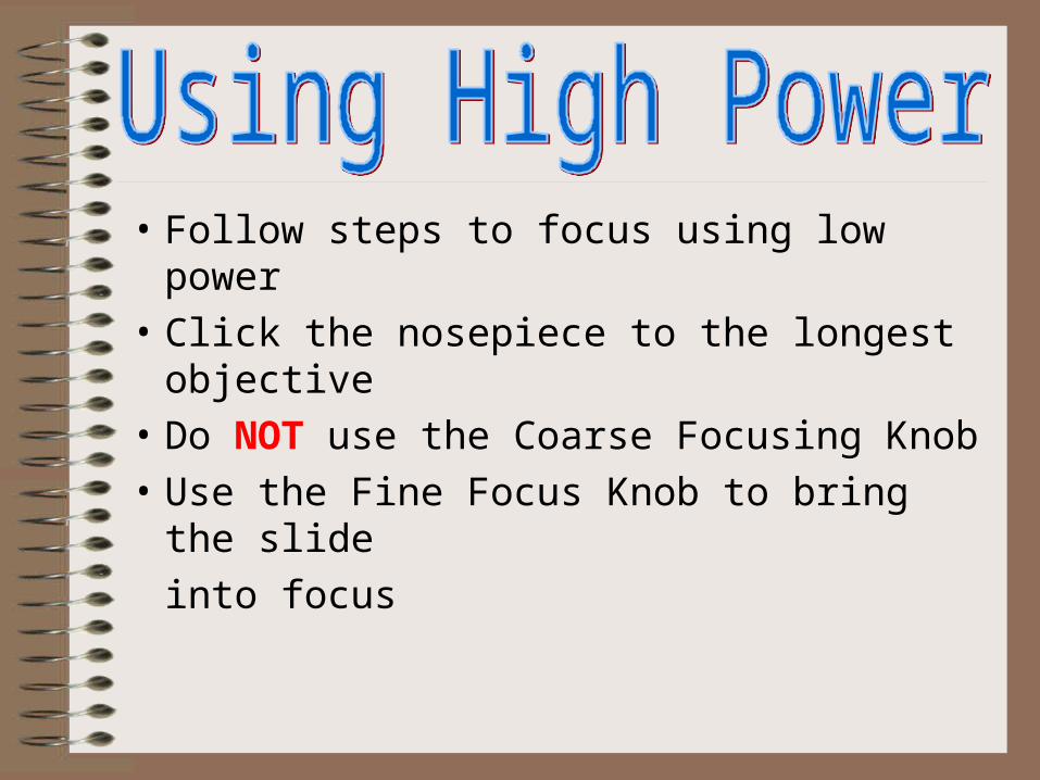
• Follow steps to focus using low power
• Click the nosepiece to the longest objective
• Do NOT use the Coarse Focusing Knob
• Use the Fine Focus Knob to bring the slide
into focus