introduction to fungal physiology - abertay university · the cell envelope in yeasts and fungi is...
TRANSCRIPT

Introduction to fungal physiology
Graeme M. Walker and Nia A. White
This is the accepted manuscript of the book chapter:Walker, G.M. and White, N.A. 2017. Introduction to fungal physiology. In: K. Kavangh, ed. Fungi: biology and applications. John Wiley & Sons.
which has been published in final form at doi: http://dx.doi.org/10.1002/9781119374312.ch1
This chapter may be used for non-commercial purposes in accordance with the Wiley Terms and Conditions for Self-Archiving

1
CHAPTER 1
INTRODUCTION TO FUNGAL PHYSIOLOGY
Graeme M. Walker & Nia A. White 1.1. Introduction
Fungal physiology refers to the nutrition, metabolism, growth, reproduction
and death of fungal cells. It also generally relates to interaction of fungi with their
biotic and abiotic surroundings, including cellular responses to environmental stress.
The physiology of fungal cells impacts significantly on the environment, industrial
processes and human health. In relation to ecological aspects, the biogeochemical
cycling of carbon in Nature would not be possible without the participation of fungi
acting as primary decomposers of organic material. Furthermore, in agricultural
operations fungi play important roles as mutualistic symbionts, pathogens and
saprophytes, where they mobilize nutrients and affect the physico-chemical
environment, or can be exploited as agents of biocontrol or as bio-fertilizers. Fungal
metabolism is also responsible for the detoxification of organic pollutants and for
bioremediating heavy metals and other recalcitrant chemicals in the environment
(including waste- and ground-waters). The production of many economically
important industrial commodities relies on the exploitation of yeast and fungal
metabolism and these include such diverse products as whole foods, food additives,
fermented beverages, antibiotics, probiotics, pigments, pharmaceuticals, biofuels,
enzymes, vitamins, organic and fatty acids and sterols. More negatively, fungi can
cause considerable disease, spoilage and decay of important artefacts, commodities
and materials, buildings and of course food supplies. In terms of human health, some
yeasts and fungi represent major opportunistic life-threatening pathogens, whilst
others are life-savers as they provide antimicrobial and chemotherapeutic agents. In

2
modern biotechnology, several yeast species are being exploited as hosts for the
expression of human therapeutic proteins following recombinant DNA and gene
editing technologies (see Chapter 9). Recently, the application of gene editing using
CRISPR/Cas is leading to a revolution in fungal genetic engineering (see Chapter 2).
Furthermore, an international synthetic biology research consortium, called Sc-2.0,
has embarked on the construction of a completely synthetic version of S. cerevisiae.
This would represent the world’s first fully synthetic eukaryotic genome! In addition
to the direct industrial exploitation of yeasts and fungi, it is important to note that
these organisms, most notably the yeast Saccharomyces cerevisiae, play increasingly
significant roles as model eukaryotic cells in furthering our fundamental knowledge
of biological and biomedical science. This is especially the case now that numerous
fungal genomes have been completely sequenced and the information gleaned from
fungal genomics and proteomics is providing valuable insight into human genetics
and heritable disorders. However, knowledge of cell physiology is essential if the
functions of many of the currently unknown fungal genes, including “synthetic” ones,
are to be fully elucidated.
It is apparent, therefore, that fungi are important organisms for human society,
health and well-being and that studies of fungal physiology are very pertinent to our
understanding, control and exploitation of this group of microorganisms. This Chapter
describes some basic aspects of fungal cell physiology, focusing primarily on
nutrition, growth and metabolism in unicellular yeasts and filamentous fungi.
1.2. Morphology of yeasts and fungi
There are a diversity of yeast and fungal cellular morphologies. Most higher
fungi are filamentous, yeasts grow as unicells, and some primitive fungi such as the

3
chytridomycota grow as individual rounded cells or dichotomous branched chains of
cells with root like rhizoids for attachment to a nutrient resource. Here we will
consider the most common growth forms, the filamentous fungi and unicellular
yeasts.
1.2.1. Filamentous fungi
The gross morphologies of macrofungi and microfungi are varied and often
apparent throughout the environment (see Plate 1.1). For example, we can easily
recognise a variety of mushrooms and toadstools, the sexual fruiting bodies of certain
macro fungi (the higher fungi Asomycota and Basidiomycota and related forms),
during a walk through pasture or woodland. Microfungi (the moulds) are also diverse
and are often observed on decaying foods and detritus, whereas many, including the
coloured rusts, smuts and mildews, are common plant pathogens. Closer inspection of
these visible structures, however, reveals that all are composed of aggregated long,
branching threads termed hyphae (singular: hypha), organised to support spores for
reproduction and dissemination. The hyphae of these aerial structures extend and
branch within the supporting substratum as a network, termed a mycelium, from
which the apically growing hyphae seek out, exploit and translocate available
nutrients. Apically growing hyphae usually have a relatively constant diameter
ranging from 1- 30m or more, depending on fungal species and growth conditions.
Filamentous fungi may be cultivated within the laboratory on a variety of different
liquid or solid media. On agar, the radially expanding colonial growth form of the
fungal mycelium is most evident, extending from an inoculum, on, within and
sometimes above the substrate, forming a near spherical 3-dimensional colony. This

4
radiating, circular pattern is also visible during the growth of fairy ring fungi in
grassland and as ringworm infections of the skin (Plate 1.1).
The hyphae of individual fungi may (theoretically) extend endlessly via
apical growth, provided they are supported with appropriate nutrients and other
environmental conditions. Eucarpic fungi are therefore spatially and temporally
indeterminate organisms, and unlike animal, plant and other microbial individuals,
have no predetermined maximum size or age. The mycelium is not, however, simply a
homogeneously extending entity, but displays considerable developmental plasticity.
Different interconnected regions of the fungal mycelium may grow, branch,
anastomose (fuse), age, die, sporulate, and display varying physiological and
biochemical activities at different times or even simultaneously, depending on local
micro-environmental conditions. Thus, colonies growing on relatively homogeneous
media may be pigmented, exhibit different morphological sectors, produce aerial
structures, grow as fast-effuse or slow-dense forms, and even exhibit rhythmic growth
As well as reproductive structures and substrate mycelium, certain higher fungi, most
notably the basidiomycetes, when growing within an environment where nutrients are
distributed heterogeneously, can differentiate into long string-like structures called
rhizomorphs or cords. These linear organs have evolved to rapidly explore for,
connect and translocate water and nutrients between patches of resource (e.g. pieces
of fallen timber on the forest floor or from tree root to tree root). Accordingly, many,
particularly mature rhizomorphs, contain internal vessel hyphae which possess a wide
diameter, forming a channel running along the organ. The peripheral hyphae are often
closely packed and melanized for insulation (Plate 1.1)

5
Filamentous fungi and yeasts are simply different styles of fungal growth
suitable for occupation of different habitats and produced by differing cell growth
polarities. Many species termed dimorphic fungi can adopt either the hyphal or
unicellular yeast forms according to environmental circumstances. For example,
certain important human and animal pathogens exist as yeast forms mobilised in body
fluids but are able to form hyphae or pseudohyphae for tissue invasion.
1.2.2. Yeasts
Yeasts are unicellular (mostly Ascomycete, Basidiomycete or members of the
Deuteromycete group) fungi that divide asexually by budding or fission and whose
individual cell size can vary widely from 2-3m to 20-50m in length and 1-10m in
width. Saccharomyces cerevisiae, commonly referred to as brewer’s or baker’s yeast,
is generally ellipsoid in shape with a large diameter of 5-10m and a small diameter
of around 5m (Figure 1.1). There is great diversity in cell shapes and modes of
cellular reproduction in the yeasts, as summarised in Tables 1.1.
<FIGURE 1.1 HERE>
The morphology of agar-grown yeasts show great diversity in terms of colour,
texture and geometry (peripheries, contours) of giant colonies. Several yeasts are
pigmented and the following colours may be visualised in surface-grown colonies:
cream (e.g. S. cerevisiae); white (e.g. Geotrichum candidum); black (e.g.
Aureobasidium pullulans); pink (e.g. Phaffia rhodozyma); red (e.g. Rhodotorula
rubra); orange (e.g. Rhodosporidium spp.) and yellow (e.g. Cryptococcus laurentii).
The pigments of some yeasts have biotechnological uses, including astaxanthin from
P. rhodozyma in aquacultural feed supplements for farmed salmon (that are unable to
synthesise these natural pink compounds).
<TABLE 1.1 HERE>

6
1.3. Ultrastructure and function of fungal cells 1.3.1 The fungal cell surface
The cell envelope in yeasts and fungi is the peripheral structure that encases the
cytoplasm and comprises the plasma membrane, the periplasm, the cell wall and
additional extracellular structural components (such as fimbriae and capsules). The
cell wall represents a dynamically forming exoskeleton that protects the fungal
protoplast from the external environment and defines directional growth, cellular
strength, shape and interactive properties (Figure 1.2). In filamentous fungi, cell wall
formation and organisation is intimately bound to the process of apical growth. Thus,
for example in Neurospora crassa, the wall is thin (approx. 50nm) at the apex but
becomes thicker (approx. 125nm) at 250m behind the tip. The plasma membrane
component of the fungal cell envelope is a phospholipid bilayer interspersed with
globular proteins that dictates entry of nutrients and exit of metabolites and represents
a selective barrier for their translocation. Ergosterol is the major sterol found in the
membranes of fungi, in contrast to the cholesterol found in the membranes of animals
and phytosterols in plants. This distinction is exploited during the use of certain
antifungal agents used to treat some fungal infections, and can be used as an assay
tool to quantify fungal growth. The periplasm, or periplasmic space, is the region
external to the plasma membrane and internal to the cell wall. In yeast cells, it
comprises secreted proteins (mannoproteins) and enzymes (such as invertase and acid
phosphatase) that are unable to traverse the cell wall. In filamentous fungi, the cell
membrane and wall may be intimately bound as hyphae are often resistant to
plasmolysis. <INSERT FIGURE 1.2>
Fungal cell surface topological features can be visualised using scanning electron
microscopy (SEM) and nanometre resolution achieved using atomic force microscopy

7
(AFM). The latter is beneficial as it can be employed with unfixed, living cells and
avoids potentially misleading artefacts that may arise when preparing cells for
electron microscopy.
Ultrastructural analysis of fungal cell walls reveals a thick, complex fibrillar
network. The cell walls of filamentous fungi are mainly composed of different
polysaccharides according to taxonomic group. For example, they may contain either
chitin, glucans, mannoproteins, chitosan, polyglucuronic acid or cellulose (absent
from true fungi), together with smaller quantities of proteins and glycoproteins (Table
1.2). Generally, the semi-crystalline microfibrillar components are organised in a
network mainly in the central cell wall region and are embedded within an amorphous
matrix. Bonding occurs between certain components behind the extending hyphal tip,
thereby strengthening the entire wall structure. The processes of endocytosis and
exocytosis occur around apical and subapical regions and serve to both shape hyphal
growth and interactions with the environment (Figure 1.2). There is evidence to
suggest that the cell wall is a dynamic structure where considerable quantitative and
qualitative differences occur not only between different fungal species, but also
between different morphological forms of the same species and even in response to
environmental stress. For example, a class of hydrophobic proteins called
hydrophobins are localised within the aerial growth or appresoria (terminal swellings
involved in infection) of certain fungi, whereas pigmented melanins are often found
within some fungal cell walls to insulate against biotic and abiotic stresses.
<INSERT TABLE 1.2 HERE>
The hyphae of higher fungi extend via tip growth followed by cross-wall
formation or septation, whereas the lower fungi remain aseptate (except when
segregating spores or in damaged colony regions). Septa may offer some structural

8
support to hyphae. Significantly, septa serve to compartmentalise hyphae but are
typically perforated, thereby permitting passage and communication of cytoplasm or
even protoplasm between compartments. However, septal pores can become blocked
by Woronin bodies or other materials. This aids morphological and biochemical
differentiation and serves to seal-off stressed or damaged hyphae from undamaged
colony regions. Again, different pore types are representative of different taxonomic
groups and species (Table 1.2).
In yeasts, the cell wall provides stability and protection to the cells and its
structure comprises polysaccharides (predominantly -glucans for rigidity), proteins
(mainly mannoproteins on the outermost layer for determining porosity), together
with some lipid, chitin (eg. in bud scar tissue) and inorganic phosphate material.
Figure 1.3 shows the composition and structure of the S. cerevisiae cell wall. Hyphal
cell walls generally contain fewer mannans than yeast cell forms, and such changes in
composition are even observed during the transition from unicellular to mycelial
growth of dimorphic fungi.
Chitin is also found in yeast cell walls and is a major constituent of bud scars
(Figure 1.1). These are remnants of previous budding events found on the surface of
mother cells following birth of daughter cells (buds). The chitin-rich bud scars of
yeast cells can be stained with fluorescent dyes (e.g. calcoflour white) and this can
provide useful information regarding cellular age, since the number of scars
represents the number of completed cell division cycles. Outside the cell wall in
fungi, several extramural layers may exist including fimbriae and capsules. Fungal
fimbriae are long, protein-containing protrusions appearing from the cell wall of
certain basidiomycetous and ascomycetous fungi that are involved in cell-cell
conjugation. Capsules are extracellular polysaccharide-containing structures found in
9
basidiomycetous fungi that are involved in stress protection. In Cryptococcus
neoformans (the pathogenic yeast state of Filobasidiella neoformans) the capsule may
determine virulence properties and evasion from macrophages. One extrahyphal
substance, the polymer pullulan, is produced commercially from Aureobasidium
pullulans, and is used in the production of oral hygiene products <FIGURE 1.3
HERE>
1.3.2. Subcellular architecture and organelle function
Transmission electron microscopy of ultrathin sections of fungal cells reveals
intracellular fine structure (Figures 1.2 & 1.4). Sub-cellular compartments
(organelles) are bathed in an aqueous cytoplasm containing soluble proteins and other
macromolecules together with low-molecular weight metabolites.
However, the hyphae of central (and therefore older) colony regions of
filamentous fungi may become devoid of protoplasm and organelles, as protoplasmic
components are driven forward or are recycled, to support the growth of actively
growing hyphal-tips. Cytoplasmic components additionally comprise microbodies,
ribosomes, proteasomes, lipid particles and a cytoskeletal network. The latter confers
structural stability to the fungal cytoplasm and consists of microtubules and
microfilaments. The following membrane-bound organelles may be found in a typical
fungal cell: nucleus, endoplasmic reticulum (ER), mitochondria, Golgi apparatus,
secretory vesicles and vacuoles. Several of these organelles form extended
membranous systems. For example, the ER is contiguous with the nuclear membrane
and secretion of fungal proteins involves inter-membrane trafficking in which the ER,
Golgi apparatus, plasma membrane and vesicles all participate. The physiological

10
function of the various fungal cell organelles is summarised in Table 1.3. <FIGURE
1.4 AND TABLE 1.3 HERE>
The nucleus is the structure that defines the eukaryotic nature of fungal cells.
It is bound by a double-membrane and encases the chromosomes in a nucleoplasm.
Most yeast and fungi are haploid (singular copies of each chromosome), although
some (e.g. S. cerevisiae) may alternate between haploidy and diploidy. Many
industrial strains of S. cerevisiae exhibit aneuploidy (odd numbers of chromosomes)
or are polyploid (multiple chromosome copies). Chromosomes comprise DNA-
protein structures that replicate and segregate to newly-divided cells or hyphal
compartments at mitosis. This, of course, ensures that genetic material is passed onto
daughter cells or septated compartments at cell division. Yeasts usually contain a
single nucleus per cell. However, the hyphal compartments of filamentous fungi may
contain one or more nuclei. Monokaryotic basidiomycetes possess one nucleus per
compartment whereas dikaryons and heterokaryons possess two or more genetically
distinct haploid nuclei. The maintenance of multiple nuclei within individual hyphal
compartments allows fungi to take advantage of both haploid and diploid life-styles.
This is discussed further in Chapter 2.
In filamentous fungi, a phase-dark near-spherical region, which also stains
with iron-haemotoxylin, is evident by light microscopy at the apex during hyphal tip
growth. The region is termed the Spitzenkörper, the apical vesicle cluster or centre or
apical body, and it consists of masses of small membrane-bound vesicles around a
vesicle-free core with emergent microfilaments and microtubules (Figure 1.2). The
Spitzenkörper contains differently sized vesicles derived from Golgi bodies, either
large vesicles or microvesicles (chitosomes), with varying composition. It orientates
to the side as the direction of tip growth changes, and disappears when growth ceases.
11
This vesicle supply centre is involved in wall extension and hence tip growth,
branching, clamp connection formation (in Basidiomycetes) and germ tube formation.
1.4. Fungal nutrition and cellular biosyntheses 1.4.1 Chemical requirements for growth
Yeasts and fungi have relatively simple nutritional needs and most species
would be able to survive quite well in aerobic conditions if supplied with glucose,
ammonium salts, inorganic ions and a few growth factors. Exceptions to this would
include for example, obligate symbionts such as the Vesicular-Arbuscular
Mycorrhizal fungi (VAM) which require growth of a plant partner for cultivation.
Macronutrients, supplied at millimolar concentrations, comprise sources of carbon,
nitrogen, oxygen, sulphur, phosphorus, potassium and magnesium; and
micronutrients, supplied at micromolar concentrations, comprise trace elements like
calcium, copper, iron, manganese and zinc would be required for fungal cell growth
(Table 1.4). Some fungi are oligotrophic, apparently growing with very limited
nutrient supply, surviving by scavenging minute quantities of volatile organic
compounds from the atmosphere. <INSERT TABLE 1.4 HERE>
Being chemoorganotrophs, fungi need fixed forms of organic compounds for
their carbon and energy supply. Sugars are widely utilised for fungal growth, and can
range from simple hexoses like glucose to polysaccharides like starch and cellulose.
Some fungi can occasionally utilise aromatic hydrocarbons (e.g. lignin by the white-
rot fungi). Table 1.5 outlines the variety of carbon sources which can be utilised by
yeasts and filamentous fungi for growth. <INSERT TABLE 1.5 HERE>
Fungi are non-diazotrophic (cannot fix nitrogen) and need to be supplied with
nitrogenous compounds, either in inorganic form such as ammonium salts, or in
organic form such as amino acids. Ammonium sulphate is a commonly used nitrogen

12
source in fungal growth media since it also provides a source of utilisable sulphur.
Many fungi (but not the yeast S. cerevisiae) can also grow on nitrate, and if able to do
so, may also utilize nitrite. Nitrate reductase, followed by nitrite reductase, are the
enzymes responsible for converting nitrate to ammonia. Most fungi can assimilate
amino acids, amines and amides as nitrogen sources. Most fungi (but not many
yeasts) are also proteolytic and can hydrolyse proteins (via extracellularly secreted
proteases) to liberate utlisable amino acids for growth. Urea utilisation is common in
fungi and some basidiomycotenous yeasts are classed as urease-positive (able to
utilise urea) whilst several ascomycotenous yeasts are urease-negative.
In terms of oxygen requirements, most fungi are aerobes and are often
described as being microaerophilic (preferring an oxygen tension below that of
normal atmospheric). Although yeasts like S. cerevisiae are sometimes referred to as
facultative anaerobes, they cannot actually grow in strictly anaerobic conditions
unless supplied with certain fatty acids and sterols (which they cannot synthesise
without molecular oxygen). In fact, there are thought to be very few yeast species that
are obligately anaerobic. Unsaturated fatty acids (e.g. oleic acid) and sterols (e.g.
ergosterol) are important constituents of the yeast cell membrane, and oxygen is
required for their synthesis and to maintain membrane functional integrity and stress
resistance. For aerobically respiring yeasts and fungi, oxygen is required as the
terminal electron acceptor where it is finally reduced to water in the electron transport
chain. Different fungal species respond to oxygen availability in diverse ways and
Table 1.6. categorises fungi into different groups on this basis. <TABLE 1.6 HERE>
Sulphur sources for fungal growth include sulphate, sulphite, thiosulphate,
methionine and glutathione with inorganic sulphate and the sulphur amino acid

13
methionine being effectively utilised. Virtually all yeasts can synthesize sulphur
amino acids from sulphate, the most oxidized form of inorganic sulphur.
Phosphorus is essential for biosynthesis of fungal nucleic acids, phospholipids,
ATP, glycophosphates and polyphosphates. Hence, the phosphate content of fungi is
considerable (e.g. in yeast cells, this accounts for around 3-5% of dry weight; the
major part of this is in the form of orthophosphate (H2PO4-) which acts as a substrate
and enzyme effector). The fungal vacuole can serve as a storage site for phosphate in
the form of complexed inorganic polyphosphates (also referred to as volutin
granules). Both nitrogen and phosphorus availability may be growth limiting in
Nature. Filamentous fungi have evolved a number of biochemical and morphological
strategies allowing capture of often poorly available phosphorus within the natural
environment. Plants exploit such efficiency during symbioses between their roots and
certain mycorrhizal fungi. The major storage form of phosphorus in plants is phytic
acid (myo-inositol hexa-dihydrogenphosphate) that is poorly utilised by monogastrics
(e.g. humans, pigs, poultry) and fungal (and yeast) phytases have applications in
reducing phytate content of foods and feeds (see Chapter 8).
Concerning requirements for minerals, potassium, magnesium and several
trace elements are necessary for fungal growth. K and Mg are macroelements
required in millimolar concentrations primarily as enzyme cofactors, whereas other
microelements (trace elements) are generally required in the micromolar range. These
include: Mn, Ca Fe, Zn, Cu, Ni, Co, Mo. Table 1.7 summarises the main metals
required for fungal growth. Toxic minerals (eg Ag, As, Ba, Cs, Cd, Hg, Li, Pb)
adversely affect fungal growth generally at concentrations greater than 100M.
<TABLE 1.7 HERE>

14
Fungal growth factors are organic compounds occasionally needed in very low
concentrations for specific enzymatic or structural roles, but not as energy sources.
These include vitamins (e.g. thiamine, biotin), purines, pyrimidines, nucleosides,
nucleotides, amino acids, fatty acids and sterols. For fungi to have a growth factor
requirement, this indicates that cells cannot synthesise the particular factor resulting in
the curtailment of growth without its provision in culture media. Some fungi (e.g.
Aspergillus niger, Penicillium chrysogenum) have very simple nutritional needs and
are able to synthesise their own growth factors from glucose.
1.4.2 Fungal cultivation media
Fungal nutritional requirements are important not only for successful
cultivation in the laboratory but also for the optimisation of industrial fermentation
processes. In the laboratory, it is relatively easy to grow yeasts and fungi on complex
culture media such as malt extract or potato-dextrose agar or broth, which are both
carbon rich and in the acidic pH range. Mushrooms are cultivated on various solid-
substrates depending on provincial availability. Therefore, Agaricus bisporus
(common button mushroom) is grown in the UK, US and France on wheat-straw; the
padi-straw mushroom (Volvariella volvacea) is grown in South-east Asia on damp
rice-straw and in Hong-Kong on cotton waste; and in Japan, the shiitake mushroom
(Lentinus edodes) is cultivated on fresh oak logs (see Chapter 6). In industry, media
for fungal fermentation purposes needs to be optimised with regard to the specific
application and production process. For some industrial processes, growth media may
already be relatively complete in a nutritional sense, such as malt wort or molasses for
brewing or baker’s yeast production, respectively (Table 1.8). However, for other
processes, supplementation of agriculturally-derived substrates like corn steep liquor,
molasses or malt broth with additional nutrients and growth factors may be necessary.

15
For example, for penicillin production by Penicillium spp. the following may
constitute a suitable fermentation medium: sucrose (3 g/L), corn steep liquor (100
g/L), KH2PO4 (1g/L), (NH4)2SO4 (12 g/L), CaCl2.2H2O (0.06 g/L), phenoxyacetic
acid (5.7 g/L) Whereas, other industrial processes such as the growth of Fusarium
graminarium for the production of Quorn™ mycoprotein, requires culture on a
completely defined medium. <TABLE 1.8 HERE>
1.4.3 Nutrient uptake and assimilation
Fungal cells utilise a diverse range of nutrients and employ equally diverse
nutrient acquisition strategies. Fungi are non-motile, saprophytic (and sometimes
parasitic), chemo-organotrophic organisms. They exhibit dynamic interactions with
their nutritional environment that may be exemplified by certain morphological
changes depending on nutrient availability. For example, the filamentous mode of
growth observed at the periphery of certain yeast colonies growing in agar is akin to a
foraging for nutrients as observed in certain eucarpic fungi. Metabolic dynamism is
also evident in yeasts which, although not avid secretors of hydrolytic enzymes like
higher fungi, are nevertheless able to secrete some enzymes to degrade polymers such
as starch (as in amylolytic yeasts like Schwanniomyces spp.).
Several cellular envelope barriers to nutrient uptake by fungal cells exist,
namely: the capsule, the cell wall, the periplasm and the cell membrane. Although not
considered as freely porous structures, fungal cell walls are relatively porous to
molecules up to an average molecular mass of around 300Da, and will generally
retain molecules greater than around 700Da. Typically, fungi absorb only small
soluble nutrients such as monosaccharides and amino acids.
The plasma membrane is the major selectively permeable barrier which
dictates nutrient entry and metabolite exit from the fungal cell. Membrane transport

16
mechanisms are important in fungal physiology since they govern the rates at which
cells metabolise, grow and divide. Fungi possess different modes of passive and
active uptake at the plasma membrane: free diffusion, facilitated diffusion, diffusion
channels and active transport (Table 1.9). Active transport of nutrients such as sugars,
amino acids, nitrate, ammonium, sulphate and phosphate in filamentous fungi
involves spatial separation of the ion pumps mostly behind the apex, whereas the
symport proteins are active close to the tip. Thus, nutrient uptake occurs at the hyphal
tip as it continuously drives into fresh resource, and the mitochondria localised behind
the apex supply ATP to support the ion pump and generate proton motive force.
<TABLE 1.9 HERE>
1.4.4. Overview of fungal biosynthetic pathways
Anabolic pathways are energy-consuming, reductive processes which lead to
the biosynthesis of new cellular material and are mediated by dehydrogenase enzymes
which predominantly use reduced NADP+ as the redox cofactor. NADPH is generated
by the hexose monophosphate pathway (or Warburg-Dickens pathway) which
accompanies glycolysis (see section 1.5a). In S. cerevisiae, up to 20% of total glucose
may be degraded via the hexose monphosphate pathway. This pathway generates
cytosolic NADPH (following the dehydrogenation of glucose 6-phosphate using
glucose 6-phosphate dehydrogenase and NADP+ as hydrogen acceptor) for
biosynthetic reactions leading to the production of fatty acids, amino acids, sugar
alcohols, structural and storage polysaccharides and secondary metabolites. Besides
generating NADPH, the hexose monophosphate pathway also produces ribose sugars
for the synthesis of nucleic acids, RNA and DNA and for nucleotide coenzymes,
NAD, NADP, FAD and FMN. This is summarised as follows:

17
Glucose 6-phosphate + 2NADP+--> Ribulose 5-phosphate + CO2 +NADPH + 2H+
and complete oxidation of glucose 6-phosphate would result in:
Glucose 6-phosphate + 12NADP+ -> 6CO2 + 12NADPH + 12H+ + Pi
Fungal growth on non-carbohydrate substrates as sole carbon sources (e.g.
ethanol, glycerol, succinate and acetate) may lead to gluconeogenesis (conversion of
pyruvate to glucose) and polysaccharide biosynthesis. Gluconeogenesis may be
regarded as a reversal of glycolysis and requires ATP as energy and NADH as
reducing power.
Concerning fungal amino acid biosynthesis, simple nitrogenous compounds
such as ammonium may be assimilated into amino acid families, the carbon skeletons
of which originate from common precursors of intermediary carbon metabolism.
The two main fungal storage carbohydrates are glycogen and trehalose.
Glycogen is similar to starch with α(1->4) glucan linear components and α(1->6)
branches. Trehalose (also known as mycose) is a disaccharide of glucose comprising
an α,α(1->1) glucoside bond between two α-glucose units. Both trehalose and
glycogen are synthesised following the formation of UDP-glucose, catalysed by UDP-
glucose pyrophosphorylase:
UTP + Glucose 1-phosphate -> UDP-glucose + Pyrophosphate
Glycogen is synthesised by glycogen synthase. Glycogen may be metabolised
by glycogen phosphorylase when nutrients become limited under starvation
conditions and this contributes to the maintenance metabolism of cells by furnishing
energy in the form of ATP. In yeast cells, glycogen breakdown is accompanied by
membrane sterol biosynthesis (in the presence of some oxygen) and this is important
for brewing yeast vitality and successful beer fermentations. The other major storage
carbohydrate, trehalose, is synthesized from glucose 6-phosphate and UDP-glucose
by trehalose 6-phosphate synthase and converted to trehalose by a phosphatase. In

18
addition to a storage role, trehalose is an important translocation material in
filamentous forms and is also involved in stress protection in yeasts and fungi,
accumulating when cells are subject to environmental insults such as heat shock or
osmotic stress, or during plant host-fungal parasite interactions. Trehalose acts by
protecting cell membranes against desiccation or thermal damage. Polyols, such as
mannitol derived from fructose phosphate and glycerol from the glycolytic
intermediate dihydroxyacetone phosphate, are also translocated by fungi. In
particular, glycerol is produced as a “compatible solute” in response to osmotic stress
to counteract the loss of intracellular water (see section 1.6.1). Glycerol is also a yeast
fermentation by-product and contributes to the viscosity or mouthfeel of alcoholic
beverages such as beer and wine. 1.4.5. Fungal cell wall growth
The structural polysaccharides in fungal cell walls include mannans, glucans
and chitin and are synthesised from sugar nucleotides substrates formed by
pyrophosphorylase enzymes. For example:
Glucose 1-phosphate + UTP ->UDP-glucose + PPi
Mannose 1-phosphate + GTP -> GDP-mannose + PPi
Glucan synthesis involves plasma membrane-associated glucan synthetases for
assembly of -1,3 linkages and -1,6 branches of cell wall glucan. Chitin (a polymer
of N-acetylglucosamine) is an important fungal cell wall structural component and is
involved in the yeast budding process and in dimorphic transitions from yeast to
filamentous forms. Chitin synthetases catalyze the transfer of N-acetylglucosamine
from UDP-N-acetylglucosamine to a growing chitin polymer within the fungal cell
wall. The mannoproteins predominantly of unicellular forms are pre-assembled within
the Golgi and are delivered to the cell wall via vesicles from the vesicle supply centre.
Various vesicles containing cell wall-synthetic enzymes, wall-lytic enzymes, enzyme

19
activators and certain pre-formed wall components, are transported to the tip where
they fuse with the plasma membrane and release their contents, which together with
substrates delivered from the cytosol, facilitate synthesis of the growing cell wall.
1.5. Fungal metabolism 1.5.1 Carbon catabolism
Being chemoorganotrophs, fungi derive their energy from the breakdown of
organic compounds. Generally speaking, fungi, but few yeast species, extracellularly
breakdown polymeric compounds by secreted enzymes prior to utilization of
monomers as carbon and energy sources. Due to their relatively large size (20-60
KDa), enzymes assembled by the Golgi are transported in vesicles to be secreted from
sites of cell growth, essentially from extending hyphal tips. Enzymes may either
become linked to the cell wall as wall bound enzymes or may diffuse externally to
decay substrates within the local environment.
Some examples follow of hydrolytic, oxidative, peroxidative and free radical
generating enzyme systems produced by fungi for the degradation of polymeric
compounds:
Pectin Pectin lyase, polygalactorunase Galacturonic acid
Starch Amylases,glucoamylase Glucose
Inulin Inulinase Fructose
Cellulose Cellulases Glucose
Hemicellulose xylanaseases,Hemicellul Xylose, Glucose
Lipids Lipases Fatty acids
Proteins Proteinases Amino acids
Chitin Chitinase N-acetylglucosamine
Lignin Ligninase; manganese peroxidase; laccase; glucose oxidase Variety of largely phenolic products

20
Several lipolytic yeasts are known (eg Candida rugosa, Yarrowia lipolytica)
which secrete lipases to degrade triacylgycerol substrates to fatty acids and glycerol.
In wood, the cellulose and hemicellulose components are embedded within a
heteropolymeric 3-D lignin matrix, thus forming a complex lignocellulose material.
Only certain filamentous basidiomycete or ascomycete fungi are able to degrade the
recalcitrant lignin component making available the cellulose or hemicellulose
components. These are known as white-rot fungi due to resultant colouration of the
delignified wood. Such fungi employ a cocktail of oxidative (including laccases) and
peroxidative enzymes, together with hydrogen peroxide generating enzyme systems,
to attack at least 15 different inter-unit bond types extant within the lignin polymer.
The manganese and lignin peroxidase enzyme systems operate by releasing highly
reactive but transient oxygen free radicals, which bombard and react with parts of the
lignin molecule, generating a chain of chemical oxidations and producing a range of
mainly phenolic end products. White-rot fungi have applications in, for example,
upgrading lignocellulose waste for animal feed, paper production and bleaching, the
bioremediation of contaminated land and water and (potentially) for biofuel
production (e.g. pre-treatment of lignocellulosic biomass for second-generation
bioethanol). Brown-rot and soft-rot (in wet wood) fungi are only able to degrade the
cellulose and hemicellulose components of wood. Cellulose decomposition involves
the synergistic activity of endoglucanases (that hydrolyse the internal bonds of
cellulose), exoglucanases (that cleave cellobiose units from the end of the cellulose
chain) and glucosidases (that hydrolyse cellobiose to glucose). Initial attack of
cellulose microfibrills within the cell wall may involve the generation of hydrogen
peroxide. Commercially available cellulolytic enzymes are produced from
filamentous fungal cultures, notably Trichoderma reesei.

21
Catabolic pathways are oxidative processes which remove electrons from
intermediate carbon compounds and use these to generate energy in the form of ATP.
The catabolic sequence of enzyme-catalyzed reactions that convert glucose to pyruvic
acid is known as glycolysis, and this pathway provides fungal cells with energy,
together with precursor molecules and reducing power (in the form of NADH) for
biosynthetic pathways. Therefore, in serving both catabolic and anabolic functions,
glycolysis is sometimes referred to as an amphibolic pathway. Glycolysis may be
summarised as follows:
Glucose + 2ADP + 2Pi + 2NAD+ 2Pyruvate + 2ATP + 2NADH+ + 2H+
During glycolysis, glucose is phosphorylated using ATP to produce fructose
1,6-biphosphate which is then split by aldolase to form two triose phosphate
compounds. Further phosphorylation occurs forming two triose diphosphates from
which four H atoms are accepted by two molecules of NAD+. In the latter stages of
glycolysis, four molecules of ATP are formed (by transfer of phosphate from the
triose diphosphates to ADP) and this results in the formation of two molecules of
pyruvic acid. ATP production (2 molecules net) during glycolysis is referred to as
substrate-level phosphorylation.
In yeast cells undergoing alcoholic fermentation of sugars under anaerobic
conditions, NAD+ is regenerated in terminal step reactions from pyruvate. In the first
of these, pyruvate is decarboxylated (by pyruvate decarboxylase) before a final
reduction, catalyzed by alcohol dehydrogenase (ADH) to ethanol. Such regeneration
of NAD+ prevents glycolysis from stalling and maintains the cell’s oxidation-
reduction balance and ATP production. Additional minor fermentation metabolites are
produced by fermenting yeast cells, including glycerol, fusel alcohols (e.g. isoamyl
alcohol), esters, (e.g. ethyl acetate) organic acids (e.g. citrate, succinate, acetate) and

22
aldehydes (e.g. acetaldehyde). Such compounds are important in flavour development
in alcoholic beverages such as beer, wine and whisky.
Aerobic dissimilation of glucose by fungi leads to respiration which is the
major energy-yielding metabolic route and involves glycolysis, the citric acid cycle,
the electron transport chain and oxidative phosphorylation. Yeasts, in particular S.
cerevisiae, are unique microorganisms in that they can switch from respiration to
fermentation, and vice versa, depending on the prevailing growth conditions. In
addition to glucose, many carbon substrates can be respired by fungi including:
pentose sugars (e.g. xylose), sugar alcohols (e.g. glycerol), organic acids (e.g. acetic
acid), aliphatic alcohols (eg methanol, ethanol), hydrocarbons (e.g. n-alkanes) and
aromatic compounds (e.g. phenol). Fatty acids are made available for fungal
catabolism following extracellular lipolysis of fats and are metabolised by-oxidation
in mitochondria.
During glucose respiration under aerobic conditions, pyruvate enters the
mitochondria where it is oxidatively decarboxylated to acetyl CoA by pyruvate
dehydrogenase which acts as the link between glycolysis and the cyclic series of
enzyme catalyzed reactions known as the citric acid cycle (or Krebs cycle). This
cycle represents the common pathway for the oxidation of sugars and other carbon
sources in yeasts and filamentous fungi and results in the complete oxidation of one
pyruvate molecule to: 2CO2, 3NADH, 1FADH2, 4H+ and 1GTP. Like glycolysis, the
citric acid cycle is amphibolic since it performs both catabolic and anabolic functions,
the latter providing intermediate precursors (e.g. oxaloacetate and ketoglutarate)
for the biosynthesis of amino acids and nucleotides. The removal of intermediates
necessitates their replenishment to ensure continued operation of the citric acid cycle.
23
The glyoxylate cycle is an example of such an anaplerotic reaction and involves the
actions of the enzymes pyruvate carboxylase:
Pyruvate +CO2 + ATP + H2O -> Oxaloacetate + ADP + Pi
and phosophoenolpyruvate carboxykinase:
Phosphoenolpyruvate + CO2 + H2O -> Oxaloacetate + H3PO4
During the citric acid cycle, dehydrogenase enzymes transfer hydrogen atoms
to the redox carriers NAD+ and FAD, which become reduced. On the inner membrane
of mitochondria, these reduced coenzymes are then re-oxidized and oxygen is reduced
to water via the electron transport chain. Energy released by electron transfer is used to
synthesize ATP by a process called oxidative phosphorylation. The chemiosmotic
theory describes proton pumping across the inner mitochondrial membrane to create a
transmembrane proton gradient (pH) and a membrane potential difference. Together,
these comprise the proton motive force that is the driving force for ATP synthesis.
Each pair of electrons in NADH yields about 2.5 ATP while residual energy is largely
dissipated as metabolic heat. Since mitochondria are impermeable to NADH, this
reduced coenzyme generated in the cytoplasm during glycolysis is “shuttled” across the
mitochondrial membrane using either the glycerophosphate shuttle (that uses NADH to
reduce dihydroxyacetone phosphate to glycerol 3-phosphate) or the malate shuttle (that
uses NADH to reduce oxaloacetate to malate). These processes enable molecules to be
oxidized within mitochondria to yield reduced cofactors which in turn are oxidized by
the electron transport chain.
Fungi use molecular oxygen as a terminal electron acceptor in aerobic
respiration in different ways (Table 1.10). Some yeasts, including S. cerevisiae,
exhibit alternative respiration characterised by insensitivity to cyanide but sensitivity
to azide. <TABLE 1.10 HERE>
1.5.2. Nitrogen metabolism

24
Fungi assimilate simple nitrogenous sources for the biosynthesis of amino
acids and proteins. For example, ammonium ions are readily utilised and can be
directly assimilated into the amino acids glutamate and glutamine that serve as
precursors for the biosynthesis of other amino acids. Proteins can also be utilised
following release of extracellular protease enzymes. Glutamate is a key compound in
both nitrogen and carbon metabolism and glutamine synthetase is important as it
catalyzes the first step in pathways leading to the synthesis of many important cellular
macromolecules. Other important enzymes of fungal nitrogen metabolism include
glutamate dehydrogenase and glutamate synthase (glutamine amide: 2-oxoglutarate-
aminotransferase, or GOGAT), the latter requiring ATP. When glutamine synthetase
is coupled with glutamate synthase this represents a highly efficient “nitrogen-
scavenging” process for fungi to assimilate ammonia into amino acids and citric acid
cycle intermediates. The particular route(s) of ammonium assimilation adopted by
fungi depend on the concentration of available ammonium ions and the intracellular
amino acid pools.
Some yeasts (but not S. cerevisiae) and fungi can use nitrate as a sole source
of nitrogen through the activities of nitrate reductase:
NO3- NO2
-
and nitrite reductase:
NO2- NH4
+
The resulting ammonium ions can then be assimilated into glutamate and
glutamine that represent end products of nitrate assimilation by yeasts.
Urea can also be utilised following its conversion to ammonium by urea
aminohydrolase (urea carboxylase plus allophanate hydrolase):

25
NH2CONH2 + ATP + HCO3- -> NH2CONHCOO- 2NH4
+ + 2HCO3-
Amino acids can either be assimilated into proteins or dissimilated by
decarboxylation, deamination, transamination and fermentation. Amino acid
degradation by yeasts and fungi yields both ammonium and glutamate. During
fermentation, yeasts may produce higher alcohols or fusel oils such as isobutanol and
isopentanol following amino acid deamination and decarboxylation. These represent
important yeast-derived flavour constituents in fermented beverages.
1.6 Fungal growth and reproduction
1.6.1 Physical requirements for growth
Most yeast and fungal species thrive in warm, sugary, acidic and aerobic
conditions. The temperature range for fungal growth is quite wide, but generally
speaking most species grow very well around 25C. Low-temperature psychrophilic
fungi and high-temperature thermophilic fungi do, however, exist in nature. Fungal
growth at various temperatures depends not only on the genetic background of the
species but also on other prevailing physical growth parameters and nutrient
availability. With regard to high temperature stress (or heat shock) on fungal cells,
thermal damage can disrupt hydrogen bonding and hydrophobic interactions leading
to general denaturation of proteins and nucleic acids. Fungi, of course, have no means
of regulating their internal temperature and the higher the temperature, the greater the
cellular damage with cell viability declining when temperatures increases beyond
growth optimal levels. Temperature optima vary greatly in fungi with those termed
"thermotolerant” growing well above 40oC. Thermotolerance relates to the transient

26
ability of cells subjected to high temperatures to survive subsequent lethal exposures
to elevated temperatures, such that intrinsic thermotolerance is observed following a
sudden heat shock (e.g. to 50oC), whereas induced thermotolerance occurs when cells
are pre-conditioned by exposure to a mild heat shock (e.g. 30 minutes at 37oC) prior
to a more severe heat shock. Heat-shock responses in fungi occur when cells are
rapidly shifted to elevated temperatures and if this is sub-lethal, induced synthesis of a
specific set of proteins, the highly conserved "heat-shock proteins" (Hsps) occurs.
Hsps play numerous physiological roles, including thermo-protection.
High water activity, aw, is required for growth of most fungi with a minimum
aw of around 0.65. Water is absolutely essential for fungal metabolism, and any
external conditions which result in reduced water availability to cells (i.e.
"osmostress") will adversely affect cell physiology. The term water potential refers to
the potential energy of water and closely relates to the osmotic pressure of fungal
growth media. Certain fungal species, for example, the yeast Zygosaccharomyces
rouxii, and some Aspergillus species are able to grow in low water potential
conditions (i.e. high sugar or salt concentrations) and are referred to as osmotolerant
or zerotolerant. By comparison, S. cerevisiae is generally regarded as a non-
osmotolerant yeast. Mild water stress, or hyper-osmotic shock, occurs in fungi when
cells are placed in a medium with low water potential brought about by increasing the
solute (e.g. salt, sugar) concentration. Conversely, cells experience a hypo-osmotic
shock when introduced to a medium of higher osmotic potential (due to reducing the
solute concentration). Fungi are generally able to survive such short-term shocks by
altering their internal osmotic potential (e.g. by reducing intracellular levels of K+ or
glycerol). Glycerol is an example of a compatible solute that is synthesised in order to
maintain low cytosolic water activity when the external solute concentration is high.

27
Glycerol can effectively replace cellular water, restore cell volume and enable fungal
metabolism to continue. Trehalose, arabitol and mannitol can similarly protect against
osmotic stress. Evidence suggests that the accumulation of compatible solutes is
attributed not only to their synthesis but also to control of membrane fluidity thus
preventing their leakage to the external environment.
As for pH, most fungi are acidiophilic and grow well between pH 4-6 but
many species are able to grow, albeit to a lesser extent, in more acidic or alkaline
conditions (around pH 3 or pH 8, respectively). Fungal cultivation media acidified
with organic acids (e.g. acetic, lactic acids) are more inhibitory to growth compared
with those acidified with mineral acids (e.g. hydrochloric, phosphoric acids) because
organic acids can lower intracellular pH (following their translocation across fungal
plasma membranes). Exposure to organic acids leads to cells exhausting their energy
(ATP) when endeavouring to maintain pH homeostasis through the activities of
proton-pumping ATPase in the plasma membrane. This forms the basis of action of
weak acid preservatives in inhibiting the growth of food spoilage fungi. Many
filamentous fungi can alter their local external pH by selective uptake and exchange
of ions (NO3- or NH4
+/ H+), or by excretion of organic acids such as oxalic acid.
Other physical parameters influencing fungal physiology include radiation
(light or UV may elicit mycelial differentiation and sporulation in some fungi that
produce airborne spores), aeration, pressure, centrifugal force and mechanical shear
stress. 1.6.2 Cellular reproduction
Fungal growth involves transport and assimilation of nutrients followed by
their integration into cellular components followed by biomass increase and eventual
cell division (as in yeasts) or septation (as in higher fungi). The physiology of

28
vegetative reproduction and its control in fungi has been most widely studied in two
model eukaryotes, the budding yeast, Saccharomyces cerevisiae, and the fission yeast,
Schizosaccharomyces pombe.
Budding is the most common mode of vegetative reproduction in yeasts and
multilateral budding is typical in ascomycetous yeasts (Table 1.11). In S. cerevisiae,
buds are initiated when mother cells attain a critical cell size and this coincides with
the onset of DNA synthesis. The budding processes results from localized weakening
of the cell wall and this, together with tension exerted by turgor pressure, allows
extrusion of cytoplasm in an area bounded by a new cell wall. Cell wall
polysaccharides are mainly synthesized by glucan and chitin synthetases. Chitin is a
polymer of N-acetylglucosamine and this material forms a ring between the mother
cell and the bud that will eventually form the characteristic bud scar after cell
division. Under optimised growth conditions, budding yeasts, typified by S.
cerevisiae, can complete their budding cell division cycle in around 2 hours.
<TABLE 1.11 HERE>
Fission yeasts, typified by Schizosaccharomyces spp, divide exclusively by
forming a cell septum, which constricts the cell into two equal-sized daughters. In
Schiz. pombe, newly divided daughter cells grow in length until mitosis is initiated
when cells reach a constant cell length (about 14m). The cell septum in Schiz. pombe
forms by lateral growth of the inner cell wall (the primary septum) and proceeds
inwardly followed by deposition of secondary septa. Cellular fission, or transverse
cleavage, is completed in a manner resembling the closure of an iris diaphragm.
In certain yeast species, the presence or absence of pseudohyphae and true
hyphae can be used as taxonomic criteria (e.g. the ultrastructure of hyphal septa may
discriminate between certain ascomycetous yeasts). Some yeasts grow with true
hyphae initiated from germ tubes (eg Candida albicans), but others (including S.

29
cerevisiae) may grow in a pseudohyphal fashion when starved of nutrients or when
subjected to environmental stress. Filamentous growth of yeasts by hyphal or
pseudohyphal extension represents a different developmental pathway that is
generally reversible. In other words, cells can revert to yeast unicellular growth in
more conducive growth conditions indicating that a filamentous mode of growth
represents an adaptation by yeast to foraging when nutrients are scarce.
What constitutes a cell in filamentous fungi is ambiguous. The apical
compartments of higher filamentous fungi are often multinucleate, and so the process
of nuclear replication and segregation into a newly extended septated hyphal
compartment is known as the duplication cycle. Thus, Aspergillus nidulans apical
compartments contain approximately 50 nuclei per compartment produced during a 2
hour duplication cycle period. Continued septation results in the formation of sub-
apical compartments containing fewer nuclei. Hyphae also commonly branch, usually
at some distance behind the leading growing hyphal tip and often just behind a septum
in higher fungi. The processes that control branching are not fully elucidated but
branch initiation is associated with the appearance of a Spitzenkörper at the site of tip
emergence and extension. Mathematical and computational models coupled with
experimental data are being used to test our understanding of fungal growth not just at
the hyphal tip but across multiple spatio-temporal scales and within communities.
Branching allows filamentous fungi to fill space in an efficient and appropriate way,
and according to local environmental circumstances. Therefore, fungi colonising
nutrient rich substrata branch frequently producing dense mycelia for resource
exploitation, whereas hyphae colonising nutrient poor substrata branch less frequently
producing effuse mycelia appropriate for resource exploration.

30
Rates of branching and tip growth are related to the cytoplasmic volume.
Thus, the Hyphal Growth Unit is a measure of the average length of hypha required to
support hyphal tip growth. It can be calculated from microscopic preparations
growing on agar media as the ratio between the total length of mycelium and the total
number of tips. The ratio becomes constant after the initial stages of growth, and is
characteristic of each fungal species or strain.
1.6.3 Population growth
When yeast or fungal cells are inoculated into a nutrient medium and
incubated under optimal physical growth conditions, a typical batch growth curve will
result comprising lag, exponential and stationary phases. The lag phase represents a
period of zero population growth and reflects the time required for inoculated cells to
adapt to their new physical and chemical growth environment (by synthesizing
ribosomes and enzymes). The exponential phase is a period of logarithmic cell (or
mycelial biomass in the case of filamentous growth) doublings and constant,
maximum specific growth rate (max, in dimensions of reciprocal time, h-1), the
precise value of which depends on the prevailing growth conditions. If growth is
optimal and cells double logarithmically, then
dx
dt max
x when integrated, this yields
lnx - lnxo = maxt (where xo is the initial cell mass)
or
x = xoe (maxt)

31
which is the fundamental equation for exponential batch growth. According to these
kinetic expressions a plot of lnx versus time is linear with the slope being max.
Calculation of the doubling time (td) of a yeast or fungal culture can be achieved
from knowledge of max as follows:
td = ln2 0.693
max max
During the exponential phase of balanced growth, cells are undergoing
primary metabolism, explicitly those metabolic pathways that are essential for growth
of the cell. Industrial fermentations requiring maximum cell biomass production or
the extraction of primary metabolites or their products, therefore aim to extend this
phase of growth, often via fed-batch culture (incremental nutrient feeding) or
continuous culture techniques (continuous nutrient input with concomitant withdrawal
of the biomass suspension).
Following the exponential phase, cells enter a period of zero population
growth rate, the stationary phase, in which the accumulated fungal or yeast biomass
remains relatively constant and the specific growth rate returns to zero. After
prolonged periods in stationary phase, individual cells may die and autolyse (see
below). The stationary phase may be defined as cellular survival for prolonged
periods (i.e. months) without added nutrients. In addition to nutrient deprivation, other
physiological causes may promote entry of fungal cells into stationary phase
including: toxic metabolites (e.g. ethanol in the case of yeasts), low pH, high CO2,
variable O2 and high temperature. During the stationary phase of unbalanced growth,
fungi may undergo secondary metabolism, specifically initiating metabolic pathways
that are not essential for growth of cells but are involved in the survival of the
organism. The industrial production of fungal secondary metabolic compounds such

32
as penicillin and the ergot alkaloids, therefore involves the controlled maintenance of
cell populations within a stationary phase of growth. Recently, S. cerevisiae has been
grown at near-zero growth rates in specialised cultivations systems called retentostats,
in which cells can retain high metabolic capacities and stress resistance. Retaining
yeast cells under such maintenance-energy metabolic conditions may have relevance
for industrial bioprocesses.
Filamentous fungi tend to grow as floating surface pellicles when cultivated in
static liquid culture. In agitated liquid culture, fungi grow either as dispersed
filamentous forms, or as pellets of aggregated mycelia subject to species, inoculum
size, agitation rate and nutrient availability. Different growth forms will locally
experience different micro-environmental conditions which will affect fungal
physiology and hence fermentation processes. In fungal biotechnology, cell
morphology may directly influence fermentation progress. For example, the
rheological properties of the growth medium, oxygen transfer and nutrient uptake may
adversely affect bioproduct formation. In the natural environment, fungal populations
interact frequently to form often complex dynamic communities which in turn shape
ecosystem functioning. Understanding the population growth and functional
(physiological) responses of fungi to their local environment is key to the
development of predictive models and to our general understanding of the resilience
and resistance of fungal communities to environmental perturbations such as climate
change.
Yeast or fungal cell immobilization onto inert carriers has many advantages
over free cell suspension culture in industrial processes. Cells may be successfully
immobilized either by entrapment, aggregation, containment, attachment or
deposition. Fungal biofilms represent a natural form of cell immobilization resulting

33
from cellular attachment to solid support materials. Yeast biofilms have several
practical applications in fermentation biotechnology and are also medically important
with regard to colonization of human tissue. Regarding the former case, with
dimorphic yeasts such as Kluyveromyces marxianus, filamentous cells with a large
surface area may be better suited to immobilization compared with ellipsoidal
unicellular yeast forms with a low surface area. In this latter case of pathogenic yeast
biofilms, Candida albicans has been shown to adhere to surgical devices such as heart
pacemakers and catheters, human epithelial cells and dental acrylic.
1.6.4 Fungal cell death
An understanding of the death of fungal cells is important from a fundamental
viewpoint because fungi, especially yeasts, represent valuable model systems for the
study of cellular ageing and apoptosis (programmed cell death). Recycling and
redeployment of cellular material also helps drive the apical growth of filamentous
fungi and the mycelium explores and extends through the environment. From a
practical perspective, cell death in fungi is pertinent in relation to the following
situations: industrial fermentation biotechnology (where high culture viabilities are
desired), food preservation (regarding inhibition of spoilage fungal growth), food
production (promotion of cellular autolysis for yeast extracts), and clinical mycology
(where fungal death is the goal in treatment of human mycoses).
Numerous physical, chemical and biological factors influence fungal cell
death, which may be defined as complete and irreversible failure of cells to reproduce.
Fungi will die if confronted with excessive heat, extreme cold, high voltage
electricity, ionizing radiation, high hydrostatic and osmotic pressures and if exposed
to chemical or biological fungicidal agents. When the cells' physiological protection

34
responses are insufficient to counteract the cellular damage caused by physical stress,
cells will die. In industrial situations, physical treatments can be used to eradicate
contaminant fungi. For example, yeasts exposed to elevated temperatures may lead to
their thermal death, and this is exploited in the pasteurization of foods and beverages
to kill spoilage yeasts.
There are numerous chemical factors influencing survival of fungi. Several
external chemical agents act as fungicides including toxic organic compounds,
oxygen free radicals and heavy metals. Chemical preservatives are commonly
employed as antifungal agents in foodstuffs, including weak acids such as sorbic,
benzoic and acetic acids. These agents, which are generally fungistatic rather than
fungicidal, act by dissipating plasma membrane proton gradients and depressing cell
pH when they dissociate into ions in the yeast cytoplasm. Similarly, sulphur dioxide
which has long been used to eliminate undesirable yeasts (and bacteria) from wine,
dissociates within the yeast cell to SO32- and HSO3- resulting in a decline in
intracellular pH and this forms the basis of its antizymotic action. Fungicidal acids
include medium-chain fatty acids (e.g. decanoic acid) which may cause rapid cell
death of yeasts and fungi by disruption of cell membrane integrity. Endogenous
chemical factors such as ethanol and other toxic metabolites (e.g. acetaldehyde)
produced by fermentative activity, excessive intracellular acidity or alkalinity,
inability to protect against oxidative damage or sequester toxic metals, may also prove
lethal to fungi. If fungal cells are unable to detoxify or counteract detrimental effects
of chemicals, they may die.
Examples of lethal biotic interactions with fungi include direct ingestion (by
insects, protozoa), engulfment and lysis (by mycoparasitising fungi), direct predation
(by haustoria-mediated processes) and intoxication (by killer toxin producing yeasts).

35
Killer yeasts secrete proteinaceous toxins that are lethal to other yeasts but to which
the killers themselves are immune. Several yeast species have now been identified as
possessing killer character, but the best known is the K1 system in S. cerevisiae. The
K1 toxin from this species acts by binding to cell wall receptors in sensitive yeast
cells, followed by plasma membrane channel formation. This latter event causes
disruption of membrane permeability, which leads to the death of sensitive cells.
Killer cells synthesise a membrane-bound immunity protein that prevents cellular
suicide. In recent years, it has been established that some killer yeasts may also
possess antimycotic activity against filamentous fungi. This has lead to the potential
use of killer yeasts and their toxins as novel antifungal biocontrol agents for
combating important fungal pathogens in agriculture. For example, the killer yeast
Pichia anomala (Wickerhamomyces anomalus) has been shown to inhibit the growth
of grain-storage fungi (Penicillium spp.) and fungal spoilage of fruits (caused by
Botrytis cinerea).
With regard to endogenous biotic factors influencing fungal cell survival,
several physiological, morphological, genetic and biochemical events may take place
leading to 'self-inflicted' death. For example, fungal autolysis may be described as
cellular self-digestion and occurs when endogenous (vacuolar) hydrolytic enzymes,
notably proteases and carbohydrases, cause dissolution of cytoplasmic proteins and
cell wall polysaccharides, respectively. Autolytic enzymatic activity is encouraged
during the production of yeast extracts in the food industry by using high
temperatures (e.g. 45C), salt (to encourage plasmolysis) and solvents (to promote
lipid dissolution). Exogenous hydrolytic enzymes such as papain can also be used to
accelerate cell wall breakdown.

36
Genetic factors also influence fungal cell death. For example, cells may
commit suicide following DNA damage, presumably to avoid the risk of producing
genetically altered progeny. Cellular ageing and apoptotic cell death has been widely
studied in yeasts, especially S. cerevisiae, which is a valuable model organism for
understanding molecular genetic basis of the ageing process in eukaryotic cells.
Beyond a certain finite limit (termed the Hayflick limit) of cell division cycles
(generally around 20 in S. cerevisiae), this yeast can generate no further progeny and
cells enter a senescent physiological state leading to death. Aged and senescent
populations of this yeast can be isolated, together with mutants displaying age-related
phenotypes. In S. cerevisiae, UTH (youth) genes have now been identified which
appear to influence both stress resistance and longevity.
1.7 Conclusion
This Chapter has highlighted the physiological diversity of yeasts and fungi in
terms of morphology, growth, metabolism and cell death. Understanding the ways in
which fungi interact with their growth environment is crucial in medical mycology to
control fungal pathogens and also in industry to exploit yeasts and fungi for
production of biotechnological commodities.
1.8 Further Reading Arora D (ed) (2003) Handbook of Fungal Biotechnology (2nd Edn). Marcel Dekker, Inc. Arora D (ed) (2004) Fungal Biotechnology in Agricultural, Food and Environmental Applications. Marcel Dekker, Inc. Boswell, G.P. and Davidson, F, A (2012) Modelling hyphal networks. Fungal Biology Reviews 26, 30-38.

37
Carlile, M.J., Watkinson, S.C. and Gooday, G.W. (2001) The Fungi. Second Edition. Academic Press, London & San Diego Daniel, H-M, Passoth, V and Walker, GM (2011) Eds. Pichia anomala Special Issue of Antonie Van Leeuwenhoek International Journal of Microbiology, Volume 99. Deak, T (2008) Handbook of Food Spoilage Yeasts (2nd Edn). CRC Press/Taylor & Francis, Boca Raton Deacon, J (2005). Fungal Biology. (4th Edition). Wiley-Blackwell Publishing. Jennings DH & Lysek G (1999) Fungal Biology; Understanding the Fungal Lifestyle. Bios. Falconer, R.E.; Otten, W. and White, N.A (2015) Towards modelling the resistance and resilience of ‘below-ground’ fungal communities: a mechanistic and trait-based approach. Advances in Applied Microbiology 93; pp. 1-44. Feldmann,H (2012) Yeast. Molecular and Cell Biology. Wiley-VCH Verlag & Co, Weinheim, Germany Huberman, L.B; Liu, J; Qin, L. & Glass, N.L. (2016) Regulation of the lignocellulytic response in filamentous fungi. Fungal Biology Reviews 30: pp 101-111. Jorgensen, H.; Nielsen, J; Villadsen, J and Mollgaard, H (1995) Metabolic flux distributions in Penicillium chrysogenum during fed-batch cultivations. Biotechnology and Bioengineering 46: 117-131 Moore, D (1998) Fungal Morphogenesis. Cambridge University Press. Moore, D. and Robson, G.D. (2011) 21st Century Guidebook to Fungi. Cambridge University Press. Querol, A and Fleet, GH eds (2006) The Yeast Handbook. Vol 1. Biodiversity and Ecophysiology of Yeasts. Vol 2. Yeasts in Food and Beverages. Springer-Verlag: Berlin & Heidelberg Sander,J.D. and Joung, J.K. (2014) CRISPR-Cas systems for editing, regulating and targeting genomes. Nature Biotechnology 32: 347-355 Satyanarayana, T & Kunze, G (2009). Yeast Biotechnology: Diversity and Applications. Springer Science & Business Media Schultzhaus, Z.S. and Shaw, B.D. (2015) Endocytosis and exocytosis in hyphal growth. Fungal Biology Reviews 29; pp 43-53. Vos,T., Hakkaart, X.D.V., de Hulster, E.A.F., van Maris, A.J.A., Pronk, J.T. and Daran-Lapujade, P. (2016) Maintenance-energy requirements and robustness of Saccharomyces cerevisiae at aerobic near-zero specific growth rates. Microbial Cell Factories 15: 111.

38
Walker, G.M. (1998) Yeast Physiology and Biotechnology. John Wiley & Sons, Chichester Walker, G.M. (2004) Metals in yeast fermentation processes. Advances in Applied Microbiology 54: 197-229. Walker, G.M. (2009) Yeasts. In: The Desk Encyclopedia of Microbiology. Ed M. Schaechter. Pp1174-1187. Elsevier Inc.: Oxford. Walker, G.M. (2010) Bioethanol: Science and Technology of Fuel Alcohol. Ventus Publishing ApS, Copenhagen. ISBN 978-87-7681-681-0 [http://bookboon.com/int/student/chemical/bioethanol-science-and-technology-of-fuel-alcohol]
Walker, G.M. (2011) Pichia anomala: cell physiology and biotechnology relative to other yeasts. Antonie Van Leeuwenhoek 99: 25-34 Walker, R.S.K. and Cai, Y. (2016) The fifth annual Sc2.0 and synthetic genomes conference: Synthetic genomes in high gear. ACS Synth. Biol. 5, 920−922 [DOI: 10.1021/acssynbio.6b00227]

39
CHAPTER 1: FIGURE LEGENDS LEGENDS TO FIGURES
Figure 1.1 Scanning electron micrograph of a typical yeast cell.
(x10,000). BS, bud scar; BirS, birth scar. (Reproduced with kind
permission of Professor Masako Osumi, Japan Women's University,
Tokyo".)
Figure 1.2 Transmission electron microscopy of ultrathin sections of a
hyphal tip of Fusarium reveals intracellular fine structure. M,
mitochondrion; V, vesicles; P, plasma membrane; MT, microtubules; SC,
smooth Golgi cisternae; 4 layers of the cell wall (1,2,3,4). The
Spitzenkörper appears as a region surrounded by vesicles containing
many small particles (from Carlile, Watkinson and Gooday, 2001)
Figure 1.3. Cell envelope structure of the yeast, S. cerevisiae (from
Walker, 1998).
Figure 1.4 Electron micrograph of a typical yeast cell. (CW, cell wall;
CM, cell membrane; CMI, cell membrane invagination; BS, bud scar; M,
mitochondrion, N, nucleus; V, vacuole; ER, endoplasmic reticulum.
(Reproduced with kind permission of Professor Masako Osumi, Japan
Women's University, Tokyo")

Table 1.1: Diversity of yeast cell shapes Cell shape Description Examples of yeast genera
Ellipsoid Ovoid shaped cells Saccharomyces
Cylindrical Elongated cells with hemi-sherical ends Schizosaccharomyces Apiculate Lemon-shaped Hanseniaspora, Saccharomycodes
Ogival Elongated cell rounded at one end and pointed at other
Dekkera, Brettanomyces
Flask-shaped Cells dividing by bud-fission Pityrosporum
Miscellaneous shapes
Triangular Trigonopsis
Curved Cryptococcus (e.g. C. cereanus) Spherical Debaryomyces
Stalked Sterigmatomyces
Pseudohyphal Chains of budding yeast cells which have elongated without detachment.
Candida (e.g. C. albicans)
Hyphal Branched or unbranched filamentous cells which form from germ tubes. Septa may be laid down by the continuously extending hyphal tip. Hyphae may give rise to blastospores
Candida albicans
Dimorphic Yeasts that grow vegetatively in either yeast or filamentous (hyphal or pseudohyphal) form.
Candida albicans Saccharomycopsis fibuligera Kluyveromyces marxianus Malassezia furfur Yarrowia lipolytica Histoplasma capsulatum
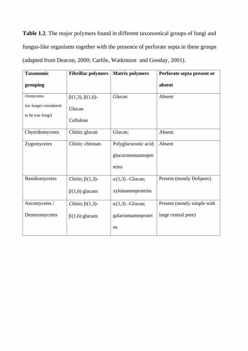
Table 1.2. The major polymers found in different taxonomical groups of fungi and
fungus-like organisms together with the presence of perforate septa in these groups
(adapted from Deacon, 2000; Carlile, Watkinson and Gooday, 2001).
Taxonomic
grouping
Fibrillar polymers Matrix polymers Perforate septa present or
absent
Oomycetes
(no longer considered
to be true fungi)
(1,3), (1,6)-
Glucan
Cellulose
Glucan Absent
Chytridomycetes Chitin; glucan Glucan; Absent
Zygomycetes Chitin; chitosan Polyglucuronic acid;
glucuronomannopro
teins
Absent
Basidiomycetes Chitin; (1,3)-
(1,6) glucans
(1,3) –Glucan;
xylomannoproteins
Present (mostly Dolipore)
Ascomycetes /
Deuteromycetes
Chitin; (1,3)-
(1,6) glucans
(1,3) –Glucan;
galactomannoprotei
ns
Present (mostly simple with
large central pore)

Table 1.3 Functional components of an idealised fungal cell Organelle or cellular structure Function Cell envelope Nucleus Mitochondria Endoplasmic reticulum Proteasome Golgi apparatus and vesicles Vacuole Peroxisome
Comprising: the plasma membrane which acts as a selectively permeable barrier for transport of hydrophilic molecules in and out of fungal cells; the periplasm containing proteins and enzymes unable to permeate the cell wall; the cell wall which provides protection, shape and is involved in cell-cell interactions, signal reception, and specialised enzyme activities; fimbriae involved in sexual conjugation; capsules to protect cells from dehydration and immune cell attack. Relatively small. Containing chromosomes (DNA-protein complexes) that pass genetic information to daughter cells at cell division and the nucleolus which is the site of ribosomal RNA transcription and processing. Site of respiratory metabolism under aerobic conditions and, under anaerobic conditions, for fatty acid, sterol and amino acid metabolism. Ribosomes on the rough ER are the sites of protein biosynthesis Multi-subunit protease complexes involved in regulating protein turnover Secretory system for import (endocytosis) and export (exocytosis) of proteins Intracellular reservoir (amino acids, polyphosphate, metal ions); proteolysis; protein trafficking; control of cellular pH. In filamentous fungi, tubular vacuoles transport materials bi-directionally along hyphae. Oxidative utilisation of specific carbon and nitrogen sources (contain catalase, oxidases). Glyoxysomes contain enzymes of the glyoxylate cycle.

Table 1.4. Elemental Requirements of Fungal Cells
Element Common Sources Cellular Functions
Carbon Sugars Structural element of fungal cells in
combination with hydrogen, oxygen and nitrogen. Energy source
Hydrogen Protons from acidic environments
Transmembrane proton motive force vital for fungal nutrition. Intracellular acidic pH (around 5-6) necessary for fungal metabolism
Oxygen Air, O2 Substrate for respiratory and other mixed-function oxidative enzymes. Essential for ergosterol and unsaturated fatty acid synthesis
Nitrogen NH4+ salts, urea,
amino acids
Structurally and functionally as organic amino nitrogen in proteins and enzymes
Phosphorus Phosphates Energy transduction, nucleic acid and membrane structure
Potassium K+ salts Ionic balance, enzyme activity
Magnesium Mg2+ salts Enzyme activity, cell and organelle structure
Sulphur Sulphates, methionine
Sulphydryl amino acids and vitamins
Calcium Ca2+ salts Possible second messenger in signal transduction
Copper Cupric salts Redox pigments Iron Ferric salts. Fe3+ is
chelated by siderophores and released as Fe2+ within the cell.
Haeme-proteins, cytochromes
Manganese Mn2+ salts Enzyme activity
Zinc Zn2+ salts Enzyme activity
Nickel Ni2+ salts Urease activity
Molybdenum Na2MoO4 Nitrate metabolism, vitamin B12

Table 1.5. Diversity of carbon sources for yeast and filamentous fungal growth (adapted from Walker, 1998)
Carbon source Typical Examples Comments
Hexose sugars D-glucose, D-galactose, Glucose metabolized by majority of
yeasts and filamentous fungi D-fructose, D-mannose If a yeast does not ferment glucose, it will
not ferment other sugars. If a yeast ferments glucose, it will also ferment fructose and mannose, but not necessarily galactose
Pentose sugars L-arabinose, D-xylose,
D-xyulose, L-rhamnose Some fungi respire pentoses better than glucose. S.cerevisiae can utilize xylulose but not xylose
Disaccharides Maltose, sucrose, lactose,
trehalose, melibiose, cellobiose, melezitose
If a yeast ferments maltose, it does not generally ferment lactose and vice versa. Melibiose utilization used to distinguish ale and lager brewing yeasts. Large number of yeasts utilize disaccharides. Few filamentous fungi (e.g. Rhizopus nigricans) cannot utilise sucrose.
Trisaccharides Raffinose, maltotriose Raffinose only partially used by
S.cerevisiae, but completely used by other Saccharomyces spp. (S.carlsbergensis, S.kluyveri)
Oligosaccharides Maltotetraose,
maltodextrins Metabolized by amylolytic yeasts, not by brewing strains
Polysaccharides Starch, inulin, cellulose,
hemicellulose, chitin, pectic substances.
Polysaccharide-fermenting yeasts are rare. Saccharomycopsis spp. and S.diastaticus can utilize soluble starch; Kluyveromyces spp. possess inulinase. Many filamentous fungi can utilise these depending on extracellular enzyme activity.
Lower aliphatic alcohols
Methanol, ethanol Respiratory substrates for many fungi. Several methylotrophic yeasts (e.g. Pichia

pastoris, Hansenula polymorpha) have industrial potential
Sugar alcohols Glycerol, glucitol Can be respired by yeasts and a few fungi.Organic acids Acetate, citrate, lactate,
malate, pyruvate, succinate
Many yeasts can respire organic acids, but few can ferment them
Fatty acids Oleate, palmitate Several species of oleaginous yeasts can
assimilate fatty acids as carbon and energy sources
Hydrocarbons n-alkanes Many yeast and a few filamentous species
grown well on C12-C18 n-alkanes
Aromatics Phenol, cresol, quinol,
resourcinol, catechol, benzoate
Few yeasts can utilize these compounds. Several n-alkane utilizing yeasts use
phenol as carbon source via the -ketoadipate pathway
Miscellaneous Adenine, uric acid,
butylamine, pentylamine, putrescine.
Some mycelial fungi and yeasts, for example, Arxula adeninivorans and A.terestre can utilize such compounds as sole source of carbon and nitrogen.
Lignin Can be decayed only by white-rot fungi (basidiomycotina). Little net energy gained directly, but makes available other polysaccharides such as cellulose and hemicellulose.
‘Hard’ keratin Keratinophilic fungi.

Table 1.6. Yeast and fungal metabolism based on responses to oxygen availability Mode of energy metabolism Examples Comments
Obligate fermentative Yeasts: Candida
pintolopesii (Saccharomyces telluris) Fungi: facultative and obligate anaerobes
Naturally occurring respiratory-deficient yeasts. Only ferment, even in presence of oxygen. No oxygen requirement for these fungi. Two categories exist with respect to the effects of air: Facultative anaerobes (e.g. Aqualinderella and Blastocladia) and obligate anaerobes (e.g. Neocallimastix)
Facultatively fermentative Crabtree-positive Saccharomyces cerevisiae Such yeasts predominantly
ferment high sugar-containing media in the presence of oxygen.
Crabtree-negative Candida utilis Such yeasts do not form ethanol under aerobic conditions and cannot grow anaerobically.
Non-fermentative Yeasts: Rhodotorula rubra
Fungi: Phycomyces
Such yeasts do not produce ethanol, either in the presence or absence of oxygen. Oxygen essential for such (obligately oxidative) fungi
Obligate aerobes Gaemannomyces graminis (the
take-all fungus) The growth of these is markedly reduced if oxygen partial pressure falls below normal atmospheric.
Adapted from Walker (1998), Deacon (2000) and Carlile, Watkinson and Gooday (2001)
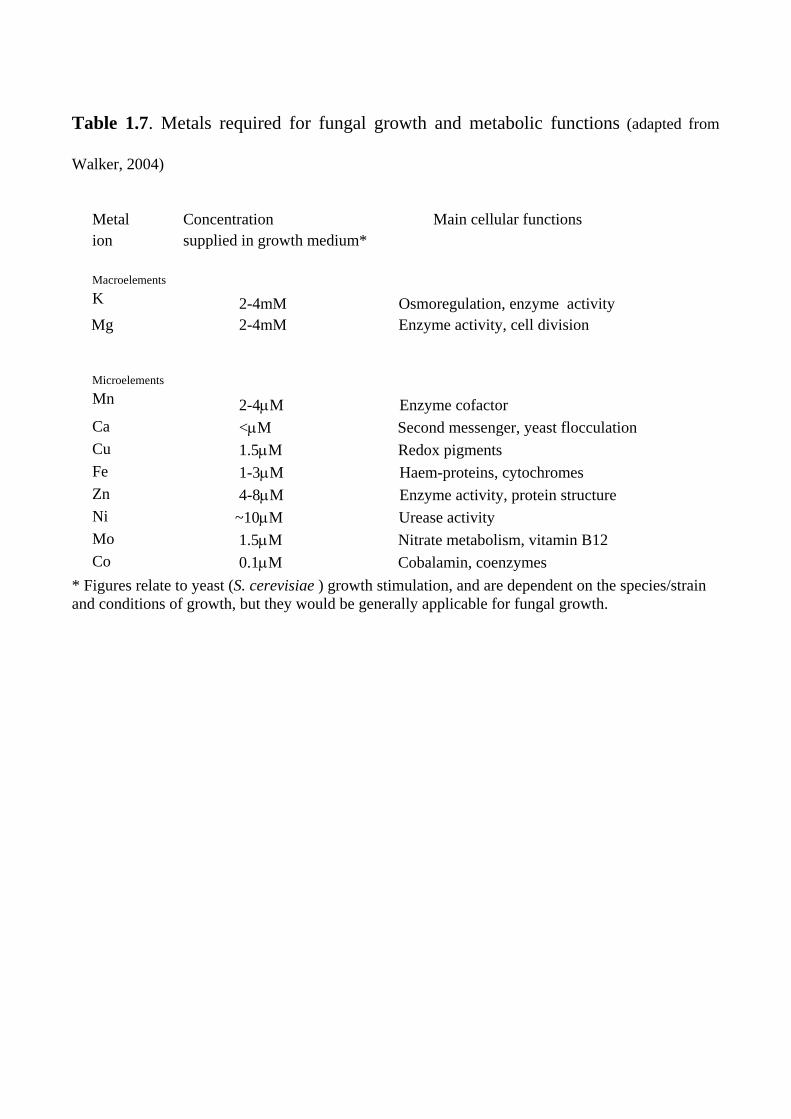
Table 1.7. Metals required for fungal growth and metabolic functions (adapted from
Walker, 2004)
Metal ion
Concentration Main cellular functions supplied in growth medium*
Macroelements
K 2-4mM Osmoregulation, enzyme activity
Mg 2-4mM Enzyme activity, cell division
Microelements
Mn
2-4M Enzyme cofactor Ca <M Second messenger, yeast flocculation Cu 1.5M Redox pigments Fe 1-3M Haem-proteins, cytochromes Zn 4-8M Enzyme activity, protein structure Ni ~10M Urease activity Mo 1.5M Nitrate metabolism, vitamin B12 Co 0.1M Cobalamin, coenzymes
* Figures relate to yeast (S. cerevisiae ) growth stimulation, and are dependent on the species/strain and conditions of growth, but they would be generally applicable for fungal growth.

Table 1.8. Principal Ingredients of Selected Industrial Media for Yeasts and Fungi
Componen
ts
Molasses Malt Wort Wine Must Cheese Whey Corn Steep
Liquor
Carbon Sources
Sucrose Fructose
Maltose Sucrose
Glucose Fructose
Lactose Glucose, other sugars
Glucose Fructose Sucrose (trace) Raffinose Glucose Maltotriose
Nitrogen Sources
Nitrogen compounds as unassimilable proteins. Nitrogen sources need to be supplemented.
Low molecular
-amino nitrogen compounds, ammonium ions and a range of amino acids.
Variable levels of ammonia nitrogen, which may be limiting. Range of amino acids.
Unassimilable globulin and albumin proteins. Low levels of ammonium and urea nitrogen.
Amino acids, protein
Minerals Supply of P, K, and S available.
High K+ levels may be inhibitory.
Supply of P, K, Mg and S available.
Supply of P, K, Mg and S available. High levels of sulphite often present.
Supply of P, K, Mg, S.
Supply of P, K, Mg, S
Vitamins Small, but generally adequate supplies. Biotin is deficient in beet molasses.
Supply of vitamins is usually adequate. High adjuct sugar
Vitamin supply generally sufficient.
Wide range of vitamins present.
Biotin, pyridoxine, thiamin

wort may be deficient in biotin.
Trace Elements
Range of trace metals present, although Mn2+
may be limiting.
All supplied,
although Zn2+ may be limiting.
Sufficient quantitities available.
Fe, Zn, Mn, Ca, Cu present.
Range of trace Elements present
Other Components
Unfermentable sugars (2-4%), organic acids, waxes, pigments, silica, pesticide residues, carmelized compounds, betaine.
Unfermentable maltodextrins, pyrazines, hop compounds.
Unfermentable pentoses. Tartaric and malic acids. Decanoic and octanoic acids may be inhibitory. May be deficient in sterols and unsaturated fatty acids.
Lipids, NaCl. Lactic and citric acids.
High levels of lactic acid present. Fat and fibre also present

Table 1.9. Modes of nutrient transport in fungi
Mode of
nutrient
transport
Description Examples of nutrients transported
Free diffusion Passive penetration of lipid-soluble solutes through the plasma membrane following the law of mass action from a high extracellular concentration to a lower intracellular concentration.
Organic acids, short-chain alkanes an long-chain fatty acids by fungi and thexport of lipophilic metabolites (e.g. ethanol) and gaseous compound
Facilitated
diffusion
Translocates solutes down a transmembrane concentration gradient in an enzyme (permease) mediated manner. As with passive diffusion, nutrient translocation continues until the intracellular concentration equals that of the extracellular medium.
In the yeast, S. cerevisiae, glucose is transported in this manner.
Diffusion
channels
These operate as voltage-dependent "gates" to transiently move certain nutrients ions down concentration gradients. They are normally closed at the negative membrane potential of resting yeast cells but open when the membrane potential becomes positive.
Ions such as potassium may be transported in this fashion.
Active
transport
The driving force is the membrane potential and the transmembrane electrochemical proton gradient generated by the plasma membrane
H+-ATPase. The latter extrudes protons using the free energy of ATP hydrolysis that enables nutrients to either enter with influxed protons, as in "symport" mechanisms, or against effluxed protons, as in "antiport" mechanisms.
Many nutrients (sugars, amino acids, ions)

Table 1.10. Respiratory chain characteristics of yeasts and fungi (Adapted from Walker,
1998) Type
Typical Species Sensitive to Insensitive to
Normal respiration
All aerobic fungi Cyanide and low azide1
SHAM2
Classic alternative
Yarrowia lipolytica (and in stationary phase cultures of several yeast species)
SHAM Cyanide, high azide
New alternative Schizosaccharomyces pombe, Saccharomyces cerevisiae Kluyveromyces lactis Williopsis saturnus
High azide Cyanide, low azide, SHAM
1The azide-sensitive pathway lacks proton transport capability and accepts electrons from NADH but not from succinate. 2SHAM = Salycil hydroxamate. The SHAM-sensitive pathway transports electrons to oxygen also without proton transport, and therefore does not phosphorylate ADP.

Table 1.11. Modes of vegetative reproduction in yeasts (adapted from Walker 1998) Mode Description Representative Yeast Genera
Multilateral budding Buds may arise at any
point on the mother cell surface, but never again at the same site. Branched chaining may occasionally follow multilateral budding when buds fail to separate.
Saccharomyces, Zygosaccharomyces, Torulaspora, Pichia, Pachysolen, Kluyveromyces, Williopsis, Debaryomyces, Yarrowia, Saccharomycopsis, Lipomyces
Bi-polar budding Budding restricted to poles of elongated cells (apiculate or lemon-shaped) along their longitudinal axis.
Nadsonia, Saccharomycodes, Haneniaspora, Wickerhamia, Kloeckera
Unipolar budding Budding repeated at same site on mother cell surface.
Pityrosporum, Trigonopsis
Monopolar budding Buds originate at only one pole of the mother cell.
Malassezia
Binary fission A cell septum (cell plate or cross-wall) is laid down within cells after lengthwise growth and which cleaves cells into two.
Schizosaccharomyces
Bud fission Broad cross-wall at base of bud forms which separates bud from mother.
Occasionally found in: Saccharomycodes, Nadsonia and Pityrosporum
Budding from stalks Buds formed on short denticles or long stalks.
Sterigmatomyces
Ballistoconidiogenesis Ballistoconidia are actively discharged from tapering outgrowths on the cell.
Bullera, Sporobolomyces
Pseudomycelia Cells fail to separate after budding or fission to produce a single filament. Pseudomycelial morphology is quite diverse and the extent of differentiation variable depending on yeast species and growth conditions.
Several yeast species may exhibit 'dimorphism' eg Candida albicans, Saccharomycopsis figuligera. Even S.cerevisiae exhibits pseudohyphal growth depending on conditions.


Figure 1.1

Figure 1.2

Figure 1.3

Figure 1.4