introduction to digestion · 2017-12-01 · form a syncitium -electrically coupled, ... properties...
TRANSCRIPT
Introduction to Digestion
Dr .sofiabadi
2015
qums
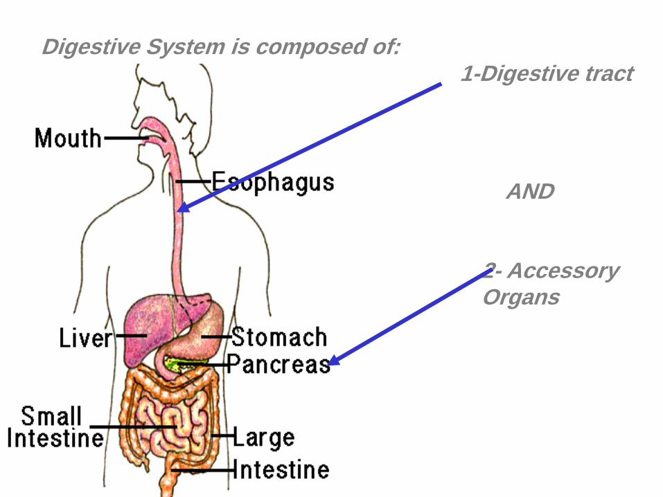
Digestive System is composed of:1-Digestive tract
AND
2- Accessory Organs

Digestive System
The primary function of the
gastrointestinal (alimentary)
tract is:
to process
ingested food and
provide the body
with
nutrients
water
electrolytes

Function of the Digestive System
(A) motility
(B) Secretion and Digestion
Dissolving and breaking-down of
nutrients into smaller units
(c) Absorption
Absorption of these units (+ water) through
the intestinal tract into the blood or lymph system

MuscularisMucosaeGland in
submucosa
Serosa
Submucosa
Lymph node
Lamina propria
Villus
Epithelium
Longitudinal
Circular

Smooth
Muscle

Form hollow tubes
Form a syncitium - electrically coupled, joined by gap
junctions contractions synchronous
Actin:myosin ratio 15:1 (skeletal muscle 2:1)
Contractile elements not arranged in sarcomeresnot
striated
Stimulated by neurotransmitter released from varicosities
Have slow wave activity
5-10m
200m
Properties of GI smooth muscle

Syncitium - electrically coupled, joined by gap
junctions
Gap junctions
Helps synchronous contractions
Electrical
Activity

0
-60
Me
mb
ran
e
po
ten
tial (m
V)
Tension
Slow Waves in GI smooth muscles
they don’t cause contraction
They are present in GI smooth muscle except in esophgagus and proximal
part of Stomach
They oscillate between -50 to -65 mV
Oscillation of ?

Slow waves are produced by
interstitial cells of CajalThey are located in a thin layer
between the longitudinal and circular
layers of muscularis externa
They have properties of
both fibroblast and smooth
muscle
They have
junctions with
both circular
and longitudinal
smooth muscle
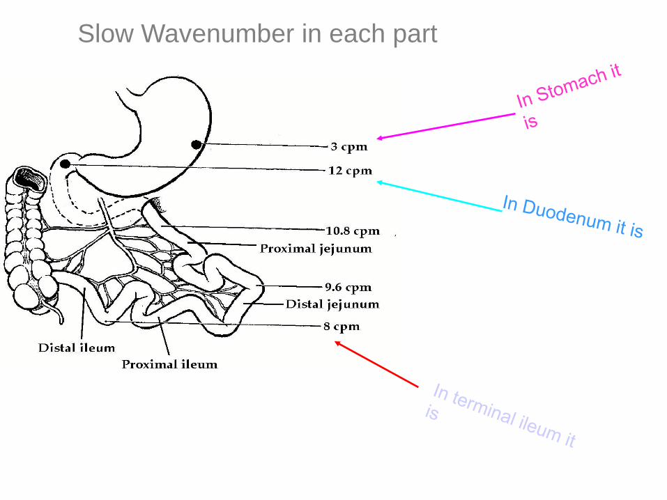
Slow Wavenumber in each part


GI Innervation
• Intrinsic (Entric Nervous
System)
• Extrinsic (Autonum)
Entric
Nervous
System

circula
r
Longitudinal
Submucosalplexus(Meissner’s)
Myentericplexus(Auerbach’s)


Functions of extrinsic nervous
system
• Parasympathetic stimulation:
A general increase in activity of the
entire enteric nervous system
• Sympathetic stimulation:
Generally inhibits GI system activity
(There are some exceptions: in
muscularis mucosa, sphincter, and GI
vessels it stimulates smooth muscle
contraction)

Afferent Nerves from Gut
• There are
sensory nerves
in epithelium or
gut wall
• They can be
stimulated by:
1- distention
2- specific
chemical
substances
3-irritation of
gut mucosa
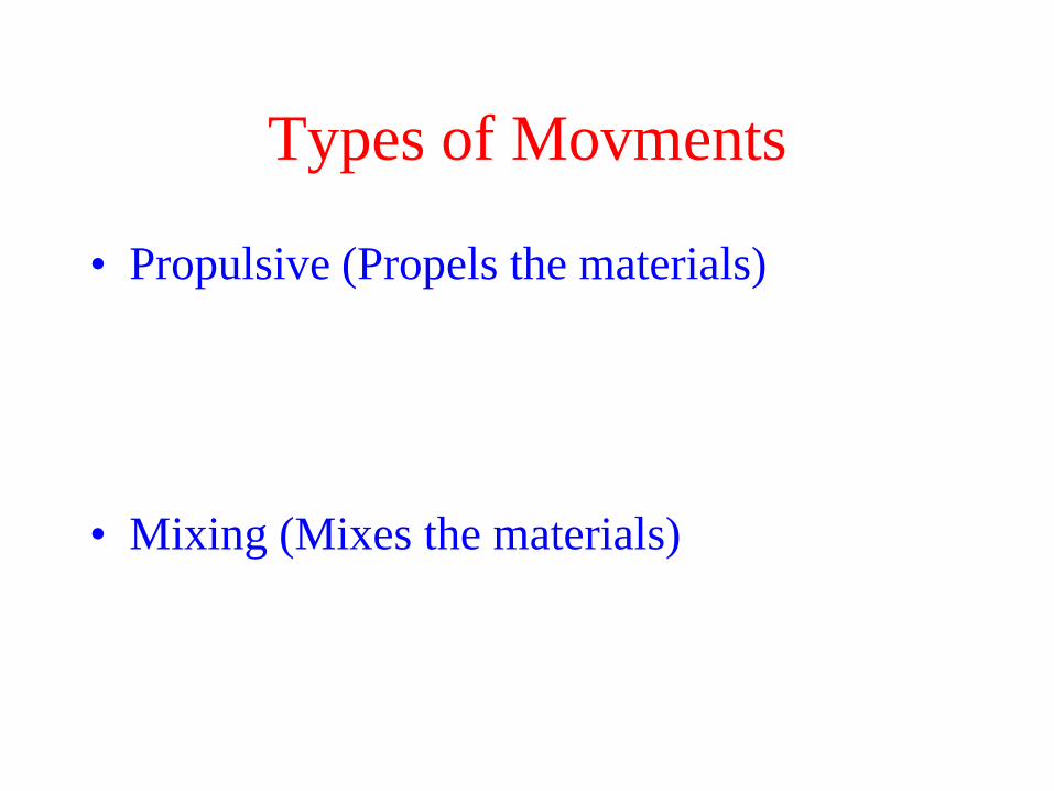
Types of Movments
• Propulsive (Propels the materials)
• Mixing (Mixes the materials)

Mechanism of Peristaltic
Contraction
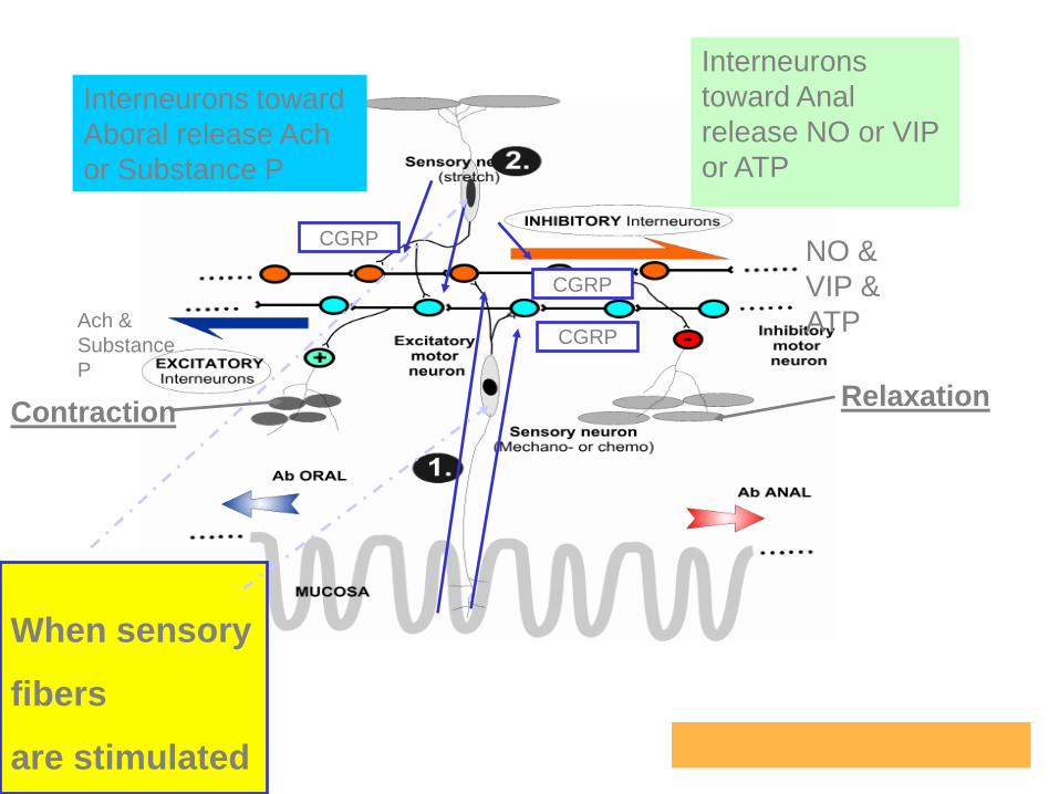
When sensory
fibers
are stimulated
CGRP
CGRP
CGRP
Interneurons toward
Aboral release Ach
or Substance P
Ach &
Substance
P
Interneurons
toward Anal
release NO or VIP
or ATP
NO &
VIP &
ATP
ContractionRelaxation

Hormonal Control of Gastrointestinal Motility
• 1) Gastrin is secreted by the "G" cells of the antrum of the stomach in response to distention of the stomach, the products of proteins, and gastrin releasing peptide, which is released by the nerves of the gastric mucosa during vagalstimulation.
• The primary actions of gastrin are : (A)stimulation of gastric acid secretion (B) stimulation of growth of the gastric mucosa.
• 2) Cholecystokinin is secreted by "I" cells in the mucosa of the duodenum and jejunum mainly in response to digestive products of fat, fatty acids, and mono glycerides in the intestinal contents. This hormone strongly contracts the gallbladder, and also inhibits stomach contraction moderately.

Hormonal Control of Gastrointestinal Motility
• 3) Secretin was the first gastrointestinal hormone discovered and is secreted by the "S" cells in the mucosa of the duodenum in response to acidic gastric juice emptying into the duodenum from the pylorus of the stomach. Secretin has a mild effect on motility of the gastrointestinal tract and acts to promote pancreatic secretion of bicarbonate which in turn helps to neutralize the acid in the small intestine.
• 4) Gastric inhibitory peptide is secreted by the mucosa of the upper small intestine, mainly in response to fatty acids and amino acids but to a lesser extent in response to carbohydrate. It has a mild effect in decreasing motor activity of the stomach and therefore slows emptying of gastric contents into the duodenum when the upper small intestine is already overloaded with food products.

Hormonal Control of Gastrointestinal Motility
• 5) Motilin is secreted by the upper duodenum during fasting, and the only known function of this hormone is to increase gastrointestinal motility. Motilin is released cyclically and stimulates waves of gastrointestinal motility called interdigestivemyoelectric complexes that move through the stomach and small intestine
• every 90 minutes in a fasted person. Motilinsecretion is inhibited after ingestion by mechanisms that are not fully understood.

Gastrointestinal Reflexes
• 1) Reflexes that are integrated entirely within the
gut wall enteric nervous system. Propulsive
Movements-Peristalsis,
• 2) Reflexes from the gut to the prevertebral
sympathetic ganglia and then back to the
gastrointestinal tract.
• (the gastrocolic reflex), (the enterogastric reflexes
(the colonoileal reflex).
• 3) Reflexes from the gut to the spinal cord or
brain stem and then back to the gastrointestinal
tract.

Gastrointestinal Reflexes
• 3) Reflexes from the gut to the spinal cord or
brain stem and then back to the gastrointestinal
tract.
• (1)(vago-vagal reflex) to control gastric motor and
secretory activity;
• (2) pain reflexes that cause general inhibition of the
entire gastrointestinal tract
• (3) defecation reflexes that travel from the colon
andrectum to the spinal cord and back again to produce
the powerful colonic, rectal, and abdominal
• contractions required for defecation .

GI blood Flow
1) Effect of Gut Activity and Metabolic Factors on Gastrointestinal
Blood Flow (oxygen concentration, adenosine)
2) Nervous Control of Gastrointestinal
Stimulation of the parasympathetic nerves increases local blood flow.
Sympathetic stimulation, decreased blood flow
3) Possible Causes of the Increased Blood Flow During
Gastrointestinal Activity:1) several vasodilator released from the mucosa of the intestinal tract:
CCK-PZ, VIP,, gastrin, and secretin.
2) kallidin and bradykinin, from Gland.
3) Hypoxia in the gut wall

Basic Digestive Processes
(1) Motility
Muscles in GI tract contract
Mix and move contents
Propulsive movements
Mix with digestive juices - greater digestion
Greater exposal to absorptive surface absorption
Food Contracted Relaxed

Chewing Reflex
• Food presence Reflex inhibition of
chewing muscles
• Lower Jaw drops
• This drops initiates a stretch reflex
Jaw muscles contract
• Continues
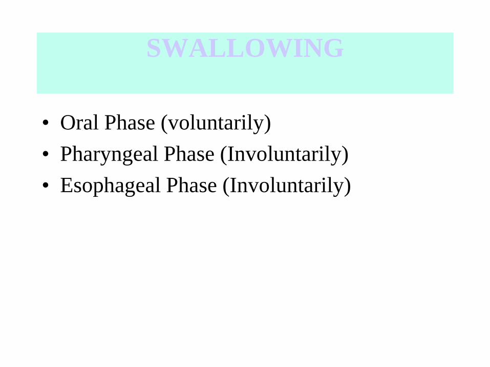
SWALLOWING
• Oral Phase (voluntarily)
• Pharyngeal Phase (Involuntarily)
• Esophageal Phase (Involuntarily)
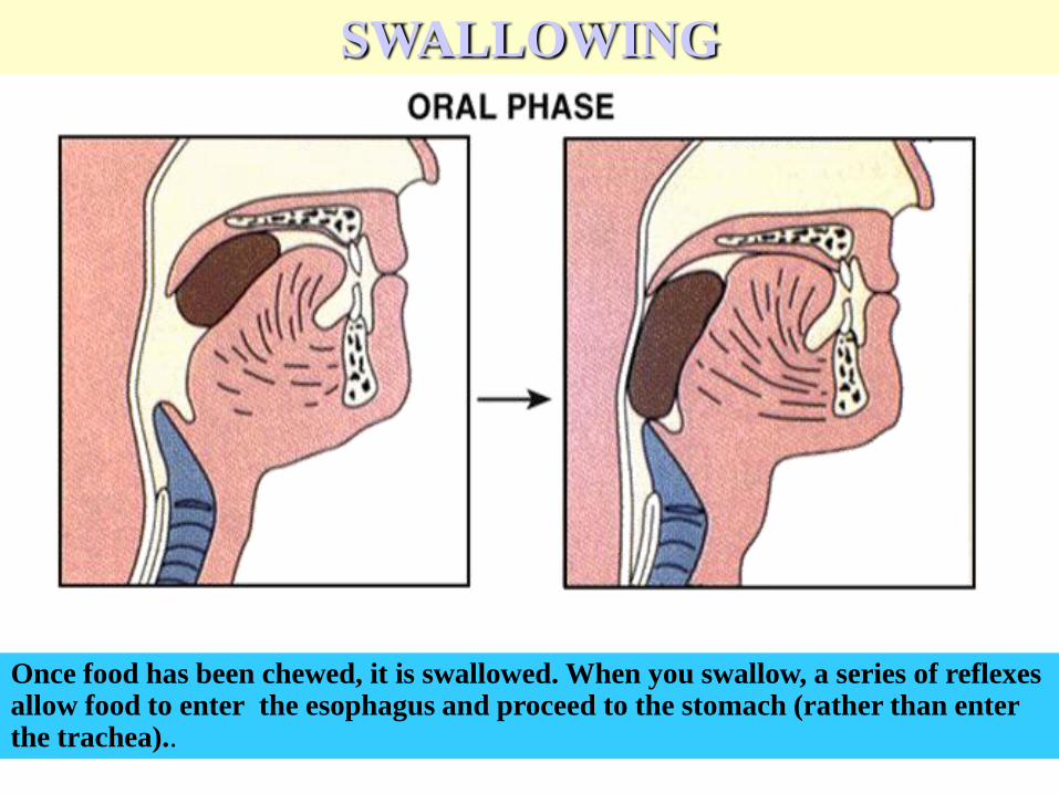
Once food has been chewed, it is swallowed. When you swallow, a series of reflexes allow food to enter the esophagus and proceed to the stomach (rather than enter the trachea)..
SWALLOWING

Esophageal Phase of Swallowing
(Pressure Changes)

Achalasia
• Problem in opening of LES ( defects in release
of NO & VIP)
• Treatment
Surgical
Balloon
Botulinum toxin

Stomach
Functions
(A) Storage
(B) Mixing
(C) Initiates secretion &
digestion
(D) Carefully controls emptying
of contents to the Small
Intestine
A
BC
D
Gastric Pits

Major Areas of the Stomach

Motor behavior of the stomach is determined by
dominant pacemaker in the corpus

Gastric Motility

Movements in Stomach

Types of Motility
• Mixing movement (Segmental
contractions)
• Propulsive movements

SEGMENTATION
In stomach it
is 3
contraction
per minute
In Duodenum
it is 12
contraction
per minute
In Jejunum it
is 12
contraction
per minute

Peristaltic Rush
• Powerful peristalsis
• Irritation of intestinal mucosa by Infectious
diarrhea
• Partly by extrinsic nervous reflexes to the brain
stem and back again to the intestine and partly by
potentiation of intrinsic myentric reflexes

Migrating Motor Complex
• From Stomach to the end of illeum
• Every 75- 90 min
• After processing of the last food

Colon
Function : Absorption of water & electrolyte
Reservior

Types of Colonic Motility
• Mixing movement (Segmental
contractions)
• Propulsive movements (Mass contraction)

Mixing movement
(Haustra contractions)
They help absorption

Mass Movements
In most cases 15 min after breakfast
1- a contracted ring in transverse colon
2- 20 Cm after the contracted ring losses its haustra and contract simultaneously

Propulsive movements (Mass contraction)
– Gastrocolic and Duodenocolic reflexes facilitate mass
movement appearance after meals
– If extrinsic autonomic nerves are dissected these
movement disappear
– Irritation of colon can also brings about mass
movement

Defecation
• Is initiated by defecation reflexes
• 1- intrinsic defecation reflexes
• 2- Parasympathetic defecation reflexes

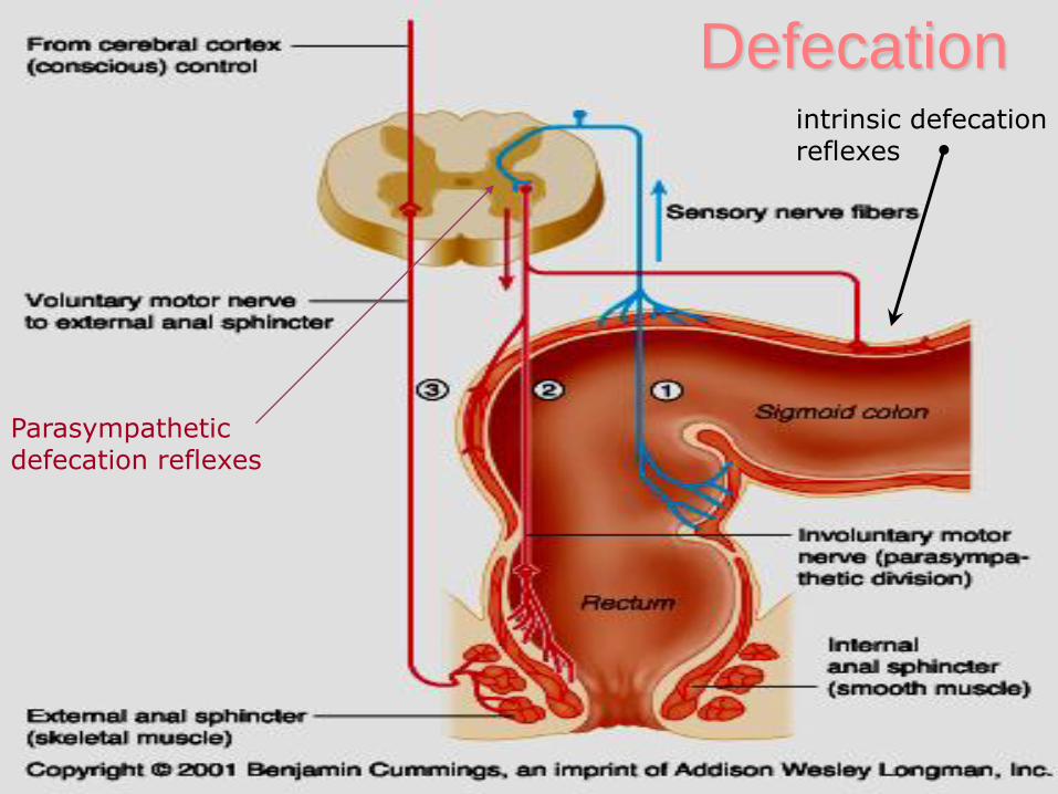
Defecationintrinsic defecation reflexes
Parasympathetic defecation reflexes

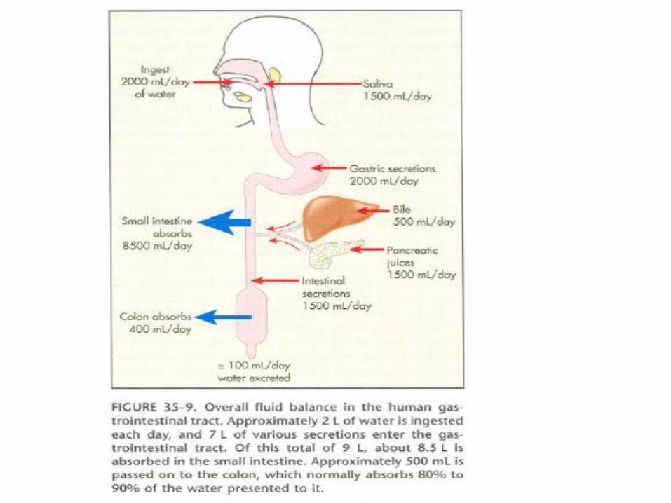
Many digestive juices are secreted into the digestive tract by glands
along the route.
Contain water, electrolytes and enzymes, bile salts, mucous etc.
Act on nutrients or start/stop other substances acting on the nutrients
Released in response to hormones or neurons
Glands in stomach
Nutrients
(2) Secretion

Salivary Secretion• FUNCTIONS:
Lubrication & Protection (Mucus)
Digestion (alpha amylase)
Speech
Tasting
Defense (IJA & Lysozym)
• GLANDS: Parotid, Submandibular, Sublingual
• FUNCTIONAL UNIT: Salivon


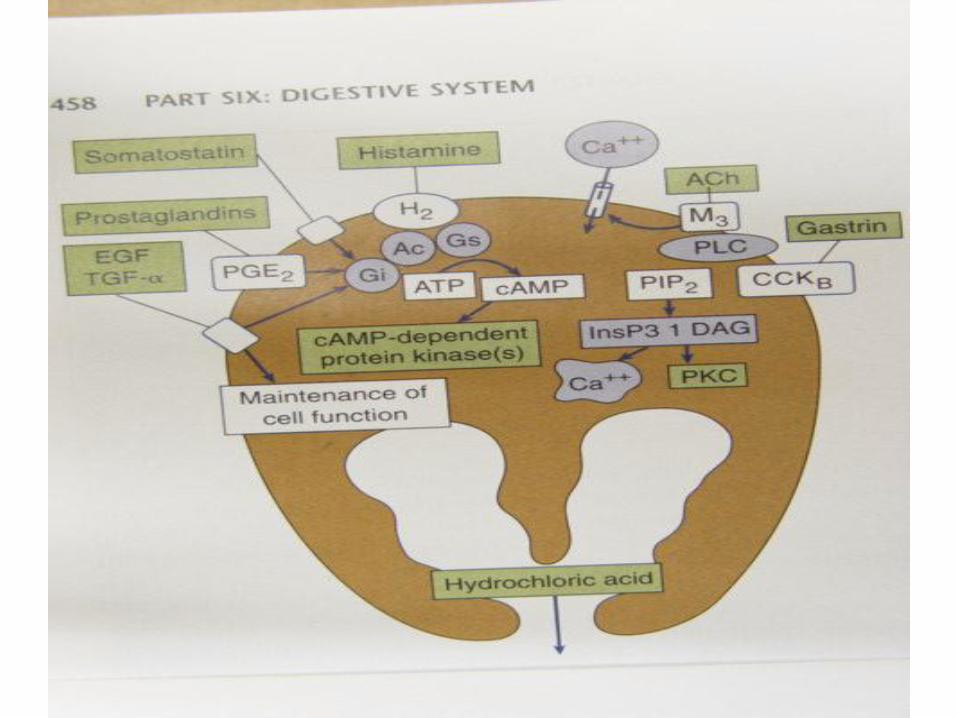
Gastric Secretion
• Acid ( Kills bacteria & Changes inactive
Pepsinogen to active form and digest protein)
• Mucus (protection)
• Bicarbonte (protection)
• Pepsinogen (enzyme)
• Intrinsic Factor (Vit B12 absorption)

Microscopic Anatomy
• The otherwise smooth
lining is dotted with
millions of gastric pits
which lead to gastric
glands that produce
gastric juice
• The glands of the
stomach body are
substantially larger and
produce the majority of
the stomach secretions


cephalic
• 1- Vagus via parasympathetic (Ach)

Gastric Phase
• Presence of food in stomach
• 1- Distention central (vagovagal) &
local reflexes Ach & Gastrin
• Amino acid & peptide Gastrin


Intestinal Phase
• At first PH>3
• Then PH<3
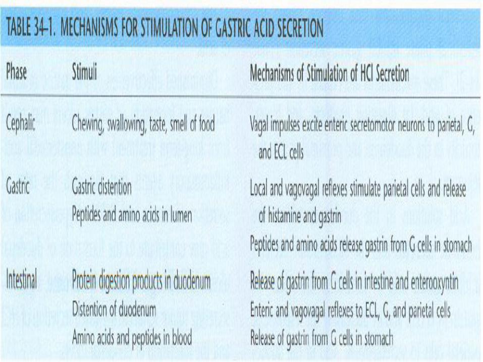

HCl
gastrin
pepsinogen
Acidic Environment
H+H+H+
pepsin
pepsinogen
Stomach lumen
protein
peptides
Gastric Pits


When the stomach contents are
emptied into small intestine they are
mixed with the secretions from the
pancreas and liverthat are emptied at the beginning of
the small intestine (duodenum)

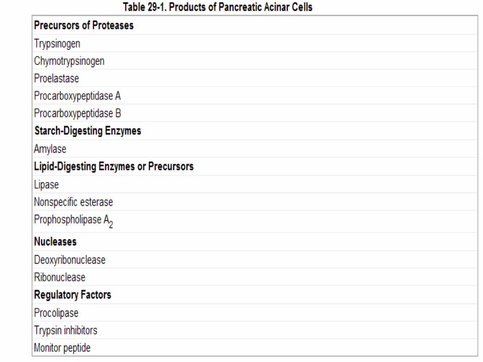
Pancreas
Pancreatic secretions
(1) Enzymes
Trypsin
Chymotrypsin
Carboxipeptidase
Amylase
Lipase
ONLY enzyme that digests FAT - Free fatty acids + Monoglycerides
Breakdown
peptides
fatty acid
fatty acidfatty acid


(2) Alkaline secretion (Sodium
Bicarbonate)
Neutralise acids from stomach
• enabling enzymes to function

Activation of pancreatic proteases
Trypsinogen Trypsin
Enterokinase
Trypsinogen
Chymotrypsinogen
Proelastase
Procarboxypeptidase
Trypsin
Chymotrypsin
Elastase
Carboxypeptidase
Active proteases inactivated by trypsin


H2O CO2CO2
Bicarbonate secretion
Cl- Cl-
HCO3-
ATP
H+
Na+Na+Na+
H2OH2O
H+
HCO3-
H2CO3
Lumen Blood

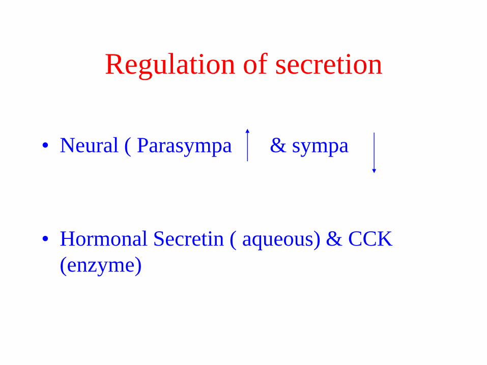
Regulation of secretion
• Neural ( Parasympa & sympa
• Hormonal Secretin ( aqueous) & CCK
(enzyme)
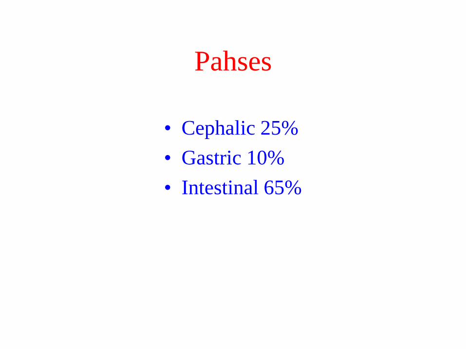
Pahses
• Cephalic 25%
• Gastric 10%
• Intestinal 65%

Peptides
Amino acids
Fat, H+
Enzymes
HCO3-
CCK
Secretin
Intestinal phase of secretionVAGUS
ACh

Liver (Bile)Liver produces bile salts & gallbladder
stores & concentrates it
Bile
(A) Converts large fat globule into an
emulsion of fat droplets - greater area
(B) Transport of fat droplets -
fat is difficult to transport
Fat
Bile
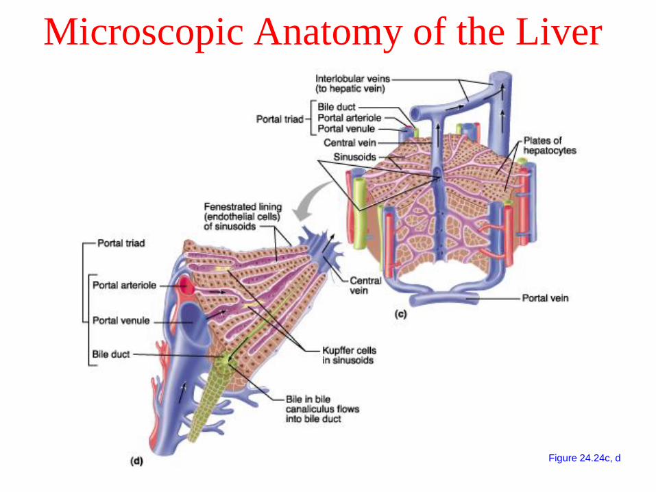
Microscopic Anatomy of the Liver
Figure 24.24c, d


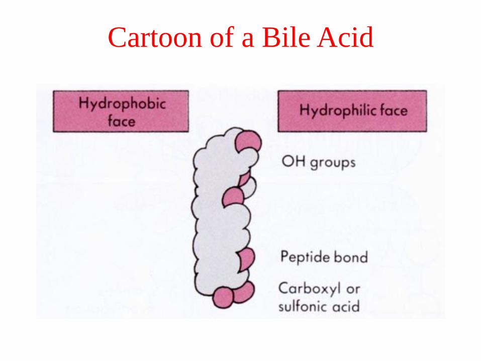
Cartoon of a Bile Acid

Form
micelle

Helps lipid absorption


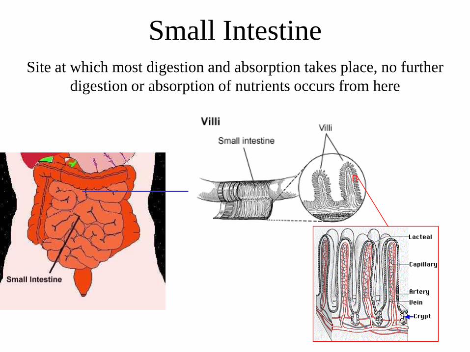
Small IntestineSite at which most digestion and absorption takes place, no further
digestion or absorption of nutrients occurs from here

Intestinal secretion

Duodenum
• Bruner’s gland HCO3 & Mucus
• Stimulated by parasympathetic &
mechanical & secretin
• Sympathetic stimulation decreases

Crypt of lieberkuhn
• Secretes Na & Cl & water
• Stimulated by:
Local reflexes & mechanical stimuli
Goblet cells:
– Mucus

a) COMPLETE DIGESTION
b) ABSORPTION
FLUID SECRETION
Absorption
and Secretion
occur
simultaneously

Histology of Large Intestine

Colonic sercretion
• Low volume
• High mucus by goblet cells
• Aqueous Rich of K & Bicarbonate
• Stimulated by mechanical & cholinergic
• Sympathetic stimulation Decrease
secretion

Stores and concentrates faecal material before defecation
Secretion of mucous for protection of mucosa
No nutrient absorption,
only salts and relatively
small amounts of water
Faeces: Water, unabsorbed
food e.g. cellulose, bacteria
Large Intestine

• Mixing and segmentation of contents - no secretion
• Brings contents in contact with epithelial surface
• Digestion in the lumen is accomplished by secretions from the
pancreas
• Digestion occurs within the epithelial cells of the intestine
• Minerals are also absorbed
Small Intestine
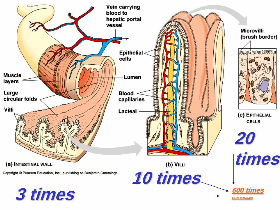
The structure of the small intestine
20 times
3 times10 times
600 timesOuzr istaiiram
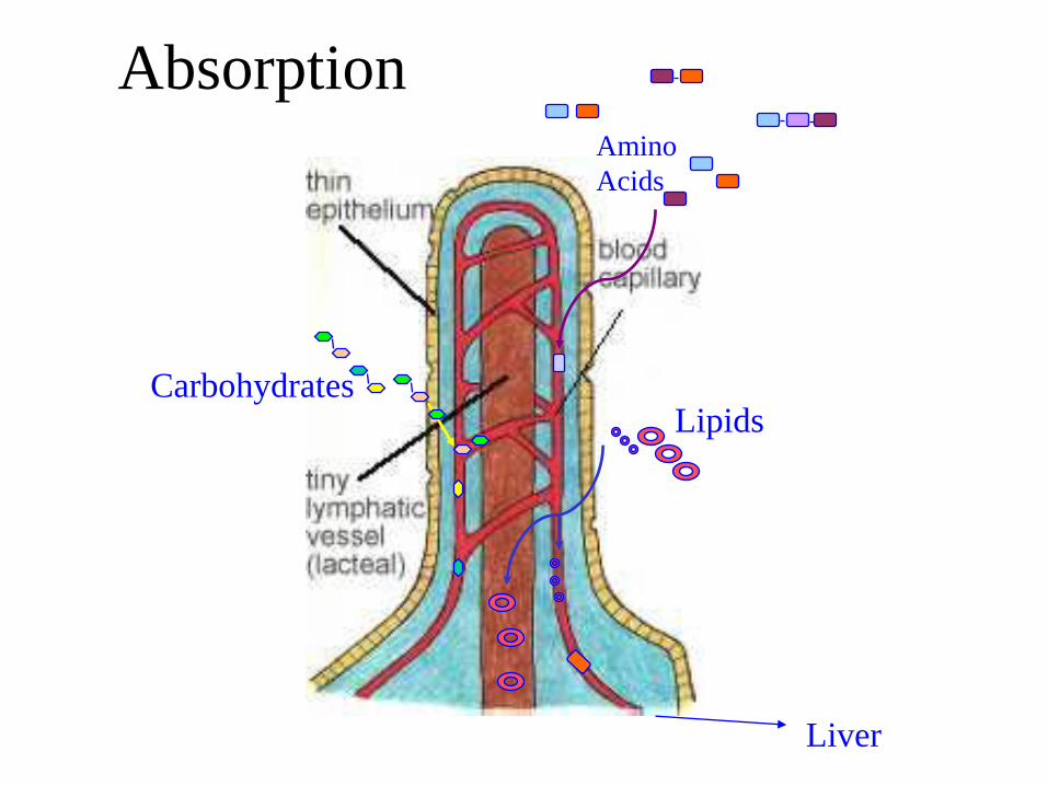
Absorption
Lipids
Amino
Acids
Carbohydrates
Liver

Small Intestinal
Lumen
Intestinal cells
Carbohydrates
H2O
Proteins
LactealCapillary
Lipids
+

Enzymes
• Biological catalysts which greatly increase the rate of a chemical reaction but are not themselves changed during the process
• Enzymes are central to the digestion process.
Digestion
Glands in stomach
Pepsin
Protein
Peptide
fragments

ProteinsStructure
Amino Acids - glycine

Amino acids combine to form peptides which combine to form proteins

Protein digestion & absorption
Protein digestion involves the enzymatic degradation of proteins to di-, or tri- peptides & finally amino acids.
• Digestion begins in the stomach with the interaction with pepsin
• Further proteolytic cleavage occurs in the intestinal lumen by pancreatic trypsin, chymotrypsin, and carboxy peptidases.
• Final degradation occurs on the membrane of the intestinal microvilli by the action of aminopeptidases.
• Protein absorption occurs through active transport.

peptidases
aminopolypeptidasetransporters
amino acidsDi/tri
peptides
Cytoplasmic peptidase
transporters
Amino acids
Protein
peptides

The result of pancreatic and brush
border enzyme
• AA & small peptides (mostly di & tri)
• They are entered into the cells
• In the cell peptides are digested
• Finally entered into blood as AA and a few
as dipeptide
3 sites for digestion of protein

How AA are entered into the cells
• Some by Na ( co-transport) (5 types)
• Some facilitated ( 2 types)
• Some diffusion

How Di & Tri peptides are entered
into the cells
• Co-transport by Na
Co transport by H

Carbohydrates
Monosaccharides
Disaccharides
Polysaccharides

CarbohydratesStructure
Monosaccharides
Disaccharides
Polysaccharides
+ Lactose
Maltose
+ Galactose
+ Fibre
Glycogen


Carbohydrate digestion & absorption
• Carbohydrate digestion involves the enzymatic degradation of
di-, tri, and polysaccharides to monosaccharides (glucose,
fructose, galactose).
• Digestion begins in the mouth with salivary amylase
• Polysaccharides are then further broken down by pancreatic
amylase in the small intestine.
• Final degradation occurs on the surface (brushborder) of the
absorptive cells in the jejunum. lactase, sucrase, and maltase.
• Monosaccharides absorbed by facilitated (membrane carrier) or
active transport (membrane carrier, Na+, and ATP).
• Fructose = facilitated diffusion
• Glucose and galactose = active transport


FatsFatty Acid
Fat (Triglyceride)

Lipid digestion and absorption
• Fats triacylglycerols (triglycerides) to monoacylglycerols
and 2 fatty acids.
• Small amount of lipase in saliva begins digestion which
continues in the stomach with (slow acting) gastric lipases.
• Pancreatic lipases and bile are mixed with hydrolyzed
product in the duodenum. The bile emulsifies the fat into
fat droplets (micelles) making it easier for colipase and
pancreatic lipase to breakdown triglycerides
• Monoacylglycerol and fatty acids are packaged into mixed
micelles (cholesterol, bile salts, and fat soluble vitamins)
and diffuse across the cellular membrane (of the jejunum).
• Triglycerides are reformed (re-esterified) then combine
with cholesterol and phospholipids to form chylomicrons
for transportation

Lipid Absorption

Intracellular Metabolism of Absorbed Lipids

Small Intestinal
Lumen
Intestinal cells
Carbohydrates
H2O
Proteins
LactealCapillary
Lipids
+

WHAT ?


