introduction to cell culture and the nano-imaging facility at the
TRANSCRIPT

Introduction to Cell Culture and the Nano-imaging Facility at the Nano Bio-systems and Bio-mimetics Lab
Dr. Scott Lenaghan (Post-doc)
March 2009

Nano Bio-systems and Bio- mimetics Lab
• Housed in Dougherty 206
• Self-sustained research lab with cell culture and nano-scale imaging capabilities
• Consists of Cell Culture, Imaging, and Office facilities
• Primary purpose: to experimentally explore the potential for “bio-inspired” nanoscale research

Offices for Laboratory Personnel
Cell Culture Facility
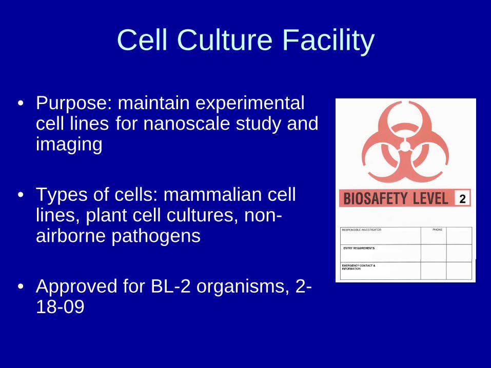
• Purpose: maintain experimental cell lines for nanoscale study and imaging
• Types of cells: mammalian cell lines, plant cell cultures, non- airborne pathogens
• Approved for BL-2 organisms, 2- 18-09
Cell Culture Facility

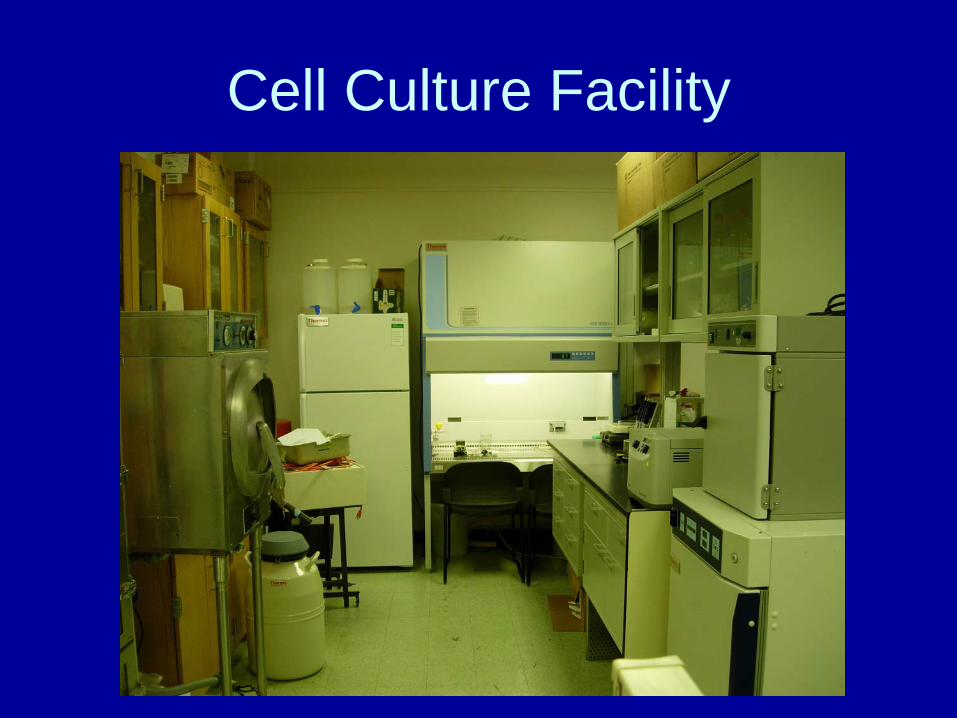
Cell Culture Facility (Equipment)• Self-sustained lab with all equipment
necessary for cell culture
• Two CO2 incubators for anaerobic cell culture
Forma Scientific Water Jacketed Incubator
Thermo Scientific Direct heat w/ Sterilization Cycle Incubator

Cell Culture Facility (Equipment)
• Thermo Scientific Revco®, Flammable and explosion proof refrigerator and freezer
VWR™ International., Gravity Convection Oven, 320c-2400C

Cell Culture Facility (Equipment)
Shel Lab Hybridization Chamber 32-65oC in 7 min, precise to 0.10C w/ rotisserie
VWR™ International water Jacketed CO2 Incubator

Cell Culture Facility (Equipment)
Thermo Scientific 1300 Series A2 Biological Safety Cabinet, with attached vacuum pump and trap

Cell Culture Facility (Sample Storage)
• Liquid Nitrogen storage for long term storage of cells
Thermolyne Locator 8, box type dewarThermo scientific 8036, cane storage and transport dewar
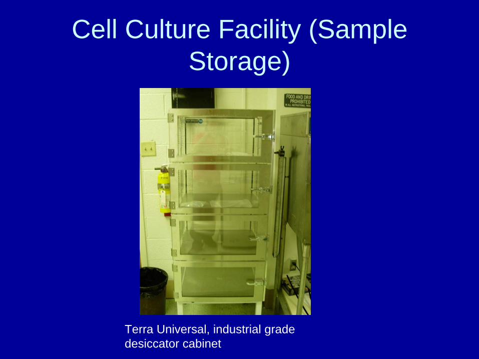
Cell Culture Facility (Sample Storage)
Terra Universal, industrial grade desiccator cabinet

Cell Culture Facility (Benchtop Equipment)
Eppendorf 5702RH, refrigerated centrifuge with swinging bucket rotor 15 and 50 ml conical tubes, max 3000xg
IEC HN-S2 Centrifuge, swinging bucket rotor for 15 and 50ml conical tubes, max ~9000xg
Cell Culture Facility (Benchtop Equipment)
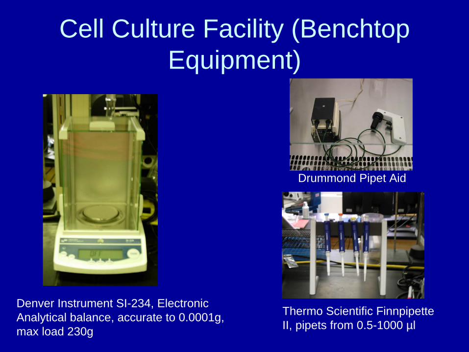
Denver Instrument SI-234, Electronic Analytical balance, accurate to 0.0001g, max load 230g
Thermo Scientific Finnpipette II, pipets from 0.5-1000 µl
Drummond Pipet Aid

Imaging Facility

Imaging Facility• Olympus Fluoview 1000 Confocal Laser Scanning
Biological Microscope on Olympus IX81 inverted base
• Agilent technologies State-of-the-art AFM

Imaging Facility
• For reduction of vibration crucial for AFM, the system is housed in a Herzan cabinet with a Herz DT- A series vibration dampening table

Olympus Fluoview FV1000• Lasers:
Multi-line Argon (457nm, 488nm, 515nm)HeNe gas (543nm)Violet diode (405mn) Diode (635nm)
Our system does not have IR laser or UV Ar (770nm, 351nm)

Olympus Fluoview FV1000 (Probes)
Cos-7 Cells
Orange = nucleusBlue = mitochondriaGreen = actin
Cos-7 Cells
Orange = nucleusGreen = actinYellow = mitochondria
(Olympus Confocal Image Gallery)

Olympus Fluoview FV1000 (Autofluorescence)
Cherry bud blossom Rubber Tree Leaf
(Olympus Confocal Image Gallery)

Olympus Fluoview FV1000
• Strengths– Allows visualization of internal structures
within cells– Can view live and fixed cells– With proper label can specifically label
structures– Can take time-lapse over long time scale to
track intracellular movements– Z-sectioning allows for 3-D sectioning

Olympus Fluoview FV1000
• Limitations– Most biological specimen need to be labeled
with a probe– Bleaching often occurs– Autofluorescence can complicate results

Olympus Fluoview FV1000
• Applications for “Bio Inspired” Nano technology– Visualization of drug delivery– Tracking of particles within cells (bio
machines)– Potential visualization of nanoparticles
(~70nm and greater)
Agilent Technologies AFM
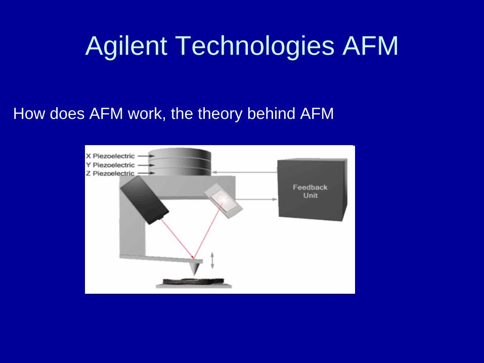
How does AFM work, the theory behind AFM
Agilent Technologies AFM
• What is unique about our system?– Increased Z-depth– Advanced laser alignment and calibration– Increased resolution due to superior design
and manufacturing– Integration with a confocal system for
simultaneous imaging of surface and internal structure of specimen

Agilent Technologies AFM
• Imaging with AFM generates surface topography
DNABreast Cancer Tissue
Pacific Nanotechnology, Inc.

Agilent Technologies AFM
• AFM can also be used for force measurements and determining adhesive strength
Agilent Technologies AFM
• Potential applications for imaging and force determination in biological systems– Imaging of membrane interactions with
foreign particles– Indentation of membranes to determine
membrane strength– Introduction of wounds with tip, and
observation of repair

Functional AFM
• Attaching a ligand, nanoparticle, receptor, etc. to a tip and then measuring adhesive forces of this functionalized tip– Can be used for receptor binding– Nanoparticle interaction with surfaces– Bonding strength of chemicals, and specific
functional groups
Agilent Technologies AFM
• Strengths of AFM– Nano resolution for imaging of particles– Can measure nano-scale force
measurements for interactions including weak forces such as van der Waals forces
– Functional AFM can measure binding kinetics of any attached particle

Agilent Technologies AFM
• Weaknesses of AFM– Cannot “see” internal cellular structure, only
topographical mapping– Samples must be strongly adhered to a
surface (can be difficult for biologicals)– Probe can damage live cells and destory
membranes

Unique Nature of Nano Bio- systems and Bio-mimetics Lab
• One of the only systems that combines simultaneous imaging of both confocal and AFM
• State-of-the-Art platform for looking at the nano-scale interior and exterior of a cell
• Diverse group with experience in aspects of engineering, biology, and chemistry
Questions?