introduction - the movement disorder society · introduction the diagnosis of parkinson disease in...
TRANSCRIPT
Introduction
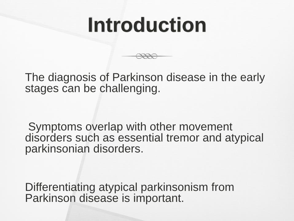
The diagnosis of Parkinson disease in the early stages can be challenging.
Symptoms overlap with other movement disorders such as essential tremor and atypical parkinsonian disorders.
Differentiating atypical parkinsonism from Parkinson disease is important.
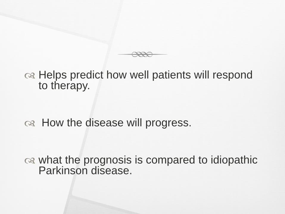
Helps predict how well patients will respond to therapy.
How the disease will progress.
what the prognosis is compared to idiopathic Parkinson disease.

Parkinsonism Diagnostic
Criteria
Tremor at rest
Bradykinesia
Rigidity
Loss of postural reflexes
Flexed posture
Freezing (motor blocks)

Definite: At least two of these
features must be present, one of them being
1 or 2.
Probable: Feature 1 or 2 alone is
present.
Possible: At least two of features 3
to 6 must be present
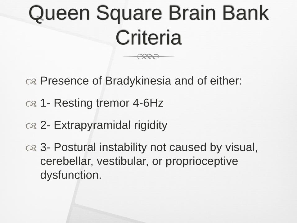
Queen Square Brain Bank
Criteria
Presence of Bradykinesia and of either:
1- Resting tremor 4-6Hz
2- Extrapyramidal rigidity
3- Postural instability not caused by visual,
cerebellar, vestibular, or proprioceptive
dysfunction.
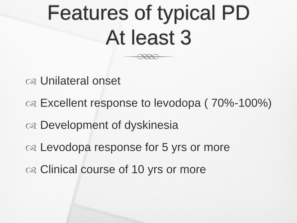
Features of typical PD
At least 3
Unilateral onset
Excellent response to levodopa ( 70%-100%)
Development of dyskinesia
Levodopa response for 5 yrs or more
Clinical course of 10 yrs or more
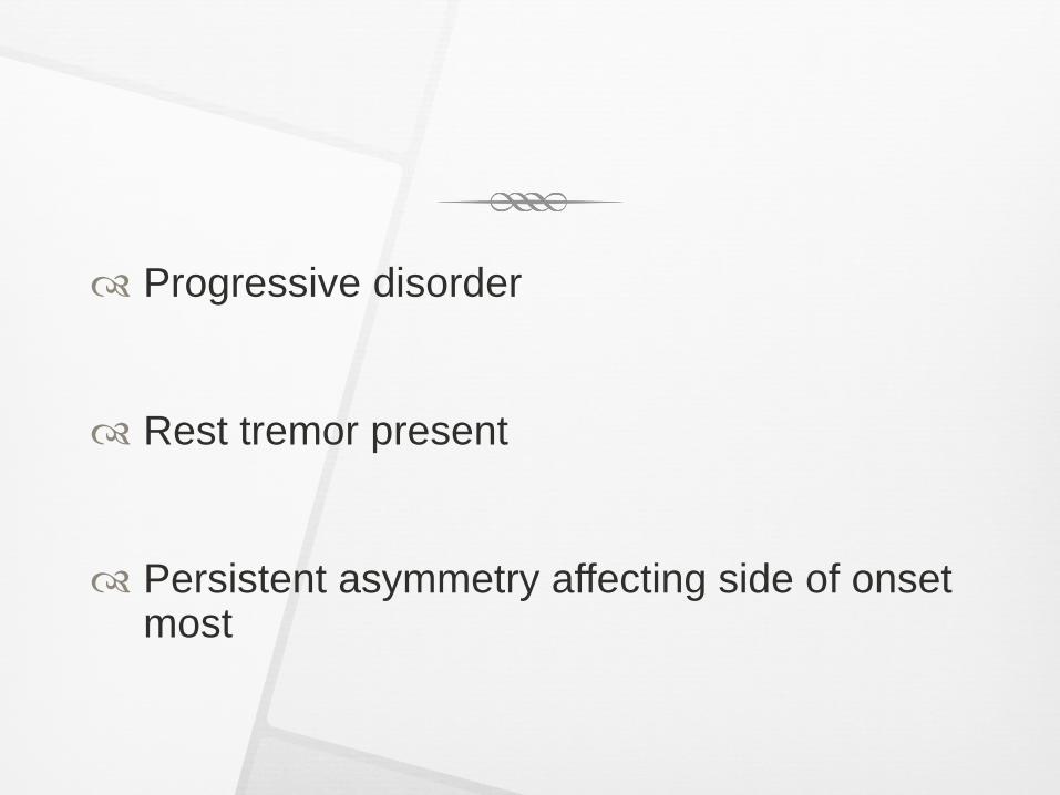
Progressive disorder
Rest tremor present
Persistent asymmetry affecting side of onset most
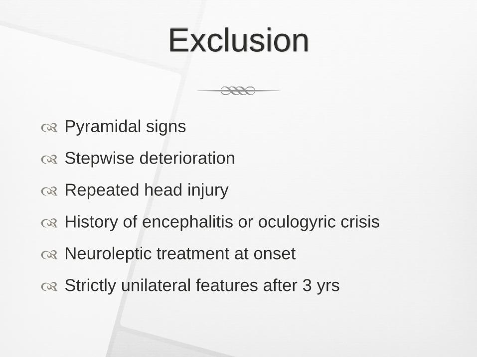
Exclusion
Pyramidal signs
Stepwise deterioration
Repeated head injury
History of encephalitis or oculogyric crisis
Neuroleptic treatment at onset
Strictly unilateral features after 3 yrs
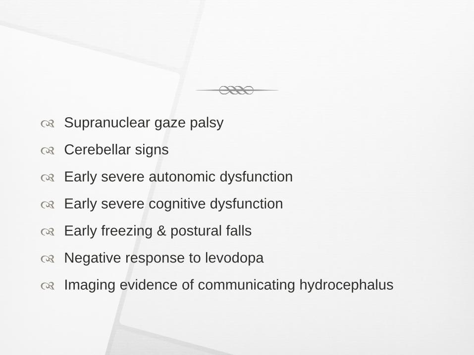
Supranuclear gaze palsy
Cerebellar signs
Early severe autonomic dysfunction
Early severe cognitive dysfunction
Early freezing & postural falls
Negative response to levodopa
Imaging evidence of communicating hydrocephalus

Parkinson’s Disease
Diagnostic Criteria
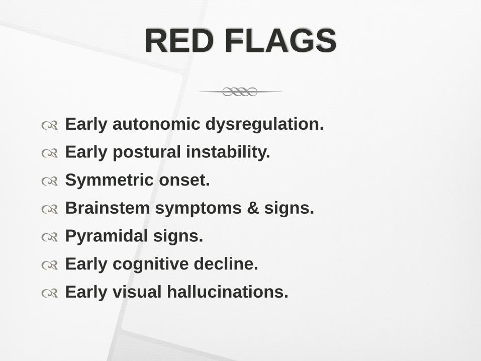
RED FLAGS
Early autonomic dysregulation.
Early postural instability.
Symmetric onset.
Brainstem symptoms & signs.
Pyramidal signs.
Early cognitive decline.
Early visual hallucinations.
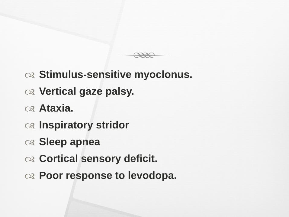
Stimulus-sensitive myoclonus.
Vertical gaze palsy.
Ataxia.
Inspiratory stridor
Sleep apnea
Cortical sensory deficit.
Poor response to levodopa.
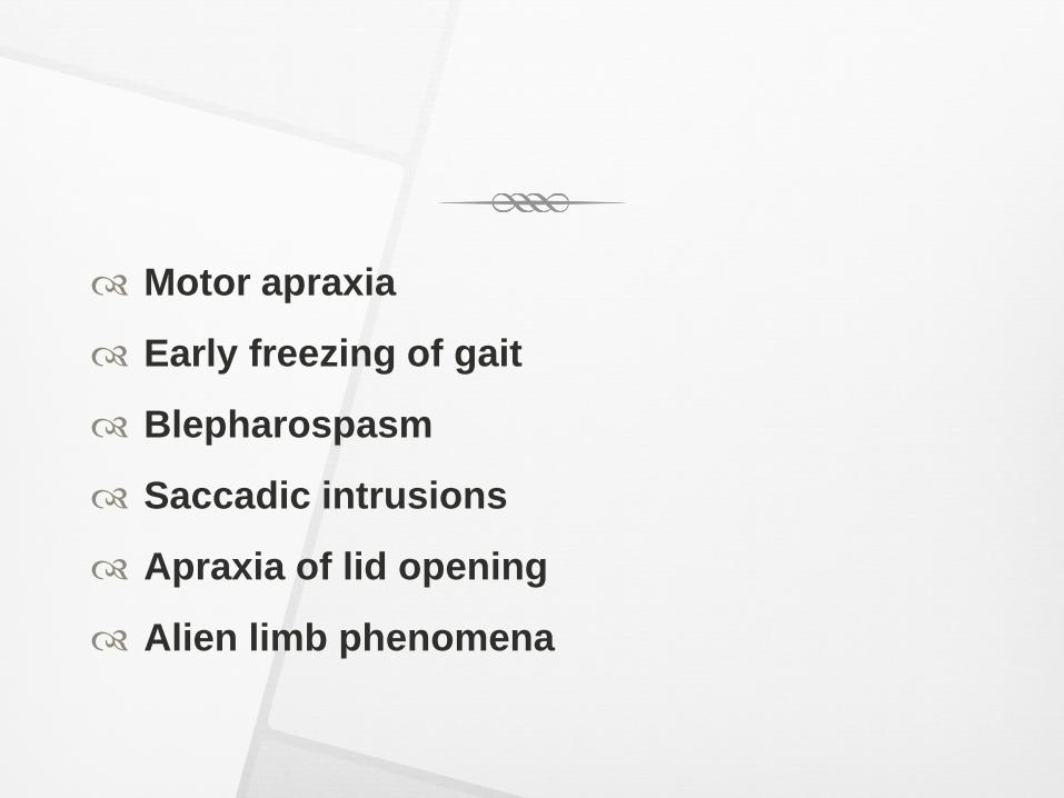
Motor apraxia
Early freezing of gait
Blepharospasm
Saccadic intrusions
Apraxia of lid opening
Alien limb phenomena

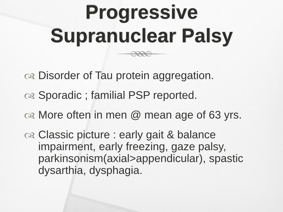
Progressive
Supranuclear Palsy
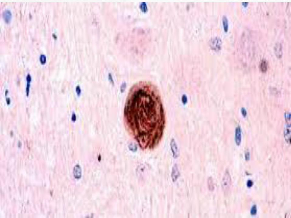
Disorder of Tau protein aggregation.
Sporadic ; familial PSP reported.
More often in men @ mean age of 63 yrs.
Classic picture : early gait & balance impairment, early freezing, gaze palsy, parkinsonism(axial>appendicular), spastic dysarthia, dysphagia.
Average annual incidence rate has been
estimated to be 5.3 new cases per 100 000
person-years (Bower et al., 1997).
The prevalence, after age adjustment to the
US population, has been estimated to be
1.39 per 100 000 (Golbe et al., 1988).
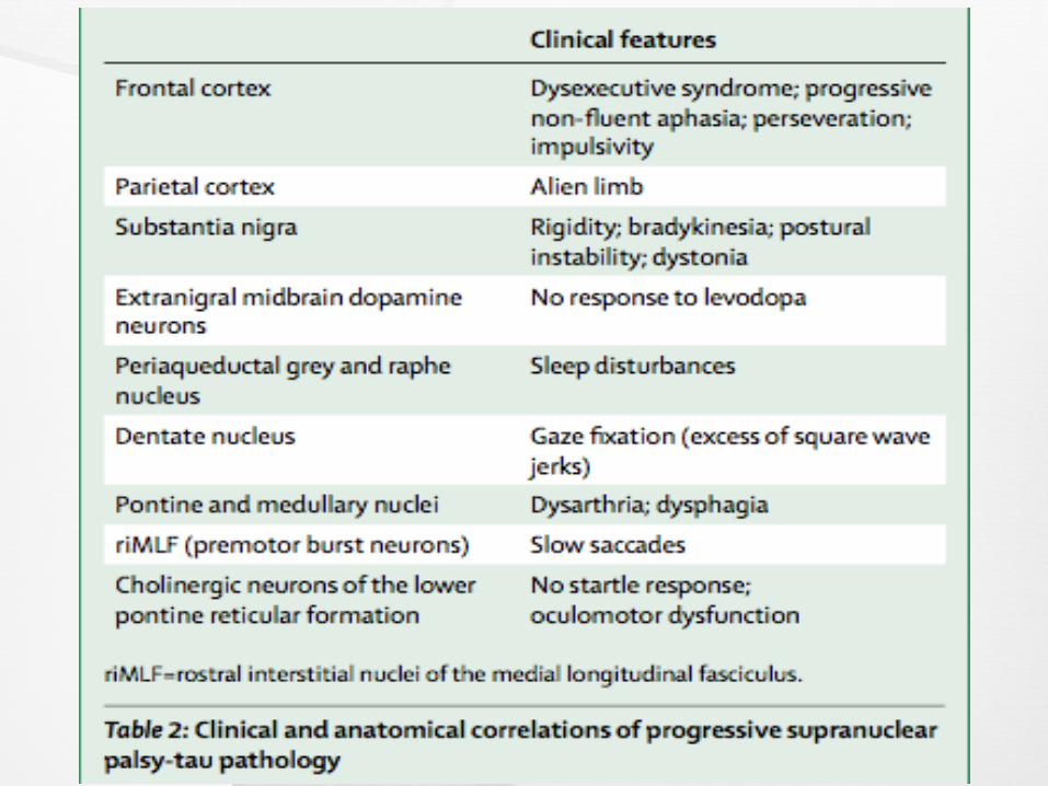
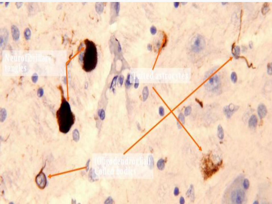
Degeneration of multiple neurotransmitter
systems.
Neuronal loss, gliosis and neurofibrillary
tangles in the pretectal area, substantia
nigra, subthalamic nucleus, globus pallidus
and superior colliculus.
Diagnosis of PSP should be considered in all patients presenting with :
1- Parkinsonism
2- NOT responding to levodopa
3- Early postural instability, falls & freezing
4- Slowing of vertical saccades ( downward>upward)

5- Supranuclear vertical gaze palsy
6- Pronounced saccadic intrusions on primary gaze fixation
7- Rapid onset of dysarthia/dysphagia
8- Early executive dysfunction, apathy, & depression
Clinical signs:
Procerus sign : Worried/astonished look
Rocket sign (darting out of the wheel chair without regard for safety)
Applause sign: (frontal disinhibition and perseverance)
Asymmetric apraxia with arm levitation (overlap with CBS)
Tremor at rest may be present in 5-10% of patients.
Dystonic posturing
Retrocollis

Characteristic eye movement abnormalities:
Slowing of vertical saccades(down>up), (OKN)
Apraxia of gaze initiation,
Saccadic pursuit, poor convergence and square wave jerks).
Blepharospasm
Apraxia of eyelid opening or closure ( Levator inhibition)
Reduced blinking
Speech abnormalities are common with spastic dysarthria, hypophonia or ataxic speech.
Patients can be uninhibited in stuffing their mouth and are at a higher risk of dysphagia and aspiration pneumonia.
Pseudobulbar symptoms in PSP patients are characterized chiefly by dysarthria, dysphagia, and “emotional incontinence”
Sleep abnormalities such as:
Primary and secondary insomnia
Sleep problems were correlated with worsening dementia.
Day time hypersomnolence
Another study showed marked reduction in percentage of REM sleep (Montplaisir et al., 1997).

Involvement of substantia innominate and
onuf’s nucleus results in :
Urinary urgency and
Urine incontinence.
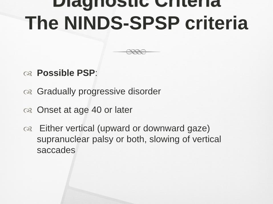
Diagnostic Criteria
The NINDS-SPSP criteria
Possible PSP:
Gradually progressive disorder
Onset at age 40 or later
Either vertical (upward or downward gaze)
supranuclear palsy or both, slowing of vertical
saccades

prominent postural instability with falls in the
first year of disease onset
No evidence of other diseases that could
explain the foregoing features, as indicated
by mandatory exclusion criteria
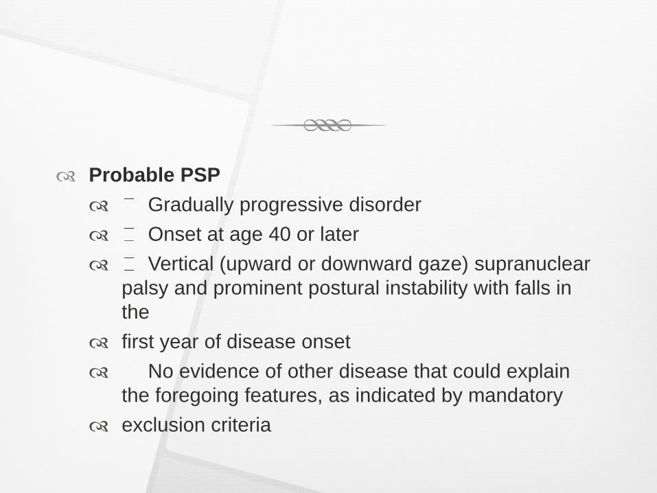
Probable PSP
Gradually progressive disorder
Onset at age 40 or later
Vertical (upward or downward gaze) supranuclear
palsy and prominent postural instability with falls in
the
first year of disease onset
No evidence of other disease that could explain
the foregoing features, as indicated by mandatory
exclusion criteria

Definite PSP
Clinically probable or possible PSP and histopathologic evidence of typical PSP
Mandatory exclusion criteria are the following:
Recent history of encephalitis
Alien limb syndrome, cortical sensory deficits, focal frontal or temporoparietal atrophy
Hallucinations or delusions unrelated to dopaminergic therapy
Cortical dementia of Alzheimer type
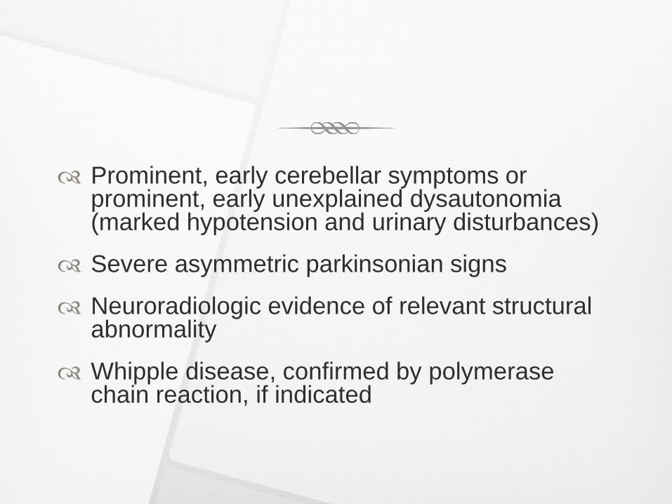
Prominent, early cerebellar symptoms or prominent, early unexplained dysautonomia (marked hypotension and urinary disturbances)
Severe asymmetric parkinsonian signs
Neuroradiologic evidence of relevant structural abnormality
Whipple disease, confirmed by polymerase chain reaction, if indicated

Supportive criteria for PSP are as follows:
Symmetric akinesia or rigidity, proximal more than distal
Abnormal neck posture, especially retrocollis
Poor or absent response of parkinsonism to levodopa therapy
Early dysphagia and dysarthria
Early onset of cognitive impairment including at least two of the following:

Apathy
Impairment in abstract thought
Decreased verbal fluency
Utilization or imitation behavior
Frontal release signs

Videos
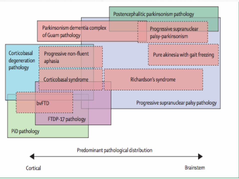

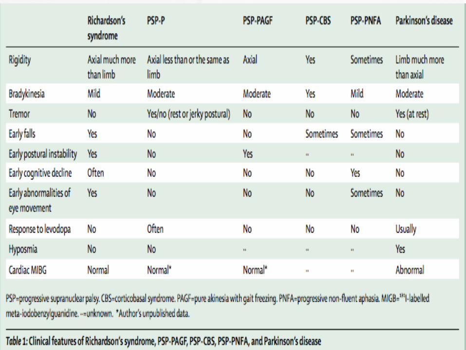
Multiple Phenotypes :
Subtypes
1- Classic phenotype: Richardson syndrome
2- Parkinson disease-like: PSP parkinsonism
3- Pure akinesia ( no appendicular rigidity): PSP-pure akinesia with gait freezing
4- Assymetric parkinsonism: PSP-corticobasal syndrome
5- Frontal-predominant dementia: PSP-frontotemporal dementia
6- PSP with speech/language dysfunction
(PSP-PNFA)
7- PSP with cerebellar ataxia ( PSP-C)
8- PSP with primary lateral sclerosis ( PSP-
PLS)
Richardson Syndrome
Mean age of onset is around 65 years
Early postural instability (backward falls)
Early personality changes (apathy)
Visual disturbances
Generalized slowness
Characteristic eye movement abnormalities:
1- Saccadic intrusions on primary gaze fixation.
2- Early loss of downward optokinetic nystagmus
3- Slowing of vertical saccades ( downward > upward)
4- Eventually vertical and horizontal gaze palsy

5- Involuntary persistence of ocular fixation
6- Difficulties in maintaining eye contact
7- Difficulties in reading
8- Diplopia & Blurred vision
9- Convergence insufficiency
10- Bilateral impairment of the antisaccade task
The ophthalmoparesis can be overcomed by
the oculocepahlic maneuvre.
Later on, vestibular-ocular reflex will be lost,
to the extent the paresis will shift to plegia.
This will not be overcomed by oculocephalic
maneuvre which indicates nuclear
involvement.
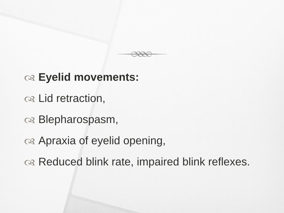
Eyelid movements:
Lid retraction,
Blepharospasm,
Apraxia of eyelid opening,
Reduced blink rate, impaired blink reflexes.
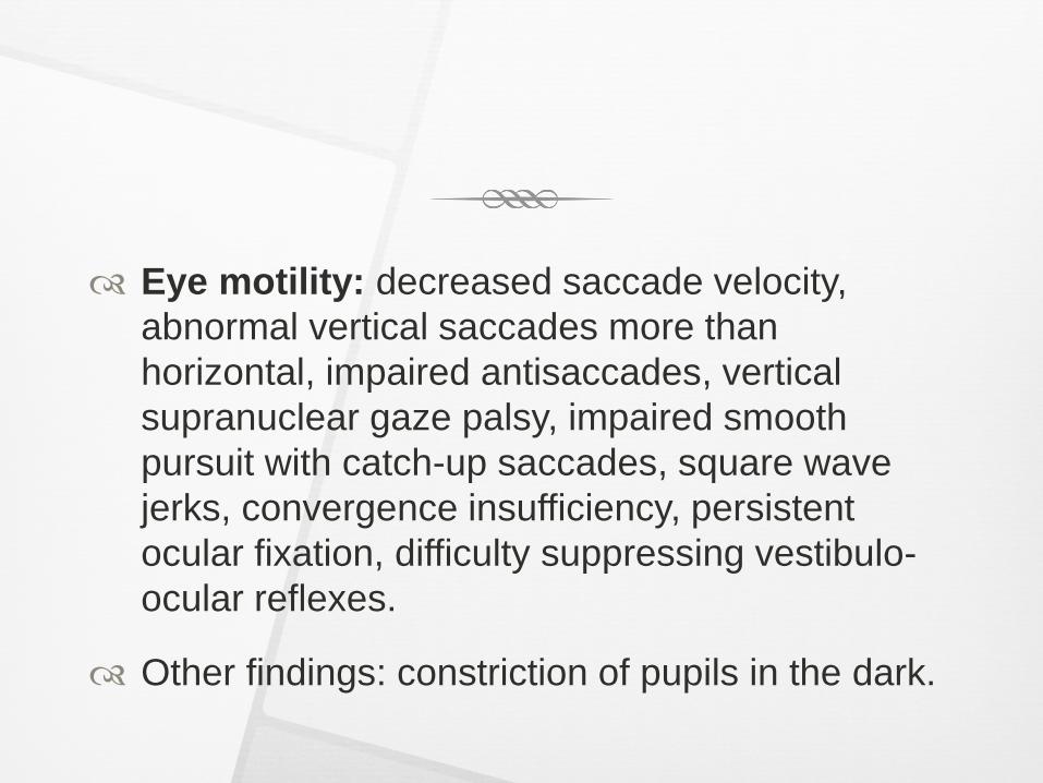
Eye motility: decreased saccade velocity,
abnormal vertical saccades more than
horizontal, impaired antisaccades, vertical
supranuclear gaze palsy, impaired smooth
pursuit with catch-up saccades, square wave
jerks, convergence insufficiency, persistent
ocular fixation, difficulty suppressing vestibulo-
ocular reflexes.
Other findings: constriction of pupils in the dark.

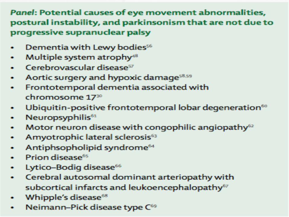
Differential: Vertical
Ophthalmoparesis
CBD
DLB
Wernike’s encepahlopathy
Dorsal midbrain syndrome
Prion disease
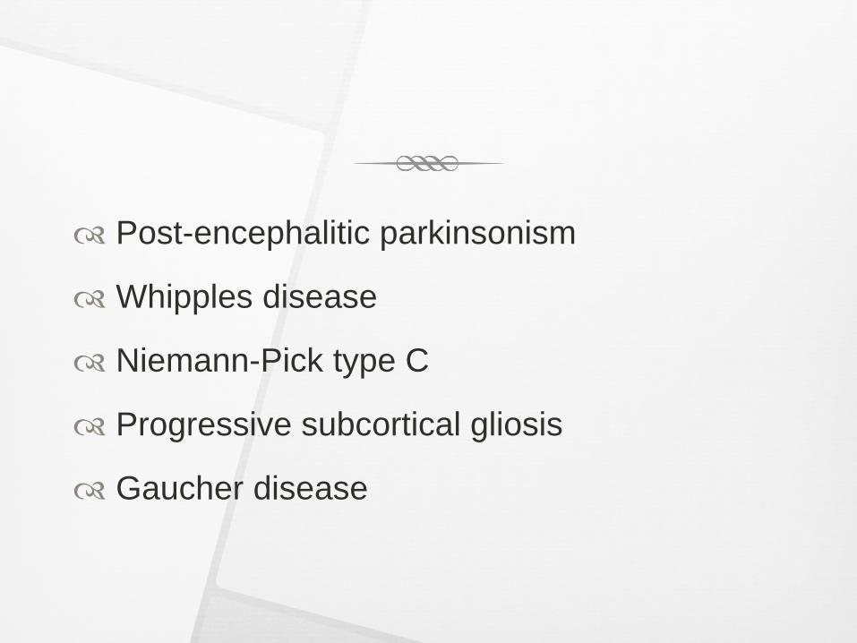
Post-encephalitic parkinsonism
Whipples disease
Niemann-Pick type C
Progressive subcortical gliosis
Gaucher disease
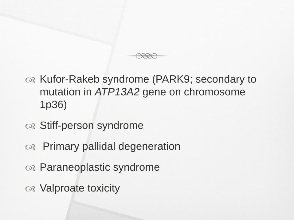
Kufor-Rakeb syndrome (PARK9; secondary to
mutation in ATP13A2 gene on chromosome
1p36)
Stiff-person syndrome
Primary pallidal degeneration
Paraneoplastic syndrome
Valproate toxicity
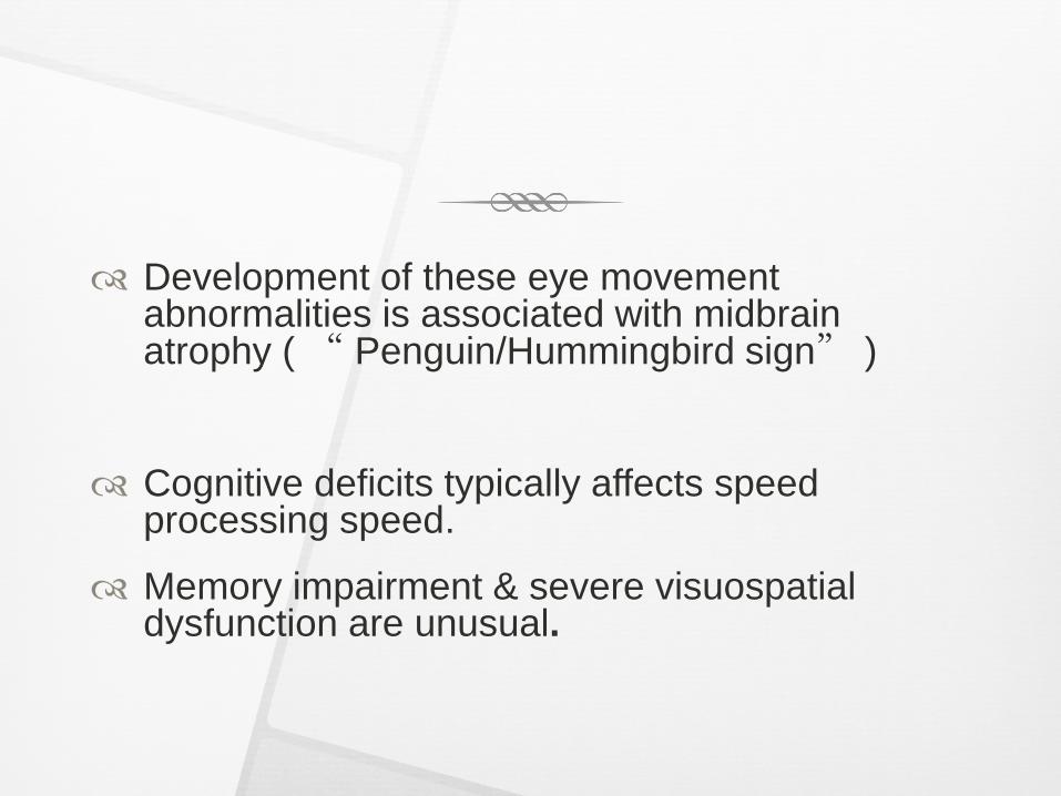
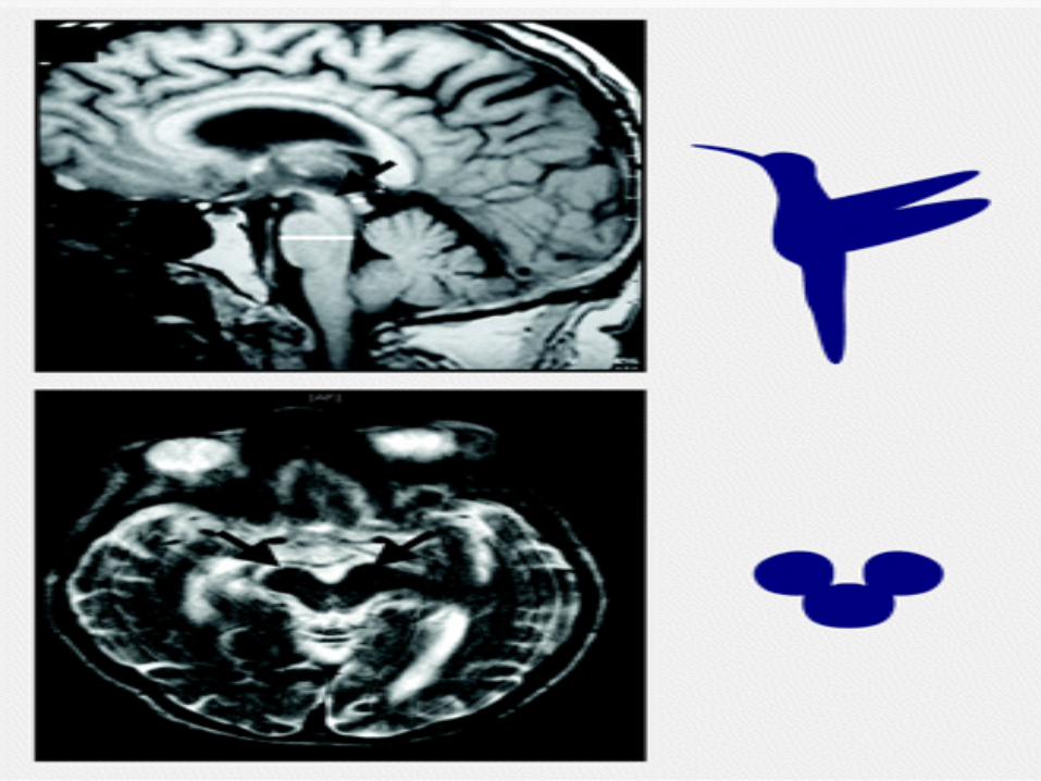
Development of these eye movement abnormalities is associated with midbrain atrophy ( “ Penguin/Hummingbird sign” )
Cognitive deficits typically affects speed processing speed.
Memory impairment & severe visuospatial dysfunction are unusual.

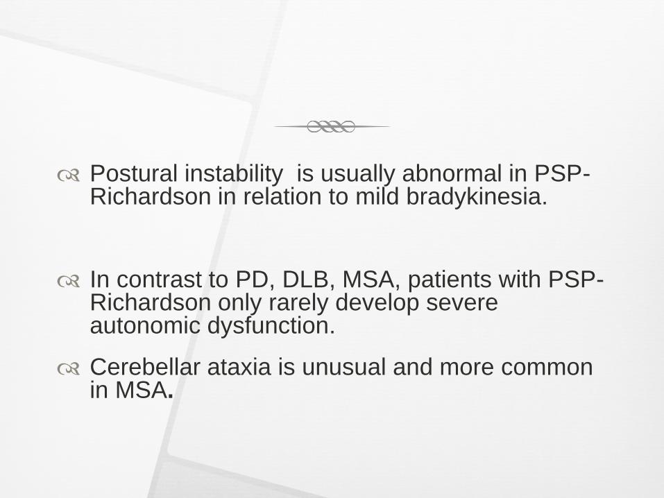
Postural instability is usually abnormal in PSP-Richardson in relation to mild bradykinesia.
In contrast to PD, DLB, MSA, patients with PSP-Richardson only rarely develop severe autonomic dysfunction.
Cerebellar ataxia is unusual and more common in MSA.

Patients usually become dependent on
others for care 3-4 yrs after disease onset.
Speech often becomes unintelligible.
Recurrent choking can lead to frequent
aspiration pneumonia.
Mean disease duration is about 7 yrs.
PSP-Parkinsonism
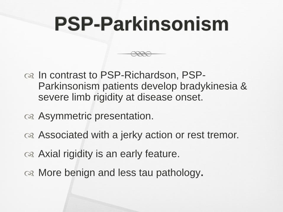
In contrast to PSP-Richardson, PSP-Parkinsonism patients develop bradykinesia & severe limb rigidity at disease onset.
Asymmetric presentation.
Associated with a jerky action or rest tremor.
Axial rigidity is an early feature.
More benign and less tau pathology.

Response to levodopa is a feature of early progressive PSP-P at a stage when postural instability, frontal cognitive impairment & vertical gaze palsy is uncommon.
Mentioned above are markers of disease severity which occur early in PSP-Richardson and later in PSP-P.
Disease duration to death is about 3 yrs.
Helpful pointers for PSP-P:
1- Rapid progression
2- Prominent axial symptoms
3- Suboptimal response to levodopa.
4- Drug-induced dyskinesias are unusual
PSP- Pure Akinesia
with Gait Freezing
Isolated bradykinesia, predominantly
affecting gait leading to freezing of gait.
Gait unsteadiness may develop upto 2 yrs
after the freezing of gait & gait initiation
failure develops.
Early hypophonia, hypomimia, and
micrographia
Axial rigidity with increasing neck stiffness in the absence of limb rigidity is a distinctive feature.
Supranuclear vertical gaze palsy and blepharospasm develop late.
In contrast to PSP-R, cognitive deficits & bradyphrenia are not prominent, but may occur late.
Mean disease duration is about 10 yrs.
PSP-Corticobasal
Syndrome
Unilateral ideomotor apraxia
Non-levodopa responsive parkinsonism
Myoclonus
Dystonia
Alien hand phenomenon

Non-motor features:
Aphasia
Cortical sensory deficit
Visuospatial deficits
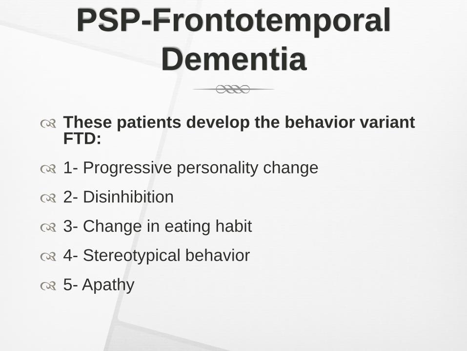
PSP-Frontotemporal
Dementia
These patients develop the behavior variant FTD:
1- Progressive personality change
2- Disinhibition
3- Change in eating habit
4- Stereotypical behavior
5- Apathy

Others develop the progressive non-fluent aphasia ( apraxia of speech) (PSP-PNFA).
These patients usually develop typical motor symptoms of PSP more than 5 yrs after presentation.
Differential Diagnosis
CBS
PD
NPH
Wilson disease
Alzheimer’s disease
Whipple’s disease
multi-infarct dementia
midbrain tumors
prion disease and syringomyelia
Niemann-Pick type C

Video: midbrain tumor
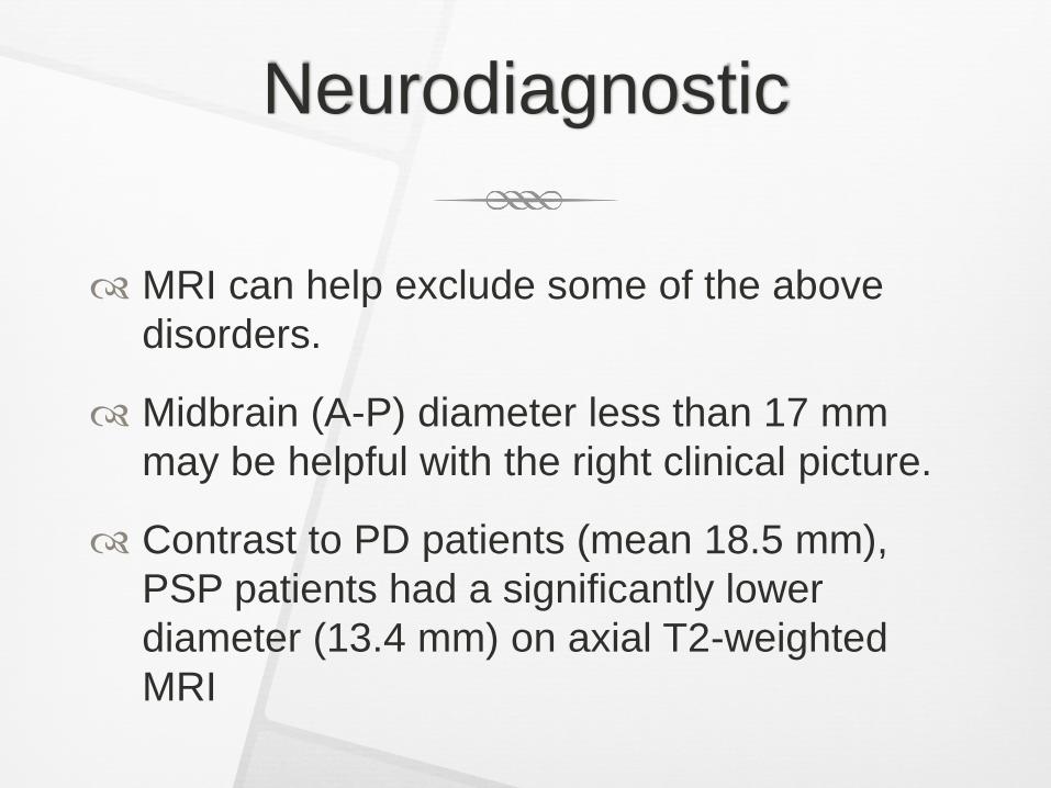
Neurodiagnostic
MRI can help exclude some of the above
disorders.
Midbrain (A-P) diameter less than 17 mm
may be helpful with the right clinical picture.
Contrast to PD patients (mean 18.5 mm),
PSP patients had a significantly lower
diameter (13.4 mm) on axial T2-weighted
MRI

“Hummingbird sign”, “Penguin sign” and
“Mickey mouse brain”, “Morning glory”
Midsagittal MRI, the average midbrain area
of patients with PSP was 56 mm2, which was
significantly smaller than that of patients with
PD (103 mm2) or MSA-P (97.2 mm2)

Using diffusion-weighted MRI (DWI-MRI)
Using voxel-based morphometry:
CBD patients had marked asymmetric (L > R) pattern of atrophy involving premotor cortex, superior parietal lobules, and striatum,
PSP was characterized by atrophy of the midbrain, pons, thalamus, and striatum.

Treatment Paradigm
Supportive therapy:
Pharmacologic
Multidisciplinary team: PT, OT, Speech pathologist, social worker, etc.
The dopaminergic meds do not improve symptoms in PSP to the same extent as PD.
There is minimal benefit in PSP-P, which may diminish for months to years.
Levodopa responsiveness can be tested by administering escalating doses ( with COMT) upto 1200 mg/d for at least 1 month.

Dopamine agonists are less effective, with high profile side-effects.
Amantadine is occassionally helpful in improving motor symptoms including:
1- Gait freezing
2- Dysphagia
3- Sialorrhea
Anticholinergics should be avoided as they may worsen cognition.
Antispasmodics for the treatment of overactive urinary bladder ( solifenacin) M3 antagonist.
Botulinum toxin used for blepharospasm, limb dystonia, apraxia of eyelid opening, and neurogenic bladder.
Mood and cognitive issues should be
addressed.
Donepezil performed poorly in a clinical trial
with worsening of motor scores.
Rivastigamine trial could be attempted.

Evaluation for swallowing difficulties is
recommended.
PEG tube placement not just for dysphagia
but also for addressing nutritional needs.
Future Directions
Drugs targeting dopaminergic, cholinergic (Physostigmine, Donepezil, Rivastigmine) or GABAergic (Zolpidem, Gabapentin) deficits are not clinically effective in PSP.
Lithium ( FAILED)
Tideglusib (GSK-3b inhibitor) (FAILED)
Davunetide (FAILED)
A phase III trial with CoQ10 (FAILED)
Rasagiline (FAILED)
Other inhibitors of Tau aggregation :
Methylene blue (under investigation)
Microtubule stabilizers (Taxol, Epothilone D and TPI-28) (under investigation)
Corticobasal
Syndrome/Degeneration
Rare, progressive neurodegenerative
disorder
Multiple phenotypes/overlap between PSP,
FTD ( Picks) , Alzheimer.
Age of onset is in the 5th to 7th decade.
Predominance in women ?
Classic presenataion:
Asymmetric parkinsonism
Myoclonus ( stimulus-sensitive)
Dystonia
Ideomotor apraxia & cortical sensory loss
Alien limb phenomena

Language disturbances:
Mild impairment to severe progressive non-fluent aphasia or even complete mutism.
Visual neglect with agnosia and or optic ataxia is seen at times.
Apathy, irritability and depression are common.
Asymmetric onset of dystonia mostly in upper limbs or hemi-dystonia along with myoclonus involving upper limbs and face can be seen.
Myoclonus can be focal with action or can be stimulus-sensitive.
Upper motor neuron signs can be present.
Dysarthria and dysphagia.
Eye motility:
Impaired convergence
Increased horizontal saccade latency with
preserved saccade velocity.
Impairment of upward gaze or vertical
saccades.

Video
Many Faces of CBD
Typical corticobasal syndrome
CBD-FTD
CBD-Aphasia/Apraxia of speech
CBD-PSP-like syndrome
CBD-Posterior cortical atrophy
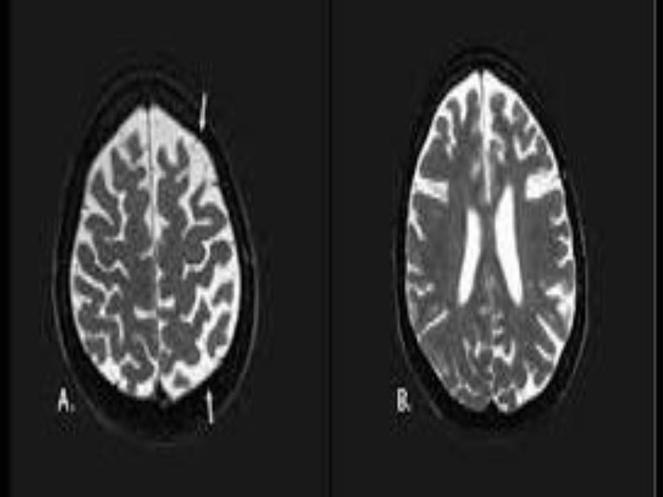
Diagnosis
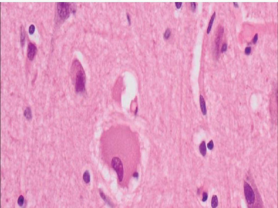
Definitive diagnosis is always
histopathological.
MRI Brain is obtained to rule out secondary
etiologies.
Cortical atrophy in frontal and parietal areas
along with ballooned and achromatic
neurons seen on microscopy.


Probable CBS: asymmetric presentation and at least two of:
a) limb rigidity or akinesia,
b) limb dystonia,
c) limb myoclonus, plus two of:
d) Orobuccal or limb apraxia, e) cortical sensory deficit, f) alien limb phenomena (more than simple levitation)
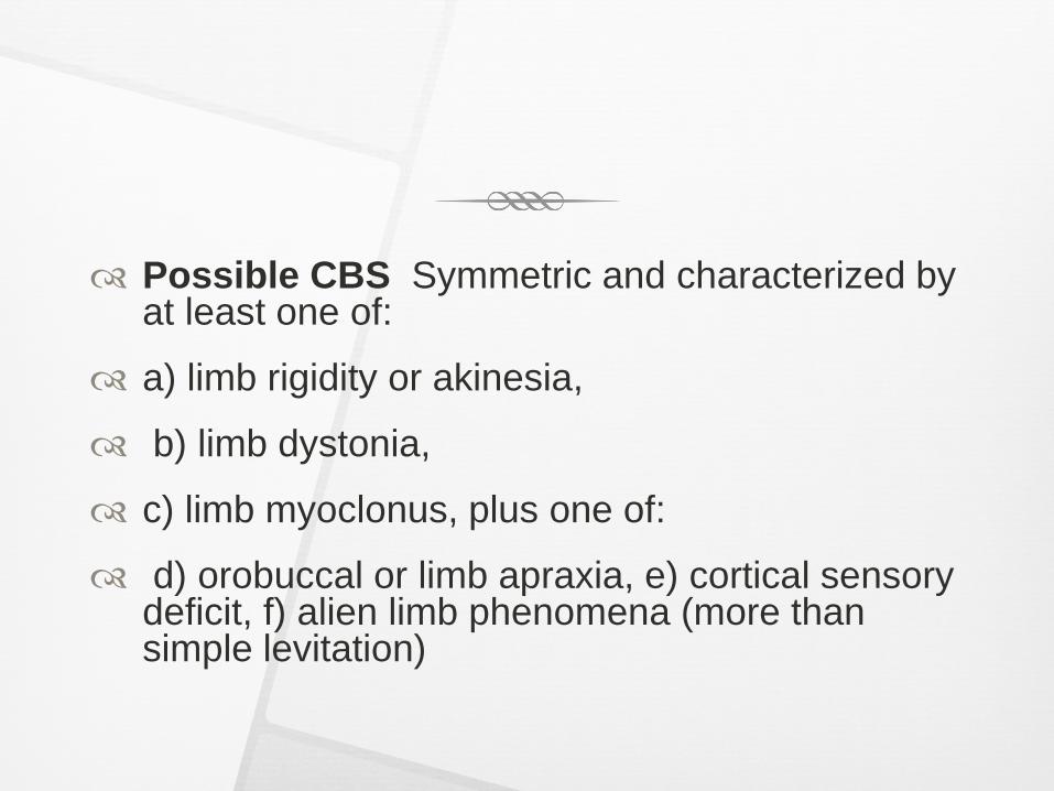
Possible CBS Symmetric and characterized by at least one of:
a) limb rigidity or akinesia,
b) limb dystonia,
c) limb myoclonus, plus one of:
d) orobuccal or limb apraxia, e) cortical sensory deficit, f) alien limb phenomena (more than simple levitation)

Frontal behavioral-spatial syndrome
(FBS): characterized by two of:
a) Executive dysfunction,
b) Behavioral or personality changes,
c) Visuospatial deficits

Nonfluent/agrammatic variant of primary progressive aphasia (naPPA),
characterized by effortful, agrammatic speech plus at least one of:
a) impaired grammar/sentence comprehension with relatively preserved single word comprehension, or b) groping, distorted speech production (apraxia of speech)
Progressive supranuclear palsy syndrome (PSPS): characterized by three of:
a) axial or symmetric limb rigidity or akinesia, b) postural instability or falls,
c) urinary incontinence,
d) behavioral changes, e) supranuclear vertical gaze palsy or decreased velocity of vertical saccades
More specific clinical research criteria for probable sporadic CBD are:
Presentation with insidious onset and gradual progression
A one-year minimum duration of symptoms
Age ≥50 years at onset
Permitted phenotypes are probable CBS, or FBS or naPPA plus at least one CBS feature (a to f above)
Management
Trial of levodopa at 1,000-1,200 mg but the
response may be lacking or diminishes with
time.
Myoclonus: valproic acid, clonazepam or
levatiracetam.
Dystonia can respond to botulinum toxin
injection.
Speech therapy (dysphagia management),
physical and occupational therapy.
Future Directions
Disease-modifying treatment is not yet
available for CBD.
Ongoing placebo controlled Phase I trial,
testing the efficacy of TPI 287 (a microtubule
inhibitor) in patients with 4 R tauopathies.
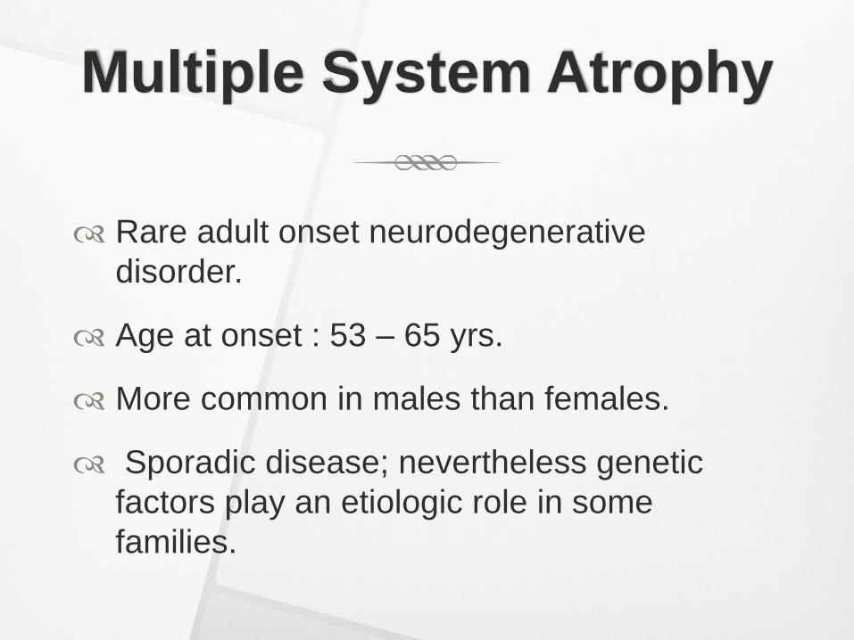
Multiple System Atrophy
Rare adult onset neurodegenerative
disorder.
Age at onset : 53 – 65 yrs.
More common in males than females.
Sporadic disease; nevertheless genetic
factors play an etiologic role in some
families.

Pathogenic mechanisms underlying MSA
remain partially unclear.
Evidence suggests that it is an
oligodendrogliopathy.
Possible genetic factors: COQ2 mutation,
SHC2 copy number loss, SNCA
multiplications or SNP’s.

Possible genetic factors: COQ2 mutation,
SHC2 copy number loss, SNCA
multiplications or SNP’s.
Possible environmental factors: agricultural
employment, organic solvents, plastic
monomers, pesticides and metal dusts.
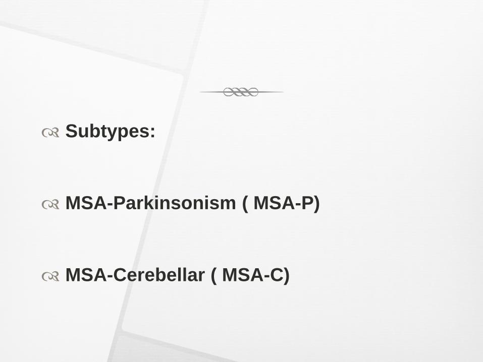
Subtypes:
MSA-Parkinsonism ( MSA-P)
MSA-Cerebellar ( MSA-C)
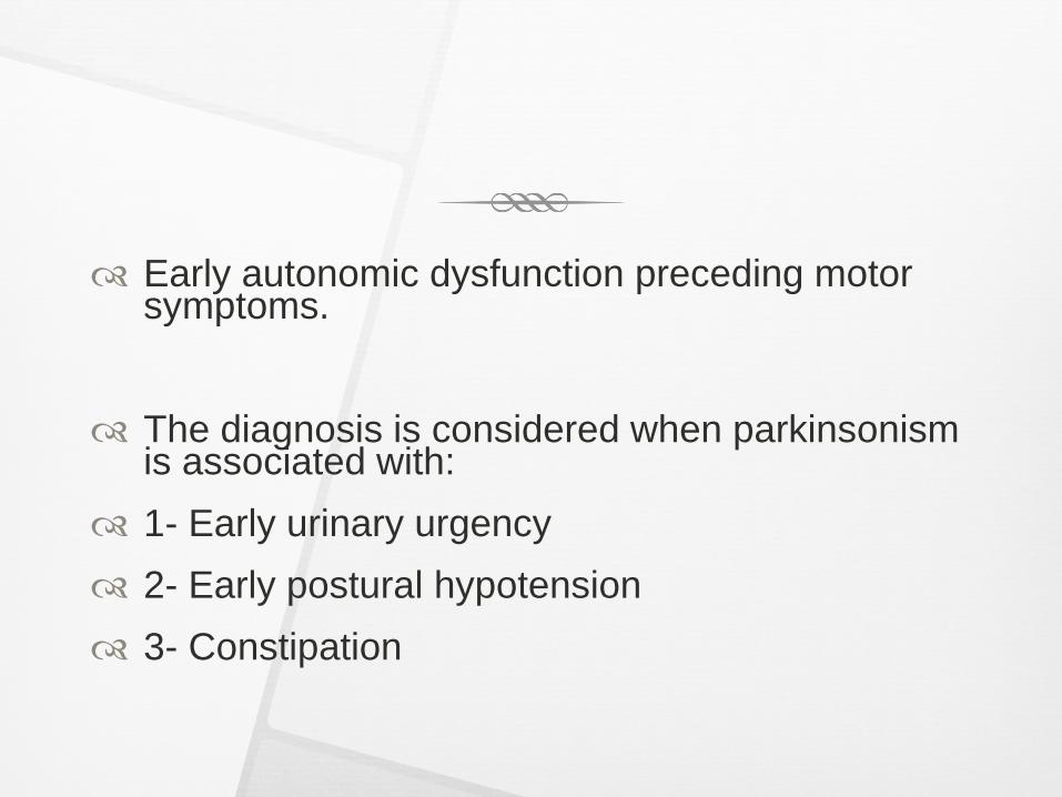
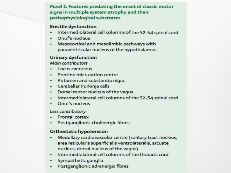
Early autonomic dysfunction preceding motor symptoms.
The diagnosis is considered when parkinsonism is associated with:
1- Early urinary urgency
2- Early postural hypotension
3- Constipation

4- Early REM sleep behavior disorder
5- Early erectile dysfunction
6- Sleep apnea
7- Early morning headaches
8- Nocturnal inspiratory stridor
9- Levodopa-induced facial/axial dyskinesias
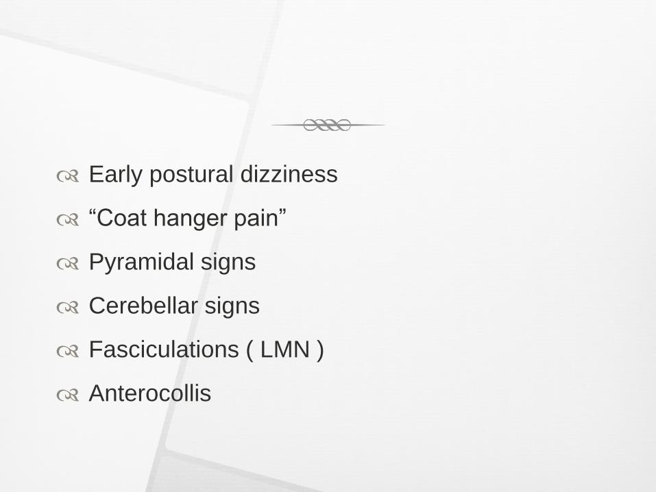
Early postural dizziness
“Coat hanger pain”
Pyramidal signs
Cerebellar signs
Fasciculations ( LMN )
Anterocollis
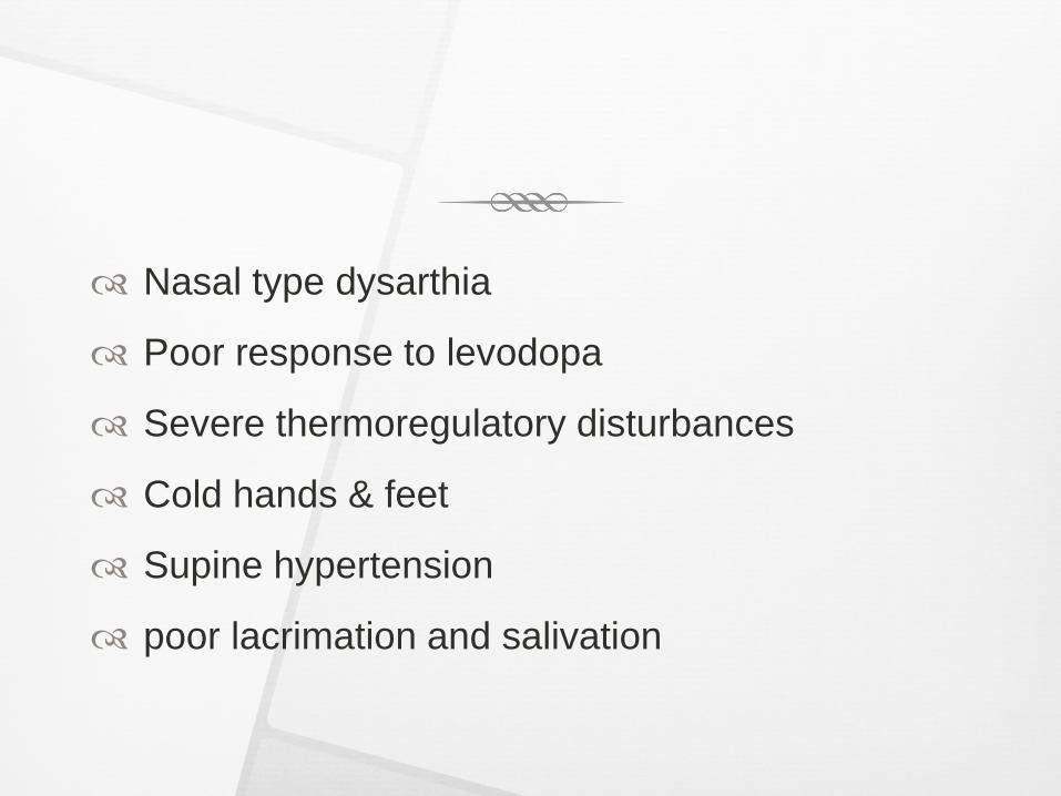
Nasal type dysarthia
Poor response to levodopa
Severe thermoregulatory disturbances
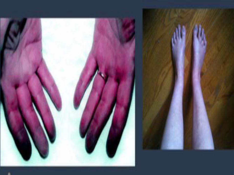
Cold hands & feet
Supine hypertension
poor lacrimation and salivation


Parkinsonism is seen in 90% of cases.
76% of patients with MSA-C have
parkinsonism and 54% of MSA-P patients
have cerebellar symptoms.
Tremor is seen in 80% of MSA patients and
is more common in MSA-P.
The parkinsonism of MSA is usually symmetric.
Approx thirty percent responds to levodopa.
Bradykinesia & rigidity progress faster than PD.
Postural instability & falls emerge within the first 3 yrs of disease onset.
Cognitive dysfunction ( prefrontal ) more severe in MSA-P than MSA-C.
Pseudobulbar affect more in MSA-C.
Polyminimyoclonus is seen.
Jerky myoclonic action tremor, stimulus-sensitive distal myoclonus is more common in MSA-P.
Cerebellar dysfunction with:
Intention tremor
Gait and appendicular ataxia
Dysarthria
Sustained gaze-evoked nystagmus with hypometric saccades is present.
Dystonia can be seen in the form of:
Laryngeal stridor and facial dystonia
(levodopa-induced).
Anterocollis, camptocormia and truncal
dystonia (“Pisa syndrome”).
Contractures of hands or feet.


REM sleep behavioral disorder can be seen
in 70% .
Central and obstructive sleep apnea can
increase risk of sudden death.
Eye motility:
Spontaneous or gaze-evoke nystagmus
Square-wave jerks
Slow and hypometric saccades
Reduced vestibulo-ocular reflex suppression, reduced vertical gaze, impaired smooth pursuit.
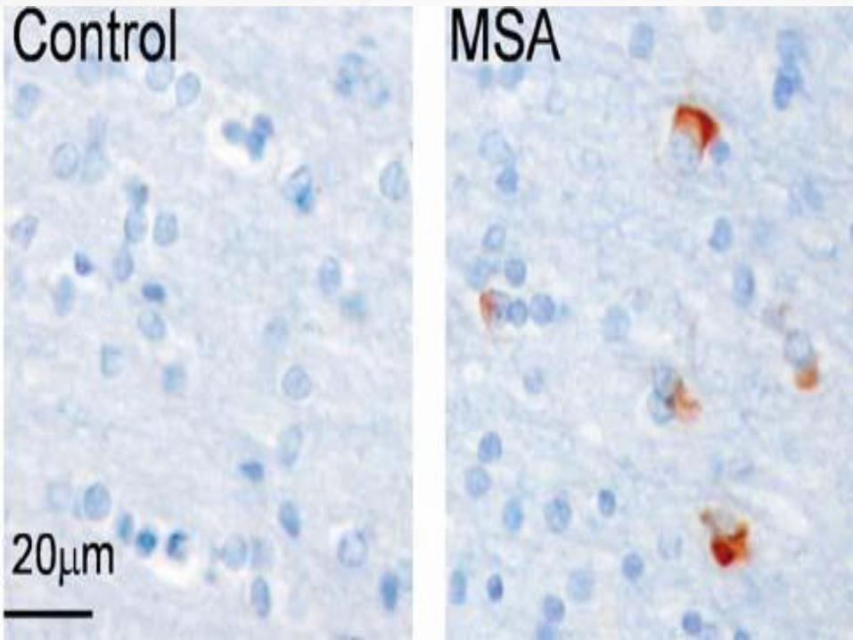
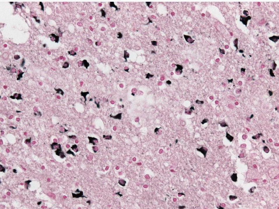
Diagnostic Criteria
A definite diagnosis requires the
neuropathologic findings of widespread and
abundant CNS-synuclein–positive glial
cytoplasmic inclusions (Papp–Lantos
inclusions).
Neurodegenerative changes in striatonigral
or olivopontocerebellar structures.
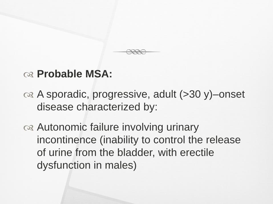
Probable MSA:
A sporadic, progressive, adult (>30 y)–onset
disease characterized by:
Autonomic failure involving urinary
incontinence (inability to control the release
of urine from the bladder, with erectile
dysfunction in males)

Or an orthostatic decrease of blood pressure within 3 min of standing by at least 30 mm Hg systolic or 15 mm Hg diastolic and
● Poorly levodopa-responsive parkinsonism (bradykinesia with rigidity, tremor, or postural instability) or ● A cerebellar syndrome (gait ataxia with cerebellar dysarthria, limb ataxia, or cerebellar oculomotor dysfunction)
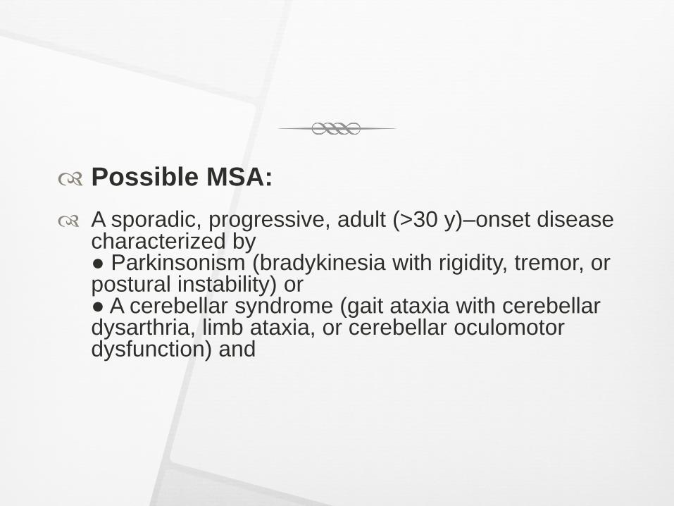
Possible MSA:
A sporadic, progressive, adult (>30 y)–onset disease characterized by ● Parkinsonism (bradykinesia with rigidity, tremor, or postural instability) or ● A cerebellar syndrome (gait ataxia with cerebellar dysarthria, limb ataxia, or cerebellar oculomotor dysfunction) and

● At least one feature suggesting autonomic
dysfunction (otherwise unexplained urinary
urgency, frequency or incomplete bladder
● At least one of the additional features
shown in table 3

Additional features of possible MSA
Possible MSA-P or MSA-C
● Babinski sign with hyperreflexia
● Stridor

Possible MSA-P
● Rapidly progressive parkinsonism ● Poor response to levodopa ● Postural instability within 3 y of motor onset ● Gait ataxia, cerebellar dysarthria, limb ataxia, or cerebellar oculomotor dysfunction ● Dysphagia within 5 y of motor onset ● Atrophy on MRI of putamen, middle cerebellar peduncle, pons, or cerebellum ● Hypometabolism on FDG-PET in putamen, brainstem, or cerebellum
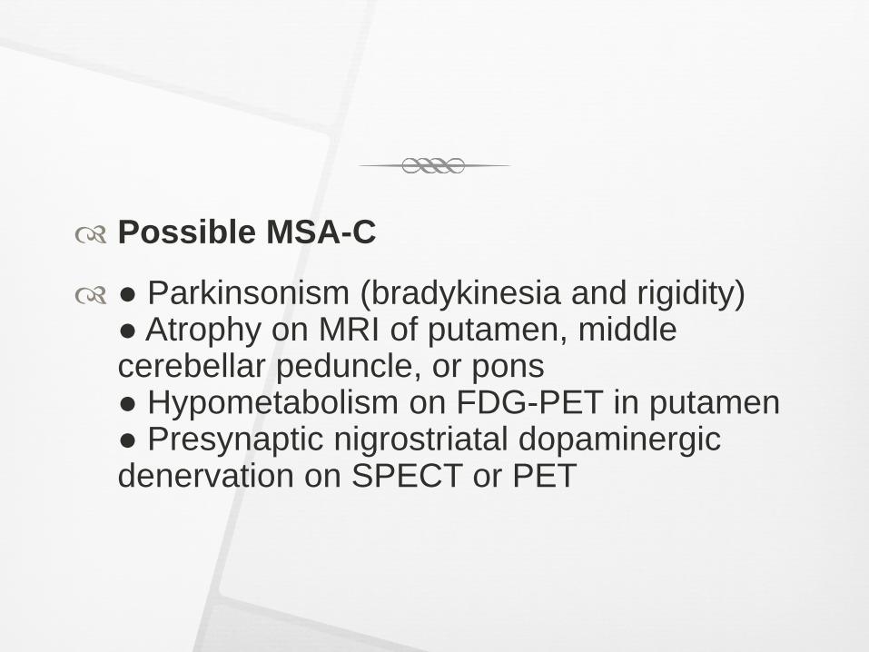
Possible MSA-C
● Parkinsonism (bradykinesia and rigidity) ● Atrophy on MRI of putamen, middle cerebellar peduncle, or pons ● Hypometabolism on FDG-PET in putamen ● Presynaptic nigrostriatal dopaminergic denervation on SPECT or PET
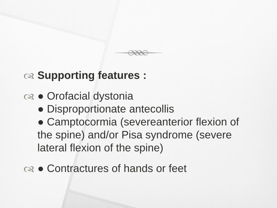
Supporting features :
● Orofacial dystonia
● Disproportionate antecollis
● Camptocormia (severeanterior flexion of
the spine) and/or Pisa syndrome (severe
lateral flexion of the spine)
● Contractures of hands or feet
● Contractures of hands or feet ● Inspiratory
sighs
● Severe dysphonia
● Severe dysarthria sclerosis
● New or increased snoring
● Cold hands and feet● Pathologic laughter
or crying
Nonsupporting features :
● Classic pill-rolling rest tremor ● Clinically significant neuropathy ● Hallucinations not induced by drugs
● Onset after age 75 y ● Family history of ataxia or parkinsonism ● Dementia (on DSM-IV) ● White matter lesions suggesting multiple
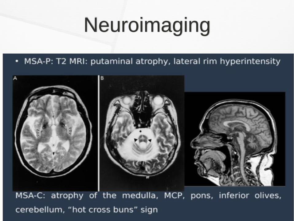
Neuroimaging


Autonomic studies reveal early abnormalities compared to PD:
Orthostatic hypotension
Abnormal QSART testing
Urinary residual volume more than 100 ml.
Abnormal norepinephrine levels along and an abnormal composite autonomic score (CASS score).
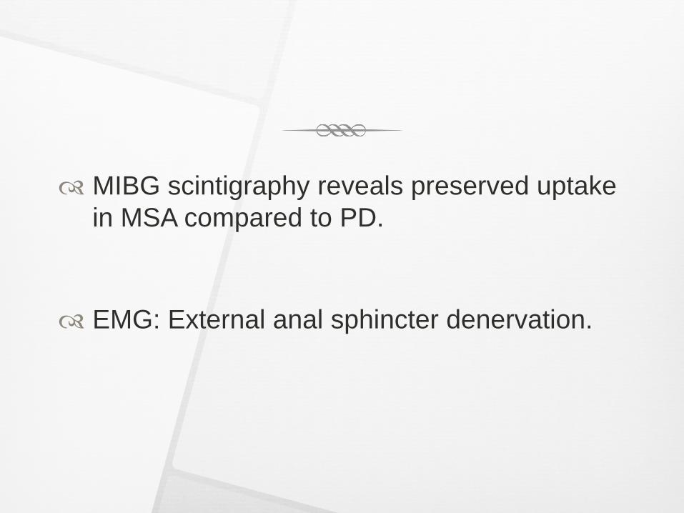
MIBG scintigraphy reveals preserved uptake
in MSA compared to PD.
EMG: External anal sphincter denervation.
The median survival of either phenotypes is about 8 yrs.
Factors predicting short disease survival:
1- Early autonomic failure.
2- Older age of onset. Usually age of onset: 50’s
3- Short interval from disease onset to frequent falling.
4- Cognitive disability.
5- Unintelligible speech.
6- Severe dysphagia.
7- Dependence on wheel chair for mobility.
8- Urinary catheter use.
9- Lack of admission to a nursing home facility.

Management
Multidisciplinary team approach.
Dopaminergic meds:
Only 30% of patients respond to levodopa
with short-lived benefit.
Dosing is limited by nausea, hypotension
and hallucinations.

Approx one-third of patients with MSA-P will
benefit.
Levodopa/carbidopa ( Sinemet 100/25 po tds) to start low by ½ tab and titrate slowly over a weekly period to the target dose.
Can reach upto 1200mg/d, according to the best response/ emerging side effects.

There is no specific therapy for cerebellar
symptoms.
Dystonia can be treated with botulinum toxin
injections.
Autonomic Dysfunction:
Management of orthostatic hypotension is critical.
Non-pharmacologic approach:
1- increasing dietary salt intake.
2- Avoid caffeinated drinks.
3- D/c or reduce the dose of any antihypertensive meds.
4- Getting up slowly from a seated/lying position.
5- Wearing thigh-high compression stockings.
6- Leg elevation while lying.
7- Monitoring for supine hypertension.

Pharmacologic:
Fludrocortisone: ( Florinef): Increases blood volume.
Midodrine: Increases peripheral vascular resistance.
If both are ineffective, indomethacin/pyridostigmine can be used.
Droxidopa: alpha/beta adrenergic agonist
Converts to norepinephrine by dopa
decarboxylase.
Dose: 100 mg po tds.
Last dose to be given 3 hrs prior to sleep to
avoid supine hypertension.
Urologic Symptoms:
Urinary retention or incontinence are usually present early in MSA.
Urodynamic studies determines the type of neurogenic urinary bladder.
Bladder spasticity responds to peripherally acting anticholinergic agents or botulinum toxin.
Occassionally, intermittent or suprapubic catheterization maybe required.
REM-BD can respond to clonazepam or
melatonin.
Sleep study with management of obstructive
sleep apnea.

Supportive therapy including allied health
care team:
Tracheostomy/gastrostomy maybe considered in
selected patients with:
Cervical, laryngeal, & pharyngeal dystonia
causing upper airway obstruction.
These procedure will improve the quality of life.
Tracheostomy:
May effectively relieve airway obstruction at
the laryngeal level
Does not eliminate the risk of sudden death
due to fatal sleep apnea.
Referral should be sent to Urology for
management of sexual dysfunction and
urinary abnormalities.
Physical, speech and occupational therapies
are part of rehabilitation with this progressive
disorder.

Future Directions
No clinical trials have proven efficacy for MSA progression.
MSA specific biomarkers are essential to facilitate the early identification
Enrollment of patients in disease- modifying clinical trials.
Blood and CSF alpha-synuclein levels (not sensitive and specific)
Riluzole, Minocycline, Rifampicin or
Rasagiline have not shown any benefit.
A global MSA registry (GLOMSAR) has been
established to facilitate accelerated
interventional target delivery, biomarker
development and early patient recruitment.
Conclusion
Atypical parkinsonism can present initially as idiopathic PD ( PSP & MSA).
Close follow up is crucial.
Don’t rush into conclusions and send patients for DBS in the very early stages.
Currently, no effective Rx that alter the natural history of disease or alter survival.
Current therapies only focus on symptomatic management.
Clinical trials have been limited by poor funding, small size, & lack of biomarkers.
Recent breakthrough in genetics, molecular biology, & neuroimaging will help in developing targeted therapies with disease-modifying properties.
Thank You
References:
Principles and practice of Movement Disorders; Fahn, Jankovic & Hallett
Ali, Khalid, and Huw R. Morris. "Parkinson's disease: chameleons and mimics." Practical neurology 15,
no. 1 (2015): 14-25.
Eschlbo ̈ck, S., F. Krismer, and G. K. Wenning. "Interventional trials in atypical parkinsonism.” Parkinsonism & related disorders (2015).
Villemagne, Victor L., Michelle T. Fodero-Tavoletti, Colin L. Masters, and Christopher C. Rowe. "Tau
imaging: early progress and future directions." The Lancet Neurology 14, no. 1 (2015): 114-124.
Progressive Supranuclear Palsy; LI Golbe, Semin Neurol, 2014
Litvan, I., Y. Agid, D. Calne, G. Campbell, B. Dubois, R. C. Duvoisin, C. G. Goetz et al. "Clinical research .