intestinal udp-glucuronosyltransferase as a potential
TRANSCRIPT
RESEARCH ARTICLE
Intestinal UDP-glucuronosyltransferase as a
potential target for the treatment and
prevention of lymphatic filariasis
Alexander F. FlynnID1, M. Gordon JoyceID
2,3, Rebekah T. Taylor4, Sasisekhar Bennuru5,
Alyssa R. LindroseID1, Spencer L. Sterling1, C. Paul MorrisID
6, Thomas B. Nutman5,
Edward MitreID1*
1 Department of Microbiology, F. Edward Hebert School of Medicine, Uniformed Services University of the
Health Sciences, Bethesda, Maryland, United States of America, 2 U.S. Military HIV Research Program,
Walter Reed Army Institute of Research, Silver Spring, Maryland, United States of America, 3 Henry M.
Jackson Foundation for the Advancement of Military Medicine, Inc., Bethesda, Maryland, United States of
America, 4 Department of Biology, Frostburg State University, Frostburg, Maryland, United States of
America, 5 National Institute of Allergy and Infectious Diseases, National Institutes of Health, Bethesda,
Maryland, United States of America, 6 Department of Pathology, Johns Hopkins Hospital, Baltimore
Maryland, United States of America
Abstract
Lymphatic filariasis (LF), a morbid disease caused by the tissue-invasive nematodes
Wuchereria bancrofti, Brugia malayi, and Brugia timori, affects millions of people worldwide.
Global eradication efforts have significantly reduced worldwide prevalence, but complete
elimination has been hampered by limitations of current anti-filarial drugs and the lack of a
vaccine. The goal of this study was to evaluate B. malayi intestinal UDP-glucuronosyltrans-
ferase (Bm-UGT) as a potential therapeutic target. To evaluate whether Bm-UGT is essen-
tial for adult filarial worms, we inhibited its expression using siRNA. This resulted in a 75%
knockdown of Bm-ugt mRNA for 6 days and almost complete suppression of detectable
Bm-UGT by immunoblot. Reduction in Bm-UGT expression resulted in decreased worm
motility for 6 days, 70% reduction in microfilaria release from adult worms, and significant
reduction in adult worm metabolism as detected by MTT assays. Because prior allergic-sen-
sitization to a filarial antigen would be a contraindication for its use as a vaccine candidate,
we tested plasma from infected and endemic normal populations for Bm-UGT-specific IgE
using a luciferase immunoprecipitation assay. All samples (n = 35) tested negative. We then
tested two commercially available medicines known to be broad inhibitors of UGTs, sulfinpy-
razone and probenecid, for in vitro activity against B. malayi. There were marked macrofilari-
cidal effects at concentrations achievable in humans and very little effect on microfilariae. In
addition, we observed that probenecid and sulfinpyrazone exhibit a synergistic macrofilarici-
dal effect when used in combination with albendazole. The results of this study demonstrate
that Bm-UGT is an essential protein for adult worm survival. Lack of prior IgE sensitization in
infected and endemic populations suggest it may be a feasible vaccine candidate. The find-
ing that sulfinpyrazone and probenecid have in vitro effects against adult B. malayi worms
suggests that these medications have promise as potential macrofilaricides in humans.
PLOS Neglected Tropical Diseases | https://doi.org/10.1371/journal.pntd.0007687 September 12, 2019 1 / 25
a1111111111
a1111111111
a1111111111
a1111111111
a1111111111
OPEN ACCESS
Citation: Flynn AF, Joyce MG, Taylor RT, Bennuru
S, Lindrose AR, Sterling SL, et al. (2019) Intestinal
UDP-glucuronosyltransferase as a potential target
for the treatment and prevention of lymphatic
filariasis. PLoS Negl Trop Dis 13(9): e0007687.
https://doi.org/10.1371/journal.pntd.0007687
Editor: Benjamin L Makepeace, University of
Liverpool, UNITED KINGDOM
Received: April 27, 2019
Accepted: August 5, 2019
Published: September 12, 2019
Copyright: This is an open access article, free of all
copyright, and may be freely reproduced,
distributed, transmitted, modified, built upon, or
otherwise used by anyone for any lawful purpose.
The work is made available under the Creative
Commons CC0 public domain dedication.
Data Availability Statement: All relevant data are
within the manuscript and its Supporting
Information files.
Funding: This work was supported by the
Uniformed Services University (Grant#
F173424117), the USU Center for Global Health
Engagement (Grant# CGHE-73-8985), and by the
Division of Intramural Research, National Institute
of Allergy and Infectious Diseases, National
Institutes of Health. The funders had no role in
Author summary
Brugia malayi is a parasitic nematode and one of the causative agents of lymphatic filaria-
sis, a disease that affects 70 million people worldwide. Currently, there are no effective
therapeutics that kill adult filarial parasites when given as a short course. This limitation
has hampered global eradication efforts. Studies have shown that the intestinal tract in
nematodes can be effectively targeted by drugs and antibodies. Given this potential, we
decided to investigate B. malayi intestinal UDP-glucuronosyltransferase as a potential
therapeutic target. We determined that this protein is essential for B. malayi adult worm
survival, as gene-expression knockdown rapidly decreased motility, fecundity, and micro-
filarial release. We also identified two FDA-approved UGT inhibitors that cause death of
adult filariae in vitro. This is a critical finding due to the need for effective macrofilaricides
and the potentially rapid translatability of these drugs for use in filaria-infected people.
Finally, we showed that serum from filarial patients does not contain specific IgE to Bm-
UGT and thus this protein would likely not induce allergic reaction if given as a vaccine
antigen to endemic populations.
Introduction
Lymphatic filariasis (LF) is a debilitating disease caused by the tissue-invasive nematodes
Wuchereria bancrofti, Brugia malayi, and Brugia timori. Currently, there are ~ 70 million peo-
ple infected worldwide and over a billion people at risk for infection [1]. Since 2000, the Global
Programme to Eliminate Lymphatic Filariasis has substantially reduced the number of people
infected or at risk for infection [1]. However, it has become apparent that new strategies must
be implemented in order to attain global eradication of LF [2, 3].
Development of new therapeutics that target adult filarial worms would greatly enhance
our ability to eliminate lymphatic filariasis. When given individually, the antifilarial drugs
diethylcarbamazine (DEC), ivermectin (IVM), and albendazole are effective against the micro-
filaria (Mf) stage but exhibit little activity against adult filarial worms [4]. Use of all three medi-
cations together appears to have a macrofilaricidal effect [5], but due to the adverse effects
caused by their potent microfilaricidal activity DEC and ivermectin cannot be used for mass
drug administration (MDA) in areas endemic for Loa loa or Onchocerca volvulus. Therefore,
development of a short-course macrofilaricidal agent or a vaccine would be very valuable for
eradication efforts.
Unlike cestodes and trematodes, nematodes have a complete intestinal tract. Over the past
15 years, intestinal proteins of Necator americanus (hookworm) and Haemonchus contortus(barber pole worms) have been shown to be effective vaccine candidates in animal models [6–
11]. Considering this work, our group performed a proteomic analysis of the intestine, body
wall, and reproductive tract of adult B. malayi worms to potentially identify novel drug and
vaccine targets for lymphatic filariasis [12]. We identified 396 proteins that were specific to the
intestinal tract of the adult worms. Of these intestinal proteins, we selected a subset for evalua-
tion as drug and vaccine candidates based on high homology with other filarial species, extra-
cellular domains with accessibility to drugs and antibody, and predicted function.
In this study, an adult B. malayi intestinal protein, UDP-glucuronosyltransferase (Bm-
UGT), was identified as a potential therapeutic target. The protein was predicted to have an
enzymatic function that could be inhibited. Furthermore, structural analysis of Bm-UGT by
InterPro revealed a large extracellular domain that could be targeted by therapeutics. We
determined that this protein was essential for worm survival using small interfering RNA
Intestinal UDP-glucuronosyltransferase as a therapeutic target of lymphatic filariasis
PLOS Neglected Tropical Diseases | https://doi.org/10.1371/journal.pntd.0007687 September 12, 2019 2 / 25
study design, data collection and analysis, decision
to publish, or preparation of the manuscript.
Competing interests: The authors have declared
that no competing interests exist.
(siRNA) to knockdown expression. Importantly, we identified two FDA-approved commer-
cially available UGT inhibitors that exhibit macrofilaricidal activity and display synergy with
albendazole in vitro. Finally, we analyzed the antibody response against Bm-UGT in filarial
patients and found that neither infected individuals nor endemic normals develop detectable
levels of IgE against Bm-UGT, suggesting it would not induce allergic reactions if used in a
vaccine.
Results
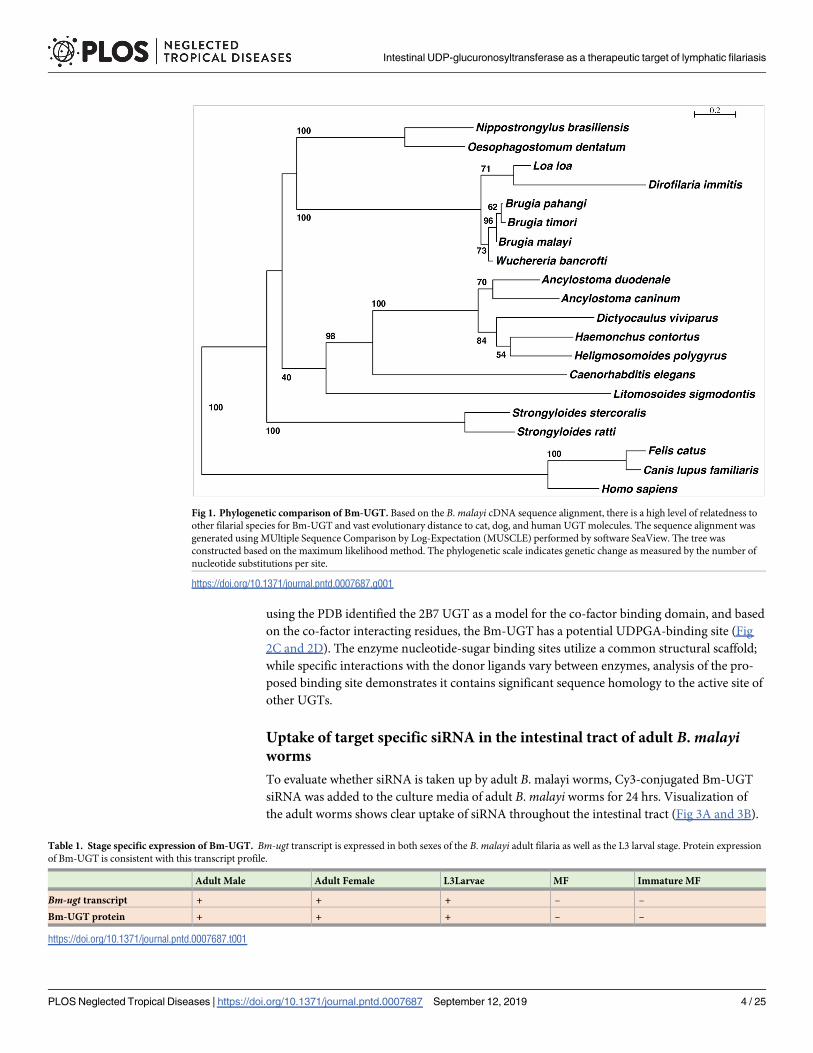
B. malayi intestinal UGT exhibits high homology to other filarial species
Previously, we reported that Bm-UGT (Bm17378) was a specific intestinal protein of B. malayiadult worms [12]. Sequence analyses indicated the presence of homologues in human filarial
worms (Brugia sp., W. bancrofti, L. loa) with significant homology (>75% identity), and to a
lesser extent (~35–40% identity) in other nematodes such as Dirofilaria immitis, Haemonchuscontortus, Ancylostoma sp., Strongyloides sp., Oesophagostomum dentatum and Toxocara canis.The most similar human proteins were UDP-glucuronosyltransferases as expected, but with
low sequence identity <27%. Given the high predicted homology of Bm-UGT between B.
malayi and D. immitis, we also evaluated orthologs in cat and dogs. Results of the sequence
analyses revealed little homology to Bm-UGT.
We then generated a phylogenetic tree by first aligning the Bm-UGT cDNA-derived pep-
tide sequences using MUltiple Sequence Comparison by Log-Expectation (MUSCLE) and
then creating a tree based on efficient maximum-likelihood estimation method by the LG
model. As seen in Fig 1, there is a high level of relatedness between Bm-UGT and several filar-
ial orthologs, including other Brugia species, W. bancrofti, L. loa, and D. immitis. Interestingly,
we could find no UGT ortholog in O. volvulus, and relatedness to the ortholog in Litomosoidessigmodontis, a common murine model of filarial infection, is low. Importantly, there is signifi-
cant evolutionary distance between the Bm-UGT and orthologs in humans, cats, and dogs.
Brugia malayi iUGT expression is stage specific
Evaluation of data available from prior transcriptomic and proteomic studies of B. malayidemonstrates that Bm-UGT is not expressed in all the lifecycle stages (Table 1). A study by Li
et al. shows that Bm-UGT transcript is only expressed in third stage larvae (L3s) and adult
female and male worms [13]. In addition, an RNAseq study by Choi et al. on various lifecycle
stages of B. malayi found that Bm-UGT was preferentially expressed during later larval stages.
Consistent with these findings, Bm-UGT protein expression was found to be specific to these
stages as well [14].
Structural analysis of Bm-UGT
Predictive analysis using the InterPro database revealed that the protein contains a large
luminally-expressed domain likely accessible to small molecules or ingested antibodies.
Sequence analysis of the Bm-UGT (Fig 2A) indicates that residues 1–20 encode a signal pep-
tide [15] followed by a two-domain UGT (residues 21–278: N-terminal domain; 278–445: co-
factor binding domain), a linker region, a transmembrane domain, and a short intra-cellular
domain. The structure of the Bm-UGT was modeled using SWISS-MODEL with 43 template
structures utilized. The final model was based on Protein Data Bank (PDB): 5NLM (the struc-
ture of the Polygonum tinctorium UGT) with a sequence identity of 19.7% [PMID: 29309053]
(Fig 2B). UGTs add a glucuronic acid moiety to a substrate by transfer of the glucuronosyl
group from uridine 5’-diphospho-glucuronic acid (UDPGA). Further structure-based searches
Intestinal UDP-glucuronosyltransferase as a therapeutic target of lymphatic filariasis
PLOS Neglected Tropical Diseases | https://doi.org/10.1371/journal.pntd.0007687 September 12, 2019 3 / 25
using the PDB identified the 2B7 UGT as a model for the co-factor binding domain, and based
on the co-factor interacting residues, the Bm-UGT has a potential UDPGA-binding site (Fig
2C and 2D). The enzyme nucleotide-sugar binding sites utilize a common structural scaffold;
while specific interactions with the donor ligands vary between enzymes, analysis of the pro-
posed binding site demonstrates it contains significant sequence homology to the active site of
other UGTs.
Uptake of target specific siRNA in the intestinal tract of adult B. malayiworms
To evaluate whether siRNA is taken up by adult B. malayi worms, Cy3-conjugated Bm-UGT
siRNA was added to the culture media of adult B. malayi worms for 24 hrs. Visualization of
the adult worms shows clear uptake of siRNA throughout the intestinal tract (Fig 3A and 3B).
Fig 1. Phylogenetic comparison of Bm-UGT. Based on the B. malayi cDNA sequence alignment, there is a high level of relatedness to
other filarial species for Bm-UGT and vast evolutionary distance to cat, dog, and human UGT molecules. The sequence alignment was
generated using MUltiple Sequence Comparison by Log-Expectation (MUSCLE) performed by software SeaView. The tree was
constructed based on the maximum likelihood method. The phylogenetic scale indicates genetic change as measured by the number of
nucleotide substitutions per site.
https://doi.org/10.1371/journal.pntd.0007687.g001
Table 1. Stage specific expression of Bm-UGT. Bm-ugt transcript is expressed in both sexes of the B. malayi adult filaria as well as the L3 larval stage. Protein expression
of Bm-UGT is consistent with this transcript profile.
Adult Male Adult Female L3Larvae MF Immature MF
Bm-ugt transcript + + + – –
Bm-UGT protein + + + – –
https://doi.org/10.1371/journal.pntd.0007687.t001
Intestinal UDP-glucuronosyltransferase as a therapeutic target of lymphatic filariasis
PLOS Neglected Tropical Diseases | https://doi.org/10.1371/journal.pntd.0007687 September 12, 2019 4 / 25
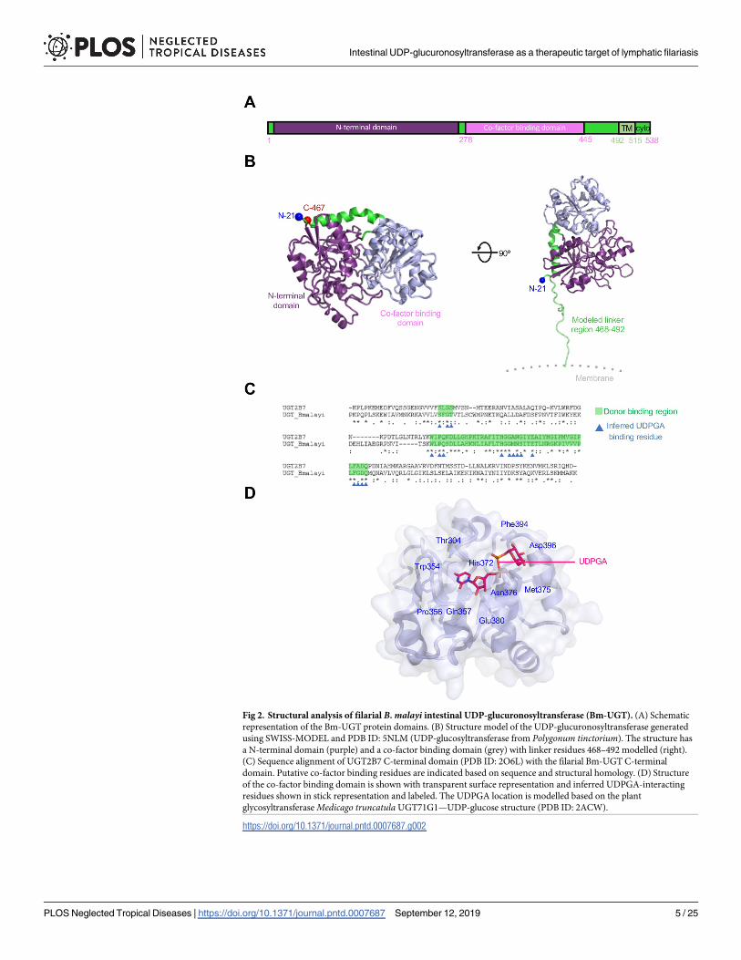
Fig 2. Structural analysis of filarial B. malayi intestinal UDP-glucuronosyltransferase (Bm-UGT). (A) Schematic
representation of the Bm-UGT protein domains. (B) Structure model of the UDP-glucuronosyltransferase generated
using SWISS-MODEL and PDB ID: 5NLM (UDP-glucosyltransferase from Polygonum tinctorium). The structure has
a N-terminal domain (purple) and a co-factor binding domain (grey) with linker residues 468–492 modelled (right).
(C) Sequence alignment of UGT2B7 C-terminal domain (PDB ID: 2O6L) with the filarial Bm-UGT C-terminal
domain. Putative co-factor binding residues are indicated based on sequence and structural homology. (D) Structure
of the co-factor binding domain is shown with transparent surface representation and inferred UDPGA-interacting
residues shown in stick representation and labeled. The UDPGA location is modelled based on the plant
glycosyltransferase Medicago truncatula UGT71G1—UDP-glucose structure (PDB ID: 2ACW).
https://doi.org/10.1371/journal.pntd.0007687.g002
Intestinal UDP-glucuronosyltransferase as a therapeutic target of lymphatic filariasis
PLOS Neglected Tropical Diseases | https://doi.org/10.1371/journal.pntd.0007687 September 12, 2019 5 / 25
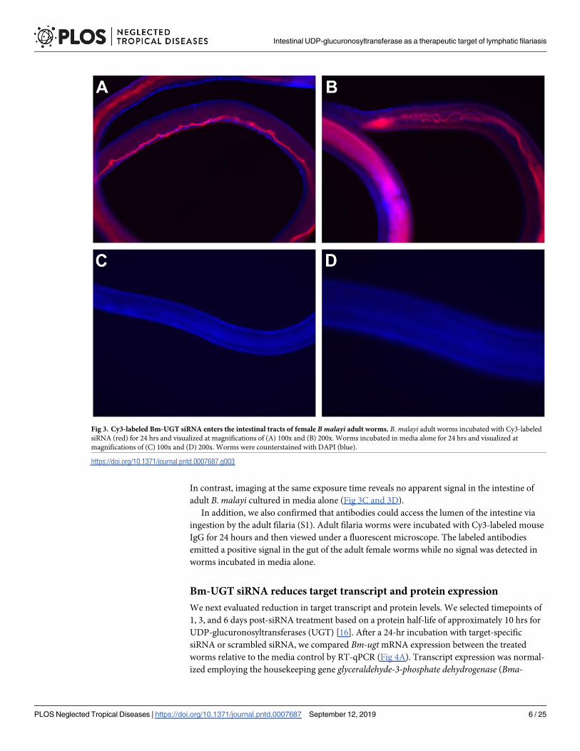
In contrast, imaging at the same exposure time reveals no apparent signal in the intestine of
adult B. malayi cultured in media alone (Fig 3C and 3D).
In addition, we also confirmed that antibodies could access the lumen of the intestine via
ingestion by the adult filaria (S1). Adult filaria worms were incubated with Cy3-labeled mouse
IgG for 24 hours and then viewed under a fluorescent microscope. The labeled antibodies
emitted a positive signal in the gut of the adult female worms while no signal was detected in
worms incubated in media alone.
Bm-UGT siRNA reduces target transcript and protein expression
We next evaluated reduction in target transcript and protein levels. We selected timepoints of
1, 3, and 6 days post-siRNA treatment based on a protein half-life of approximately 10 hrs for
UDP-glucuronosyltransferases (UGT) [16]. After a 24-hr incubation with target-specific
siRNA or scrambled siRNA, we compared Bm-ugt mRNA expression between the treated
worms relative to the media control by RT-qPCR (Fig 4A). Transcript expression was normal-
ized employing the housekeeping gene glyceraldehyde-3-phosphate dehydrogenase (Bma-
Fig 3. Cy3-labeled Bm-UGT siRNA enters the intestinal tracts of female B malayi adult worms. B. malayi adult worms incubated with Cy3-labeled
siRNA (red) for 24 hrs and visualized at magnifications of (A) 100x and (B) 200x. Worms incubated in media alone for 24 hrs and visualized at
magnifications of (C) 100x and (D) 200x. Worms were counterstained with DAPI (blue).
https://doi.org/10.1371/journal.pntd.0007687.g003
Intestinal UDP-glucuronosyltransferase as a therapeutic target of lymphatic filariasis
PLOS Neglected Tropical Diseases | https://doi.org/10.1371/journal.pntd.0007687 September 12, 2019 6 / 25
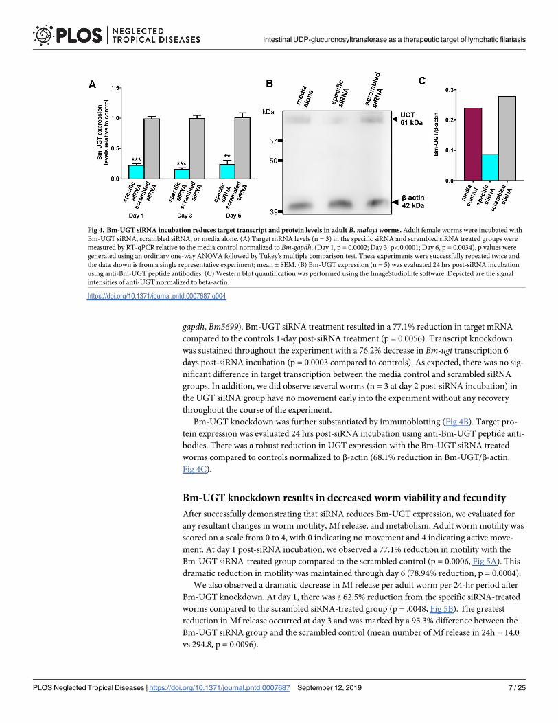
gapdh, Bm5699). Bm-UGT siRNA treatment resulted in a 77.1% reduction in target mRNA
compared to the controls 1-day post-siRNA treatment (p = 0.0056). Transcript knockdown
was sustained throughout the experiment with a 76.2% decrease in Bm-ugt transcription 6
days post-siRNA incubation (p = 0.0003 compared to controls). As expected, there was no sig-
nificant difference in target transcription between the media control and scrambled siRNA
groups. In addition, we did observe several worms (n = 3 at day 2 post-siRNA incubation) in
the UGT siRNA group have no movement early into the experiment without any recovery
throughout the course of the experiment.
Bm-UGT knockdown was further substantiated by immunoblotting (Fig 4B). Target pro-
tein expression was evaluated 24 hrs post-siRNA incubation using anti-Bm-UGT peptide anti-
bodies. There was a robust reduction in UGT expression with the Bm-UGT siRNA treated
worms compared to controls normalized to β-actin (68.1% reduction in Bm-UGT/β-actin,
Fig 4C).
Bm-UGT knockdown results in decreased worm viability and fecundity
After successfully demonstrating that siRNA reduces Bm-UGT expression, we evaluated for
any resultant changes in worm motility, Mf release, and metabolism. Adult worm motility was
scored on a scale from 0 to 4, with 0 indicating no movement and 4 indicating active move-
ment. At day 1 post-siRNA incubation, we observed a 77.1% reduction in motility with the
Bm-UGT siRNA-treated group compared to the scrambled control (p = 0.0006, Fig 5A). This
dramatic reduction in motility was maintained through day 6 (78.94% reduction, p = 0.0004).
We also observed a dramatic decrease in Mf release per adult worm per 24-hr period after
Bm-UGT knockdown. At day 1, there was a 62.5% reduction from the specific siRNA-treated
worms compared to the scrambled siRNA-treated group (p = .0048, Fig 5B). The greatest
reduction in Mf release occurred at day 3 and was marked by a 95.3% difference between the
Bm-UGT siRNA group and the scrambled control (mean number of Mf release in 24h = 14.0
vs 294.8, p = 0.0096).
Fig 4. Bm-UGT siRNA incubation reduces target transcript and protein levels in adult B. malayi worms. Adult female worms were incubated with
Bm-UGT siRNA, scrambled siRNA, or media alone. (A) Target mRNA levels (n = 3) in the specific siRNA and scrambled siRNA treated groups were
measured by RT-qPCR relative to the media control normalized to Bm-gapdh, (Day 1, p = 0.0002; Day 3, p<0.0001; Day 6, p = 0.0034). p values were
generated using an ordinary one-way ANOVA followed by Tukey’s multiple comparison test. These experiments were successfully repeated twice and
the data shown is from a single representative experiment; mean ± SEM. (B) Bm-UGT expression (n = 5) was evaluated 24 hrs post-siRNA incubation
using anti-Bm-UGT peptide antibodies. (C) Western blot quantification was performed using the ImageStudioLite software. Depicted are the signal
intensities of anti-UGT normalized to beta-actin.
https://doi.org/10.1371/journal.pntd.0007687.g004
Intestinal UDP-glucuronosyltransferase as a therapeutic target of lymphatic filariasis
PLOS Neglected Tropical Diseases | https://doi.org/10.1371/journal.pntd.0007687 September 12, 2019 7 / 25
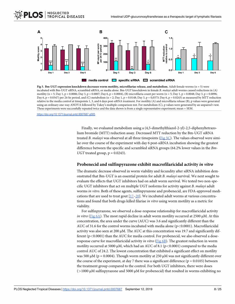
Finally, we evaluated metabolism using a (4,5-dimethylthiazol-2-yl)-2,5-diphenyltetrazo-
lium bromide (MTT) reduction assay. Decreased MTT reduction by the Bm-UGT siRNA
treated B. malayi was observed at all three timepoints (Fig 5C). The values observed were simi-
lar over the course of the experiment with day 6 post-siRNA incubation showing the greatest
difference between the specific and scrambled siRNA groups (64.2% lower values in the Bm-UGT treated group, p = 0.0243).
Probenecid and sulfinpyrazone exhibit macrofilaricidal activity in vitroThe dramatic decrease observed in worm viability and fecundity after siRNA inhibition dem-
onstrated that Bm-UGT is an essential protein for adult B. malayi survival. We next sought to
evaluate the effects that UGT inhibitors had on adult worm survival. We tested two non-spe-
cific UGT inhibitors that act on multiple UGT isoforms for activity against B. malayi adult
worms in vitro. Both of these agents, sulfinpyrazone and probenecid, are FDA-approved medi-
cations that are used to treat gout [17–20]. We incubated adult worms at various concentra-
tions and found that both drugs killed filariae in vitro using worm motility as a metric for
viability.
For sulfinpyrazone, we observed a dose-response relationship for macrofilaricidal activity
in vitro (Fig 6A). The most rapid decline in adult worm motility occurred at 2500 μM. At this
concentration, the area under the curve (AUC) was 3.6 and significantly different than the
AUC of 31.6 for the control worms incubated with media alone (p<0.0001). Macrofilaricidal
activity was also seen at 200 μM. The AUC at this concentration was 19.7 and significantly dif-
ferent (p<0.0001) than the AUC for media control. For probenecid, we also observed a dose-
response curve for macrofilaricidal activity in vitro (Fig 6B). The greatest reduction in worm
motility occurred at 5000 μM, which had an AUC of 8.1 (p<0.0001) compared to the media
control AUC of 24.2. The lowest concentration that exhibited a significant effect on motility
was 500 μM (p = 0.0004). Though worm motility at 250 μM was not significantly different over
the course of the experiment, at day 7 there was a significant difference (p = 0.0105) between
the treatment group compared to the control. For both UGT inhibitors, there were doses
(>1000 μM sulfinpyrazone and 5000 μM for probenecid) that resulted in worms exhibiting no
Fig 5. Bm-UGT expression knockdown decreases worm motility, microfilariae release, and metabolism. Adult female worms (n = 5) were
incubated with Bm-UGT siRNA, scrambled siRNA, or media alone. Bm-UGT knockdown in female B. malayi adult worms caused reductions in (A)
motility (n = 5; Day 1, p = 0.0006; Day 3, p = 0.0007; Day 6, p = 0.0004), (B) microfilaria count per worm (n = 5; Day 1, p = 0.0048; Day 3, p = 0.0096;
Day 6, p = 0.0347) per 24-hr period, and (C) metabolism (n = 2; Day 1, p = 0.0148; Day 3, p = 0,0373; Day 6, p = 0.0243) as measured by MTT reduction
relative to the media control at timepoints 1, 3, and 6 days post-siRNA treatment. For motility (A) and microfilaria release (B), p values were generated
using an ordinary one-way ANOVA followed by Tukey’s multiple comparison test. For metabolism (C), p values were generated by an unpaired t-test.
These experiments were successfully repeated twice and the data shown is from a single representative experiment; mean + SEM.
https://doi.org/10.1371/journal.pntd.0007687.g005
Intestinal UDP-glucuronosyltransferase as a therapeutic target of lymphatic filariasis
PLOS Neglected Tropical Diseases | https://doi.org/10.1371/journal.pntd.0007687 September 12, 2019 8 / 25
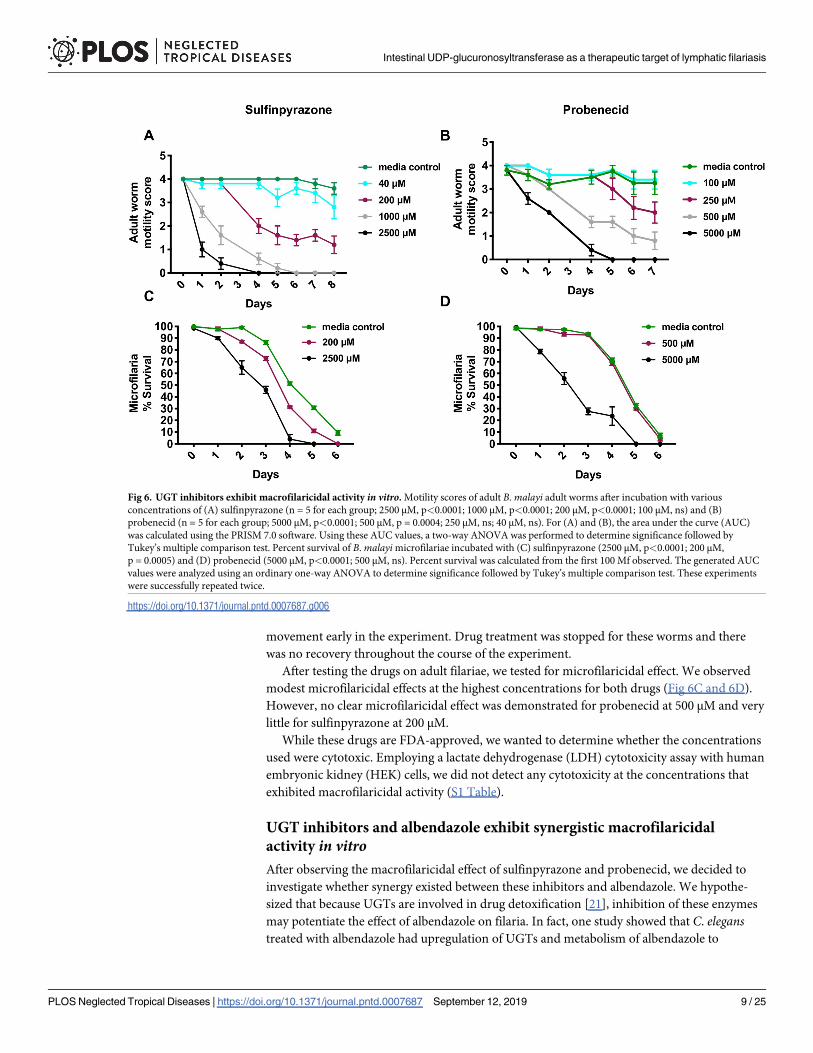
movement early in the experiment. Drug treatment was stopped for these worms and there
was no recovery throughout the course of the experiment.
After testing the drugs on adult filariae, we tested for microfilaricidal effect. We observed
modest microfilaricidal effects at the highest concentrations for both drugs (Fig 6C and 6D).
However, no clear microfilaricidal effect was demonstrated for probenecid at 500 μM and very
little for sulfinpyrazone at 200 μM.
While these drugs are FDA-approved, we wanted to determine whether the concentrations
used were cytotoxic. Employing a lactate dehydrogenase (LDH) cytotoxicity assay with human
embryonic kidney (HEK) cells, we did not detect any cytotoxicity at the concentrations that
exhibited macrofilaricidal activity (S1 Table).
UGT inhibitors and albendazole exhibit synergistic macrofilaricidal
activity in vitroAfter observing the macrofilaricidal effect of sulfinpyrazone and probenecid, we decided to
investigate whether synergy existed between these inhibitors and albendazole. We hypothe-
sized that because UGTs are involved in drug detoxification [21], inhibition of these enzymes
may potentiate the effect of albendazole on filaria. In fact, one study showed that C. eleganstreated with albendazole had upregulation of UGTs and metabolism of albendazole to
Fig 6. UGT inhibitors exhibit macrofilaricidal activity in vitro. Motility scores of adult B. malayi adult worms after incubation with various
concentrations of (A) sulfinpyrazone (n = 5 for each group; 2500 μM, p<0.0001; 1000 μM, p<0.0001; 200 μM, p<0.0001; 100 μM, ns) and (B)
probenecid (n = 5 for each group; 5000 μM, p<0.0001; 500 μM, p = 0.0004; 250 μM, ns; 40 μM, ns). For (A) and (B), the area under the curve (AUC)
was calculated using the PRISM 7.0 software. Using these AUC values, a two-way ANOVA was performed to determine significance followed by
Tukey’s multiple comparison test. Percent survival of B. malayi microfilariae incubated with (C) sulfinpyrazone (2500 μM, p<0.0001; 200 μM,
p = 0.0005) and (D) probenecid (5000 μM, p<0.0001; 500 μM, ns). Percent survival was calculated from the first 100 Mf observed. The generated AUC
values were analyzed using an ordinary one-way ANOVA to determine significance followed by Tukey’s multiple comparison test. These experiments
were successfully repeated twice.
https://doi.org/10.1371/journal.pntd.0007687.g006
Intestinal UDP-glucuronosyltransferase as a therapeutic target of lymphatic filariasis
PLOS Neglected Tropical Diseases | https://doi.org/10.1371/journal.pntd.0007687 September 12, 2019 9 / 25
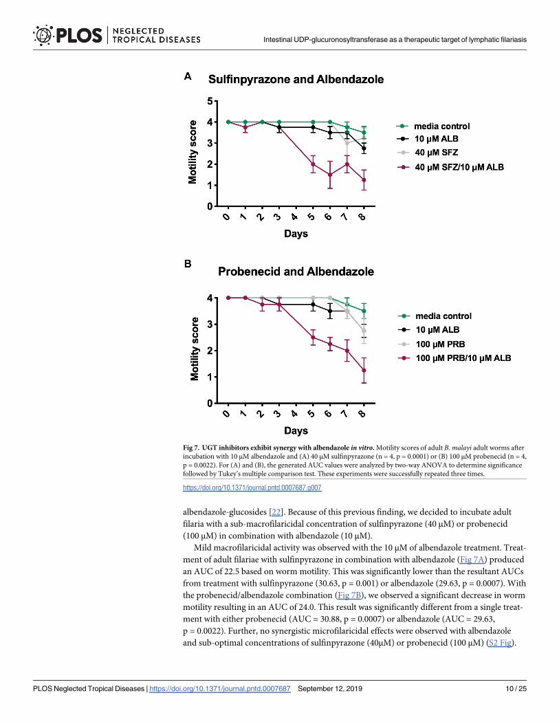
albendazole-glucosides [22]. Because of this previous finding, we decided to incubate adult
filaria with a sub-macrofilaricidal concentration of sulfinpyrazone (40 μM) or probenecid
(100 μM) in combination with albendazole (10 μM).
Mild macrofilaricidal activity was observed with the 10 μM of albendazole treatment. Treat-
ment of adult filariae with sulfinpyrazone in combination with albendazole (Fig 7A) produced
an AUC of 22.5 based on worm motility. This was significantly lower than the resultant AUCs
from treatment with sulfinpyrazone (30.63, p = 0.001) or albendazole (29.63, p = 0.0007). With
the probenecid/albendazole combination (Fig 7B), we observed a significant decrease in worm
motility resulting in an AUC of 24.0. This result was significantly different from a single treat-
ment with either probenecid (AUC = 30.88, p = 0.0007) or albendazole (AUC = 29.63,
p = 0.0022). Further, no synergistic microfilaricidal effects were observed with albendazole
and sub-optimal concentrations of sulfinpyrazone (40μM) or probenecid (100 μM) (S2 Fig).
Fig 7. UGT inhibitors exhibit synergy with albendazole in vitro. Motility scores of adult B. malayi adult worms after
incubation with 10 μM albendazole and (A) 40 μM sulfinpyrazone (n = 4, p = 0.0001) or (B) 100 μM probenecid (n = 4,
p = 0.0022). For (A) and (B), the generated AUC values were analyzed by two-way ANOVA to determine significance
followed by Tukey’s multiple comparison test. These experiments were successfully repeated three times.
https://doi.org/10.1371/journal.pntd.0007687.g007
Intestinal UDP-glucuronosyltransferase as a therapeutic target of lymphatic filariasis
PLOS Neglected Tropical Diseases | https://doi.org/10.1371/journal.pntd.0007687 September 12, 2019 10 / 25
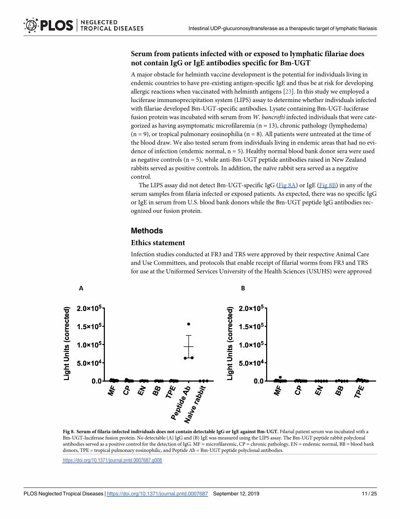
Serum from patients infected with or exposed to lymphatic filariae does
not contain IgG or IgE antibodies specific for Bm-UGT
A major obstacle for helminth vaccine development is the potential for individuals living in
endemic countries to have pre-existing antigen-specific IgE and thus be at risk for developing
allergic reactions when vaccinated with helminth antigens [23]. In this study we employed a
luciferase immunoprecipitation system (LIPS) assay to determine whether individuals infected
with filariae developed Bm-UGT-specific antibodies. Lysate containing Bm-UGT-luciferase
fusion protein was incubated with serum from W. bancrofti infected individuals that were cate-
gorized as having asymptomatic microfilaremia (n = 13), chronic pathology (lymphedema)
(n = 9), or tropical pulmonary eosinophilia (n = 8). All patients were untreated at the time of
the blood draw. We also tested serum from individuals living in endemic areas that had no evi-
dence of infection (endemic normal, n = 5). Healthy normal blood bank donor sera were used
as negative controls (n = 5), while anti-Bm-UGT peptide antibodies raised in New Zealand
rabbits served as positive controls. In addition, the naïve rabbit sera served as a negative
control.
The LIPS assay did not detect Bm-UGT-specific IgG (Fig 8A) or IgE (Fig 8B) in any of the
serum samples from filaria infected or exposed patients. As expected, there was no specific IgG
or IgE in serum from U.S. blood bank donors while the Bm-UGT peptide IgG antibodies rec-
ognized our fusion protein.
Methods
Ethics statement
Infection studies conducted at FR3 and TRS were approved by their respective Animal Care
and Use Committees, and protocols that enable receipt of filarial worms from FR3 and TRS
for use at the Uniformed Services University of the Health Sciences (USUHS) were approved
Fig 8. Serum of filaria-infected individuals does not contain detectable IgG or IgE against Bm-UGT. Filarial patient serum was incubated with a
Bm-UGT-luciferase fusion protein. No detectable (A) IgG and (B) IgE was measured using the LIPS assay. The Bm-UGT peptide rabbit polyclonal
antibodies served as a positive control for the detection of IgG. MF = microfilaremic, CP = chronic pathology, EN = endemic normal, BB = blood bank
donors, TPE = tropical pulmonary eosinophilic, and Peptide Ab = Bm-UGT peptide polyclonal antibodies.
https://doi.org/10.1371/journal.pntd.0007687.g008
Intestinal UDP-glucuronosyltransferase as a therapeutic target of lymphatic filariasis
PLOS Neglected Tropical Diseases | https://doi.org/10.1371/journal.pntd.0007687 September 12, 2019 11 / 25
by the USUHS Animal Care and Use Committee. The generation of peptide antibodies by
Genscript was on a protocol approved by the Genscript institutional animal care and use com-
mittee. Blood samples were obtained from patients and healthy volunteers who provided writ-
ten consent under protocols approved by the NIAID’s Institutional Review board. All human
subjects were adults.
Parasites and culture
Female B. malayi adults used in this study were obtained from the NIH/NIAID Filariasis
Research Reagent Resource Center (FR3) and TRS Laboratories in Athens, Georgia, USA. The
worms were cultured in Dulbecco’s Modified Eagle’s Medium (Corning cellgro) supplemented
with 10% heat-inactivated fetal bovine serum (Atlanta Biologicals), 100 units/mL of penicillin,
100 ug/mL of streptomycin, and 1% L-glutamine (Sigma) for 24 hrs at 37˚C in 5% CO2 prior
to siRNA treatment. Microfilariae were obtained from adult female worms cultured in vitro.
Phylogenetic tree analysis
Orthologs in other nematode species were identified in WormBase Parasite based on a
BLAST query [24] against the Bm-UGT protein sequence (Bm17378). The following are the
accession numbers of each ortholog as identified in WormBase Parasite: Brugia timori(BTMF_0001026401), Wuchereria bancrofti (WBA_0000030501), Brugia pahangi(BPAG_0000208101), Loa loa (LOAG_03428), Dirofilaria immitis (nDi.2.2.2.t06727),
Litomosoides sigmodontis (nLs.2.1.2.t00666-RA), Ancylostoma caninum (ANCCAN_05977),
Anyclostoma duodenale (ANCDUO_14383), Dictyocaulus viviparous (NDV.1.0.1.g111112),
Haemonchus contortus (HCON_00121250), Heligmosomoides polygyrus (HPOL_0001615101),
Nippostrongylus brasiliensis (NBR_0001252501), Caenorhabditis elegans (Y37E11AR), Strongy-loides ratti (SRAE_2000477000), Strongyloides stercoralis (SSTP_0001129400), and Oesopha-gostomum dentatum (OESDEN_03545).
Orthologs in selected mammals were identified in the National Center of Biotechnology
Information (NCBI) databases based on a BLAST query against the Bm-UGT peptide
sequence. The following are the orthologs selected for analyses: Homo sapiens (NP_066307),
Canis lupus familiaris (XP_005635657), and Felis catus (BAA2492).
Structural analysis of Bm-UGT
The Bm-UGT sequence was initially analyzed for properties including signal peptide sequence,
and potential transmembrane sequence using InterPro, SignalP4.1 (PMID: 28451972) and TM
servers [15]. Using the SWISS-MODEL homology modelling server [PMID: 29788355], the
iUGT sequence was used to search against the SWISS-MODEL template library using BLAST
and HHBlits for structures that matched the target sequence. The model was visualized using
PyMOL [25] and COOT [PMID: 20383002], with residues 468–492 manually built using
COOT. Based on sequence and structure identity, the co-factor binding domain was further
analyzed, utilizing the inferred UDPGA-binding site of Bm-UGT mapped using the UGT 2B7
structure [PMID: 17442341] as a model and visualized using COOT [PMID: 20383002].
siRNA design
Using the BLOCK-iT™ RNAi Designer, we selected the top three Bm-UGT siRNA duplexes for
gene silencing activity and specificity. The Bm-UGT siRNA and corresponding scrambled
siRNA were synthesized by Life Technologies and purified by standard desalting methods.
The 5’-3’ sequences of the Bm-UGT siRNA strands were as follows:
Intestinal UDP-glucuronosyltransferase as a therapeutic target of lymphatic filariasis
PLOS Neglected Tropical Diseases | https://doi.org/10.1371/journal.pntd.0007687 September 12, 2019 12 / 25
Bm-UGT siRNA 1
sense: 5’ GCCUAACGAAACUAAGCAAdTdT 3’
antisense: 5’ UUGCUUAGUUUCGUUAGGCdTdT 3’
Bm-UGT siRNA 2
sense: 5’ GGCUUCCACAAUCUGAUUUdTdT 3’
antisense: 5’ AAAUCAGAUUGUGGAAGCCdTdT 3’
Bm-UGT siRNA 3
sense: 5’ GGUGGUAUGAAUAGCAUAAdTdT 3’
antisense: 5’ UUAUGCUAUUCAUACCACCdTdT 3’
Demonstration of siRNA uptake and antibody ingestion by fluorescence
microscopy
For demonstration of siRNA uptake, adult female worms were incubated in 5 μM of 5’ Cy3-la-
beled Bm-UGT siRNA 3 (Sigma Aldrich) for 24 hrs. Adult worms incubated in media alone
were used as a negative control. Both groups of worms were then stained with 10 μg/mL of
DAPI (Sigma-Aldrich) in PBS. Fluorescent images were captured by a Nikon Eclipse E600
fluorescent microscope and converged using NIS-Elements software. For experiments to test
ingestion of antibody, adult female worms were incubated with 100 μg of mouse Cy3-labeled
IgG isotype control in 2 mL of culture media. The worms were imaged 24 hrs later using the
TRITC filter on a Zeiss Axio Observer.A1.
siRNA incubation of B. malayi female adult worms
siRNA inhibition of Bm-UGT in B. malayi adult female worms followed a protocol established
by Aboobaker et al. with minor modifications [26]. siRNA inhibition in filarial worms has
well-known variability and difficulty [27, 28]. For this study, we analyzed data for experiments
that received greater than 60% knockdown. For each timepoint, 5 adult female worms were
soaked in an equal mixture of the Bm-UGT siRNAs at a final concentration of 5 μM in 850 μL
of culture media in a 5000 MWCO Pur-A-Lyzer™ dialysis tube (Sigma-Aldrich). This concen-
tration of siRNA was shown in multiple studies to be sufficient at silencing gene expression
[26, 29–31]. The dialysis tubes were placed in 1 L beakers with 500 mL of culture media for 24
hrs at 37˚C in 5% CO2. Similarly, 5 adult female worms were soaked in media alone or scram-
bled siRNA (5 μM) in dialysis tubes for each timepoint as experimental controls. After the
24-hr incubation, the worms for each group were carefully extracted from the dialysis tubes
and individually placed into wells with 1 mL of media. The worms were evaluated at time-
points 1, 3, and 6 days post-incubation for transcript knockdown, worm motility, MTT reduc-
tion, and microfilariae release.
Worm motility
Worms were visualized with a dissecting microscope by an observer blinded to treatment cate-
gory. Motility of the adult female B. malayi worms was rated based on the following scale
4 = active movement, 3 = modest reduction in movement, 2 = severe reduction in movement,
1 = twitching, and 0 = no movement.
Intestinal UDP-glucuronosyltransferase as a therapeutic target of lymphatic filariasis
PLOS Neglected Tropical Diseases | https://doi.org/10.1371/journal.pntd.0007687 September 12, 2019 13 / 25
MTT reduction
Metabolic function of the adult female worms was assessed by reduction of (4,5-dimethylthia-
zol-2-yl)-2,5-diphenyltetrazolium bromide (MTT) from Sigma using a protocol established by
Comley et al [32]. For each group per timepoint, 2 worms were incubated in 0.5 mL of phos-
phate buffered solution (PBS) pH 7.4 with 0.5 mg/mL of MTT for 30 minutes at 37˚C in 5%
CO2. The worms were then transferred into separate wells of a 96-well plate containing 200 μL
of DMSO and incubated at room temperature for 1 hr. MTT reduction was quantified by
absorbance relative to a DMSO blank at 570 nm using a Synergy HTX multi-mode plate reader
(BioTek).
Microfilaria release
For each timepoint, adult worms were placed in new culture media 24 hrs prior to
enumeration of microfilariae. After the overnight incubation, the worms were then
removed for processing by the MTT reduction assay and RT-qPCR. The Mf in the well con-
taining expended culture media (1 mL) were counted under a light microscope at high
magnification.
RNA extraction and analysis of RNA levels by RT-qPCR
For each condition, adult B. malayi female worms (n = 3) were homogenized in TRIzol
(Thermo Fischer Scientific) after three freeze/thaw cycles using Matrix D lysis tubes (MP Bio-
medicals) agitated by a FastPrep™-24 Biopulverizer (MP Biomedicals) for 7 minutes at 6 m/s.
Chloroform was added to the homogenate, transferred to Phase Lock Gel tubes (5Prime), and
phase separated at 11,900 g for 15 minutes at 4˚C. The aqueous phase was collected and cold
isopropanol was added to precipitate the RNA, which was then pelleted at 12,000 g for 1 hr
and washed twice using 75% ethanol. The RNA pellet was resuspended in nuclease-free water
and quantified using a NanoDrop 1000 (Thermo Fischer Scientific). cDNA was prepared
using Superscript IV (Thermo Fischer Scientific) as per the manufacturer’s protocol. The
cDNA levels of Bm-UGT and B. malayi house-keeping gene gapdh were assessed in duplicate
20 uL reactions using 1 μL of 20X TaqMan™ gene expression assay (Thermo Fischer), 1 μL of
cDNA, and 18 μL of TaqMan™ gene expression master mix (Applied Biosystems). PCR condi-
tions were 2 min at 50˚C, 10 min at 95˚C, 40 cycles of 15 sec at 95˚C, and 1 min at 60˚C cycle
of 50˚C with a 7500 Real-Time PCR System (Applied Biosystems). The primers used were as
follows:
Bma-gapdh:
Forward primer: 5’ TTGATCTCACTTGCCGACTC 3’
Reverse primer: 5’ TGGTCTTCGGTGTATTCCAA 3’
Internal probe: 5’ CAGCTAATGGACCGATGAAGGGGA 3’
Bm-ugt:
Forward primer: 5’ TATCATTCGGCACCGTTACA 3’
Reverse primer: 5’ ATTCATACCACCATGCGTCA 3’
Internal probe: 5’ TCGCTGAGGGACGTCCAAACG 3’
Intestinal UDP-glucuronosyltransferase as a therapeutic target of lymphatic filariasis
PLOS Neglected Tropical Diseases | https://doi.org/10.1371/journal.pntd.0007687 September 12, 2019 14 / 25
Generation of rabbit polyclonal antibodies against Bm-UGT peptides
Polyclonal anti-Bm-UGT peptide antibodies were generated in New Zealand rabbits by Gen-
script using Bm-UGT peptide sequences conjugated to keyhole limpet hemocyanin (KLH).
The peptide sequences used were as follows: CYEKDEHLIAEGRPN, DSTGSKLAKTVKIDC,
and CGQIANFDPYGRKMS. Cysteines were added at either the N- or C-terminus to facilitate
KLH conjugation.
Immunoblotting
B. malayi adult worms (n = 5) were incubated in 5 μM combination of Bm-UGT siRNA for
24 hrs using the previously mentioned method and transferred into individual wells with 1
mL of media. The adult worms were cultured for an additional 24 hrs and then homogenized
in PBS (pH 7.4) and 4 μL Halt™ Protease Inhibitor Cocktail (Thermo Scientific) using Matrix
D lysis tubes (MP Biomedicals) agitated by a FastPrep™-24 Biopulverizer (MP Biomedicals)
for 3 minutes at 4 m/s. Protein levels were quantified by the Bradford protein assay (Bio-
Rad). For immunoblot analysis, 10 μg of protein was separated on 10% Bis-Tris NuPAGE gel
(Invitrogen) and blotted onto 0.2 μm nitrocellulose filter paper (Bio-Rad). After blocking
overnight in 5% bovine serum albumin (BSA) in tris-buffered saline with 0.1% Tween
20 (TBS-T), the membrane was incubated with 1:4000 anti-UGT peptide antibodies (Gen-
script) and 1:1000 rabbit anti-β actin antibodies (Abcam) for 1 hr. Following this, the filter
paper was washed with TBS-T and then incubated with 1:2000 horseradish peroxidase con-
jugated goat anti-rabbit IgG for 1 hr. The membrane subsequently washed and incubated in
Chemiluminescent reagent, SuperSignal™ West Pico PLUS (Thermo Scientific), to visual the
bands.
Adult B. malayi worm incubation with probenecid and sulfinpyrazone
Sulfinpyrazone (ChemCruz) and probenecid (Invitrogen, water soluble formulation), broadly
acting UGT inhibitors, were evaluated for macrofilaricidal activity in vitro. Sulfinpyrazone was
resuspended in 1X PBS (pH 7.4) and 1% dimethylsulfoxide (DMSO, v/v) while probenecid
was resuspended in deionized water. When testing sulfinpyrazone, adult B. malayi female
worms were incubated in culture media with the drug for 8 days at concentrations of 2500 μM,
1000 μM, 200 μM, 40 μM, and 8 μM. For probenecid, adult female worms were incubated in
culture media with the drug for 7 days at concentrations of 5000 μM, 500 μM, 250 μM, and
100 μM. Worms were transferred into new media with corresponding drug concentrations
every day except day 4. As a negative control, worms were incubated in culture media alone
with a similar volume of vehicle. Worm motility was scored using the previously mentioned
scale for the course of the experiment. Worms that were scored a zero stopped receiving UGT
inhibitor treatment.
For the albendazole synergy experiments, adult filariae were incubated in culture media
with 40 μM of sulfinpyrazone or 100 μM of probenecid in combination with 10 μM of albenda-
zole, which was resuspended in 1X PBS (pH 7.4) and 1% DMSO (v/v). The worms were scored
for motility for 8 days and were transferred into new media with corresponding drug concen-
trations every day except day 4.
B. malayi microfilariae incubation with UGT inhibitors
The above UGT inhibitors were evaluated for microfilaricidal activity in vitro. For each drug,
experiments were performed in triplicate at a concentration of 2 x 10⁴ Mf/mL in culture
media. Viability was determined by quantifying the number of motile larvae from 100
Intestinal UDP-glucuronosyltransferase as a therapeutic target of lymphatic filariasis
PLOS Neglected Tropical Diseases | https://doi.org/10.1371/journal.pntd.0007687 September 12, 2019 15 / 25
randomly selected Mf per well. The concentrations used for sulfinpyrazone were 2500 μM and
200 μM while the concentrations used for probenecid were 5000 μM and 500 μM. As a nega-
tive control, larvae were incubated in culture media alone.
UGT inhibitor Cytotoxicity Assay
We measured cytotoxicity of the UGT inhibitors using a Pierce LDH Cytotoxicity Assay Kit
(Thermo Scientific). HEK cells were seeded at 5 x 10⁴ per well in DMEM (Quality Biological)
with 10% Hyclone Cosmic Calf Serum (Thermo Fischer), 200 μM of L-glutamine (Quality Bio-
logical), and 50 μg/mL of gentamicin (Quality Biological) at 37˚C in 5% CO2. We then incu-
bated the cells with various concentrations of the UGT inhibitors overnight. Following this, we
transferred 50 μL of media from each well to a new 96-well plate and then added 50 μL of reac-
tion buffer. We incubated the mixture for 30 minutes and then added 50 μL of stop solution.
We measured absorbance at 490 nm and 680 nm. We employed the following controls: a spon-
taneous LDH activity control which was incubated with the vehicle only and a maximum LDH
activity control which was incubated with nothing but later lysed prior to incubation with the
reaction buffer. We calculated absorbance for each well by subtracting the 680 nm absorbance
value (background) from the 490 nm absorbance value. We then calculated percent cytotoxic-
ity using the following equation:
%Cytotoxicity ¼ðUGT inhibitor LDH activity � Spontaneous LDH actvityÞðMaximum LDH activity � Spontaneous LDH activityÞ
Generation of Ruc-antigen fusion proteins
Bm-UGT was expressed as a Renilla reniformis luciferase (Ruc) fusion protein by cloning the
Bm-UGT coding sequence in pREN2 (Genscript). The Bm-UGT signal sequence as predicted
by signalP was removed prior to synthesis. Plasmid encoding the fusion protein was used to
transformed TOP10 cells (Thermo Fischer) and plasmid DNA was obtained from colonies
selected on kanamycin (50 μg/ml) as per the manufacturer’s guidelines (Qiagen Midi-Prep).
293F cells grown in 293 Freestyle Medium as suspension cultures were transfected with 30 μg
of Bm-UGT-Ruc plasmid, at a final concentration of 1 μg per 1 x 10⁶ cells (Thermo Fischer
Sceintific) per mL, and cultured at 37˚C with 8%CO2 on a rotary shaker at 125 rpm. After
72hrs, the cells were pelleted and sonicated in LIPS lysis buffer (20 mM Tris pH 7.5, 100 mM
NaCl, 5 mM MgCl2, 1% TritonX-100, 50% glycerol, protease inhibitors (Mini from Roche)).
The lysate was centrifuged to remove cellular debris and supernatant containing the Bm-UGT-
Ruc fusion proteins were stored at -80˚C for later use.
Luciferase immunoprecipitation system
Antibody titers were measured using a luciferase immunoprecipitation system (LIPS) assay
[33–35]. For IgG and IgE quantification, serum was diluted to 1:100 and 1:10 respectively in
50 μL of LIPS master mix (20 mM Tris pH 7.5, 100 mM NaCl, 5mM MgCl2, 1% Triton X-100)
and mixed with 50 μL of the UGT-Ruc fusion containing 1 x 10⁶ light units (LU) of protein in
PBST (PBS with 0.05% Tween-20). The reaction mixture was incubated in a 96-well polypro-
pylene plate for 10 minutes at room temperature and transferred to a 96-well high throughput
screening filter plate (Milipore) containing 5 μL of a 50% suspension of Ultralink protein A/G
(Pierce) or Ultralink anti-human IgE beads in PBS and incubated for an additional 15 minutes
at room temperature. The plates were washed under vacuum with 200 μL of LIPS master 3x
followed by PBS once. The relative light units (RLU) were measured with a Berthold LB 960
Intestinal UDP-glucuronosyltransferase as a therapeutic target of lymphatic filariasis
PLOS Neglected Tropical Diseases | https://doi.org/10.1371/journal.pntd.0007687 September 12, 2019 16 / 25
Centro microplate luminometer with 50 μL of coelenterzine solution (Promega). For these
experiments, samples were run in duplicate, and the calculated RLU was adjusted for the mea-
sured RLU of UGT fusion protein without serum.
Serum samples
All human serum samples were obtained following written informed consent from all
subjects using Institutional Review Board-approved protocols that have been registered
(NCT00001345, NCT00090662, NCT00342576). Patients were grouped into clinical categories
as previously detailed [36].
Statistical analysis
The siRNA and UGT inhibitor experiments were repeated two times under similar conditions.
Data shown is from a single representative experiment. For the siRNA experiments, data was
analyzed using one-way analysis of variance (ANOVA) or T-test by PRISM 7.0. Following
ANOVA, individual comparisons of mean values were performed using Tukey’s multiple com-
parisons test. For the UGT inhibitor experiments, we performed AUC analysis followed by
one-way ANOVA to determine significance. Statistical significance between the experimental
and control groups was designated as follows: � for p values<0.05, �� for p values<0.01, and��� for p values<0.001.
Discussion
UDP-glucuronosyltransferases (UGT) are enzymes important for detoxification of xenobiotics
and homeostasis of endogenous molecules [21]. Specifically, these phase II enzymes increase
the solubility of hydrophobic molecules by attaching sugar moieties such as glucuronic acid.
Since this sugar molecule is negatively charged at physiological pH, anion efflux pumps are
able to transport these molecules outside the cell [37]. In C. elegans, studies demonstrated that
RNAi of detoxification enzymes results in lethality, sluggish movement, or impaired growth
[38–40]. In addition, one study showed that glycosylation by phase II enzymes was important
for the detoxification of albendazole in C. elegans [22]. While UGTs in helminths have not
been studied extensively, there is evidence from intestinal helminths to suggest that these
enzymes play a critical role in drug resistance [41–43].
In this study, we showed that B. malayi intestinal UGT expression could be silenced by spe-
cific siRNA. This knockdown caused a significant reduction in worm motility, fecundity, and
metabolism. While these metrics alone do not determine worm survival, we are fairly confi-
dent these are appropriate surrogates. We observed several worms with a scored motility of
zero early into the siRNA experiments that did not show any recovery over the course of the
experiment. Thus, Bm-UGT appears essential for adult B. malayi worm survival. It is possible
that the observed phenotypic changes occurred due to an imbalance in endogenous molecules.
Past studies in mice and rats demonstrated that UGTs were critical for protection against free
radicals, which if left unchecked could mediate damage to DNA, lipid membranes, and amino
acids [44–47]. The reduction in microfilaria release was most likely due to decreased adult
worm viability because Bm-UGT is not expressed in the Mf stage [13, 14]. We suspect that tar-
geting Bm-UGT in vivo would leave the worms not only susceptible to endogenous free radi-
cals but also to those released by host immune cells.
Interestingly, it should be noted that the Brugia intestinal UGT was predicted by InterPro
to be localized to the plasma membrane, which would be a novel location for this family of
enzymes that are typically found in the endoplasmic reticulum. The prediction software also
Intestinal UDP-glucuronosyltransferase as a therapeutic target of lymphatic filariasis
PLOS Neglected Tropical Diseases | https://doi.org/10.1371/journal.pntd.0007687 September 12, 2019 17 / 25
determined that this protein has a large extracellular domain, which potentially makes it read-
ily accessible to drugs or antibodies.
Development of short-course macrofilaricidal agents would greatly enhance LF eradication
efforts. Because the aim of this study was to evaluate Bm-UGT as a potential drug and vaccine
target, we investigated the effects of non-specific UGT inhibitors on adult filariae. We found
two UGT inhibitors that are also FDA-approved to treat gout [17–20]. Both drugs, sulfinpyra-
zone and probenecid, exhibited macrofilaricidal activity in vitro. For worms that were scored a
zero for motility, we did not see any recovery after cessation of the UGT inhibitor treatment.
The lowest effective concentration for sulfinpyrazone was 200 μM. A previous study investigat-
ing sulfinpyrazone showed a maximum concentration (Cmax) in humans of 79.9 μM for a 400
mg dose [48]. Given that the daily maximum recommended dose for humans is 800 mg, we
believe a Cmax similar to our lowest effective sulfinpyrazone concentration may be achievable
in humans [49]. Therefore, we speculate that this drug could serve as a novel therapeutic
against adult filariae. Likewise, we believe that the same applies to probenecid, which demon-
strated robust macrofilaricidal activity in vitro at 500 μM. We also observed a significant
reduction in motility by day 7 at 250 μM suggesting that this concentration may be effective at
killing adult worms if given over a longer time course. In the context of physiological rele-
vance, one pharmacokinetic study showed the peak concentration in humans given a single 2
g oral dose of probenecid to be 148.6 μg/mL (520.7 μM) with minimal adverse events [50].
Based on our in vitro data, this level could rapidly kill adult filarial worms.
While the UGT inhibitors displayed macrofilaricidal activity, it is possible that probenecid
and sulfinpyrazone may act on adult B. malayi worms through mechanisms independent of
effects on Bm-UGT. In addition to inhibiting UGTs, probenecid and sulfinpyrazone also
inhibit organic anion transporters (OATs) [51, 52] and pannexins [53]. OATs function to
transport negatively charged molecules and are likely important for adult filarial worm sur-
vival. Innexins, which are present in invertebrate organisms, are structurally very similar to
pannexins and primarily function as membrane channels that communicate with the extracel-
lular space [54]. Interestingly, probenecid has been shown to impair touch responses in C. ele-gans by inhibiting mechanosensitive innexin channels [55]. Elucidating the mechanisms by
which probenecid and sulfinpyrazone kill adult filarial worms will be the focus of future
studies.
While we are interested in developing a drug that kills adult filarial worms, the absence of
microfilaricidal activity also presents advantages. Current antifilarial therapeutics such as
diethylcarbamazine (DEC) and ivermectin (IVM) are extremely effective at clearing microfi-
lariae. However, their use is contraindicated in areas co-endemic for loiasis and onchocerciasis
because rapid killing of microfilariae in these infections can lead to severe adverse outcomes
[56]. Individuals with loiasis when treated with IVM or DEC have a significantly higher risk of
experiencing severe neurologic events such as encephalopathy due to rapid Mf death in the
vasculature [57, 58]. Similarly, DEC can induce adverse systemic reactions such as skins
lesions, fever, polyarthritis, and ocular reactions in patients with onchocerciasis as determined
by Mf load [59, 60]. Therefore, probenecid, which demonstrated macrofilaricidal but not
microfilaricidal activity at 500 μM in vitro, may be an attractive LF treatment candidate in
these co-endemic areas.
Because UGTs are involved in drug metabolism, we suspected that there may be synergy
between the UGT inhibitors and albendazole. A recent study demonstrated that overexpres-
sion of UGT-22 in C. elegans resulted in albendazole resistance [61]. This, coupled with an ear-
lier study by Laing et al showing that upregulation of UGTs is associated with metabolism of
albendazole into various glucuronide products [22], suggests that UGTs may play a critical
role in nematode metabolism of albendazole. In support of this hypothesis, our data
Intestinal UDP-glucuronosyltransferase as a therapeutic target of lymphatic filariasis
PLOS Neglected Tropical Diseases | https://doi.org/10.1371/journal.pntd.0007687 September 12, 2019 18 / 25
demonstrate a synergistic effect of sub-macrofilaricidal concentrations with sulfinpyrazone or
probenecid in combination with albendazole against adult filaria in vitro. These results suggest
that combination therapy with albendazole and either probenecid or sulfinpyrazone may be
highly effective in treating filarial infections in people. Future studies will determine whether
Bm-UGT functions to metabolize albendazole in filarial worms and whether this metabolism
is inhibited by UGT inhibitors. We also plan to test the efficacy of combination therapy in ani-
mal models of infection.
As previously mentioned, these probenecid and sulfinpyrazone inhibitors are FDA-
approved and have been shown to be safe in humans [62], with probenecid characterized as a
pregnancy category B drug. If these medicines demonstrate macrofilaricidal activity in vivo,
translation into human use could occur quickly.
In addition to being a potential drug target for filariasis, we postulate that Bm-UGT could
serve as a vaccine candidate as well. One of the challenges in helminth vaccine development is
the risk that the vaccine may induce an allergic response in endemic populations. Indeed, gen-
eralized urticaria was seen in several Brazilian patients immunized against Ancylostoma-
secreted protein 2 during hookworm vaccine trials [23]. A solution to this obstacle is to iden-
tify “hidden antigens” which are not exposed to the immune system during natural infection
yet are essential to worm survival and present in an anatomical location accessible to host
antibodies [63, 64]. In theory, these proteins would not elicit an IgE-mediated response, and,
as postulated by Munn, these antigens may be especially vulnerable to the immune system
due to a lack of evolutionary pressure to evade it [64]. There is evidence to support that the
intestinal tract of nematodes contains hidden antigens. Studies have demonstrated the
absence of pre-existing IgE in serum from endemic populations against hookworm intestinal
antigens APR-1 and GST [8, 9, 65]. Furthermore, studies have shown that these antigens are
protective in animal models [8, 10, 11]. There is also evidence of hidden antigens in H. contor-tus seen with the lack of an antibody response against H11, a glycosylated intestinal protein
[66]. In this study, we observed no detectable Bm-UGT-specific IgE in the serum from indi-
viduals infected with or exposed to lymphatic filariae, which suggests this antigen may be safe
to administer as a vaccine candidate in endemic populations. However, due to the low num-
bers of patient samples tested, if vaccine work using Bm-UGT progresses, then future studies
would need to evaluate potential for allergic responses by testing far larger numbers of
individuals.
After demonstrating that recombinant Bm-UGT would not induce an allergic response in
endemic populations, we investigated whether adult B. malayi worms could ingest antibody
as there is a degree of uncertainty about whether filarial worms use their intestine to feed.
Only the adult and L4 stages of filariae have a fully formed intestinal tract, while the Mf, L2,
and L3 stages have an immature intestine inaccessible to nutrients [67]. Furthermore, past
studies have already shown that Brugia worms are able to absorb nutrients such as nucleo-
tides, amino acids, sugars, and vitamins through their cuticle, which calls into question the
purpose of the intestinal tract [68]. Notwithstanding these findings, Attout et al. investigated
the intestinal tract of L. sigmodontis and demonstrated that young adult worms (25–56 weeks
post-infection) ingested red blood cells [69]. This suggests that intestinal feeding may occur
but only at the early adult stage. Another study showed that D. immitis can ingest labeled
serum [70]. With our study, we were able to show that adult B. malayi worms can ingest
Cy3-labeled IgG (S1 Fig). This serves as clear evidence that circulating antibody can poten-
tially access the intestine of adult filarial worms. However, not all the worms ingested the
labeled antibody, which indicates that, at least during in vitro conditions, adult worms do not
feed through the intestine continuously. Future studies will work towards recombinant
Intestinal UDP-glucuronosyltransferase as a therapeutic target of lymphatic filariasis
PLOS Neglected Tropical Diseases | https://doi.org/10.1371/journal.pntd.0007687 September 12, 2019 19 / 25
expression of Bm-UGT in order to test its ability to induce protective immune responses in
animal models of filariasis.
On the basis of the phylogenetics analyses we conducted, we expect that Bm-UGT may also
be essential in W. bancrofti and B. timori given the overall high homology shared between
these species and B. malayi. Additionally, there is a high level of sequence homology (> 70%)
between the B. malayi intestinal UGT and the orthologs found in D. immitis and L. loa and
thus the UGT orthologs in these filaria species may also serve as novel therapeutic targets.
Interestingly, we did not find an ortholog of Bm-UGT in O. volvulus and speculate this could
be the result of evolutionary pressure to lose this gene.
In summary, we believe that Bm-UGT is an essential intestinal protein in B. malayi adult
worms that does not induce IgE antibodies in endemic populations. Importantly, we found
that sulfinpyrazone and probenecid, two commercially available, FDA-approved medications
in use for gout, exhibit strong macrofilaricidal activity in vitro at concentrations that are
achievable in humans. This promising data warrants future investigation in animal models of
Brugia infection as well as assessment of whether these UGT inhibitors exhibit macrofilaricidal
activity in vitro against other filarial species. Furthermore, we demonstrated that probenecid
and sulfinpyrazone may potentiate the effect of albendazole on filariae, suggesting that combi-
nation therapy may be an ideal approach to obtain macrofilaricidal effect. In terms of vaccine
development, future studies will focus on recombinantly expressing Bm-UGT and then testing
whether it is protective in animal models of filariasis. Finally, the results of this study suggest
that the intestinal tract of filarial nematodes may serve as a rich source of essential proteins
that can serve as important therapeutic targets.
Supporting information
S1 Table. LDH Cytotoxicity Assay for the UGT inhibitors. 2 x 10⁴ HEK cells were seeded in
various concentrations of sulfinpyrazone and probenecid. LDH activity was measured by
absorbance at 490 nm and 680 nm and was calculated by subtracting LDH activity at 680 nm
from activity at 490 nm. Percent cytotoxicity was calculated against the maximum LDH activ-
ity (lysed cells) using spontaneous LDH activity (vehicle only) as a background control.
(TIF)
S1 Fig. Adult B. malayi ingest mouse polyclonal IgG. Adult worms were cultured for 24 hrs
with Cy3-labeled mouse polyclonal IgG or in media alone. Ingested antibody is visible in the
intestinal of the adult filaria.
(TIF)
S2 Fig. UGT inhibitors do not exhibit synergy with albendazole against B. malayi microfi-
lariae. Percent survival of microfilariae incubated with 10 μM albendazole and 40 μM sulfin-
pyrazone (ns) and 100 μM probenecid (ns). Percent survival was calculated wells containing
2.0 x 10⁶ Mf similar to Fig 6. The generated AUC values were analyzed using an ordinary one-
way ANOVA to determine significance followed by Tukey’s multiple comparison test. This
experiment was only performed once.
(TIF)
Acknowledgments
We thank Belinda Jackson (Uniformed Services University) and Laura Kropp for their assis-
tance with the worm motility scoring. In addition, we would like to thank Roopa Biswas (Uni-
formed Services University) for her assistance with the RNAi experiments.
Intestinal UDP-glucuronosyltransferase as a therapeutic target of lymphatic filariasis
PLOS Neglected Tropical Diseases | https://doi.org/10.1371/journal.pntd.0007687 September 12, 2019 20 / 25
Author Contributions
Conceptualization: Alexander F. Flynn, Rebekah T. Taylor, Sasisekhar Bennuru, C. Paul Mor-
ris, Thomas B. Nutman, Edward Mitre.
Data curation: Alexander F. Flynn.
Formal analysis: Alexander F. Flynn, M. Gordon Joyce, Edward Mitre.
Funding acquisition: Edward Mitre.
Investigation: Alexander F. Flynn, M. Gordon Joyce, Rebekah T. Taylor, Sasisekhar Bennuru,
Alyssa R. Lindrose, Spencer L. Sterling.
Methodology: Alexander F. Flynn, M. Gordon Joyce, Rebekah T. Taylor, Sasisekhar Bennuru,
Spencer L. Sterling.
Project administration: Alexander F. Flynn, Alyssa R. Lindrose, Edward Mitre.
Resources: Alexander F. Flynn, M. Gordon Joyce, Thomas B. Nutman, Edward Mitre.
Software: M. Gordon Joyce, Rebekah T. Taylor.
Supervision: Thomas B. Nutman, Edward Mitre.
Validation: Alexander F. Flynn, Edward Mitre.
Visualization: Alexander F. Flynn, M. Gordon Joyce, Rebekah T. Taylor, Spencer L. Sterling.
Writing – original draft: Alexander F. Flynn, M. Gordon Joyce, Edward Mitre.
Writing – review & editing: Alexander F. Flynn, M. Gordon Joyce, Rebekah T. Taylor, Sasise-
khar Bennuru, Alyssa R. Lindrose, Spencer L. Sterling, C. Paul Morris, Thomas B. Nutman,
Edward Mitre.
References
1. Ramaiah KD, Ottesen EA. Progress and impact of 13 years of the global programme to eliminate lym-
phatic filariasis on reducing the burden of filarial disease. PLoS Negl Trop Dis. 2014; 8(11):e3319. Epub
2014/11/21. https://doi.org/10.1371/journal.pntd.0003319 PMID: 25412180
2. Bennuru S, O’Connell EM, Drame PM, Nutman TB. Mining Filarial Genomes for Diagnostic and Thera-
peutic Targets. Trends Parasitol. 2018; 34(1):80–90. Epub 2017/10/17. https://doi.org/10.1016/j.pt.
2017.09.003 PMID: 29031509
3. Bockarie MJ, Deb RM. Elimination of lymphatic filariasis: do we have the drugs to complete the job?
Curr Opin Infect Dis. 2010; 23(6):617–20. Epub 2010/09/18. https://doi.org/10.1097/QCO.
0b013e32833fdee5 PMID: 20847694.
4. Ottesen EA. Lymphatic Filariasis: Treatment, Control and Elimination. In: Molyneux DH, editor.
Advances in Parasitology. 61: Academic Press; 2006. p. 395–441.
5. King CL, Suamani J, Sanuku N, Cheng YC, Satofan S, Mancuso B, et al. A Trial of a Triple-Drug Treat-
ment for Lymphatic Filariasis. N Engl J Med. 2018; 379(19):1801–10. Epub 2018/11/08. https://doi.org/
10.1056/NEJMoa1706854 PMID: 30403937
6. Bassetto CC, Silva BF, Newlands GF, Smith WD, Amarante AF. Protection of calves against Hae-
monchus placei and Haemonchus contortus after immunization with gut membrane proteins from H.
contortus. Parasite Immunol. 2011; 33(7):377–81. Epub 2011/05/04. https://doi.org/10.1111/j.1365-
3024.2011.01295.x PMID: 21535018.
7. Loukas A, Bethony JM, Williamson AL, Goud GN, Mendez S, Zhan B, et al. Vaccination of dogs with a
recombinant cysteine protease from the intestine of canine hookworms diminishes the fecundity and
growth of worms. J Infect Dis. 2004; 189(10):1952–61. Epub 2004/05/04. https://doi.org/10.1086/
386346 PMID: 15122534.
8. Pearson MS, Bethony JM, Pickering DA, de Oliveira LM, Jariwala A, Santiago H, et al. An enzymatically
inactivated hemoglobinase from Necator americanus induces neutralizing antibodies against multiple
hookworm species and protects dogs against heterologous hookworm infection. FASEB J. 2009;
23(9):3007–19. Epub 2009/04/22. https://doi.org/10.1096/fj.09-131433 PMID: 19380510
Intestinal UDP-glucuronosyltransferase as a therapeutic target of lymphatic filariasis
PLOS Neglected Tropical Diseases | https://doi.org/10.1371/journal.pntd.0007687 September 12, 2019 21 / 25
9. Pearson MS, Pickering DA, Tribolet L, Cooper L, Mulvenna J, Oliveira LM, et al. Neutralizing antibodies
to the hookworm hemoglobinase Na-APR-1: implications for a multivalent vaccine against hookworm
infection and schistosomiasis. J Infect Dis. 2010; 201(10):1561–9. Epub 2010/04/07. https://doi.org/10.
1086/651953 PMID: 20367477.
10. Zhan B, Liu S, Perally S, Xue J, Fujiwara R, Brophy P, et al. Biochemical characterization and vaccine
potential of a heme-binding glutathione transferase from the adult hookworm Ancylostoma caninum.
Infect Immun. 2005; 73(10):6903–11. Epub 2005/09/24. https://doi.org/10.1128/IAI.73.10.6903-6911.
2005 PMID: 16177370
11. Zhan B, Perally S, Brophy PM, Xue J, Goud G, Liu S, et al. Molecular cloning, biochemical characteriza-
tion, and partial protective immunity of the heme-binding glutathione S-transferases from the human
hookworm Necator americanus. Infect Immun. 2010; 78(4):1552–63. Epub 2010/02/11. https://doi.org/
10.1128/IAI.00848-09 PMID: 20145100
12. Morris CP, Bennuru S, Kropp LE, Zweben JA, Meng Z, Taylor RT, et al. A Proteomic Analysis of the
Body Wall, Digestive Tract, and Reproductive Tract of Brugia malayi. PLoS Negl Trop Dis. 2015; 9(9):
e0004054. Epub 2015/09/15. https://doi.org/10.1371/journal.pntd.0004054 PMID: 26367142
13. Li BW, Wang Z, Rush AC, Mitreva M, Weil GJ. Transcription profiling reveals stage- and function-
dependent expression patterns in the filarial nematode Brugia malayi. BMC Genomics. 2012; 13:184.
Epub 2012/05/16. https://doi.org/10.1186/1471-2164-13-184 PMID: 22583769
14. Bennuru S, Meng Z, Ribeiro JM, Semnani RT, Ghedin E, Chan K, et al. Stage-specific proteomic
expression patterns of the human filarial parasite Brugia malayi and its endosymbiont Wolbachia. Proc
Natl Acad Sci U S A. 2011; 108(23):9649–54. Epub 2011/05/25. https://doi.org/10.1073/pnas.
1011481108 PMID: 21606368
15. Hofmann K, Stoffel W. TMbase-a database of membrane spanning proteins segments. Biological
Chemistry Hoppe-Seyler. 1993; 374(166).
16. Emi Y, Omura S, Ikushiro S, Iyanagi T. Accelerated degradation of mislocalized UDP-glucuronosyl-
transferase family 1 (UGT1) proteins in Gunn rat hepatocytes. Arch Biochem Biophys. 2002; 405
(2):163–9. Epub 2002/09/11. https://doi.org/10.1016/s0003-9861(02)00351-x PMID: 12220528.
17. Domenjoz R. THE PHARMACOLOGY OF PHENYLBUTAZONE ANALOGUES. Annals of the New
York Academy of Sciences. 1960; 86(1):263–91. https://doi.org/10.1111/j.1749-6632.1960.tb42811.x
18. Uchaipichat V, Mackenzie PI, Elliot DJ, Miners JO. Selectivity of substrate (trifluoperazine) and inhibitor
(amitriptyline, androsterone, canrenoic acid, hecogenin, phenylbutazone, quinidine, quinine, and sulfin-
pyrazone) "probes" for human udp-glucuronosyltransferases. Drug Metab Dispos. 2006; 34(3):449–56.
Epub 2005/12/31. https://doi.org/10.1124/dmd.105.007369 PMID: 16381668.
19. Cunningham RF, Israili ZH, Dayton PG. Clinical pharmacokinetics of probenecid. Clin Pharmacokinet.
1981; 6(2):135–51. Epub 1981/03/01. https://doi.org/10.2165/00003088-198106020-00004 PMID:
7011657.
20. Uchaipichat V, Mackenzie PI, Guo XH, Gardner-Stephen D, Galetin A, Houston JB, et al. Human
udp-glucuronosyltransferases: isoform selectivity and kinetics of 4-methylumbelliferone and 1-naph-
thol glucuronidation, effects of organic solvents, and inhibition by diclofenac and probenecid. Drug
Metab Dispos. 2004; 32(4):413–23. Epub 2004/03/25. https://doi.org/10.1124/dmd.32.4.413 PMID:
15039294.
21. Rowland A, Miners JO, Mackenzie PI. The UDP-glucuronosyltransferases: their role in drug metabolism
and detoxification. Int J Biochem Cell Biol. 2013; 45(6):1121–32. Epub 2013/03/19. https://doi.org/10.
1016/j.biocel.2013.02.019 PMID: 23500526.
22. Laing ST, Ivens A, Laing R, Ravikumar S, Butler V, Woods DJ, et al. Characterization of the xenobiotic
response of Caenorhabditis elegans to the anthelmintic drug albendazole and the identification of novel
drug glucoside metabolites. Biochem J. 2010; 432(3):505–14. Epub 2010/10/12. https://doi.org/10.
1042/BJ20101346 PMID: 20929438.
23. Diemert DJ, Pinto AG, Freire J, Jariwala A, Santiago H, Hamilton RG, et al. Generalized urticaria
induced by the Na-ASP-2 hookworm vaccine: implications for the development of vaccines against hel-
minths. J Allergy Clin Immunol. 2012; 130(1):169–76 e6. Epub 2012/05/29. https://doi.org/10.1016/j.
jaci.2012.04.027 PMID: 22633322.
24. Altschul SF, Madden TL, Schaffer AA, Zhang J, Zhang Z, Miller W, et al. Gapped BLAST and PSI-
BLAST: a new generation of protein database search programs. Nucleic Acids Res. 1997; 25
(17):3389–402. Epub 1997/09/01. https://doi.org/10.1093/nar/25.17.3389 PMID: 9254694
25. DeLano WL. The PyMOL Molecular Graphics System. 2.0 ed: Schrodinger, LLC.
26. Aboobaker AA, Blaxter ML. Use of RNA interference to investigate gene function in the human filarial
nematode parasite Brugia malayi. Mol Biochem Parasitol. 2003; 129(1):41–51. Epub 2003/06/12.
https://doi.org/10.1016/s0166-6851(03)00092-6 PMID: 12798505.
Intestinal UDP-glucuronosyltransferase as a therapeutic target of lymphatic filariasis
PLOS Neglected Tropical Diseases | https://doi.org/10.1371/journal.pntd.0007687 September 12, 2019 22 / 25
27. Dalzell JJ, Warnock ND, McVeigh P, Marks NJ, Mousley A, Atkinson L, et al. Considering RNAi experi-
mental design in parasitic helminths. Parasitology. 2012; 139(5):589–604. Epub 2012/01/06. https://doi.
org/10.1017/S0031182011001946 PMID: 22216952.
28. Ratnappan R, Vadnal J, Keaney M, Eleftherianos I, O’Halloran D, Hawdon JM. RNAi-mediated gene
knockdown by microinjection in the model entomopathogenic nematode Heterorhabditis bacteriophora.
Parasit Vectors. 2016; 9:160. Epub 2016/03/20. https://doi.org/10.1186/s13071-016-1442-4 PMID:
26993791
29. Kushwaha S, Singh PK, Shahab M, Pathak M, Bhattacharya SM. In vitro silencing of Brugia malayi tre-
halose-6-phosphate phosphatase impairs embryogenesis and in vivo development of infective larvae in
jirds. PLoS Negl Trop Dis. 2012; 6(8):e1770. Epub 2012/08/21. https://doi.org/10.1371/journal.pntd.
0001770 PMID: 22905273
30. Misra S, Gupta J, Misra-Bhattacharya S. RNA interference mediated knockdown of Brugia malayi UDP-
Galactopyranose mutase severely affects parasite viability, embryogenesis and in vivo development of
infective larvae. Parasit Vectors. 2017; 10(1):34. Epub 2017/01/21. https://doi.org/10.1186/s13071-
017-1967-1 PMID: 28103957
31. Singh PK, Kushwaha S, Mohd S, Pathak M, Misra-Bhattacharya S. In vitro gene silencing of indepen-
dent phosphoglycerate mutase (iPGM) in the filarial parasite Brugia malayi. Infect Dis Poverty. 2013;
2(1):5. Epub 2013/07/16. https://doi.org/10.1186/2049-9957-2-5 PMID: 23849829
32. Comley JC, Rees MJ, Turner CH, Jenkins DC. Colorimetric quantitation of filarial viability. Int J Parasi-
tol. 1989; 19(1):77–83. Epub 1989/02/01. https://doi.org/10.1016/0020-7519(89)90024-6 PMID:
2707965.
33. Burbelo PD, Goldman R, Mattson TL. A simplified immunoprecipitation method for quantitatively mea-
suring antibody responses in clinical sera samples by using mammalian-produced Renilla luciferase-
antigen fusion proteins. BMC Biotechnol. 2005; 5:22. Epub 2005/08/20. https://doi.org/10.1186/1472-
6750-5-22 PMID: 16109166
34. Burbelo PD, Ramanathan R, Klion AD, Iadarola MJ, Nutman TB. Rapid, novel, specific, high-throughput
assay for diagnosis of Loa loa infection. J Clin Microbiol. 2008; 46(7):2298–304. Epub 2008/05/30.
https://doi.org/10.1128/JCM.00490-08 PMID: 18508942
35. Drame PM, Meng Z, Bennuru S, Herrick JA, Veenstra TD, Nutman TB. Identification and Validation of
Loa loa Microfilaria-Specific Biomarkers: a Rational Design Approach Using Proteomics and Novel
Immunoassays. MBio. 2016; 7(1):e02132–15. Epub 2016/02/18. https://doi.org/10.1128/mBio.02132-
15 PMID: 26884435
36. Bennuru S, Maldarelli G, Kumaraswami V, Klion AD, Nutman TB. Elevated levels of plasma angiogenic
factors are associated with human lymphatic filarial infections. Am J Trop Med Hyg. 2010; 83(4):884–
90. Epub 2010/10/05. https://doi.org/10.4269/ajtmh.2010.10-0039 PMID: 20889885
37. Guillemette C. Pharmacogenomics of human UDP-glucuronosyltransferase enzymes. Pharmacogeno-
mics J. 2003; 3(3):136–58. Epub 2003/06/20. https://doi.org/10.1038/sj.tpj.6500171 PMID: 12815363.
38. Simmer F, Moorman C, van der Linden AM, Kuijk E, van den Berghe PV, Kamath RS, et al. Genome-
wide RNAi of C. elegans using the hypersensitive rrf-3 strain reveals novel gene functions. PLoS Biol.
2003; 1(1):E12. Epub 2003/10/14. https://doi.org/10.1371/journal.pbio.0000012 PMID: 14551910
39. Kamath RS, Fraser AG, Dong Y, Poulin G, Durbin R, Gotta M, et al. Systematic functional analysis of
the Caenorhabditis elegans genome using RNAi. Nature. 2003; 421(6920):231–7. Epub 2003/01/17.
https://doi.org/10.1038/nature01278 PMID: 12529635.
40. Maeda I, Kohara Y, Yamamoto M, Sugimoto A. Large-scale analysis of gene function in Caenorhabditis
elegans by high-throughput RNAi. Curr Biol. 2001; 11(3):171–6. Epub 2001/03/07. https://doi.org/10.
1016/s0960-9822(01)00052-5 PMID: 11231151.
41. Vokral I, Bartikova H, Prchal L, Stuchlikova L, Skalova L, Szotakova B, et al. The metabolism of fluben-
dazole and the activities of selected biotransformation enzymes in Haemonchus contortus strains sus-
ceptible and resistant to anthelmintics. Parasitology. 2012; 139(10):1309–16. Epub 2012/06/22. https://
doi.org/10.1017/S0031182012000595 PMID: 22717022.
42. Vokral I, Jirasko R, Stuchlikova L, Bartikova H, Szotakova B, Lamka J, et al. Biotransformation of alben-
dazole and activities of selected detoxification enzymes in Haemonchus contortus strains susceptible
and resistant to anthelmintics. Vet Parasitol. 2013; 196(3–4):373–81. Epub 2013/04/17. https://doi.org/
10.1016/j.vetpar.2013.03.018 PMID: 23587403.
43. Kotze AC, Ruffell AP, Ingham AB. Phenobarbital induction and chemical synergism demonstrate the
role of UDP-glucuronosyltransferases in detoxification of naphthalophos by Haemonchus contortus lar-
vae. Antimicrob Agents Chemother. 2014; 58(12):7475–83. Epub 2014/10/08. https://doi.org/10.1128/
AAC.03333-14 PMID: 25288079
44. Attia SM. Deleterious effects of reactive metabolites. Oxid Med Cell Longev. 2010; 3(4):238–53. Epub
2010/10/26. https://doi.org/10.4161/oxim.3.4.13246 PMID: 20972370
Intestinal UDP-glucuronosyltransferase as a therapeutic target of lymphatic filariasis
PLOS Neglected Tropical Diseases | https://doi.org/10.1371/journal.pntd.0007687 September 12, 2019 23 / 25
45. Kim PM, Wells PG. Genoprotection by UDP-glucuronosyltransferases in peroxidase-dependent, reac-
tive oxygen species-mediated micronucleus initiation by the carcinogens 4-(methylnitrosamino)-1-(3-
pyridyl)-1-butanone and benzo[a]pyrene. Cancer Res. 1996; 56(7):1526–32. Epub 1996/04/01. PMID:
8603397.
46. Kim PM, Winn LM, Parman T, Wells PG. UDP-glucuronosyltransferase-mediated protection against in
vitro DNA oxidation and micronucleus formation initiated by phenytoin and its embryotoxic metabolite 5-
(p-hydroxyphenyl)-5-phenylhydantoin. J Pharmacol Exp Ther. 1997; 280(1):200–9. Epub 1997/01/01.
PMID: 8996197.
47. Barbier O, Villeneuve L, Bocher V, Fontaine C, Torra IP, Duhem C, et al. The UDP-glucuronosyltrans-
ferase 1A9 enzyme is a peroxisome proliferator-activated receptor alpha and gamma target gene. J Biol
Chem. 2003; 278(16):13975–83. Epub 2003/02/13. https://doi.org/10.1074/jbc.M300749200 PMID:
12582161.
48. Rosenkranz B, Fischer C, Jakobsen P, Kirstein Pedersen A, Frolich JC. Plasma levels of sulfinpyrazone
and of two of its metabolites after a single dose and during the steady state. Eur J Clin Pharmacol.
1983; 24(2):231–5. Epub 1983/01/01. https://doi.org/10.1007/bf00613823 PMID: 6840173.
49. Perez-Ruiz F, Maria Herrero-Beites A, Atxotegi Saenz de Buruaga J. Uricosuric Therapy of Hyperurice-
mia in Gout2012. 148–53 p.
50. Selen A, Amidon GL, Welling PG. Pharmacokinetics of probenecid following oral doses to human volun-
teers. J Pharm Sci. 1982; 71(11):1238–42. Epub 1982/11/01. https://doi.org/10.1002/jps.2600711114
PMID: 7175716.
51. Bach MH, Simkin PA. Uricosuric drugs: the once and future therapy for hyperuricemia? Current opinion
in rheumatology. 2014; 26(2):169–75. https://doi.org/10.1097/BOR.0000000000000035 PMID:
24445479.
52. Robbins N, Koch SE, Tranter M, Rubinstein J. The history and future of probenecid. Cardiovasc Toxicol.
2012; 12(1):1–9. https://doi.org/10.1007/s12012-011-9145-8 PMID: 21938493.
53. Silverman W, Locovei S, Dahl G. Probenecid, a gout remedy, inhibits pannexin 1 channels. Am J Phy-
siol Cell Physiol. 2008; 295(3):C761–7. https://doi.org/10.1152/ajpcell.00227.2008 PMID: 18596212
54. Dahl G, Muller KJ. Innexin and pannexin channels and their signaling. FEBS Lett. 2014; 588(8):1396–
402. https://doi.org/10.1016/j.febslet.2014.03.007 PMID: 24632288.
55. Sangaletti R, Dahl G, Bianchi L. Mechanosensitive unpaired innexin channels in C. elegans touch neu-
rons. Am J Physiol Cell Physiol. 2014; 307(10):C966–77. https://doi.org/10.1152/ajpcell.00246.2014
PMID: 25252948
56. Boussinesq M, Fobi G, Kuesel AC. Alternative treatment strategies to accelerate the elimination of
onchocerciasis. Int Health. 2018; 10(suppl_1):i40–i8. Epub 2018/02/23. https://doi.org/10.1093/
inthealth/ihx054 PMID: 29471342
57. Twum-Danso NA. Loa loa encephalopathy temporally related to ivermectin administration reported
from onchocerciasis mass treatment programs from 1989 to 2001: implications for the future. Filaria J.
2003; 2 Suppl 1:S7. Epub 2004/02/21. https://doi.org/10.1186/1475-2883-2-S1-S7 PMID: 14975064
58. Wanji S, Eyong EJ, Tendongfor N, Ngwa CJ, Esuka EN, Kengne-Ouafo AJ, et al. Ivermectin treatment
of Loa loa hyper-microfilaraemic baboons (Papio anubis): Assessment of microfilarial load reduction,
haematological and biochemical parameters and histopathological changes following treatment. PLoS
Negl Trop Dis. 2017; 11(7):e0005576. Epub 2017/07/08. https://doi.org/10.1371/journal.pntd.0005576
PMID: 28686693
59. Albiez EJ, Newland HS, White AT, Kaiser A, Greene BM, Taylor HR, et al. Chemotherapy of onchocer-
ciasis with high doses of diethylcarbamazine or a single dose of ivermectin: microfilaria levels and side
effects. Trop Med Parasitol. 1988; 39(1):19–24. Epub 1988/03/01. PMID: 3291074.
60. Awadzi K, Gilles HM. Diethylcarbamazine in the treatment of patients with onchocerciasis. Br J Clin
Pharmacol. 1992; 34(4):281–8. Epub 1992/10/01. https://doi.org/10.1111/j.1365-2125.1992.tb05632.x
PMID: 1457260
61. Fontaine P, Choe K. The transcription factor SKN-1 and detoxification gene ugt-22 alter albendazole
efficacy in Caenorhabditis elegans. Int J Parasitol Drugs Drug Resist. 2018; 8(2):312–9. Epub 2018/05/
25.
62. Caravati EM, McGuigan MA, Whyte IM, Dawson AH, Seifert SA, Schonwald S, et al. Uricosuric Agents.
In: Dart RC, editor. Medical Toxicology. Philadelphia, PA 19106 USA: Lippincott Williams &Wilkins;
2003. p. 916–8.
63. Bennuru S. Understanding Hidden Antigens and Targeting Parasitic Nematodes. EBioMedicine. 2015;
2(9):1010–1. Epub 2015/10/27. https://doi.org/10.1016/j.ebiom.2015.09.021 PMID: 26501090
64. Munn EA. Rational design of nematode vaccines: hidden antigens. Int J Parasitol. 1997; 27(4):359–66.
Epub 1997/04/01. https://doi.org/10.1016/s0020-7519(97)00003-9 PMID: 9184927.
Intestinal UDP-glucuronosyltransferase as a therapeutic target of lymphatic filariasis
PLOS Neglected Tropical Diseases | https://doi.org/10.1371/journal.pntd.0007687 September 12, 2019 24 / 25
65. Diemert DJ, Freire J, Valente V, Fraga CG, Talles F, Grahek S, et al. Safety and immunogenicity of the
Na-GST-1 hookworm vaccine in Brazilian and American adults. PLoS Negl Trop Dis. 2017; 11(5):
e0005574. Epub 2017/05/04. https://doi.org/10.1371/journal.pntd.0005574 PMID: 28464026
66. Newton SE, Morrish LE, Martin PJ, Montague PE, Rolph TP. Protection against multiply drug-resistant
and geographically distant strains of Haemonchus contortus by vaccination with H11, a gut membrane-
derived protective antigen. Int J Parasitol. 1995; 25(4):511–21. Epub 1995/04/01. https://doi.org/10.
1016/0020-7519(94)00143-c PMID: 7635627.
67. Munn EA, Munn PD. Feeding and Digestion. In: Lee DL, editor. The Biology of Nematodes. London:
Taylor & Francis; 2002. p. p. 211–33.
68. Scott AL. Lymphatic-dwelling filariae. London: Imperial College Press; 2000. p. 5–39.
69. Attout T, Babayan S, Hoerauf A, Taylor DW, Kozek WJ, Martin C, et al. Blood-feeding in the young
adult filarial worms Litomosoides sigmodontis. Parasitology. 2005; 130(Pt 4):421–8. Epub 2005/04/16.
https://doi.org/10.1017/s0031182004006651 PMID: 15830816.
70. McGonigle S, Yoho ER, James ER. Immunisation of mice with fractions derived from the intestines of
Dirofilaria immitis. Int J Parasitol. 2001; 31(13):1459–66. Epub 2001/10/12. https://doi.org/10.1016/
s0020-7519(01)00272-7 PMID: 11595233.
Intestinal UDP-glucuronosyltransferase as a therapeutic target of lymphatic filariasis
PLOS Neglected Tropical Diseases | https://doi.org/10.1371/journal.pntd.0007687 September 12, 2019 25 / 25