udp-glucuronosyltransferase 2b7 glucuronidation of …
TRANSCRIPT

The Pennsylvania State University
The Graduate School
The Department of Pharmacology
UDP-GLUCURONOSYLTRANSFERASE 2B7 GLUCURONIDATION
OF THE ACTIVE TAMOXIFEN METABOLITES
A Dissertation in
Integrative Biosciences
by
Andrea S. Blevins Primeau
2011 Andrea S. Blevins Primeau
Submitted in Partial Fulfillment of the Requirements
for the Degree of
Doctor of Philosophy
December 2011

The dissertation of Andrea S. Blevins Primeau was reviewed and approved* by the following:
Harriet C. Isom Distinguished Professor of Microbiology and Immunology Professor of Pathology Chair of Committee Dissertation Advisor
John P. Richie, Jr. Professor of Pharmacology and Public Health Sciences Thomas E. Spratt Associate Professor of Biochemistry and Molecular Biology Henry J. Donahue Michael and Myrtle Baker Professor of Orthopaedics Melvin L. Billingsley Professor of Pharmacology Kent E. Vrana Elliot S. Vesell Professor and Chair of Pharmacology
*Signatures are on file in the Graduate School

iii
ABSTRACT
Tamoxifen (TAM) is a non-steroidal selective estrogen receptor modulator
that was approved by the FDA in 1977 for the treatment of breast cancer.
Although it is generally well tolerated, significant adverse effects have been
reported, including severe hot flashes and an increased risk for venous
thromboembolism and endometrial cancer. The phase I metabolism of TAM is
primarily performed by CYP2D6 and CYP3A4/5, resulting in the major, active
metabolites N-desmethyl-4-hydroxy-tamoxifen (endoxifen) and 4-hydroxy-
tamoxifen (4-OH-TAM). Interestingly, CYP2D6 variant genotypes that result in
an inactive or less active phenotype results in greater levels of circulating
endoxifen and is also associated with clinical outcomes. However, despite
adjusting for CYP2D6 genotype, large variability in the circulating levels of
endoxifen are still observed, indicating that additional mechanisms, such as other
metabolizing pathways, are involved. The UDP-glucuronosyltransferases (UGT)
are a super family of phase II metabolizing enzymes that conjugate a glucuronic
acid moiety to a substrate, increasing the polarity and thereby facilitating
excretion. The present dissertation identified UGTs 1A8, 1A10, and 2B7 as the
most active UGTs against trans-endoxifen, in vitro. In addition, UGT2B7
genotype is associated with the glucuronidation phenotype of human liver
microsomes (HLM) against both trans-endoxifen and trans-4-OH-TAM. HLM
specimens that were hetero- or homozygous for the polymorphic UGT2B7268Tyr
allele exhibited a significant decrease in the glucuronidation of trans-endoxifen
and trans-4-OH-TAM. A previous study reported the phosphorylation of UGT2B7

iv
by the non-receptor tyrosine kinase, Src, which altered UGT2B7 enzyme activity
against the endogenous substrate, 4-hydroxy-estrone. Therefore, the effect of
over-expression of Src in UGT2B7 cells on the glucuronidation of trans-endoxifen
and trans-4-OH-TAM was examined. Stable over-expression of Src in the wild-
type UGT2B7 cells resulted in a significant decrease in the glucuronidation of
both TAM metabolites, similar to the level observed in cell lines only stably
expressing the polymorphic UGT2B7268Tyr. Interestingly, over-expression of Src
in the variant UGT2B7268Tyr cell line did not alter glucuronidation activity. The
evidence presented in this dissertation provides additional knowledge of the
metabolism of TAM and specifically, how the pharmacogenetics of the UGT
family of phase II metabolizing enzymes cause inter-individual differences in
TAM metabolism.

v
TABLE OF CONTENTS
List of Figures…………………………………………………………………………..........x
List of Tables………………………………………………………………………………....xv
List of Abbreviations……………………………………………………………………….…xvii
Acknowledgements………………………………………………………………………….xx
Chapter 1: Literature Review……………………………………………………………..1
A. Abstract…………………………………………………………………………… 2
B. Introduction to cancer biology………………………………………………….. 3
i. Cancer epidemiology…………………………………………………………. 3
ii. The molecular basis of cancer………………………………………………. 3
C. Introduction to breast cancer .......................................................................... 5
i. Breast cancer epidemiology………………………………………………….. 5
ii. Causes of breast cancer……………………………………………………... 5
D. Drug metabolism ............................................................................................. 6
i. Phase I metabolism…………………………………………………………… 6
ii. Phase II metabolism…………………………………………………………. 6
E. The role of estrogen in breast cancer ............................................................. 7
F. Tamoxifen ....................................................................................................... 7
i. Introduction to tamoxifen……………………………………………………... 7
ii. Mechanism of action of tamoxifen………………………………………….. 8
iii. Tamoxifen metabolism………………………………………………………. 9
iv. The major, active metabolites of tamoxifen……………………………….. 10
v. Tamoxifen resistance………………………………………………………… 12
iv. Tamoxifen pharmacogenetics………………………………………….14

vi
G. The UDP-glucuronosyltransvferases ..................................................... 15
i. UGT function……………………………………………………………… 15
ii. UGT nomenclature……………………………………………………… 16
iii. UGT family and gene structure……………………………………….. 19
iv. UGT polymorphisms…………………………………………………… 26
v. UGT structure……………………………………………………………. 26
vi. UGT localization………………………………………………………… 28
vii. UGT pharmacogenetics………………………………………………. 29
H. UGT2B7 phosphorylation by Src………………………………………… 30
i. Introduction to Src……………………………………………………….. 30
ii. Src in cancer…………………………………………………………….. 31
iii. UGT phosphorylation…………………………………………………… 32
I. UGT pharmacogenetics is clinically significant ..................................... 33
i. Hyperbilirubemia is caused by UGT1A1 polymorphisms……………. 33
ii. The UGT1A1 TATAA box polymorphism………………………………34
iii. UGT pharmacogenetics of anti-cancer agents………………………. 35
J. Hypothesis and aims………………………………………………………. 36
Chapter 2: Identification of the UGTs that are active against trans-endoxifen…………………………………………………………….. 37
A. Abstract……………………………………………………………………… 38
B. Introduction…………………………………………………………………. 40
i. Tamoxifen………………………………………………………………… 40
ii. Tamoxifen metabolism…………………………………………………. 40
iii. Hypothesis and goals………………………………………………….. 41
C. Materials and Methods……………………………………………………. 43

vii
i. Chemicals and materials………………………………………………... 43
ii. Generation of the UGT stably expressing cell lines…………………. 43
iii. Glucuronidation activity assays and kinetic analyses………………. 53
iv. Statistical analysis……………………………………………………… 54
D. Results……………………………………………………………………… 55
i. Glucuronidation activity screen of the UGTs against
trans-endoxifen…………………………………………………………. 55
ii. Kinetic analyses of the UGTs capable of glucuronidating
trans-endoxifen…………………………………………………………. 55
iii. Kinetic analyses of UGT variants and trans-endoxifen
glucuronidation………………………………………………………….. 60
E. Discussion………………………………………………………………….. 62
Chapter 3: UGT2B7 genotype and glucuronidation phenotype of
trans-endoxifen and trans-4-OH-TAM………………………………………. 66
A. Abstract……………………………………………………………………… 67
B. Introduction…………………………………………………………………. 68
i. Tamoxifen pharmacogenetics…………………………………………. 68
ii. UDP-glucuronosyltransferases in the metabolism of tamoxifen…… 69
iii. Hypothesis……………………………………………………………… 70
C. Materials and methods……………………………………………………. 72
i. Chemicals and materials……………………………………………….. 72
ii. Human liver microsome (HLM) preparation…………………………. 72
iii. Human breast homogenate and microsome preparation………….. 74
iv. Glucuronidation assays……………………………………………….. 74
v. UGT genotyping………………………………………………………… 75

viii
vi. UGT2B7 mRNA expression analysis………………………………… 78
vii. Immunoblot analyses…………………………………………………..79
viii. Statistical analysis……………………………………………………. 79
D. Results………………………………………………………………………. 80
i. Glucuronidation activities of HLM……………………………………… 80
ii. UGT genotype and glucuronidation phenotype analysis…………… 82
iii. Glucuronidation activity of breast tissue…………………………….. 85
iv. Immunoblot analysis of breast tissue…………………………………89
E. Discussion………………………………………………………………….. 91
Chapter 4: Src over-expression alters UGT2B7 enzyme activity against the
major, active metabolites of tamoxifen metabolites……………………….. 97
A. Abstract……………………………………………………………………… 98
B. Introduction…………………………………………………………………. 100
i. Phosphorylation by Src………………………………………………… 100
ii. Phosphorylation of UGT2B7 by Src…………………………………. 101
iii. Hypothesis……………………………………………………………… 101
C. Materials and methods……………………………………………………. 102
i. Chemicals and materials………………………………………………... 102
ii. Src over-expressing cell lines………………………………………….. 102
iii. Glucuronidation assays………………………………………………… 106
iv. Src inhibitor treatment………………………………………………….. 107
v. Immunoblot analyses…………………………………………………… 107
vi. Statistical analysis……………………………………………………… 108
D. Results………………………………………………………………………. 109
i. Over-expression of Src in UGT2B7 stably expressing cell lines…... 109

ix
ii. Kinetic analyses of UGT2B7 and Src over-expressing cell lines
against trans-4-OH-TAM, trans-endoxifen, and 4-OH-estrone……..109
iii. Treatment of UGT2B7 and Src over-expressing cell lines with
Src inhibitor-1………………………………………………………….. 115
E. Discussion…………………………………………………………………... 117
Chapter 5: Final considerations and clinical implications………………………. 127
A. Final conclusions…………………………………………………………… 128
B. Future directions……………………………………………………………. 130
i. Chapter 4……………………………………………………………….. 130
ii. in vivo studies………………………………………………………….. 132
C. Clinical implications………………………………………………………...132
i. Acquired tamoxifen resistance……………………………………….. 132
ii. Multidrug resistance pathways in tamoxifen resistance…………... 133
iii. UGT2B7 genotype and MDR of tamoxifen………………………… 134
iv. A personalized medicine approach to tamoxifen…………………. 135
v. Aromatase inhibitors compared to tamoxifen……………………….135
vi. CYP2D6 extensive metabolizers…………………………………….139
vii. CYP2D6 intermediate metabolizers……………………………….. 140
viii. CYP2D6 poor metabolizers…………………………………………140
ix. Endoxifen as the parent drug………………………………………...142
D. Conclusion………………………………………………………………….. 142
References…………………………………………………………………………... 144
x
LIST OF FIGURES
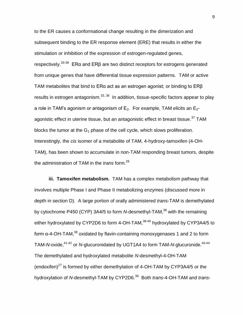
Figure 1-1. A simplified schematic of the tamoxifen metabolism pathway. Tamoxifen is administered in the trans configuration and undergoes
extensive metabolism by a variety of enzymes. Cytochrome P450’s hydroxylate or demethylate tamoxifen to from the major, active metabolites 4-
hydroxy-N-desmethyl-tamoxifen (endoxifen) and 4-hydroxy-tamoxifen (4-OH- TAM) which are subsequently glucuronidated by the UGTs resulting in
deactivation and elimination. All species are shown in the trans configuration, but are able to convert to the cis configuration ...................................................... 11
Figure 1-2. Glucuronide conjugation of a nucleophilic substrate by a UGT. The glucuronic acid moiety of uridine-5’-diphospho-α-D-glucuronic acid is
conjugated to a nucleophilic substrate by UGT enzymes to produce a glucuronide-conjugate of the parent substrate and uridine diphosphate. The
product is generally more easily excreted and generally inactivated .................... 17
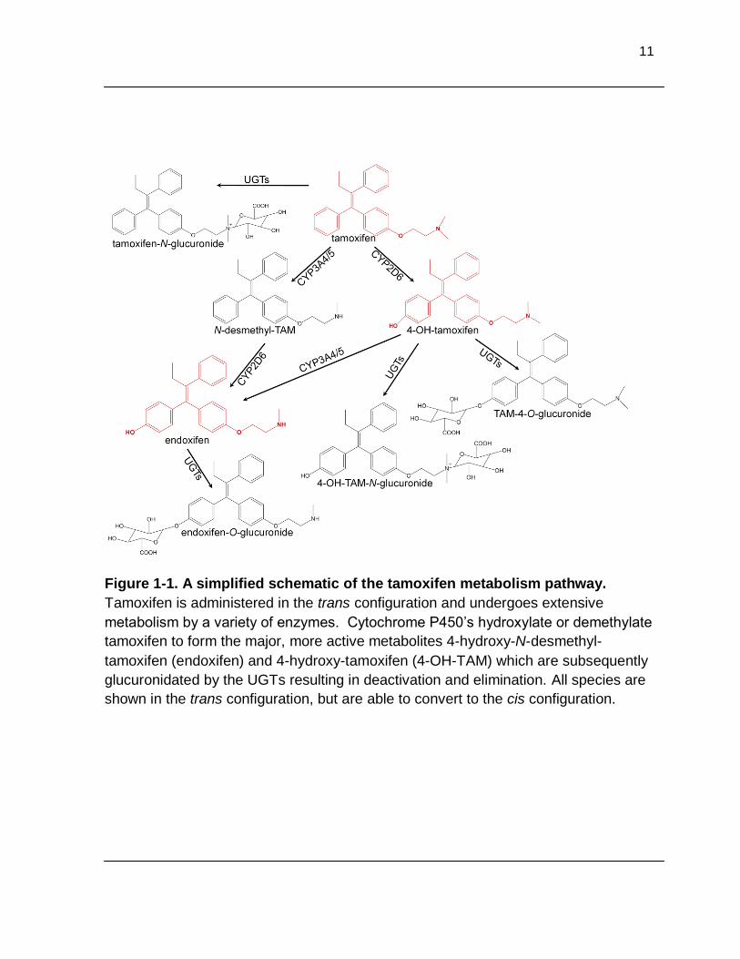
Figure 1-3. A dendogram illustration of UGT family homology. The dendogram illustrates the UGT gene clusters and relative homology. The UGT families all share at least 40 percent homology and share at least 60 percent homology within the individual families. This figure was adapted from Mackenzie 2005 ...................................................................................................... 18
Figure 1-4. The UGT1A gene locus on chromosome 2q37.The promoter element of each unique first exon initiates transcription to the common exons 2-5, resulting in 245 shared amino acids. To date, nine translational and four
pseudogenes in the UGT1A family have been identified at the 2q37 locus. This figure was modified from Girard 2007 ............................................................ 20
Figure 1-5. Alternative splicing of the UGT1A gene locus results in 18 UGT1A isoforms. Newly discovered exon 5b can be spliced instead of, or in
addition to, exon 5a resulting in two gene variants that produce the same protein isoform. All i2 variant enzymes are inactive against a variety of substrates and can negatively regulate the i1 proteins. This figure was modified from Girard 2007……………………………………………………………. 22
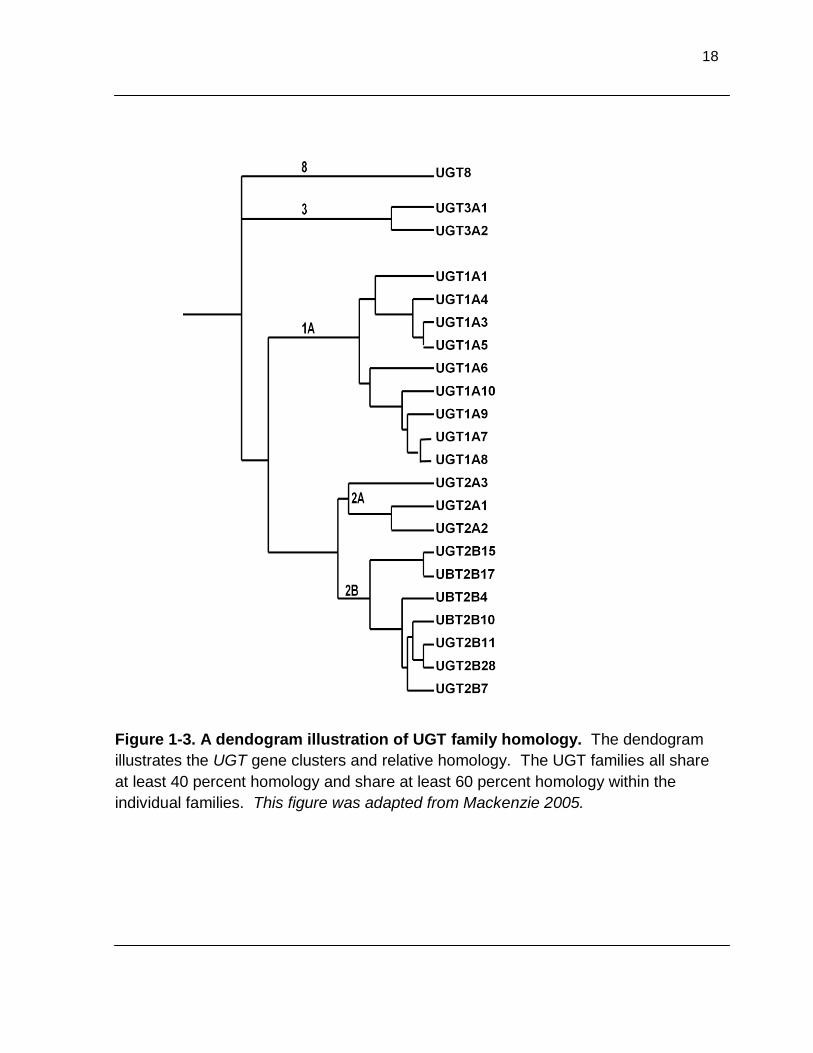
Figure 1-6. Gene map of the UGT2 family. The UGT2B family is located on chromosome 4q13 and each member consists of 6 exons. The UGT2B family includes 7 genes and 5 pseudogenes that are single, unique genes. The UGT2A family includes UGTs 2A1 and 2A2, which are similar to the UGT1A family in that a unique first exon is joined with the common 2-6 exons. Similar to the UGT2B family, UGT2A3 is a single, unique gene. This figure was modified from Mackenzie 2005…………………………………………………. 24
Figure 1-7. A ribbon diagram of the UGT2B7 partial crystal structure. The C-terminal amino acids 285-481 of UGT2B7 was crystallized to a 1.8 Å
resolution and includes the proposed UDPGA binding site. This figure was modified from Miley 2007…………………………………………………………….. 27

xi
Figure 2-1. PCR products of UGTs 1A8 and 2B17. A)The UGT1A8 PCR
product was generated from the cDNA of UGT1A8 stably expressing cell line with missense polymorphisms. B) The UGT2B17 PCR product was generated from HLM specimens 247 and 256 cDNA. HLM 145 represents the positive control, β-actin, and the negative control contained water instead of cDNA. All PCR product bands were excised and the correct sequence was verified by sequencing. The prime sequences used for each gene can be found in Table 2-1…………………………………………………………………. 46
Figure 2-2. Diagram of the pcDNA3.1 vector for stable expression of the UGTs. The pcDNA3.1/V5-His-TOPO vector manufactured by Invitrogen (Carlsbad, CA) contains an ampicillin resistance gene for bacterial selection and a neomycin resistance gene for mammalian cell selection. A CMV promoter is located upstream of the PCR product insertion site. This figure was modified from Invitrogen……………………………………………………...... 47
Figure 2-3. Vector digestion with restriction digest enzymes for determination of PCR product orientation. PCR products were ligated into the pcDNA3.1 vector and transformed into bacteria. Multiple independent clones (represented by numbers) were cultured. Following DNA extraction, DNA was incubated with a restriction digest enzyme to determine the orientation of the PCR product by RFLP. A) The UGT1A8 containing vector was incubated with the enzyme KpnI (except lane 11 which was uncut) resulting in 1491 base pair (bp) and 5633 bp fragments if the orientation was correct. B) The UGT2B17 containing vector was incubated with the enzymes BSrGI and XbaI resulting in 1100 bp and 6000 bp fragments, if the orientation was correct………..…………………………………… 49
Figure 2-4. Vector digestion with restriction digest enzymes following site- directed mutagenesis. Vectors containing the putative variant UGT gene were incubated with a restriction digest enzyme to verify the success of the
previously performed SDM or correct PCR product ligation. A) The UGT1A8 wild-type containing vector was incubated with the enzyme AluI, which cuts the wild-type containing vector 4 times resulting in 4 bands. B) The UGT1A8277Tyr containing vector was incubated with enzyme KpnI resulting in 1491 bp and 5633 bp fragments, if the gene contained the polymorphism and was in the correct orientation. Sequencing confirmed lanes 4, 5, and 6 contained the correct sequences……………………………………………………. 50
Figure 2-5. Representative immunoblot of UGT1A8 and UGT1A8173Gly protein expression. 30 µg of protein that was extracted from cell
homogenates and varying concentrations of UGT1A1 standard were loaded. Densitometry was performed using β-actin as the loading control.
Immunoblot analysis was performed independently in triplicate…………………. 52

xii
Figure 2-6. Representative HPLC chromatograms of the most active UGTs against trans-endoxifen and 4-MU. A) 500 µg of UGT1A8, B) 250 µg of
UGT1A10, and C) 1 mg of UGT2B7 homogenate were incubated with
trans-endoxifen for 60 min at 37ºC. D) 1 mg of variant UGT1A8173Ala/277Tyr homogenates were incubated with 4-MU for 120 min and analyzed by
HPLC. Peak 1 represents UDPGA, the glucuronic acid co-factor; peak 2 represents the endoxifen-O-glucuronide conjugate; peak 3 represents free
trans-endoxifen; peak 4 represents 4-MU-O-glucuronide; and peak 5
represents free 4-MU………………………………………………………………… 56
Figure 2-7. Representative kinetic curves of the glucuronidation of the most active UGTs against trans-endoxifen. Varying concentrations of trans- endoxifen, ranging from 8 to 594 µM, were incubated with A) 500 µg of
UGT1A8, B) 250 µg of UGT1A10, and C) 1 mg of UGT2B7 homogenate and analyzed by HPLC. Michaelis-Menten kinetics were determined by GraphPad Prism software…………………………………………………………………………. 58
Figure 3-1. Schematic of the procedure for microsome preparation. Adjacent normal human liver and breast tissue were homogenized with an electric homogenizer on ice. Aliquots of breast homogenate were saved and stored at -80ºC. Homogenates were centrifuged at 4ºC at 9,000 g for 30 min. The pellet was saved for DNA extraction and stored at -80ºC. The supernatant was subjected to two rounds of ultracentrifugation at 4ºC at 105,000 g for 60 min each. The supernatant, considered to be the cytosolic
fraction, was saved and stored at -80ºC. The pellet was considered to be the microsomal fraction and was resuspended with 0.25 M sucrose, flash- frozen in an ethanol and dry-ice bath and stored at -80………………………….. 73
Figure 3-2. Representative UPLC chromatograms and mass spectra of HLM activity against trans-endoxifen and trans-4-OH-TAM. Glucuronidation activity assays were performed with 40 µg of HLM and A) 30 µM of trans-endoxifen or B) 4 µM of trans-4-OH-TAM. Tandem MS/MS confirmed the glucuronide peaks of HLM glucuronidation reactions against C) trans-endoxifen and D) trans-4-OH-TAM. Peak 1 represents trans- TAM-4-O-glucuronide; peak 2 represents cis-TAM-4-O-glucuronide; peak 3
represents trans-4-OH-TAM-N+-glucuronide; peak 4 represents free trans-4- OH-TAM; peak 5 represents trans-endoxifen-O-glucuronide; and peak 6 represents free trans-endoxifen……………………………………………………… 81
Figure 3-3. UGT2B7 genotype association with HLM glucuronidation phenotype of trans-endoxifen and trans-4-OH-TAM . The rate of O-
glucuronidation of A) trans-endoxifen and B) trans-4-OH-TAM was stratified by UGT2B7 genotype. C) UGT2B7 mRNA expression levels as measured by real-time PCR, were stratified by UGT2B7 genotype. *p < 0.002, **P < 0.001, error bars represent standard deviation……………………………… 83

xiii
Figure 3-4. HLM glucuronidation activity stratified by UGT1A4 and UGT1A1 genotype. HLM N+-glucuronidation activity against trans-4-OH-TAM
stratified by A) UGT1A424Thr/48Leu genotype and B) UGT1A424Pro/48Val genotype. HLM O-glucuronidation activity against C) trans-endoxifen and D) trans-4- OH-TAM stratified by UGT1A1*28 genotype. Error bars represent standard
deviation………………………………………………………………………………… 86
Figure 3-5. Representative mass spectra of 4-MU glucuronidation in human breast tissue. A) HLM glucuronidation of 4-MU, as a positive control for UGT activity. 4-MU glucuronidation by human breast tissue B)
microsomes and C) homogenates. Mass spectral channels recognized the mass of 4-MU (177.06) and 4-MU conjugated to glucuronic acid (353.09), +1 m/z. Peak 1 represents 4-MU-O-glucuronide and peak 2 represents free 4-MU…………………………………………………………………………………….. 87
Figure 3-6. Representative mass spectra of the breast tissue glucuronidation activity assays against trans-endoxifen. A) 20 µg of HLM was incubated with trans-endoxifen as a positive control and compared to B) 670 µg of breast tissue homogenate that was incubated with trans-
endoxifen in a 100 µL reaction, and then vacuum-dried and resuspended in 6 µL prior to MS analysis. Breast tissue homogenate glucuronidation was not
observed. Peak 1 represents cis-endoxifen-O-glucuronide; peak 2 represents trans-endoxifen-O-glucuronide; and peak 3 represents residual
unconjugated trans-endoxifen……………………………………………………….. 88
Figure 3-7. Immunoblot analysis of UGT protein expression in human breast tissue. Protein was extracted from human breast tissue homogenate and microsomes and 75 µg of protein was analyzed for A)
UGT1A family member protein expression in UGT1A1 standard (250 ng of UGT protein), breast homogenate (BH) specimen 1, BH specimen 2, HLM as a positive control, HK293 as a negative control, and breast microsome (BM) specimen 2. B) UGT2B family member expression was examined in
purified UGT2B7 standard (250 ng of UGT protein), BH specimen 1, BH specimen 2, HLM, and BM specimen 2………………………………………………90
Figure 4-1. Diagram of the pcDNA vector for stable expression of c-Src and v-Src in the UGT2B7-HEK293 cell line. The pcDNA6.2/V5/GW/D-TOPO
vector manufactured by Invitrogen (Carlsbad, CA) contains an ampicillin resistance gene for bacterial selection and a blasticidin resistance gene for
mammalian cell selection. A CMV promoter is located upstream of the PCR product insertion site. c-Src and v-Src with the lead sequence CACC were ligated to the vector, resulting in a 451 or 437 of translatable codons. This figure was modified from Invitrogen……………………………………………........ 105

xiv
Figure 4-2. Immunoblot analysis of c-Src and v-Src over-expression in UGT2B7-HEK293 cell lines. Protein was extracted from the parent UGT2B7268His (2B7H) and UGT2B7268Tyr (2B7Y) cell lines and over- expressing c-Src and v-Src and 100 µg was analyzed for A) Src protein expression with β-actin as the loading control and C) activated Src protein
expression with calnexin as the loading control. B) The relative expression levels of Src in UGT2B7H or UGT2B7Y cell lines from panel A, as measured by ImageJ software and expressed in units relative to β-actin…………………… 110
Figure 4-3. Lineweaver-Burk graphs of the glucuronidation of trans- endoxifen, trans-4-OH-TAM, and 4-OH-estrone by UGT2B7 cells over-expressing Src. 15 or 500 µg of A) wild-type UGT2B7268Hi or B) the variant UGT2B7268Tyr stably expressing cell lines were incubated with varying concentrations of trans-endoxifen (column I), trans-4-OH-TAM (column II), or 4-OH-estrone (column III) for 15 min to 1 hr at 37ºC. The black lines with boxes represents the parent cell line, the red lines with triangles represents the cell lines over-expressing c-Src, and the green lines with up-side down triangles represents the cell lines over-expressing v-Src. Kinetic analyses were performed by GraphPad Prism software and are summarized in Tables 4-2 and 4-3. The error bars represent the standard deviation of three
independent experiments………………………………………………………. 112
Figure 5-1. Illustration highlighting the location of the UGTs most active against TAM metabolites. TAM (blue arrows) is ingested orally and
absorbed in the small intestine, where UGTs 1A8 and 1A10 are expressed. However, probably only a minimal amount of endoxifen or 4-OH-TAM is glucuronidated at this point because TAM must first be metabolized by the
CYP2D6 and/or CYP3A4/5. Following absorption, TAM and its metabolites travel to the liver, where UGT2B7and CYP2D6 are expressed. Following
metabolism in the liver, TAM and its metabolites (blue arrows) travel systemically, including to the target tissue of TAM, the breast, where UGTs 1A8 and 2B7 are expressed. Research presented in this dissertation found that polymorphic UGT1A8 and 2B7 glucuronidated trans-endoxifen and trans-4-OH-TAM at a reduced rate, as compared to their wild-type counterparts. A decreased rate of glucuronidation would increase the concentration of circulating endoxifen and 4-OH-TAM, potentially affecting clinical response……………………………………………………………………….129
Figure 5-2. P-glycoprotein transport of doxorubicin and endoxifen. In MDR tumor cells, low concentrations of doxorubicin (DOX) or endoxifen potentially induce the over-expression of P-glycoprotein. DOX or endoxifen enter the cell by diffusion through the plasma membrane and are rapidly removed from the cell by P-glycoprotein, preventing the therapeutic effect of the drug. This figure was modified from Sawant, R……………………………………………….... 136
Figure 5-3. Chemical structures of TAM metabolites and AIs. The major,
active metabolites of TAM, endoxifen and 4-OH-TAM, as well as the aromatase inhibitors (AI) letrozole, anastrozole, exemestane, and exemestane’s active metabolite, 17-dihydroexemestane, are illustrated……….. 138

xv
LIST OF TABLES
Table 1-1. Tissue expression of the human UGTs. The tissue site of UGT expression is listed with associated literature references. The methods of detection included quantitative PCR and/or real-time PCR and/or immunoblot… 21
Table 2-1. Primers utilized for UGT cloning. The sense and anti-sense primers used for cloning UGTs 1A8, 1A8173Gly, 1A8277Tyr, and 2B17 are listed with the
primer location relative to the translationall ATG start site .................................... 45
Table 2-2. Screening results of UGT homogenates against trans-endoxifen. 500 µg of cell homogenates were incubated with trans-endoxifen from 3 hrs to
overnight at 37ºC and analyzed by HPLC............................................................. 57 Table 2-3. Kinetic analyses of the glucuronidation of trans-endoxifen. Michaelis-Menten kinetics were determined with the GraphPad Prism software based on the reaction rate of cell homogenates incubated with varying amounts of trans-endoxifen. Values are per µg of UGT protein, as
previously determined by three independent immunoblots and values are representative of three independent experiments………………………………… 59
Table 2-4. Kinetic analyses of the variant isoforms of the most active UGTs
against trans-endoxifen. Activity assays of the wild-type(UGTs 2B7268His, 1A10139Glu, and 1A8173Ala/277Cys) and variant cell homogenates were performed simultaneously under similar conditions. The values are per µg of UGT
protein, as previously determined by three independent immunoblots and values are representative of three independent experiments. No activity was
detected in homogenates of UGT1A8173Ala/277Tyr against this substrate however, 4-MU glucuronidation was detected……………………………………... 61
Table 2-5. Frequencies of relevant SNPs in UGTs 1A8, 1A10, and 2B7………….64
Table 3-1. Primer sequences utilized for UGT genotyping……………………….. 77
Table 4-1. Primer sequences utilized for the cloning of c-Src and v-Src………..104
Table 4-2. Kinetic analyses of the glucuronidation of trans-endoxifen, trans-4-OH-TAM, and 4-OH-estrone by Src over-expressing UGT2B7268His cell lines. 15 or 500 µg of cell homogenate proteins were incubated with varying concentrations of trans- endoxifen (8-536 µM), trans-
4-OH-TAM (1-172µM), and 4-OH-estrone (2-140 µM) for 15 to 60 min and analyzed by UPLC. Kinetic analyses were performed by GraphPad Prism software and experiments were performed in triplicate. *p ≤ 0.009, ** p ≤ 0.0002, †p ≤ 0.05, ††p ≤ 0.02………………………………………………………….. 111

xvi
Table 4-3. Kinetic analyses of the glucuronidation of trans-endoxifen, trans-4-OH-TAM, and 4-OH-estrone by variant UGT2B7268Tyr cell lines over-expressing Src. 15 or 500 µg of cell homogenate protein was incubated with varying concentrations of trans-endoxifen (8-268 µM), trans- 4-OH-TAM (4-516 µM), and 4-OH-estrone (2-279 µM) for 15 to 60 min and analyzed by UPLC. Kinetic analyses were performed by GraphPad Prism software and experiments were performed in triplicate……………………………. 114 Table 5-1. Proposed clinical matrix for estrogen receptor-positive breast cancer treatment……………………………………………………………………… 141
xvii
LIST OF ABBREVIATIONS
Å Angstrom(s)
AhR arylhydrocarbon receptor AI aromatase inhibitor Ala alanine ANOVA analysis of variance between groups AR androgen receptor ARE arylhydrocarbon responsive element ATP adenosine triphosphate BH breast homogenate BIG 1-98 Breast International Group 1-98 Trial BM breast microsome bp base pair BRCA1 breast cancer type 1 susceptibility protein CAS Crk- and Src-associated substrate CHK Csk homologous kinase COS-1 CV-1 in Origin, carrying SV-40 Crk adaptor protein; proto-oncogene Csk c-Src kinase c-Src cellular-Src CYP450 cytochrome P450 Cys cystine DDT dichlorodiphenyltrichloroethane DHEA dehydroepiandrosterone DHT androgen dyhydrotestosterone DMEM Dulbecco’s modified eagle medium DMSO dimethyl sulfoxide DNA deoxyribonucleic acid DOX doxorubicin E2 17β-estradiol endoxifen N-desmethyl-4-hydroxy-tamoxifen EPR endoplasmic reticulum ER estrogen receptor ERE estrogen receptor element FAK focal adhesion kinase FDA Food and Drug Administration FXR farnesoid X-receptor Gly glycine GST glutathione-S-transferase GT-1 glycosyltransferase 1 G418 geneticin HEK293 human embryonic kidney, clone 293 His histidine HLM human liver microsomes HPLC high performance liquid chromatography HRP horse-radish peroxidase kDa kilo Dalton

xviii
Km Michaelis-Menten equilibrium constant Leu leucine Lys lysine MCF-7 Michigan Cancer Foundation-7 cell line MDRP multidrug resistant protein m/z mass-to-charge ratio NAT N-acetyltrasnferase NCCTG North Central Cancer Treatment Group Trial NNAL 4-(methylnitrosamino)-1-(3-pyridyl)-1-butanol Nrf2 Nuclear factor-like 2 NST nucleotide sugar transporter PAH polycyclic aromatic hydrocarbon PBS phosphate-buffered saline PCR polymerase chain reaction PI3K phosphoinositide 3-kinase PKC protein kinase C PM plasma membrane PP1 4-Amino-5-(methylphenyl)-7-(t-butyl)pyrazolo-(3,4-d)pyrimadine PP2 4-Amino-3-(4-chlorophenyl)-1-(t-butyl)-1H-pyrazolo[3,4-d]pyrimidine Pro proline PTP protein tyrosine phosphatase PVDF polyvinylidene fluoride PXR pregnane X-receptor RFLP restriction fragment length polymorphism RNA ribonucleic acid RT-PCR reverse transcription- PCR SAHA suberoylanilide hydroxamic acid SAM68 Src-associated in mitosis, 68 kDa SDM site-directed mutagenesis SDS-PAGE sodium dodecyl sulfate polyacrilamide gel electrophoresis SEER Surveillance Epidemiology and End Results Ser serine SERM selective estrogen receptor modulator SH SRC homology domain SNP single nucleotide polymorphism SSRI selective serotonin reuptake inhibitor SULT sulfotransferase SYF-/- mouse cells deficient in Src, Yes, and Fyn T-47D Human ductal breast epithelial tumor cell line TAM tamoxifen TBST tris-buffered saline with Tween-20 Thr threonine TK tyrosine kinase Tyr tyrosine UDP uridine diphosphate UDPGA uridine-5’-diphospho-α-D-glucuronic acid UGT UDP-glucuronosyltransferase UPLC ultra performance liquid chromatography Val valine
xix
Vmax maximal velocity v-Src viral-Src 4-OH-estrone 4-hydroxy-estrone 4-OH-TAM 4-hydroxy-tamoxifen 4-MU 4-methylumbelliferone

xx
ACKNOWLEDGEMENTS
First and foremost, I would like to thank my family for their unfailing love
and support. I strongly believe that it was through the effort of parenthood by
Ralph and Sharon Blevins--their encouragement, stories, and discipline--that I
am successful today. My husband, Dave Primeau, has had to endure graduate
school (at times, painfully) as I toiled my way through the class work, and more
importantly, the research projects. At the very end of it all, my sweet baby girl,
Kahlan, was tagging along, and in a way, encouraging me to finish. To Mom,
Dad, and Dave—thank you so much for your loving support; I certainly could not
have made it through all of this without you all. To Kahlan—thank you.
I also deeply appreciate the guidance and support of many colleagues and
faculty members at the College of Medicine. My fellow graduate students made
everyday life fun and it would have been difficult to continue on without the
knowing humor. To my fellow graduate students, research technicians, post-
docs, and assistant professors, past and present—thank you for all the laughs,
teaching, troubleshooting, and debates. A special thank you to Kristine Olson,
Ph.D., who kindly proofread and provided her thoughts on the entire dissertation.
I would like to thank my committee members, Harriet Isom, Ph.D., Mel
Billingsley, Ph.D., Hank Donahue, Ph.D., Tom Spratt, Ph.D., and John Richie,
Ph.D. for their guidance, support, and encouragement.
Finally, I would like to acknowledge Kent Vrana, Ph.D., Jong Yun, Ph.D.,
and Harriet Isom, Ph.D. for their help during a particularly challenging time for me
and their continued support. I may not have finished this thesis without their
support. Thank you!

1
Chapter 1
Literature Review

2
A. Abstract
Cancer is a complex disease that affects over 1.5 million individuals each year
in the United States. Physiological pathways, such as those involved in cell growth
and division, apoptosis, cell motility, angiogenesis, and many other areas are
continually investigated as potential targets for anti-cancer agents. This discussion
will introduce the basic concepts of cancer and more specifically, the mechanisms
involved in breast cancer and the treatment of breast cancer. The main focus will be
to introduce the anti-breast cancer agent, tamoxifen, and its mechanism of action, as
well as the pathway for its metabolism. Finally, the UDP-glucuronsyltransferases
(UGTs) will be introduced. The discussion of the UGT superfamily of phase II drug
metabolizing enzymes will include a foundation of their genetic characteristics, tissue
expression, structure, function, regulation, and clinical importance.

3
B. Introduction to cancer biology
i. Cancer epidemiology. Cancer is a complex disease etiology that affects a
large portion of the population. Surveillance Epidemiology and End Results (SEER)
data estimate that, in 2010, 1,529,560 people were diagnosed with and 569,490 died
from cancer in the United States. This translates to an age-adjusted incidence rate
for all races of 461.6 for diagnosis and 183.8 for death rate per 100,000 individuals in
the United States between 2003 and 2007. For all races, the top five cancer sites
were prostate (154/100,000), female breast (121/100,000), lung and bronchus
(68/100,000), colon and rectum (49/100,000), and corpus (body of uterus) and uterus
(24/100,000).1
ii. The molecular basis of cancer. Cancer is a result of uncontrolled cellular
proliferation caused by multiple mutations in the DNA that result in alteration of
normal cell processes, such as cell signaling, cell cycle checkpoints, and apoptosis.
Genomic mutations may be due to environmental or dietary exposures to
carcinogens, endogenous genotoxic products, and inherited aberrations in normal
cellular processes, such as DNA repair, apoptosis or drug metabolism. Hanahan and
Weinberg suggest that six essential characteristics can be identified in the over 100
unique types of cancers.2 The essential characteristics include self-perpetuating
growth signals, desensitization to anti-growth signals, absence of apoptosis,
constitutive replication ability, angiogenesis, and invasion of adjacent tissue and
subsequent metastasis.2
The development of cancer, or tumorigenesis, appears to be a multi-step
process. Cancer begins as a neoplasm, which Pitot defines as a heritably altered and

4
autonomous growth of tissue.3 The alterations are passed on to the cell’s progeny
and are relatively autonomous. A neoplasm undergoes initiation, the process
whereby an irreversible change in a single cell is caused by exposure to a chemical,
physical or biological agent that is mutagenic to DNA.3-4 Metabolism, DNA repair,
and cell proliferation are three important processes for this stage. The effectiveness
of an initiating agent appears to be dependent on the metabolism of xenobiotics, or
compounds that are not typically found endogenously. The agent has been shown to
be most effective when it is present during the DNA synthesis phase of the cell cycle
both in vitro and in vivo. In addition, at least one round of cell division in the
presence of the initiation factor is required in order for the cell to become truly
initiated.3
An initiated cell must often be promoted in order to progress to a malignancy.
Promotion is the reversible stage of progression where an agent that is able to alter
gene expression and/or apoptosis supports the growth of initiated cells, but does not
directly interact with DNA.3-4 A promoting agent selectively augments cell replication
of pre-neoplastic cells, yet inhibits apoptosis of neoplastic cells.3 In the promotion
phase, cells require the constant presence of the promoting agent.3-4
The clinical disease state of a malignant neoplasm is the progression stage,
where a number of initiated and promoted cells transition to rapidly growing, virulent,
and malignant cells with the hallmark characteristic of irreversible karyotypic
instability.3 In this stage, it is common to observe mutations in proto-oncogenes,
such as c-Src, tumor suppressor genes and irreversible changes in gene expression,
such as those due to alterations in DNA methylation.3

5
C. Introduction to breast cancer
i. Breast cancer epidemiology. Breast cancer is the most diagnosed cancer
in women in the United States. In 2010, approximately 207,090 women were
diagnosed with and 39,840 died of breast cancer. Currently, one in every eight
women will be diagnosed with breast cancer in their lifetime.1 About 60 to 65 percent
of all diagnosed breast cancer cases consist of estrogen receptor (ER) positive
tumors and of these, 60 to 65 percent will respond to hormonal treatment.5
ii. Causes of breast cancer. Although the exact causes of breast cancer are
unknown in many cases, several prominent theories have been suggested. Only
about 5 percent of breast cancer cases can be directly attributed to a germ line
mutation, such as in the case of a BRCA1 deletion or mutation.6 Clearly, additional
factors beyond genetics are involved in breast carcinogenesis.
A major theory attributes steroid hormones, both endogenous and xenobiotic
in nature, as the cause of many breast cancers. It has been hypothesized that
endogenous or xenobiotic steroid hormones can either directly bind to endogenous
hormones or other factors, or alter the levels and ratios of endogenous hormone
metabolites. Both routes result in the aberrant proliferation of breast cells.7
Several forms of evidence support the theory that hormones can cause breast
cancer. The breast cancer cell line, MCF-7, exhibits increased rates of proliferation
when treated with 17β-estradiol (E2).8 Treatment of the murine cell line C57/MG with
16α-hydroxyestrone caused DNA damage, an increase in proliferation, and
production of soft agar colonies, indicating attachment-free growth.9 Increased levels
of cell cycle entry markers were observed when MCF-7 and T-47D cells were treated
6
with E2 or the xenoestrogens (synthetic estrogens) DDT or Red Number 3.10 At the
in vivo level, the genotoxic metabolite, 4-hydroxy-catecholestrogen, has been
observed at particularly high levels in breast tumors as compared to normal breast
tissue.11 In addition, high estrogen levels in post-menopausal women are associated
with an increased risk for breast cancer.12
D. Drug Metabolism
i. Phase I metabolism. Endogenous compounds, such as hormones,
bilirubin, and bile acids, and exogenous compounds, such as carcinogens and drugs,
are metabolized by the body to eliminate activity and facilitate excretion. Phase I
metabolizing reactions create a more polar, and often more active, compound by
adding or unmasking a functional group such as OH-, NH2-, or SH-.13 The CYP450s
are responsible for the phase I metabolism of 80 percent of clinical drugs14 and are
localized to the endoplasmic reticulum (EPR) membrane.3
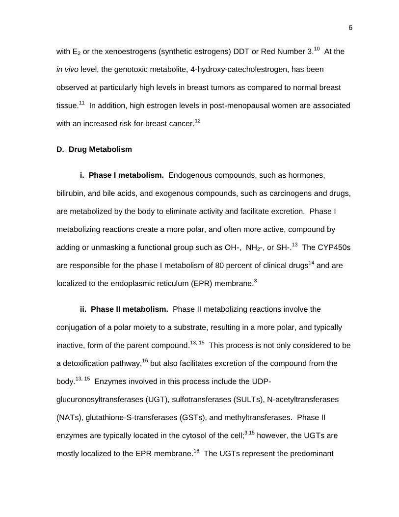
ii. Phase II metabolism. Phase II metabolizing reactions involve the
conjugation of a polar moiety to a substrate, resulting in a more polar, and typically
inactive, form of the parent compound.13, 15 This process is not only considered to be
a detoxification pathway,16 but also facilitates excretion of the compound from the
body.13, 15 Enzymes involved in this process include the UDP-
glucuronosyltransferases (UGT), sulfotransferases (SULTs), N-acetyltransferases
(NATs), glutathione-S-transferases (GSTs), and methyltransferases. Phase II
enzymes are typically located in the cytosol of the cell;3,15 however, the UGTs are
mostly localized to the EPR membrane.16 The UGTs represent the predominant

7
enzyme family involved in drug metabolism as 40 to 70 percent of clinical drugs are
glucuronidated.13, 17
E. The role of estrogen in breast cancer
In the classic genomic signaling of estrogen, E2 passively diffuses through the
plasma and/or nuclear membrane, where it binds to the ER located in the cytosol and
nucleus and causes a conformational change in tertiary and quaternary structure18
that results in dimerization of the ER.19 This forms an active ligand-receptor complex
that is able to bind to DNA response elements that are located upstream of steroid-
responsive genes20 and often causes transcription,19, 21 but may also repress
transcription.22
High circulating estrogen levels have been associated with cancers of the
breast, ovary, and endometrium.23 Phase II metabolism via the UGTs (discussed
further in section G) and other enzymes has been hypothesized to play an important
role in circulating steroid levels. In a small study of 170 healthy, premenopausal
women, variant UGT1A1 and UGT2B4 genotypes were associated with increased E2
and the androgen, dehydroepiandrosterone (DHEA) levels, respectively.23
F. Tamoxifen
i. Introduction to tamoxifen. Tamoxifen (TAM; 1-[4-(2-
dimethylaminoethoxy)phenyl]-1,2-diphenylbut-1(Z)-ene) is a nonsteroidal
triphenylethylene antiestrogen that was approved by the Federal Drug Administration
(FDA) in 1977 for the treatment of breast cancer. A 47 percent annual reduction in
recurrence rate and a 26 percent annual reduction in death rate are observed in

8
women taking TAM for 5 years.24 The typical prescribed dose is 20 mg per day, as a
higher dose has not been found to provide additional benefit.25 TAM is readily
absorbed by the body following oral administration and the serum half-life is 7 to 14
days, with steady state levels reached in 4 weeks.26 The major route of elimination is
feces, although a small amount is excreted through the urine.27 The drug is
administered to patients in the trans-isomer form, due to its higher affinity for the
ER.28-29 The anti-estrogenic moiety of the compound is thought to be the
dimethylaminoethoxy side chain and the trans configuration.28-29
TAM is generally considered to be a well-tolerated therapy; however,
treatment can produce many side effects, some of which may cause compliance
issues and discontinuation of treatment. Side effects include menopausal-like
symptoms, such as hot flashes (occurs in at least 50 percent of patients), vaginal
dryness and discharge, irregular menses, nausea, insomnia, depression, fatigue, as
well as retinopathy, increased risk of endometrial cancer, endometrial hyperplasia,
endometrial polyps, increased endometrial thickness, ovarian cysts, and
thromboembolic events.29 In 1996, the International Agency for Research on Cancer
classified TAM as a carcinogen for the endometrium. Studies have found the hazard
ratio to be 2.4 for endometrial cancer and the increased risk is associated with
duration and dose of TAM usage.24, 30-31
ii. The mechanism of action of tamoxifen. The mechanism of action of
TAM is similar to that of estrogen signaling and depending on the tissue, can act as
either an agonist or antagonist of E2. TAM is a selective estrogen receptor modulator
(SERM),32 which competes with E2 for binding at both ERα and ERβ. Binding of TAM
9
to the ER causes a conformational change resulting in the dimerization and
subsequent binding to the ER response element (ERE) that results in either the
stimulation or inhibition of the expression of estrogen-regulated genes,
respectively.33-36 ERα and ERβ are two distinct receptors for estrogens generated
from unique genes that have differential tissue expression patterns. TAM or active
TAM metabolites that bind to ERα act as an estrogen agonist; or binding to ERβ
results in estrogen antagonism.33, 36 In addition, tissue-specific factors appear to play
a role in TAM’s agonism or antagonism of E2. For example, TAM elicits an E2-
agonistic effect in uterine tissue, but an antagonistic effect in breast tissue.37 TAM
blocks the tumor at the G1 phase of the cell cycle, which slows proliferation.
Interestingly, the cis isomer of a metabolite of TAM, 4-hydroxy-tamoxifen (4-OH-
TAM), has been shown to accumulate in non-TAM responding breast tumors, despite
the administration of TAM in the trans form.29
iii. Tamoxifen metabolism. TAM has a complex metabolism pathway that
involves multiple Phase I and Phase II metabolizing enzymes (discussed more in
depth in section D). A large portion of orally administered trans-TAM is demethylated
by cytochrome P450 (CYP) 3A4/5 to form N-desmethyl-TAM,38 with the remaining
either hydroxylated by CYP2D6 to form 4-OH-TAM,38-40 hydroxylated by CYP3A4/5 to
form α-4-OH-TAM,38 oxidated by flavin-containing monoxygenases 1 and 2 to form
TAM-N-oxide,41-42 or N-glucuronidated by UGT1A4 to form TAM-N-glucuronide.43-44
The demethylated and hydroxylated metabolite N-desmethyl-4-OH-TAM
(endoxifen)27 is formed by either demethylation of 4-OH-TAM by CYP3A4/5 or the
hydroxylation of N-desmethyl-TAM by CYP2D6.38 Both trans-4-OH-TAM and trans-

10
endoxifen can be converted to the cis isomer either spontaneously45 or by
CYP1B1.39, 46 In addition, TAM, 4-OH-TAM, and endoxifen are found conjugated to
glucuronic acid in the urine, bile, and feces of women taking TAM.47-48
iv. The major, active metabolites of tamoxifen. Early studies found 4-OH-
TAM to have a greater affinity for the ER than TAM itself49 and both 4-OH-TAM and
endoxifen have up to 100-fold greater potency than TAM at inhibiting the estrogen-
dependent proliferation of cells.50 In addition, 4-OH-TAM and endoxifen have been
shown to be essentially equal in affinity for ER binding, inhibition of estrogen-
dependent cell line proliferation,5 antagonism of E2-induced expression of the
progesterone receptor,51 and induction of estrogen-responsive global gene
expression in MCF-7 cell lines.52 Importantly, both metabolites are abundant in the
plasma of TAM-treated women, although endoxifen is often present at levels 5- to 10-
fold higher than 4-OH-TAM.27, 47, 53 These data suggest that TAM is a prodrug and
that 4-OH-TAM and endoxifen are the main, active metabolites that elicit a response
in breast tumors. As mentioned previously, TAM and its metabolites exist in the trans
or cis isomeric configuration and can interconvert spontaneously or due to catalysis
by CYP1B1.39, 45-46, 54
In general, the trans, but not the cis, configuration is considered to have anti-
estrogenic activity, although the data are not consistent on this point. For example,
an early study utilizing rat uterus observed trans-4-OH-TAM to have much greater
affinity for the ER than that of cis-4-OH-TAM.55 Similarly, the trans isomer of 4-OH-
TAM preferentially accumulates in MCF-7 cells, while the cis isomer remains in the
11
Figure 1-1. A simplified schematic of the tamoxifen metabolism pathway.
Tamoxifen is administered in the trans configuration and undergoes extensive
metabolism by a variety of enzymes. Cytochrome P450’s hydroxylate or demethylate
tamoxifen to form the major, more active metabolites 4-hydroxy-N-desmethyl-
tamoxifen (endoxifen) and 4-hydroxy-tamoxifen (4-OH-TAM) which are subsequently
glucuronidated by the UGTs resulting in deactivation and elimination. All species are
shown in the trans configuration, but are able to convert to the cis configuration.

12
cell media, leading to the almost exclusive association of the trans isomer with the
ER.54 In contrast, or perhaps due to unknown mechanisms, a later study found that
cis-TAM had estrogenic activity in MCF-7 cells, but cis-4-OH-TAM and trans-4-OH-
TAM had anti-estrogenic activity.56 A more recent report illustrated that both the
trans and cis isomers of 4-OH-TAM and endoxifen exerted equal inhibitory effects on
progesterone receptor induction by E2.57 Clearly, more investigation will be required
to resolve these experimental discrepancies.
v. Tamoxifen resistance. Although up to 65 percent of ER-positive tumors
respond to hormonal therapy such as TAM,5 many of these tumors will acquire
resistance.58 Multiple hypotheses exist to explain the mechanism behind resistance
to TAM therapy including alterations in the number or responsiveness of the ER,
decreased dependency on E2, reduced cellular accumulation of active TAM species,
increased tumor accumulation of E2, and multidrug resistance.58-59
Acquired TAM resistance does not appear to be related to the loss of the
ER, as a majority of resistant tumors maintain ER expression. In this case, it is
possible that despite the presence of the ER, other signaling pathways may become
dominant. In addition, some breast cancer tumors appear to become dependent on
TAM for growth, similar to the concept of tumor dependency on E2. It is probable that
in this case ER signaling occurs, but TAM adopts an agonist role due to unknown
mechanisms. However, TAM-driven tumor growth is observed in a limited number of
tumors and typically other hormonal therapy is effective.58
An early study provided evidence that MCF-7 tumors in athymic mice become
TAM resistance after 4 to 6 weeks of TAM treatment due to both decreased

13
accumulation of trans-TAM in the tumor and increased accumulation of cis-tamoxifen.
Decreased tumor dependency on E2 was not observed.59 In addition, the
accumulation of cis-4-OH-TAM was observed in human breast cancer patients
receiving TAM therapy. Although TAM levels within tumors varied, a general trend of
decreased levels was associated with TAM resistance.60 Interestingly, when TAM
and its metabolites are converted to analogs that cannot be isomerized, tumor
dependency can still be acquired in mice.61 Clearly, other mechanisms instead of, or
in addition to, accumulation of the less active and agonistic cis isomer, cause TAM
resistance.
Multidrug resistance (MDR) is a phenomenon whereby a tumor acquires
resistance to multiple anticancer drugs due to increased efficiency of drug
transporters, enabling the tumor cells to remove drugs at an increased rate. This
mechanism protects the tumor from the toxic effects of the drug. MDR can be
accomplished by induction of the efflux transporters by the particular anticancer drug.
Alternatively, drug treatment has been observed to result in over-expression62 and/or
mutations that alter substrate specificity in transporter genes associated with MDR.63-
64 In addition, MDR-associated transporters, such as Abcg2, can influence
methotrexate pharmacokinetics in mice.65
Alterations in MDR-associated genes have been associated with acquired
TAM resistance. The major mechanism for this is by induction of MDR-associated
genes resulting in the increased expression of proteins that provide transport and
efflux functions to cancer cells. For example, the multidrug resistance-associated
protein (MRP) is expressed at higher levels in TAM resistant MCF-7 cells, as

14
compared to TAM sensitive MCF-7 cells.66 Expression of multidrug resistant protein
8 (MRP8, or more commonly ABCC11) is also increased in TAM resistant MCF-7
cells. Interestingly, treatment of MCF-7 cells with E2 reduced ABCC11 mRNA
expression, which was reversed when the cells were treated with TAM.67
Implications of MDR in TAM resistance have been observed in vivo; variant ABCC11
genotype has been associated with longer recurrence-free survival in breast cancer
patients treated with TAM.68
vi. Tamoxifen pharmacogenetics. Polymorphisms, such as single
nucleotide polymorphisms (SNPs), are heritable changes in the germ-line DNA that
can occur in intronic or exonic regions and may alter mRNA expression or protein
function. The CYP450 demethylation and hydroxylation of TAM has been found to
be an important pathway that can be affected by CYP450 polymorphisms. As
previously stated, CYP2D6 hydroxylation of TAM and N-desmethylTAM is critical in
the formation of 4-OH-TAM and endoxifen.38, 69 CYP2D6 is a highly polymorphic
gene, with 19 inactive and 7 reduced activity alleles currently identified in the
population.69 An inactive or less active enzyme would result in less TAM converted
to 4-OH-TAM and endoxifen. Indeed, in vivo studies have found that CYP2D6 status
in women treated with TAM was associated with endoxifen plasma concentrations.
Namely, CYP2D6 genotypes that resulted in a reduced activity or an inactive enzyme
had lower plasma levels of endoxifen.50, 69-70 In addition, TAM treated breast cancer
patients that were CYP2D6 poor metabolizers, due to reduced activity or inactive
CYP2D6, experienced increased recurrence, mortality rates and fewer side effects as
compared to patients that were extensive metabolizers.50, 69 In this counterintuitive

15
situation, individuals who have reduced metabolism produce less endoxifen, the
active product that is responsible for therapeutic benefit and side effects. Similarly,
breast cancer patients treated with serotonin reuptake inhibitors (SSRIs) for hot
flashes (in addition to TAM), have lower plasma levels of the active metabolite,
endoxifen, because SSRIs are CYP2D6 inhibitors.71
Despite adjusting for CYP2D6 status, wide variability of 4-OH-TAM and
endoxifen plasma concentrations are still detected in women treated with TAM.69-70
This suggests that additional mechanisms, such as other metabolic pathways, are
important in 4-OH-TAM and endoxifen plasma concentrations and, potentially, patient
outcome.
Glucuronidation is a major detoxification pathway that inactivates TAM
metabolites57, 72 and facilitates their excretion from the body. Similar to the CYP450s,
the UGTs are known to be highly polymorphic. Therefore, it is important to
characterize which UGTs are responsible for the metabolism of 4-OH-TAM and
endoxifen and to subsequently determine how polymorphisms in these
specific enzymes may be playing a role in patient response. This is the central
focus of the present dissertation research program.
G. The UDP-glucuronosyltransferases
i. UGT function. The UGTs predominately catalyze the conjugation of
uridine 5’-diphospho-α-D-glucuronic acid (UDPGA) to a nucleophilic functional group
such as a hydroxyl, amino, sulfuryl, carboxyl, or carbonyl moiety (Figure 1-2).13, 16, 73
However, O- and N-linked glucuronides are the predominant species. All UGT
isoforms have been found to perform O-linked glucuronidation, which mainly forms

16
aryl-O-(phenolic)-glucuronides, but also acyl-O-glucuronides (at a carboxylic acid)
and alkyl-O-(enolic)-glucuronides (such as in coumarin). N-linked glucuronidation is
typically performed by UGT1A4 and UGT2B10 and can occur at non-quaternary
amines, such as heterocyclic amines and primary and secondary amines. Moreover,
tertiary amines, such as cyclic tertiary amines, alicyclic tertiary amines, and aromatic
heterocyclic amines, can be conjugated.16
ii. UGT nomenclature. The first Arabic number in UGT nomenclature
represents the family to which the enzyme belongs, the letter represents the
subfamily, and the second Arabic number represents the specific gene. The UGT
families share at least 40 percent nucleic acid homology and each enzyme within a
family shares at least 60 percent.13 The UGT enzymes are classified into four
families; 1, 2, 3 and 8 (Figure 1-3). The family names were assigned by the UDP
Glycosyltransferase Nomenclature Committee in the early 1990’s, although only two
families had been discovered. UGT8 was the first family name reserved for any non-
drug metabolizing UGT family, and families 3-7, which were not yet discovered, were
reserved for the anticipated discovery of drug metabolizing UGT families.74 Currently,
only one additional drug metabolizing UGT family has been identified; the UGT3
family.
The gene clustering that is observed in the UGT families is an indication of
gene duplication events. However, the UGT2B family in primates does not cluster
with that of rodents, suggesting that separate and independent gene duplication
events occurred following speciation.16

17
Figure 1-2. Glucuronide conjugation of a nucleophilic substrate by a UGT. The
glucuronic acid moiety of uridine-5’-diphospho-α-D-glucuronic acid is conjugated to a
nucleophilic substrate by UGT enzymes to produce a glucuronide-conjugate of the
parent substrate and uridine diphosphate. The product is generally more easily
excreted and generally inactivated.
18
Figure 1-3. A dendogram illustration of UGT family homology. The dendogram
illustrates the UGT gene clusters and relative homology. The UGT families all share
at least 40 percent homology and share at least 60 percent homology within the
individual families. This figure was adapted from Mackenzie 2005.

19
iii. UGT family and gene structure. The UGT1A family spans approximately
160 kb and is located on chromosome 2-q37 (Figure 1-4). The family members
consist of 5 exons; a unique first exon that is transcribed and spliced to the common
2-5 exons, that results in 245 shared amino acids.16, 75 The 5’ region of each exon 1
contains the promoter elements necessary for transcription and the 3’ boundary
region to each exon 1 contains the consensus sequence necessary for splicing via
the RNA splicesome. The process of transcribing the individual genes of the UGT1A
family is often referred to as alternative splicing, but current evidence suggests
differently. Each transcript is independently produced following transcription initiation
due to regulatory sequences flanking each exon 1 and alternative splicing is not
involved. True alternative spicing is considered to occur when exons are spliced
differentially after transcription has occurred. The UGT1A family encodes the
following proteins: UGT1A1, 1A3, 1A4, 1A5, 1A6, 1A7, 1A8, 1A9, and 1A10. Of
these, UGTs 1A7, 1A8 and 1A10 have been found to have exclusive extra-hepatic
expression (Table 1-1).16
In 2006, Levesque and colleagues discovered an additional exon located at
the 3’ end of the UGT1A gene locus that results in translation of a protein that is
inactive.76 Subsequently, the new exon was named exon 5b (as 5a is the original 5th
exon), that can be spliced to exon 4 in lieu of, or in addition to, exon 5a. Exon 5b
encodes a stop codon and therefore both variant genes translate into the same
protein isoform, as the open reading frames are identical. Therefore, there are an
additional 8 UGT1A protein isoforms, for a total of 16 translated UGT1A enzymes in
humans (Figure 1-5). The variant gene products are referred to as “variants” and

20
Figure 1-4. The UGT1A gene locus on chromosome 2q37. The promoter element
of each unique first exon initiates transcription to the common exons 2-5, resulting in
245 shared amino acids. To date, nine translational and four pseudogenes in the
UGT1A family have been identified at the 2q37 locus. This figure was modified from
Girard 2007.

21
Table 1-1. Tissue expression of the human UGTs. The tissue site of UGT
expression is listed with the associated literature references. The methods of
detection included quantitative PCR and/or real-time PCR and/or immunoblot.
UGT Tissue Reference
1A1 liver, stomach, small intestine, colon, bladder Nakamura 2008; Ohno 2008
1A3 liver, small intestine, colon, bladder Nakamura 2008; Ohno 2008
1A4 liver, stomach, small intestine, colon, kidney, bladder, ovary Nakamura 2008; Ohno 2008
1A5 liver, esophagus, stomach, small intestine, colon, brain, kidney, bladder, breast, uterus, prostrate, cervix, heart, trachea
Nakamura 2008; Ohno 2008
1A6 liver, stomach, small intestine, colon, kidney, bladder, adrenal gland, trachea
Nakamura 2008; Ohno 2008
1A7 liver, esophagus, small intestine, colon, kidney, bladder, thymus, cervix, trachea, adipose
Nakamura 2008; Ohno 2008
1A8 small intestine, colon, kidney, bladder, adrenal gland, breast, trachea
Nakamura 2008; Ohno 2008
1A9 liver, esophagus, small intestine, colon, kidney, bladder,adrenal gland, testis
Nakamura 2008; Ohno 2008
1A10 esophagus, stomach, small intestine, colon, kidney, bladder, adrenal gland, ovary, uterus, trachea
Nakamura 2008; Ohno 2008
2A1 lung, larnyx, trachea, tonsil, colon, floor of mouth Bushey 2010
2A2 liver Izukawa 2009
2A3 liver Izukawa 2009
2B4 liver, esophagus, lung, kidney, bladder, adrenal gland, breast, uterus, testis, thymus, prostate, heart, trachea
Nakamura 2008; Ohno 2008
2B7 liver, lung, stomach, small intestine, colon, kidney, bladder, adrenal gland, breast, ovary, uterus, testis
Nakamura 2008; Ohno 2008
2B10 liver, bladder, ovary, uterus, testis Nakamura 2008; Ohno 2008
2B11 liver, lung, stomach, small intestine, colon, kidney, bladder, breast, ovary, uterus
Nakamura 2008; Ohno 2008
2B15 liver, stomach, small intestine, colon, kidney, bladder, adrenal gland, breast, ovary, uterus, testis, prostate, trachea
Nakamura 2008; Ohno 2008
2B17 liver, lung, stomach, small intestine, colon, kidney, adrenal gland, breast, ovary, testis, brain, placenta, heart, trachea, spleen, adipose
Nakamura 2008; Ohno 2008
2B28 liver, bladder, breast Nakamura 2008
3A1 liver, kidney, stomach, duodenum, colon, testes Meech 2010
3A2 kidney, testes, thymus, trachea, spleen, prostate Meech 2010; Mackenzie 2010

22
Figure 1-5. Alternative splicing of the UGT1A gene locus results in 18 UGT1A
isoforms. Newly discovered exon 5b can be spliced instead of, or in addition to,
exon 5a resulting in two gene variants that produce the same protein isoform. All i2
variant enzymes are inactive against a variety of substrates and can negatively
regulate the i1 proteins. This figure was modified from Girard 2007.

23
denoted as UGT1A_v1 for the wild-type variant containing the original exon 5a and
UGT1A_v2 or UGT1A_v3 for the exon 5b or exon 5a and 5b containing variants,
respectively. The protein is referred to as an “isoform” and denoted as UGT1A_i1 for
the wild-type isoform and UGT1A_i2 for the product of UGT1A_v2 or UGT1A_v3.
The truncated isoforms have no glucuronidating activity against a variety of
substrates and co-expression studies have found that they can negatively regulate
the active isoform.77 The isoforms were observed to form both homo-oligomeric and
hetero-oligomeric complexes, where the i2-i2 complexes are inactive and the i1-i2
complexes have reduced activity, due to a decrease in maximal velocity (Vmax).78-79
The UGT2A family is not yet well characterized, although initial reports have
begun to investigate the expression and substrate targets of UGT2A1 and 2A2.80-82
UGT2A1 and 2A2 are located on chromosome 4 and have 6 exons (Figure 1-6).
Similar to the UGT1A family, the first exon is unique and the remaining 5 exons are
shared.83 The first transcripts were cloned from nasal mucosa, but recent studies
have found UGT2A1 to be well expressed in the lung, larynx, trachea, tonsil, and
colon.80 Activity assays have demonstrated that UGT2A1 effectively glucuronidated
polycyclic aromatic hydrocarbons (PAHs).80
The UGT2B family is located on chromosome 4-q13 and consists of 6 unique
exons (Figure 1-6).16, 75 Interestingly, three additional exons were recently identified
in UGT2B4 that form three inactive splice-variant isoforms and are well expressed in
human liver, gastrointestinal tract, and other tissues. The three inactive isoforms also
decrease the activity of the wild-type, active isoform when co-over-expressed in
mammalian cells.84
24
Figure 1-6. Gene map of the UGT2 family. The UGT2B family is located on
chromosome 4q13 and each member consists of 6 exons. The UGT2B family
includes 7 genes and 5 pseudogenes that are single, unique genes. The UGT2A
family includes UGTs 2A1 and 2A2, which are similar to the UGT1A family in that a
unique first exon is joined with the common 2-6 exons. Similar to the UGT2B family,
UGT2A3 is a single, unique gene. This figure was modified from Mackenzie 2005.

25
The UGT3 family was the last to be identified and its function was recently
determined to be that of an N-acetylglucosaminyltransferase. The two genes,
UGT3A1 and 3A2, are located on chromosome 5p13.2 and share approximately 80
percent sequence homology, but only 40 percent to UGT1A1, and are comprised of 7
exons that result in a 50-53 kDa protein of 523 amino acids. Interestingly, UGT3A1
utilizes UDP-N- acetylglucosamine85 and UGT3A2 utilizes UDP-glucose and UDP-
xylose, as opposed to UDPGA, as the sugar donor.86 A secondary bile acid and
several estrogens are N-acetylglucosaminidated by UGT3A1 and it has been found
to be expressed in the liver and some tissues of the gastrointestinal tract.85, 87
UGT3A2 conjugates a larger variety of substrates than UGT3A1, such as 4-
methylumbelliferone (4-MU), 1-hydroxypyrene, bioflavones, and estrogens and has
been found to be well expressed in the kidney, thymus, and testes.86
The UGT8 family encodes the UDP-galactose ceramide galactosyltransferase
enzyme, which is critical in the biosynthesis of important components of myelin
including glycosphingolipids, cerebrosides, and sulfatides. As indicated by the name,
this enzyme utilizes UDP-galactose as the co-substrate and it is located in the EPR
membrane. The gene consists of 5 exons, is located on chromosome 4q26, and is
expressed in oligodendrocytes and Schwann cells.88
The UGT1 and 2 families are the main UGT families that perform drug
metabolism13 and are the focus of this dissertation. Therefore, the remainder
of this dissertation is written in the context of the UGT1 and 2 families and the
use of the abbreviation “UGT” will refer to only these two families.
26
iv. UGT polymorphisms. The UGT superfamily is known to be highly
polymorphic, with both synonymous and non-synonymous single-nucleotide
polymorphisms (SNPs) occurring frequently. Gene deletions and microsatellite
polymorphisms have also been discovered. Several examples of the clinical
importance of UGT polymorphisms are highlighted in section G.vii.
Non-synonymous SNPs cause an amino acid change in the protein, that often
lead to alterations in enzyme activity, although, this is not always the case. Many
SNPs and other polymorphisms cause a decrease or increase in enzyme activity or
expression, which is often dependent on the substrate. For example the
UGT2B7268Tyr variant exhibited decreased activity against substrates such as 4-
(methylnitrosamino)-1-(3-pyridyl)-1-butanol (NNAL).89 Conversely, the UGT2B7268Tyr
variant exhibited an increase in activity against some morphine derivatives90, 4-
hydroxyestrone (4-OH-estrone), and 4-hydroxyestradiol.91 Some studies have
observed the wild-type and variant isoforms of UGT2B7 to exhibit similar activity
levels, such as with epirubicin92 and mycophenolic acid.93
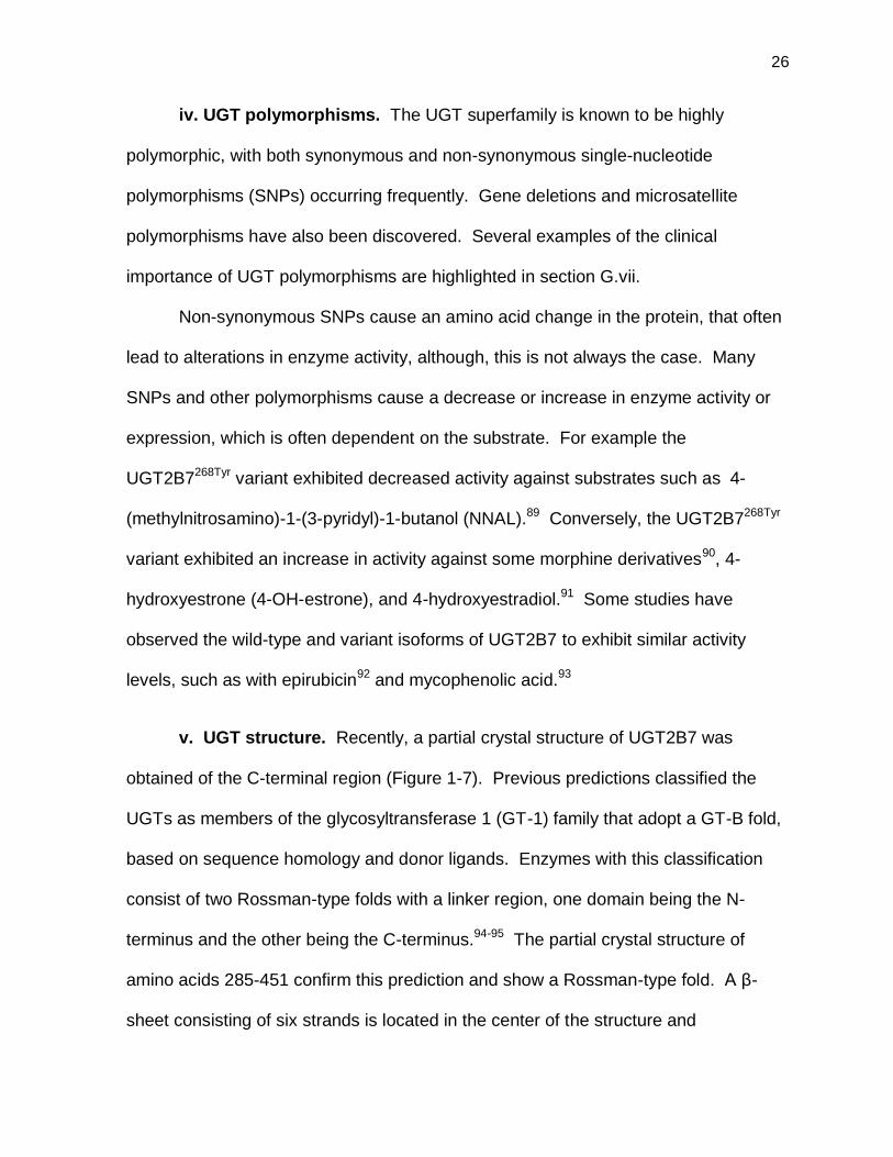
v. UGT structure. Recently, a partial crystal structure of UGT2B7 was
obtained of the C-terminal region (Figure 1-7). Previous predictions classified the
UGTs as members of the glycosyltransferase 1 (GT-1) family that adopt a GT-B fold,
based on sequence homology and donor ligands. Enzymes with this classification
consist of two Rossman-type folds with a linker region, one domain being the N-
terminus and the other being the C-terminus.94-95 The partial crystal structure of
amino acids 285-451 confirm this prediction and show a Rossman-type fold. A β-
sheet consisting of six strands is located in the center of the structure and

27
Figure 1-7. A ribbon-diagram of the UGT2B7 partial crystal structure. The C-
terminal amino acids 285-481 of UGT2B7 was crystallized to a 1.8 Å resolution and
includes the proposed UDPGA binding site. Figure was modified from Miley 2007.

28
surrounded by seven α-helices. Because the crystal structure is of the C-terminal
end of the enzyme, the proposed UDPGA binding site is observed and is similar to
that of glycosyltransferases in bacteria and plants. Unfortunately, the polypeptide of
the crystal structure did not include the amino acid that is altered in the highly
prevalent UGT2B7 polymorphism, His268Tyr.96
Despite the slightly different genetic organization of the UGTs, only the 280
amino-terminal amino acids are divergent. For example, all UGTs, except for
UGT1A10, have the N-terminal EPR-retention signal peptide, that is cleaved off
following enzyme insertion into the EPR membrane.16 However, studies of UGT1A6
localization to the EPR found that the EPR-retention signal is not required for EPR
retention, but that amino acids 140-240 are important and that a dilysine motif
located at the C-terminal stop-transfer sequence alone is sufficient for retention.97-98
vi. UGT localization. The UGTs are transmembrane proteins generally
localized to the EPR membrane with the catalytic site positioned within the EPR
lumen with a short C-terminal cytosolic segment.13, 75, 87, 97, 99 A study has reported
localization to the nuclear membrane as well.99 This orientation requires that
substrates and UDPGA be transported into the lumen of the EPR, by unknown
mechanisms and the nucleotide sugar transporters (NSTs), respectively.100-102 The
NSTs are antiporters that transport UDPGA into the lumen in exchange for UDP-N-
acetylglucosamine.101-102 Evidence suggests that newly conjugated glucuronides are
rapidly transported out of the EPR lumen and into the cytosol by facilitated diffusion
using organic anion transporters.100

29
vii. UGT pharmacogenetics. The UGTs are well expressed in a variety of
tissues at levels that vary among individuals (Table 1-1). In the liver, a recent study
of 25 different human liver samples identified the following UGT mRNA expression by
real-time PCR: UGT1A1, 1A3, 1A4, 1A5, 1A6, 1A9, 2B4, 2B7, 2B10, 2B15 and 2B17.
Barely detectable levels of expression were found for UGTs 1A5, 1A7, 1A8, 1A10,
2B11, and 2B28, and this low level of expression is probably not physiologically
relevant.103
Inter-individual variation in UGT expression and activity is common and Bock
has suggested that it is driven by three main factors; 1) genetic diversity due to
polymorphisms, alternate splicing events, and epigenetics, 2) liver-enriched
transcription factors, and 3) ligand-activated transcription factors.75 In addition to
these factors, recent evidence suggests that post-translational factors such as
dimerization104-107 and phosphorylation108-111 may also play an important regulatory
role in UGT activity.
UGT expression is inducible and several transcription factor ligands have been
linked to this process, such as the aryl hydrocarbon receptor (AhR), the farnesoid X-
receptor (FXR), and the pregnane X-receptor (PXR).112-113 In addition, some UGT-
targeted substrates have been shown to regulate the transcription of the active UGT,
suggesting UGTs are involved in feedback loops.114 For example, bilirubin, the by-
product of heme catabolism, was observed to elicit a time and dose-dependent rise in
UGT1A1 mRNA levels when incubated with rat liver microsomes.112, 114 In addition,
UGT1A1 transcription has been shown to be regulated by the AhR and known
ligands for AhR upregulate the transcription of UGT1A1. A recent study has linked

30
the previous data and illustrated that the response element in the UGT1A1 promoter
is activated by bilirubin via an AhR-mediated pathway.114-115 Other transcription
factor ligands, such as the bile acid and the FXR and the PXR, as well as the
androgen dihydrotestosterone (DHT) and the androgen receptor (AR), have also
been found to regulate the expression of some UGTs.114 Interestingly, a promoter
polymorphism in linkage disequilibrium with the UGT2B7*2 variant prevented
induction by Nrf2, a transcription factor that induces wild-type UGT2B7 via an ARE-
like promoter element.113
Induction of the UGTs by exogenous compounds has also been reported.
UGT1A1 has been induced by drugs such as dexamethasone, a synthetic
glucocorticoid,116 rifampicin, clotrimazole, carcinogens such as benzo[a]pyrene117
and dietary components such as the red wine polyphenol resveratrol, curcumin,118
the isothiocyanate sulforaphane,119 chrysin and multiple other flavonoids.117-118
Interestingly, exogenous compounds selectively induce the UGTs. For example,
treatment of Caco-2 cells with chrysin results in the induction of UGT1A1, but not
UGTs 1A6, 1A9, and 2B7.120 Induction activities have been found to be regulated by
enhancer elements,117 DNA response elements often mediated by the aryl
hydrocarbon receptor (AhR) pathway,121 and cell signaling pathways.119
H. UGT2B7 phosphorylation by Src
i. Introduction to Src. Src, a non-receptor tyrosine kinase originally
discovered as viral-Src (v-Src), a viral gene of the Rous Sarcoma virus found in
chickens, is able to trigger cellular transformation.122-123
Cellular Src (c-Src) is a
physiological gene that is present in all animals but not yeast, bacteria, or plants, and

31
is ubiquitously expressed in all tissues and cell-types. c-Src is a proto-oncogene,
and can become an oncogene when altered or over-expressed. Aberrant gene
expression of c-Src can cause dysregulation of the cell cycle and has been
associated with cancer. However, c-Src also plays a critical role in a variety of
normal cellular processes, such as differentiation, proliferation, cell division,124-125
survival,126 cell adhesion, cell motility,126-128 morphology, and bone remodeling and
reabsorption.124, 129-130 During most of the cell cycle, c-Src is dormant. c-Src only
becomes activated during the G2/M transition and is required for cell division in
fibroblasts.125 In fibroblasts, c-Src is found bound to endosomes, perinuclear
membranes, secretory vesicles, and the cytoplasmic face of the plasma
membrane,131-135 as well as in the cytoplasm and perinuclear region of the Golgi
apparatus.134-135
ii. Src in cancer. Many studies have reported increased levels of c-Src in
human cancers such as breast, colon, gastric, lung, pancreatic, neural, and
ovarian.123 Cell lines that express high levels of activated Src become more invasive
in vivo136 and are associated with metastasis in animal models.137 In addition, breast
cancer exhibits increased Src activity as compared to normal issue.138 Interestingly,
cells that acquire TAM resistance exhibit increased levels of Src and there is an
association in ER-positive patients of activated Src in the cytoplasm of their breast
tumor and reduced survival time with endocrine therapy. MCF-7 cells that over-
express active Src develop a greater migratory and invasive behavior and their
growth is not inhibited when treated with TAM. When these cells are treated with a

32
Src specific inhibitor, their morphology changes so that the cells appear to regain
their cell-to-cell contacts and both migratory and invasive behavior is decreased.139
iii. UGT phosphorylation. Recent studies have suggested that the UGT1A
family is phosphorylated by protein kinase C (PKC)108-109 and UGT2B7 is
phosphorylated by Src.111 A database search with UGT2B7 identified three putative
PKC sites, located at Thr123, Ser132, and Ser437 and two tyrosine kinase (TK) sites,
located at Tyr236 and Tyr438. Curcumin, which is both a PKC and a Src inhibitor,
decreased UGT2B7 activity without affecting PKC-site phosphorylation. In addition,
site-directed mutations of the PKC sites did not alter enzyme activity, suggesting that
PKC site phosphorylation is not important for UGT2B7 activity. However, site-
directed mutations at either or both TK sites resulted in decreased enzyme activity.
Transient co-transfection of UGT2B7 with Src caused a 1.5-fold increase in UGT2B7
activity against 4-OH-estrone, an endogenous substrate for UGT2B7.111 These data
suggest that UGT2B7 is phosphorylated by Src and that phosphorylation is important
for enzyme activity.
In contrast, Abe and colleagues reported that UGT1A protein, but not mRNA,
levels were decreased when treated with PKC inhibitors, such as curcumin, and not
tyrosine kinase inhibitors. In addition, immunoprecipitation studies did not provide
evidence of phosphorylation of the UGT1As. This study found that a decrease in 4-
MU glucuronidation was a result of reduced protein and probably not
phosphorylation.140 Clearly, more research is needed to determine if phosphorylation
is truly playing a role in UGT activity.

33
I. UGT pharmacogenetics is clinically significant
i. Hyperbilirubemia is caused by UGT1A1 polymorphisms. Several
clinically significant syndromes have been observed that are a result of inherited
polymorphisms in the UGTs. The most prominent are those characterized by
hyperbilirubemia, a disorder where blood levels of bilirubin reach 35 µM/L or greater.
If circulating bilirubin levels exceed 300 µM/L, it will cross the blood-brain barrier and
cause fatal necrosis of neurons and glial tissue. Bilirubin is the natural breakdown
product of heme, 80 percent of which is due to the catalysis of hemoglobin and 20
percent is due to free circulating heme and the breakdown of heme-containing
proteins such as CYP450s, tryptophan pyrrolase, and catalase.16
Bilirubin is glucuronidated almost exclusively by UGT1A1 and polymorphisms
in the UGT1A1 gene can lead to serious clinical outcomes.141 The most severe is the
autosomal recessive Crigler-Najjar Syndrome16 which can be caused by up to 50
different mutations in UGT1A1.13 Crigler-Najjar Syndrome is further classified into
two types, based on the amount of UGT1A1 enzyme activity remaining. Type I is
characterized by the absence of UGT1A1 activity 13 and causes nonhemolytic icterus
and kerinicterus, the accumulation of bilirubin in glial cells and nerve terminals, within
the first several days of life.16 Homozygotes of the syndrome die in early childhood
and the only available treatment is immediate liver transplant.16 Crigler-Najjar
Syndrome Type II is characterized by a severe deficiency in UGT1A1, but at least 10
percent of enzyme activity remains, probably due to impaired transcription,142 which
results in a milder hyperbilirubemia and individuals who are affected live into

34
adulthood without neurological impairment.13, 16 Treatment includes induction
therapy (such as with phenobarbitol) or phototherapy.16
The third hyperbilirubemia syndrome is the autosomal dominant Gilbert
Syndrome which is primarily caused by a polymorphism in the TATAA box region of
the UGT1A1 promoter that is present in about 10 percent of the population.13, 143 It is
clinically benign, as only a mild form of hyperbilirubinemia results,13 with an onset
typically due to physiological stress, infection, fasting or physical activity.16
Individuals affected by Gilbert Syndrome do not require treatment.16
ii. The UGT1A1 TATAA box polymorphism. The UGT1A1*28, *36 and *37
alleles are characterized by variant TA repeat(s) between nucleotides -23 and -38,
upstream of the ATG start-site. The wild-type promoter that results in full
transcription of UGT1A1 contains A(TA)6TAA. The initial study that reported that an
additional TA repeat caused Gilbert’s Syndrome determined that the repeat caused
an 82 to 67 percent decrease in luciferase activity, most likely due to a decrease in
the binding ability of the transcription factor IID. In addition, every Gilbert’s Syndrome
patient studied had the A(TA)7TAA element in the UGT1A1 promoter
(UGT1A1*28).141 Subsequent studies have found 8 TA repeats in some Gilbert’s
Syndrome patients (UGT1A1*37), which also resulted in decreased luciferase
expression, and 5 TA repeats (UGT1A1*36), that caused an increase in the
luciferase activity as compared to the wild-type 6 TA repeats. Interestingly, the
prevalence of this polymorphism varies in different populations. The 7 TA repeats
occur in about 39 percent of Caucasians, 43 percent of blacks, and 16 percent of
35
Asians and 5 and 8 TA repeats are predominately found in blacks at 3.5 percent and
6.9 percent, respectively.144-145
iii. UGT pharmacogenetics of anti-cancer agents. The UGTs are critical in
the metabolism and ultimate excretion of a variety of clinical drugs. A prominent
example of this is the drug irinotecan. Irinotecan and its major, active metabolite,
SN-38, is an FDA-approved topoisomerase I inhibitor for the treatment of colon
cancer as a monotherapy or in combination. The information pamphlet for irinotecan
has been updated to include a warning about the association between UGT1A1*28
genotype and potentially fatal neutropenia. SN-38 is mainly glucuronidated by
UGT1A1, but other UGT1A family members have been found to also be involved in
the metabolism pathway. Diarrhea and neutropenia are the most common potentially
fatal side effects and both have been linked to polymorphisms in the UGTs. The
UGT1A1*28 allele is associated with irinotecan-induced neutropenia and the FDA
now recommends that clinicians test patient UGT1A1*28 status to aid in determining
the proper irinotecan dosage.146
The promising FDA-approved histone deacetylase suberoylanilide hydroxamic
acid (SAHA) is glucuronidated in the liver primarily by UGT2B17. The deletion
polymorphism of UGT2B17 was recently observed to significantly reduce SAHA
glucuronidation in human liver microsomes (HLM).147 Interestingly, stratification by
sex revealed that males exhibited greater UGT2B17 glucuronidation against SAHA,
as well as other compounds.148

36
J. Hypothesis and Aims
An overarching goal of Dr. Philip Lazarus’ laboratory is to study how the
pharmacogenetics of the UGTs effect the metabolism of a variety of agents. The
information presented in this chapter identifies several important points in regard to
the UGTs and tamoxifen metabolism. When this research began, much of the role of
the UGTs in tamoxifen metabolism was unknown, yet this phase II family has been
determined to be important in the overview of tamoxifen and its pharmacotherapeutic
action. The overall hypothesis for this dissertation is that the UGTs play an important
role in the phase II metabolism of the major, active metabolites of tamoxifen and that
polymorphisms in the UGTs most active against these metabolites significantly alter
enzyme activity. The aims of this dissertation are:
1. To identify which UGTs are important in the metabolism of trans-endoxifen
and to assess how the polymorphic isoforms alter enzyme activity. The
UGTs active against trans-4-OH-TAM were previously determined in the
laboratory.149-150
2. To test the hypothesis that UGT genotypes are associated with human liver
microsomes (HLM) phenotype in the glucuronidation activities of trans-
endoxifen and trans-4-OH-TAM.
3. To test the hypothesis that the potential phosphorylation of UGT2B7 by Src
alters enzyme activity against trans-endoxifen and trans-4-OH-TAM.

37
Chapter 2 Identification of the UGTs that are active against trans-endoxifen
NOTE: The data reported in this chapter are based on the published manuscripts:
Sun D, Sharma AK, Dellinger RW, Blevins-Primeau AS, Balliet RM, Chen G, Boyiri T, Amin S, Lazarus, P. Glucuronidation of active tamoxifen metabolites by the human UDP glucuronosyltransferases. Drug Metab Dispos.2007. 35(11): 2006-2014.
Blevins-Primeau, AS, Sun D, Chen G, Sharma AK, Gallagher CJ, Amin S, Lazarus P. Functional significance of UDP-glucuronosyltransferase variants in the metabolism of active tamoxifen metabolites. Cancer Res. 2009. 69(5): 1892-1900.
All figures presented in this chapter are the work of A.S.B-P., except the kinetic analyses of UGT1A8173Gly and UGT1A8277Tyr, which were performed by D.S. Immunoblot analysis of UGT1A8277Tyr was also performed by D.S. (results not shown).
38
A. Abstract
Tamoxifen (TAM) is a non-steroidal selective estrogen receptor modulator
that was approved by the FDA in 1977 for the treatment of breast cancer. Although it
is generally well tolerated, significant adverse effects have been reported, including
severe hot flashes and an increased risk for venous thromboembolism and
endometrial cancer. The phase I metabolism of TAM is primarily catalyzed by
CYP2D6 and CYP3A4/5, resulting in the major, active metabolites N-desmethyl-4-
hydroxy-tamoxifen (endoxifen) and 4-hydroxy-tamoxifen (4-OH-TAM). Interestingly,
the presence of CYP2D6 variant genotypes having an inactive or less active
phenotype result in greater levels of circulating endoxifen and are also associated
with adverse clinical outcomes. The phase II metabolism of TAM is not yet well
characterized. However, glucuronide conjugates of TAM and its metabolites have
been observed in the plasma and urine of women treated with TAM. In addition,
previous studies have found TAM 4-OH-TAM to be N+-glucuronidated by UGT1A4.
The goal of the present study was to identify which UGTs are involved the O-
glucuronidation of trans-endoxifen. All of the UGT1A family member isoforms
catalyzed O-glucuronidation of trans-endoxifen, except for UGT1A4. In contrast, only
UGT2B7 of the UGT2B family was able to perform O-glucuronidation of the
metabolite. Kinetic analyses indicated that the extra-hepatic UGTs 1A8 and 1A10
and the hepatic UGT2B7 were the most active UGTs against trans-endoxifen. In
addition, the UGT1A8173Lys, UGT1A8277Tyr, and UGT2B7268Tyr variants exhibited
reduced activity against trans-endoxifen, as compared to the wild-type isoforms. The
UGT1A10139Lys variant demonstrated similar activity as compared to its wild-type

39
counterpart. These data suggest that UGTs 1A8, 1A10, and 2B7 may play an
important role in TAM metabolism. In particular, women with a UGT1A8 or UGT2B7
variant genotype may glucuronidate trans-endoxifen at a reduced rate, resulting in
increased circulating levels of a major, active metabolite, that may ultimately affect
therapeutic response.

40
B. Introduction
i. Tamoxifen. Tamoxifen (TAM; TAM, 1-[4-(2-dimethylaminoethoxy)phenyl]-
1,2-diphenylbut-1(Z)-ene) is a nonsteroidal triphenylethylene antiestrogen that was
approved by the Federal Drug Administration (FDA) in 1977 for the treatment of
breast cancer. A 47 percent annual reduction in recurrence rate and a 26 percent
annual reduction in death rate are observed in women taking TAM for 5 years.24 The
drug is administered to patients in the trans-isomer form, due to its higher affinity for
the estrogen receptor (ER).28-29
TAM is generally considered to be a well-tolerated therapy; however,
treatment can produce many side effects, some of which may cause compliance
issues and discontinuation of treatment. Side effects include menopausal-like
symptoms, such as hot flashes (occurs in at least 50 percent of patients), vaginal
dryness and discharge, irregular menses, nausea, insomnia, depression, fatigue, as
well as retinopathy, increased risk of endometrial cancer, endometrial hyperplasia,
endometrial polyps, increased endometrial thickness, ovarian cysts, and
thromboembolic events.29
ii. Tamoxifen metabolism. A large portion of orally administered trans-TAM
is metabolized by the CYP450s, including demethylation by CYP 3A4/5 to form N-
desmethyl-TAM38 and hydroxylation by CYP2D6 to form 4-OH-TAM.38-40 The
demethylated and hydroxylated metabolite N-desmethyl-4-OH-TAM (endoxifen)27 is
formed by either demethylation of 4-OH-TAM by CYP3A4 or the hydroxylation of N-
desmethyl-TAM catalyzed by CYP2D6.38
Both trans-4-OH-TAM and trans-endoxifen
can be converted to the cis isomer either spontaneously45 or by CYP1B1.39, 46 In

41
addition, TAM, 4-OH-TAM, and endoxifen are found conjugated to glucuronic acid in
the urine, bile, and feces of women taking TAM.47-48 Reports indicate that the trans
isomers of 4-OH-TAM and endoxifen are more abundant than the cis isomers,
possibly at a ratio of 70:30, at physiological pH.45, 72
A potentially important route of elimination and detoxification of TAM and its
metabolites is via glucuronidation. TAM is excreted largely through the bile which is
primarily facilitated by the conjugation of glucuronic acid.47 Glucuronide conjugates
of TAM and its metabolites have been identified in the urine and serum of women
administered TAM therapy.27, 47 N+-glucuronidation by UGT1A4 has been
demonstrated to occur for both TAM and 4-OH-TAM. Moreover, the UGT1A448Val
variant was shown to exhibit increased N+-glucuronidation activity in vitro against 4-
OH-TAM as compared with the wild-type UGT1A448Leu isoform.44
An important factor associated with endoxifen plasma concentrations in vivo is
CYP2D6 genotype.50, 69-70 Despite adjusting for CYP2D6 status, previous studies still
detected wide variability of 4-OH-TAM and endoxifen plasma concentrations in the
plasma of women treated with TAM.69-70 This suggests that additional mechanisms,
such as other metabolic pathways, are important in determining 4-OH-TAM and
endoxifen plasma concentrations and, potentially, patient treatment outcomes.
iii. Hypothesis and goals. The identification of the UGTs that are active
against trans-4-OH-TAM is reported elsewhere.149-150 The hypothesis of the present
study is that the UGTs perform the O-glucuronidation of trans-endoxifen and that
UGT polymorphisms alter enzyme activity against trans-endoxifen. The first goal of
the present study is to identify which UGTs glucuronidate trans-endoxifen. With this
42
information, the second goal is to determine if prevalent SNPs alter the activity of
important O-glucuronidating UGTs against the trans isomers of 4-OH-TAM and
endoxifen, and could therefore play an important role in patient response to TAM.
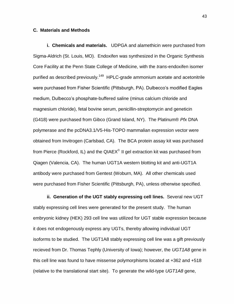
43
C. Materials and Methods
i. Chemicals and materials. UDPGA and alamethicin were purchased from
Sigma-Aldrich (St. Louis, MO). Endoxifen was synthesized in the Organic Synthesis
Core Facility at the Penn State College of Medicine, with the trans-endoxifen isomer
purified as described previously.149 HPLC-grade ammonium acetate and acetonitrile
were purchased from Fisher Scientific (Pittsburgh, PA). Dulbecco’s modified Eagles
medium, Dulbecco’s phosphate-buffered saline (minus calcium chloride and
magnesium chloride), fetal bovine serum, penicillin-streptomycin and geneticin
(G418) were purchased from Gibco (Grand Island, NY). The Platinum® Pfx DNA
polymerase and the pcDNA3.1/V5-His-TOPO mammalian expression vector were
obtained from Invitrogen (Carlsbad, CA). The BCA protein assay kit was purchased
from Pierce (Rockford, IL) and the QIAEX II gel extraction kit was purchased from
Qiagen (Valencia, CA). The human UGT1A western blotting kit and anti-UGT1A
antibody were purchased from Gentest (Woburn, MA). All other chemicals used
were purchased from Fisher Scientific (Pittsburgh, PA), unless otherwise specified.
ii. Generation of the UGT stably expressing cell lines. Several new UGT
stably expressing cell lines were generated for the present study. The human
embryonic kidney (HEK) 293 cell line was utilized for UGT stable expression because
it does not endogenously express any UGTs, thereby allowing individual UGT
isoforms to be studied. The UGT1A8 stably expressing cell line was a gift previously
recieved from Dr. Thomas Tephly (University of Iowa); however, the UGT1A8 gene in
this cell line was found to have missense polymorphisms located at +362 and +518
(relative to the translational start site). To generate the wild-type UGT1A8 gene,
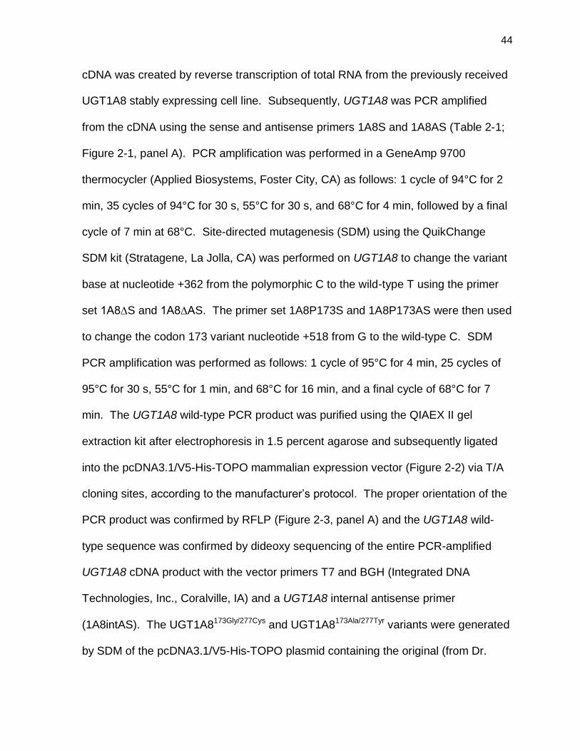
44
cDNA was created by reverse transcription of total RNA from the previously received
UGT1A8 stably expressing cell line. Subsequently, UGT1A8 was PCR amplified
from the cDNA using the sense and antisense primers 1A8S and 1A8AS (Table 2-1;
Figure 2-1, panel A). PCR amplification was performed in a GeneAmp 9700
thermocycler (Applied Biosystems, Foster City, CA) as follows: 1 cycle of 94°C for 2
min, 35 cycles of 94°C for 30 s, 55°C for 30 s, and 68°C for 4 min, followed by a final
cycle of 7 min at 68°C. Site-directed mutagenesis (SDM) using the QuikChange
SDM kit (Stratagene, La Jolla, CA) was performed on UGT1A8 to change the variant
base at nucleotide +362 from the polymorphic C to the wild-type T using the primer
set 1A8∆S and 1A8∆AS. The primer set 1A8P173S and 1A8P173AS were then used
to change the codon 173 variant nucleotide +518 from G to the wild-type C. SDM
PCR amplification was performed as follows: 1 cycle of 95°C for 4 min, 25 cycles of
95°C for 30 s, 55°C for 1 min, and 68°C for 16 min, and a final cycle of 68°C for 7
min. The UGT1A8 wild-type PCR product was purified using the QIAEX II gel
extraction kit after electrophoresis in 1.5 percent agarose and subsequently ligated
into the pcDNA3.1/V5-His-TOPO mammalian expression vector (Figure 2-2) via T/A
cloning sites, according to the manufacturer’s protocol. The proper orientation of the
PCR product was confirmed by RFLP (Figure 2-3, panel A) and the UGT1A8 wild-
type sequence was confirmed by dideoxy sequencing of the entire PCR-amplified
UGT1A8 cDNA product with the vector primers T7 and BGH (Integrated DNA
Technologies, Inc., Coralville, IA) and a UGT1A8 internal antisense primer
(1A8intAS). The UGT1A8173Gly/277Cys and UGT1A8173Ala/277Tyr variants were generated
by SDM of the pcDNA3.1/V5-His-TOPO plasmid containing the original (from Dr.
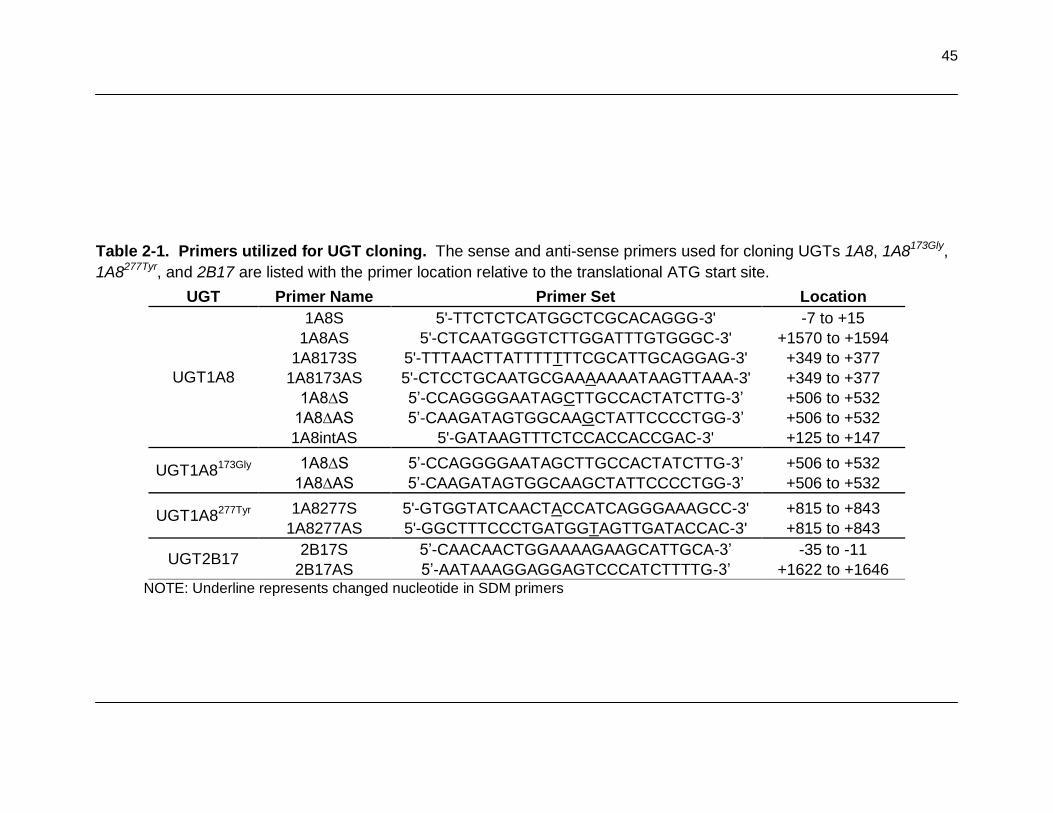
45
Table 2-1. Primers utilized for UGT cloning. The sense and anti-sense primers used for cloning UGTs 1A8, 1A8173Gly,
1A8277Tyr, and 2B17 are listed with the primer location relative to the translational ATG start site.
UGT Primer Name Primer Set Location
UGT1A8
1A8S 5'-TTCTCTCATGGCTCGCACAGGG-3' -7 to +15
1A8AS 5'-CTCAATGGGTCTTGGATTTGTGGGC-3' +1570 to +1594
1A8173S 5'-TTTAACTTATTTTTTTCGCATTGCAGGAG-3' +349 to +377
1A8173AS 5'-CTCCTGCAATGCGAAAAAAATAAGTTAAA-3' +349 to +377
1A8∆S 5’-CCAGGGGAATAGCTTGCCACTATCTTG-3’ +506 to +532
1A8∆AS 5’-CAAGATAGTGGCAAGCTATTCCCCTGG-3’ +506 to +532
1A8intAS 5'-GATAAGTTTCTCCACCACCGAC-3' +125 to +147
UGT1A8173Gly 1A8∆S 5’-CCAGGGGAATAGCTTGCCACTATCTTG-3’ +506 to +532
1A8∆AS 5’-CAAGATAGTGGCAAGCTATTCCCCTGG-3’ +506 to +532
UGT1A8277Tyr 1A8277S 5'-GTGGTATCAACTACCATCAGGGAAAGCC-3' +815 to +843
1A8277AS 5'-GGCTTTCCCTGATGGTAGTTGATACCAC-3' +815 to +843
UGT2B17 2B17S 5’-CAACAACTGGAAAAGAAGCATTGCA-3’ -35 to -11
2B17AS 5’-AATAAAGGAGGAGTCCCATCTTTTG-3’ +1622 to +1646 NOTE: Underline represents changed nucleotide in SDM primers
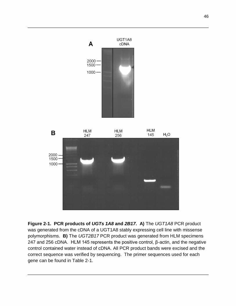
46
Figure 2-1. PCR products of UGTs 1A8 and 2B17. A) The UGT1A8 PCR product
was generated from the cDNA of a UGT1A8 stably expressing cell line with missense
polymorphisms. B) The UGT2B17 PCR product was generated from HLM specimens
247 and 256 cDNA. HLM 145 represents the positive control, β-actin, and the negative
control contained water instead of cDNA. All PCR product bands were excised and the
correct sequence was verified by sequencing. The primer sequences used for each
gene can be found in Table 2-1.

47
Figure 2-2. Diagram of the pcDNA3.1 vector for stable expression of the UGTs.
The pcDNA3.1/V5-His-TOPO vector manufactured by Invitrogen (Carlsbad, CA)
contains an ampicillin resistance gene for bacterial selection and a neomycin resistance
gene for mammalian cell selection. A CMV promoter is located upstream of the PCR
product insertion site. This figure was modified from Invitrogen.

48
Tephly) or newly created wild-type UGT1A8 gene, respectively. To remove the
missense polymorphism at +362, the SDM primers 1A8∆S and 1A8∆AS were used.
The missense polymorphism at +518 was that of the codon 173 polymorphism and was
not altered. The SDM primers used to create UGT1A8277Tyr were 1A8277S and
1A8277AS (Table 2-1).
The UGT2B17 stably expressing cell line was generated by reverse transcription
of adjacent normal human liver total RNA and subsequent PCR amplification (Figure 2-
1, panel B) from the cDNA using the sense and antisense primers 2B17S and 2B17AS,
respectively (Table 2-1). PCR amplification was performed in a GeneAmp 9700
thermocycler (Applied Biosystems, Foster City, CA) as follows: 1 cycle of 94°C for 4
min, 40 cycles of 94°C for 30 s, 59°C for 30 s, and 68°C for 2 min, followed by a final
single cycle of 7 min at 68°C. The UGT2B17 PCR product was purified using the
QIAEX II gel extraction kit as above and subsequently ligated via T/A cloning into the
pcDNA3.1/V5-His-TOPO mammalian expression vector according to the manufacturer’s
protocol. The proper orientation of the PCR product was confirmed by RFLP (Figure 2-
3, panel B) and the UGT2B17 sequence was confirmed by dideoxy sequencing of the
entire PCR-amplified UGT2B17 cDNA product using the vector primers T7 and BGH
(Integrated DNA Technologies, Inc., Coralville, IA). The cloned wild-type UGT1A8 and
UGT2B17, as well as the variant UGT1A8, inserts were compared with sequences
described in GenBank and were confirmed to be 100 percent identical to their relative
sequences.
The UGT-containing plasmids were transfected into HEK293 cells by standard

49
Figure 2-3. Vector digestion with restriction digest enzymes for determination of
PCR product orientation. PCR products were ligated into the pcDNA3.1 vector and
transformed into bacteria. Multiple independent clones (represented by numbers) were
cultured. Following DNA extraction, DNA was incubated with a restriction digest enzyme
to determine the orientation of the PCR product by RFLP. A) The UGT1A8 containing
vector was incubated with the enzyme KpnI (except lane 11 which was uncut), resulting
in 1491 base pair (bp) and 5633 bp fragments if the orientation was correct. B) The
UGT2B17 containing vector was incubated with the enzymes BSrGI and XbaI resulting
in 1100 bp and 6000 bp fragments, if the orientation was correct.

50
Figure 2-4. Vector digestion with restriction digest enzymes following site-
directed mutagenesis. Vectors containing the putative variant UGT gene were
incubated with a restriction digest enzyme to verify the success of the previously
performed SDM or correct PCR product ligation. A) The UGT1A8 wild-type containing
vector was incubated with the enzyme AluI, which cuts the wild-type containing vector 4
times resulting in 4 bands. Sequencing confirmed lanes 2, 3, and 4 contained the
correct gene. B) The UGT1A8277Tyr containing vector was incubated with enzyme KpnI
resulting in 1491 bp and 5633 bp fragments, if the gene contained the polymorphism
and was in the correct orientation. Sequencing confirmed lanes 4, 5, and 6 contained
the correct sequences.

51
electroporation techniques in a GenePulser Xcell (Bio-Rad, Hercules, CA) using 10 µg
of pcDNA3.1/V5-His-TOPO/UGT plasmid DNA with 5x10⁶ of HEK293 cells in serum-
free DMEM media, with electroporation at 250 V and 1000 µF. Following transfection,
cells were grown in 5 percent CO2 to 80 percent confluence in DMEM supplemented
with 4.5 mM glucose, 10 mM HEPES, 10 percent fetal bovine serum, 100 U/ml
penicillin, 100 µg/ml streptomycin, and 700 µg/mL of G418 for the batch selection of
G418-resistant cells, with selection medium changed every 3 to 4 days. A small
number of cells survived selection and were maintained until medium changes and cell
detachment from the plate no longer caused cell death. Subsequently, cells were
stored with DMEM containing 10 percent sterile DMSO in liquid nitrogen until required.
When needed, cells were gently thawed in a 37°C water bath and plated with fresh
supplemented DMEM medium, as above, and changed at 24 hours to remove the
DMSO. Cells were passaged until an adequate number of plates, typically 20-25, were
attained.
Additional cell lines stably expressing the UGT1A and UGT2B isoforms analyzed
in this study were previously described.151-153 All UGT stably expressing cell lines were
maintained in DMEM with G418 as described above and were grown to 80 percent
confluence before the preparation of cell homogenates by resuspending pelleted cells in
Tris-buffered saline (25 mM Tris base, 138 mM NaCl, and 2.7 mM KCl, pH 7.4) and
subjecting them to three rounds of freeze-thaws before gentle homogenization with a
glass dounce homogenizer. Cell homogenates were stored at -80°C in 200 µL aliquots.
Total homogenate protein concentrations were measured using the BCA protein assay.

52
Figure 2-5. Representative immunoblot of UGT1A8 and 1A8173Gly protein expression. 30
µg of protein that was extracted from cell homogenates and varying concentrations of UGT1A1
standard were loaded. Densitometry was performed using β-actin as the loading control.
Immunoblot analysis was performed independently, in triplicate.
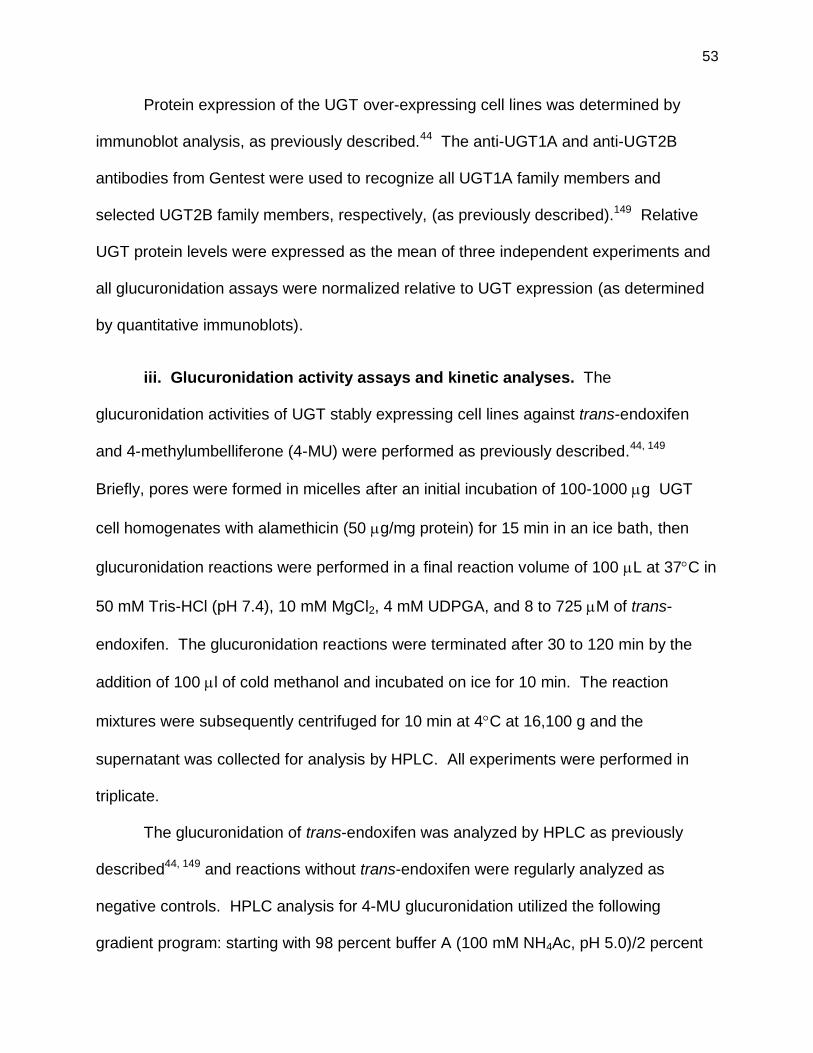
53
Protein expression of the UGT over-expressing cell lines was determined by
immunoblot analysis, as previously described.44 The anti-UGT1A and anti-UGT2B
antibodies from Gentest were used to recognize all UGT1A family members and
selected UGT2B family members, respectively, (as previously described).149 Relative
UGT protein levels were expressed as the mean of three independent experiments and
all glucuronidation assays were normalized relative to UGT expression (as determined
by quantitative immunoblots).
iii. Glucuronidation activity assays and kinetic analyses. The
glucuronidation activities of UGT stably expressing cell lines against trans-endoxifen
and 4-methylumbelliferone (4-MU) were performed as previously described.44, 149
Briefly, pores were formed in micelles after an initial incubation of 100-1000 g UGT
cell homogenates with alamethicin (50 g/mg protein) for 15 min in an ice bath, then
glucuronidation reactions were performed in a final reaction volume of 100 L at 37C in
50 mM Tris-HCl (pH 7.4), 10 mM MgCl2, 4 mM UDPGA, and 8 to 725 M of trans-
endoxifen. The glucuronidation reactions were terminated after 30 to 120 min by the
addition of 100 l of cold methanol and incubated on ice for 10 min. The reaction
mixtures were subsequently centrifuged for 10 min at 4C at 16,100 g and the
supernatant was collected for analysis by HPLC. All experiments were performed in
triplicate.
The glucuronidation of trans-endoxifen was analyzed by HPLC as previously
described44, 149 and reactions without trans-endoxifen were regularly analyzed as
negative controls. HPLC analysis for 4-MU glucuronidation utilized the following
gradient program: starting with 98 percent buffer A (100 mM NH4Ac, pH 5.0)/2 percent

54
buffer B (100 percent acetonitrile) for 5 min, a linear gradient to 70% buffer B over 17.5
min was performed and then maintained at 70 percent for 10 min. The rate of
glucuronide formation was measured based on the ratio of the glucuronide-conjugate
and the free parent compound.
iv. Statistical analysis. The Student's t-test (2-sided) was used for comparing
kinetic values of glucuronidation formation for UGT wild-type versus variant stably
expressing cell lines. Kinetic constants were determined using GraphPad Prism4
software (GraphPad Software, La Jolla, CA).

55
D. Results
i. Glucuronidation activity screen of the UGTs against trans-endoxifen.
In order to determine which UGTs were able to glucuronidate trans-endoxifen, a
glucuronidation activity screen with the homogenates of all UGT stably expressing cell
lines was performed (Figure 2-6, panels A-C). Glucuronide peaks were confirmed by
mass spectrometry, as reported elsewhere.149 Only O-glucuronidation was observed,
which is consistent with previous studies that demonstrated that UGT1A4 is the only
UGT able to perform the N+-glucuronidation of TAM and 4-OH-TAM44 and that
endoxifen is not N-glucuronidated.153 The UGTs 1A4, 2B4, 2B10, 2B15, and 2B17 did
not exhibit glucuronidation activity against trans-endoxifen (Table 2-2). Glucuronidation
of trans-endoxifen was observed in the homogenates of UGTs 1A1, 1A3, 1A7, 1A8,
1A9, 1A10, and 2B7 and the homogenates containing these UGTs were subsequently
used for kinetic analyses. UGT1A5 was not available for analysis. Interestingly,
UGT2B7 was the only UGT2B family member that was active against trans-endoxifen.
ii. Kinetic analyses of the UGTs capable of glucuronidating trans-
endoxifen. To examine the relative UGT enzyme glucuronidation capabilities of trans-
endoxifen, kinetic analyses of the active UGTs were performed (Figure 2-7, panels A-
C). As discussed in sections C.ii. and C.iii., the Vmax was calculated per µg of UGT
protein, as determined by immunoblot analyses of UGT stably expressing cell lines.
UGT1A10 exhibited the lowest KM value and UGTs 1A8 and 2B7 exhibited the second
lowest KM values (Table 2-2). In contrast, UGT1A1 demonstrated the greatest KM value
in the glucuronidation of trans-endoxifen. UGTs 1A8 and 1A10 demonstrated the

56
Figure 2-6. Representative HPLC chromatograms of the most active UGTs against
trans-endoxifen and 4-MU. A) 500 µg of UGT1A8, B) 250 µg of UGT1A10, and C) 1
mg of UGT2B7 homogenates were incubated with trans-endoxifen for 60 min at 37ºC.
D) 1 mg of variant UGT1A8173Ala/277Tyr homogenates were incubated with 4-MU for 120
min and analyzed by HPLC. Peak 1 represents UDPGA, the glucuronic acid co-factor;
peak 2 represents the trans-endoxifen-O-glucuronide conjugate; peak 3 represents free
trans-endoxifen; peak 4 represents 4-MU-O-glucuronide; and peak 5 represents free 4-
MU.

57
Table 2-2. Screening results of UGT homogenates against trans-endoxifen. 500
µg of cell homogenates were incubated with trans-endoxifen from 3 hrs to overnight at
37ºC and analyzed by HPLC.
UGT Activity
1A1 +
1A3 -
1A4 -
1A5 N.D.
1A6 -
1A7 +
1A8 +
1A9 +
1A10 +
2B4 -
2B7 +
2B10 -
2B11 -
2B15 -
2B17 - N.D., not determined
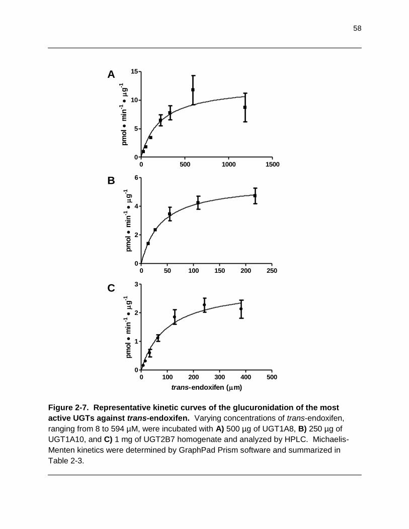
58
Figure 2-7. Representative kinetic curves of the glucuronidation of the most
active UGTs against trans-endoxifen. Varying concentrations of trans-endoxifen,
ranging from 8 to 594 µM, were incubated with A) 500 µg of UGT1A8, B) 250 µg of
UGT1A10, and C) 1 mg of UGT2B7 homogenate and analyzed by HPLC. Michaelis-
Menten kinetics were determined by GraphPad Prism software and summarized in
Table 2-3.
0 50 100 150 200 2500
2
4
6
pm
ol m
in-1
g-1
0 100 200 300 400 5000
1
2
3
trans-endoxifen (m)
pm
ol m
in-1
g-1
0 500 1000 15000
5
10
15
pm
ol m
in-1
g-1
A
B
C
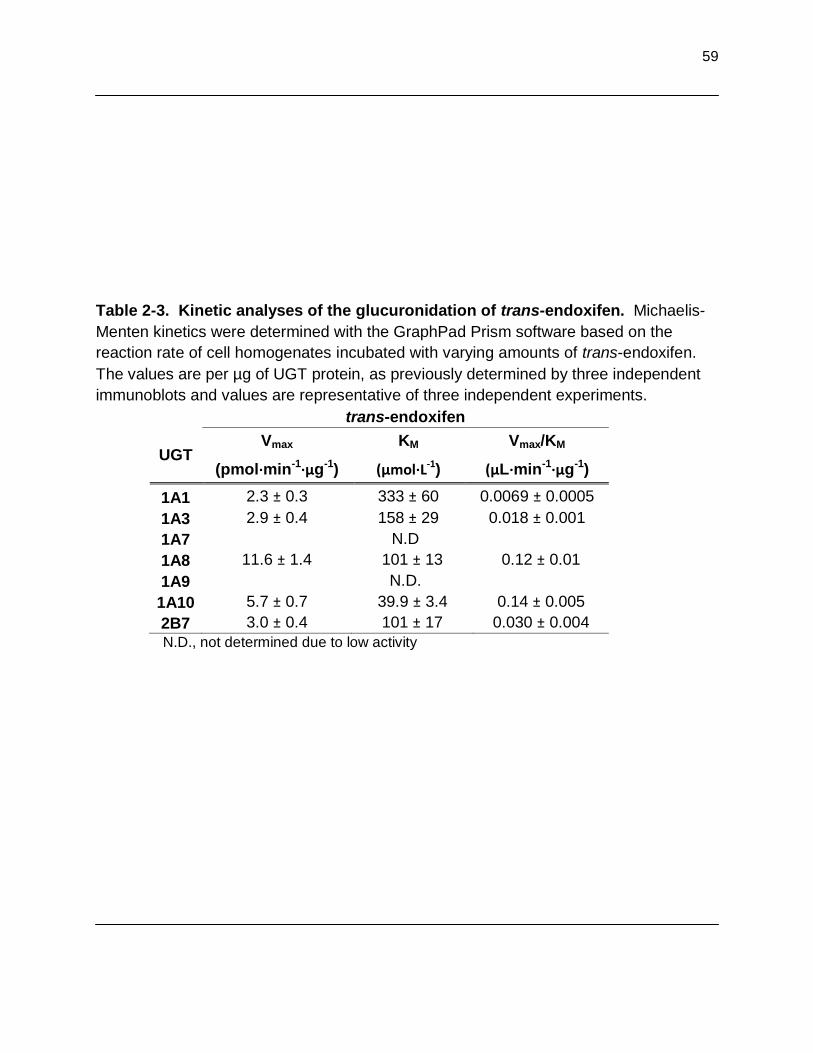
59
Table 2-3. Kinetic analyses of the glucuronidation of trans-endoxifen. Michaelis-
Menten kinetics were determined with the GraphPad Prism software based on the
reaction rate of cell homogenates incubated with varying amounts of trans-endoxifen.
The values are per µg of UGT protein, as previously determined by three independent
immunoblots and values are representative of three independent experiments.
trans-endoxifen
UGT Vmax KM Vmax/KM
(pmol·min-1·µg-1) (µmol·L-1) (µL·min-1·µg-1)
1A1 2.3 ± 0.3 333 ± 60 0.0069 ± 0.0005
1A3 2.9 ± 0.4 158 ± 29 0.018 ± 0.001
1A7 N.D
1A8 11.6 ± 1.4 101 ± 13 0.12 ± 0.01
1A9 N.D.
1A10 5.7 ± 0.7 39.9 ± 3.4 0.14 ± 0.005
2B7 3.0 ± 0.4 101 ± 17 0.030 ± 0.004
N.D., not determined due to low activity

60
greatest turn-over rate, based on the Vmax values, and UGT1A1 had the lowest.
According to the Vmax/KM value, a measure of overall enzyme activity, the trans-
endoxifen glucuronidating activities of the UGTs were: 1A10 > 1A8 > 2B7 > 1A3 > 1A1.
UGTs 1A7 and 1A9 demonstrated such low activity that kinetic analyses could not be
performed. UGTs 1A8 and 1A10 are not expressed in the liver; therefore, UGT2B7 is
the most active UGT expressed in the liver.
iii. Kinetic analyses of UGT variants and trans-endoxifen
glucuronidation. To determine if there were functional effects of prevalent UGT
polymorphisms on trans-endoxifen glucuronidation, kinetic analyses were performed.
The variant isoforms of UGTs 1A8, 1A10, and 2B7 were selected for this analysis due to
the high activity of their wild-type counterparts against trans-endoxifen (Table 2-4). The
variant UGT1A8173Gly/277Cys exhibited a significant 1.5-fold decrease (p < 0.05) in
glucuronidating activity as compared to the wild-type UGT1A8173Ala/277Cys due to an
increase in KM. Interestingly, the UGT1A8173Ala/277Tyr variant demonstrated no detectable
glucuronidating activity against trans-endoxifen, but did glucuronidate 4-MU (Figure 2-6,
panel D), a well known substrate of UGT glucuronidation and a positive control for
activity. The UGT1A10139Lys variant and the wild-type UGT1A10139Gly isoforms
demonstrated similar overall enzyme activity, despite a significant 3-fold reduction in
Vmax and a 13-fold reduction in KM (p < 0.01). A significant 5-fold decrease (p< 0.01) in
overall enzyme activity was observed in the homogenates of UGT2B7268Tyr, as
compared to the wild-type UGT2B7268His. This difference was driven by a significant
5.5-fold decrease in Vmax (p < 0.01).

61
Table 2-4. Kinetic analyses of the variant isoforms of the most active UGTs
against trans-endoxifen. Activity assays of the wild-type (UGTs 2B7268His, 1A10139Glu,
and 1A8173Ala/277Cys) and variant cell homogenates were performed simultaneously under
similar conditions. The values are per µg of UGT protein, as previously determined by
three independent immunoblots and values are representative of three independent
experiments. No activity was detected in homogenates of UGT1A8173Ala/277Tyr against
this substrate however, 4-MU glucuronidation was detected.
trans-endoxifen
UGT isoform
Vmax KM Vmax/KM
(pmol·min-1·µg-1) (µmol·L-1) (µL·min-1·µg-1)
UGT2B7268His 3.0 ± 0.44 101 ± 17 0.030 ± 0.004
UGT2B7268Tyr 0.55 ± 0.01* 101 ± 15 0.006 ± 0.001*
UGT1A10139Glu 5.7 ± 0.7 40 ± 3 0.14 ± 0.005
UGT1A10139Lys 1.9 ± 0.2* 13 ± 2* 0.14 ± 0.004
UGT1A8173Ala/277Cys 5.4 ± 0.2 98 ± 9 0.060 ± 0.004
UGT1A8173Gly/277Cys 5.9 ± 0.2 135 ± 26 0.040 ± 0.005**
UGT1A8173Ala/277Tyr no detectable activity
*p < 0.01, **p < 0.05

62
E. Discussion
Glucuronide conjugates of TAM and TAM metabolites have been observed in the
plasma and urine of women treated with TAM.47-48 In addition, previous studies
demonstrated that UGT1A4 performed N+-glucuronidation of TAM and a major, active
metabolite, 4-OH-TAM, in vitro.43-44 Demethylation of the dimethylaminoethoxy side
chain appears to inhibit N-glucuronidation, as only O-glucuronide conjugates have been
observed for endoxifen, the other major, active metabolite of TAM.153
The goal of the present study was to identify which UGTs perform O-
glucuronidation of trans-endoxifen, in vitro. Initially, the homogenates of all UGT stably
expressing cell lines were screened for activity against these substrates. Only O-
glucuronidation was observed, which was consistent with previous reports.153
Interestingly, all isoforms of the UGT1A family, except for UGT1A4, performed O-
glucuronidation against trans-endoxifen. In contrast, only UGT2B7 of the UGT2B family
was able to glucuronidate trans-endoxifen.
UGTs that demonstrated activity were further examined by kinetic analysis.
Based on the Vmax/KM values, an indication of overall enzyme activity for a given
substrate, the exclusively extra-hepatic enzymes UGT1A8 and UGT1A10 were the most
active UGTs against trans-endoxifen. UGT2B7 was the third most active UGT, but the
most active of the hepatically-expressed UGTs. Interestingly, UGTs 1A8 and 2B7
expression have been observed in breast tissue,113, 154-155 the target of tissue of TAM.
UGT2B7 is highly expressed in the liver,113, 155 the major detoxification organ.
Therefore, the activity of these three UGT isoforms may play an important role in the

63
inactivation and elimination of the major, active metabolites of TAM, both systemically
and at the target site of TAM.
The second goal of the present study was to examine how the previously
identified polymorphisms (SNPs) in UGTs 1A8, 1A10, and 2B7 (Table 2-5)156-158 alter
the glucuronidation of trans-endoxifen and trans-4-OH-TAM. Kinetic analyses were
performed on the variant UGT stably expressing cell lines. Interestingly, homogenates
of the polymorphic UGT1A10139Lys exhibited similar glucuronidation activity as the
homogenate of the wild-type UGT1A10 stably expressing cell line. In addition, the
UGT1A10139Lys variant is observed at very low prevalence in Caucasians (< 1
percent).157 The prevalences of the polymorphic UGT1A8173Gly and 1A8277Tyr are up to
40 percent93, 158 and 2.4 percent,91, 93, 158 respectively (Table 2-5). In contrast to the
polymorphic UGT1A10139Lys, the variants UGT1A8173Gly, UGT1A8277Tyr, and
UGT2B7268Tyr resulted in a decrease in glucuronidation activity. These data suggest
that SNPs in UGT1A8 and UGT2B7 result in a reduced activity enzyme and therefore,
decreased O-glucuronidation of trans-endoxifen and trans-4-OH-TAM.
The pharmacogenetics of the phase I metabolism of TAM by CYP2D6 was
previously examined. Lower plasma levels of endoxifen in women treated with TAM are
associated with intermediate and poor metabolizers of TAM due to the presence of
variant CYP2D6 genotypes. 50, 69, 71, 159
In addition, when the dose of TAM is increased
in patients based on their CYP2D6 genotypes, endoxifen levels also increase.160
However, large variability in endoxifen plasma levels remain following stratification with
CYP2D6 genotype, suggesting that other mechanisms may be important in determining
endoxifen plasma levels.69, 159

Table 2-5. Frequencies of relevant SNPs in UGTs 1A8, 1A10, and 2B7.
mRNA
amino acid frequency
(%)** UGT gene location* change
codon change source
UGT1A8 +518 C > G 173 Ala > Gly 14.5 - 40.6 Huang et al., 2002; Bernard et. al. 2006; HapMap
UGT1A8 +830 G > A 277 Cys > Tyr 1.2 - 2.4 Thibaudeau et al., 2006; Huang et. al., 2002; Bernard et. al., 2006
UGT1A10 +415 G > A
139 Glu > Lys < 1† Elahi et al., 2003; HapMap
UGT2B7 +802 T > C 268 His>Tyr 49 - 54 Lampe et al., 2000; Wiener et al., 2004; Mehlotra et al., 2007
*from ATG translational start site, **in Caucasian populations
†2.5 - 4% in blacks (Elahi et al., 2003 and HapMap)

65
The pharmacogenetics of the phase II metabolism of the trans-endoxifen and
trans-4-OH-TAM was examined in the present chapter of this dissertation.
Polymorphic UGTs 1A8 and 2B7 have significantly reduced enzyme activity against
trans-endoxifen and trans-4-OH-TAM, in vitro. Decreased UGT enzyme activity may
cause an increase in plasma levels of endoxifen or 4-OH-TAM in women treated with
TAM who are heter- or homozygous for the variant UGT1A8 and/or UGT2B7
genotype. Higher levels of circulating endoxifen or 4-OH-TAM may improve the
clinical response to TAM, but may also result in greater or more severe adverse
events.
In summary, the present data identified the extra-hepatic UGTs 1A8 and 1A10,
and the hepatic UGT2B7, as having important roles in the metabolism of the major,
active metabolites of TAM. Individuals who have variant genotypes for UGT1A8 and
UGT2B7 may glucuronidate trans-endoxifen and trans-4-OH-TAM at a reduced rate,
thereby resulting in higher circulating levels of the TAM metabolites. Long-term
clinical studies are needed to fully assess the outcomes of women that bear variant
UGT genotypes and undergoing TAM therapy, to better personalize treatment
regimens.

66
Chapter 3
UGT2B7 genotype and glucuronidation phenotype of trans-endoxifen and trans-4-OH-TAM
NOTE: The results reported in this chapter represent unpublished data and portions of the published manuscript:
Blevins-Primeau, AS, Sun D, Chen G, Sharma AK, Gallagher CJ, Amin S, Lazarus P. Functional significance of UDP-glucuronosyltransferase variants in the metabolism of active tamoxifen metabolites. Cancer Res. 2009. 69(5): 1892-1900.
All figures presented in this chapter are the work of A.S.B.-P., except the mass spectra of trans-endoxifen-O-glucuronide and trans-4-OH-TAM-N+-glucuronide in HLM and breast tissue, which were performed by G.C., and the UGT2B7 mRNA expression analysis by real-time PCR, which was performed by C.J.G.

67
A. Abstract
Tamoxifen (TAM) is a selective estrogen receptor modulator widely used in the
prevention and treatment of breast cancer. A major mode of metabolism of the major
active metabolites of TAM, 4-OH-TAM and endoxifen, is by glucuronidation via the
UDP-glucuronosyltransferase (UGT) family of enzymes. A previous report and
Chapter 2 of this dissertation indicated that hepatic UGTs 1A4 and 2B7 are highly
active against the TAM metabolites. In addition, their respective variants have
significantly altered glucuronidation activity. To determine if UGT genotype
influences human liver microsome (HLM) phenotype, glucuronidation of trans-
endoxifen and trans-4-OH-TAM by 111 HLM specimens was performed. The rate of
O-glucuronidation against trans-4-OH-TAM and trans-endoxifen was 28% (p<0.001)
and 27% (p=0.002) lower, respectively, in HLM homozygous for the polymorphic
UGT2B7268Tyr genotype, as compared to specimens that were homozygous for the
wild-type UGT2B7268His genotype. There was a significant trend for decreasing O-
glucuronidation activity against trans-4-OH-TAM (p<0.001) and trans-endoxifen
(p=0.009) with increasing numbers of the polymorphic UGT2B7268Tyr allele. In
contrast, HLM homozygous for the variant UGT1A424Thr and UGT1A448Val genotypes
exhibited similar N+-glucuronidation rates of trans-4-OH-TAM as HLM homozygous
for wild-type UGT1A424Pro/48Leu. These results suggest that functional polymorphisms
in TAM-metabolizing UGTs, particularly in UGT2B7, may be important in inter-
individual variability in TAM metabolism and ultimately, response to TAM therapy.

68
B. Introduction
i. Tamoxifen pharmacogenetics. TAM is extensively metabolized by several
enzymes, which allows the potential for polymorphic enzymes to affect TAM and
TAM metabolite levels, and therefore influence patient outcomes. As described in
section 1.E.iii, TAM undergoes phase I metabolism, mostly by CYP2D6 and/or
CYP3A4/5 to form endoxifen and 4-OH-TAM. Early studies found 4-OH-TAM to have
a greater affinity for the estrogen receptor (ER) than the parent drug TAM49 and both
4-OH-TAM and endoxifen exhibit up to 100-fold greater potency than TAM at
inhibiting the estrogen-dependent proliferation of cells.50 In addition, 4-OH-TAM and
endoxifen have been shown to be essentially equal in affinity for ER binding,
inhibition of estrogen-dependent cell line proliferation,5 antagonism of estradiol (E2)-
induced expression of the progesterone receptor,51 and induction of estrogen-
responsive global gene expression in MCF-7 cell lines.52 Importantly, both
metabolites are abundant in the plasma of TAM-treated women, although endoxifen
is often present at levels 5- to 10-fold higher than 4-OH-TAM.27, 47, 53 This evidence
supports the theory that TAM is a pro-drug and endoxifen and 4-OH-TAM are major
contributors to TAM’s therapeutic benefits. Therefore, the activity of the enzymes
that convert TAM to its active metabolites and the activity of the enzymes responsible
for the inactivation and elimination of endoxifen and 4-OH-TAM are important in
determining plasma levels of these compounds and ultimately, therapeutic response.
The pharmacogenetics of TAM due to CYP2D6 genotype has been
recognized by the FDA, although an advisory committee was undecided as to
recommending genotyping or to offer genotyping as an option for women who are

69
candidates for TAM therapy.161 CYP2D6 is a highly polymorphic gene, with 19
inactive and 7 reduced activity alleles currently identified in the population.69 An
inactive or less active enzyme would result in less TAM converted to 4-OH-TAM and
endoxifen. Indeed, in vivo studies have found that CYP2D6 status in women treated
with TAM was associated with changes in endoxifen plasma concentrations.
Namely, CYP2D6 genotypes that resulted in reduced activity or an inactive enzyme
had lower plasma levels of endoxifen.50, 69-70 In addition, TAM treated breast cancer
patients that were CYP2D6 poor metabolizers, due to reduced activity or inactive
CYP2D6, experienced increased recurrence, mortality rates and fewer side effects as
compared to patients that were extensive metabolizers.50, 69 However, despite the
apparent importance of CYP2D6 genotype, large variability in the plasma levels of
endoxifen and 4-OH-TAM are still observed in women treated with TAM, despite
stratification by CYP2D6 genotype,69-70 suggesting that additional factors influence
plasma levels.
ii. UDP-glucuronosyltransferases in the metabolism of tamoxifen.
TAM is administered to patients in the trans-isomer form, due to its higher affinity for
the estrogen receptor (ER).28-29 Previous studies demonstrated the glucuronidation
of both trans-endoxifen and trans-4-OH-TAM by the UGTs.44, 149 Wild-type
UGT1A424Pro/48Leu and the variant UGT1A424Thr/48Leu performed similar levels of N+-
glucuronidation of trans-4-OH-TAM, whereas the polymorphic UGT1A424Pro/48Val
exhibited increased enzyme activity against trans-4-OH-TAM, as compared to the
wild-type UGT1A424Pro/48Leu enzyme activity.44 In addition, all of the UGT1A family
members, except UGT1A4, and UGT2B7 performed O-glucuronidation of trans-

70
endoxifen and trans-4-OH-TAM, as demonstrated in Chapter 2 of this dissertation
and elsewhere.149 Kinetic analyses indicated that, of the hepatically-expressed
UGTs, UGT2B7 was the most active against trans-endoxifen and trans-4-OH-TAM.149
Interestingly, the polymorphic UGT2B7268Tyr results in a significant decrease in
enzyme activity against both substrates.150
UGTs 1A8 and 1A10 demonstrated the highest overall O-glucuronidation
activity against trans-endoxifen and trans-4-OH-TAM.149 Both enzymes are
exclusively extra-hepatic.113, 155 The UGT1A10 polymorphism resulting in a Glu-to-
Lys amino acid change at codon 139 has a very low frequency in Caucasians and
occurs up to 4 percent in Blacks (Table 2-5).157 However, the variant UGT1A8173Gly
has an allelic frequency up to 40 percent in Caucasians93, 158 and results in a
significant decrease in trans-endoxifen and trans-4-OH-TAM glucuronidation.150
Interestingly, UGTs 1A8 and 2B7 expression have been detected in human breast
tissue,91, 162 the target organ of TAM.
iii. Hypothesis. Our previous results indicated that UGT2B7
glucuronidation activity is significantly impaired by the His-to-Tyr change at codon
268 in the homogenates of UGT2B7 stably expressing cell lines.150 Therefore, the
hypothesis of the present study was that HLM prepared from specimens that
harbored the polymorphic UGT2B7268Tyr genotype would exhibit decreased O-
glucuronidation activity against trans-endoxifen and trans-4-OH-TAM, as compared
to wild-type UGT2B7268His glucuronidation activity. In addition, HLM that were
polymorphic for UGT1A424Pro/48Val were expected to exhibit an increase in N+-
glucuronidation of trans-4-OH-TAM.

71
UGT1A8 and UGT2B7 protein have been previously detected in human breast
tissue.91, 162 Due to the high activity of UGTs 1A8 and 2B7 against trans-endoxifen
and trans-4-OH-TAM,149-150 a second hypothesis for the current study is that the
polymorphic UGT1A8173Gly, UGT1A8277Tyr, and UGT2B7268Tyr decrease the rate of
glucuronidation of trans-endoxifen and trans-4-OH-TAM in human breast
homogenates. The overall goal of the present study was to determine if there was an
association between UGT genotype and trans-endoxifen and trans-4-OH-TAM
glucuronidation phenotype in HLM and human breast homogenates.

72
C. Materials and Methods
i. Chemicals and materials. trans-4-OH-TAM (98% pure), UDPGA,
alamethicin, and bovine serum albumin were purchased from Sigma-Aldrich (St.
Louis, MO). Endoxifen was synthesized in the Organic Synthesis Core Facility at the
Penn State College of Medicine, with the trans-endoxifen isomer purified as
previously described.149 HPLC-grade ammonium acetate and acetonitrile were
purchased from Fisher Scientific (Pittsburgh, PA). Dulbecco’s modified Eagles
medium, Dulbecco’s phosphate-buffered saline (minus CaCl2 and MgCl2), fetal
bovine serum, penicillin-streptomycin and geneticin (G418) were purchased from
Gibco (Grand Island, NY). The BCA protein assay kit was purchased from Pierce
(Rockford, IL). The human UGT1A and UGT2B Western blotting kits were purchased
from Gentest (Woburn, MA). All other chemicals used were purchased from Fisher
Scientific (Pittsburgh, PA), unless otherwise specified.
ii. Human liver microsome (HLM) preparation. Normal human liver tissue
specimens (n=111) were obtained from the Tissue Procurement Facility at the H. Lee
Moffitt Cancer Center (Tampa, FL) and include 78 liver specimens that were
examined in previous studies.163-164 HLM were prepared as previously described
(Figure 3-1)164 and stored at -80°C. Matching genomic DNA was prepared from
nuclei isolated during the microsomal differential centrifugation preparation procedure
for each specimen using standard phenol chloroform techniques. Microsomal protein
concentrations were measured using the BCA protein assay. Matching total RNA
was obtained for each specimen directly from Moffitt’s Tissue Procurement Facility
and was stored at -80oC.

73
Figure 3-1. Schematic of the procedure for microsome preparation. Adjacent normal human liver and breast tissue
were homogenized with an electric homogenizer on ice. Aliquots of breast homogenate were saved and stored at -80ºC.
Homogenates were centrifuged at 4ºC at 9,000 g for 30 min. The pellet was saved for DNA extraction and stored at -
80ºC. The supernatant was subjected to two rounds of ultracentrifugation at 4ºC at 105,000 g for 60 min each. The
supernatant, considered to be the cytosolic fraction, was saved and stored at -80ºC. The pellet was considered to be the
microsomal fraction and was resuspended with 0.25 M sucrose, flash-frozen in an ethanol and dry-ice bath and stored at -
80ºC.

74
iii. Human breast homogenate and microsome preparation. Visibly
adjacent normal human breast tissues were obtained from the Pennsylvania State
University College of Medicine during breast tumor excision surgery and flash-frozen
in liquid nitrogen within 15 min. Tissues were stored at -80ºC until homogenate and
microsome preparation. Visible adipose tissue was manually removed while the
tissue was on ice prior to the preparation of microsomes, similar to the HLM
procedure (Figure 3-1), except that aliquots of the homogenized tissue were saved
as breast tissue homogenate. Homogenate and microsomal protein concentrations
were determined by the BCA protein assay.
iv. Glucuronidation assays. The glucuronidation activities of HLM and
breast tissue homogenate and microsomes against trans-4-OH-TAM and trans-
endoxifen were performed as previously described.44, 149 HLM (2.5 g total protein),
breast homogenate (250 µg total protein), or breast microsome (250 µg total protein)
were incubated with alamethicin (50 g/mg protein) for 15 min in an ice bath to create
pores in the micelles. Glucuronidation reactions were performed in a final reaction
volume of 25 L at 37C in 50 mM Tris-HCl (pH 7.4), 10 mM MgCl2, 4 mM UDPGA,
and the subsaturating concentrations of 4 μM or 30 μM of trans-4-OH-TAM or trans-
endoxifen, respectively, for HLM and breast homogenate or microsome
glucuronidation assays. 4-methylumbelliferone (4-MU) was used as a positive
control at 100 µM with breast homogenate and microsomes. Kinetic analysis of HLM
from subjects with varying UGT2B7 genotypes was performed using 0.5-15 μM trans-
4-OH-TAM and 2-60 μM trans-endoxifen. This analysis was performed with three
randomly chosen HLM. Reactions were terminated by the addition of 25 l cold

75
methanol on ice. Mixtures were centrifuged for 10 min at 4C at 16,100 g prior to the
collection of the supernatant. All HLM glucuronidation assays were performed in
duplicate independent experiments.
Assays of TAM metabolite formation were performed by UPLC/MS/MS using a
Waters (Milford, MA) ACQUITY UPLC consisting of a binary gradient pump, an
autosampler maintained at 4ºC, and a UV detector operated at 254 nm, attached to a
Waters TQD triple quadrupole mass spectrometer. Similar to that described
previously,72 each sample was injected onto an ACQUITY UPLC BEH C18 1.7 μM,
2.1X100 mm column (Waters) with the following gradient elution conditions for trans-
4-OH-TAM and 4-MU: starting with 69% buffer A (0.01mol/L NH4Ac, pH 5.0)/31%
acetonitrile for 2 min with a subsequent linear gradient to 75% acetonitrile over 2 min.
The gradient elution conditions for trans-endoxifen, using the same buffers, were as
follows: starting with 30% acetonitrile for 4 min and a subsequent linear gradient to
75% acetonitrile for 2 min. The elution flow rate was 0.5 mL/min and 5 μL of the
reaction was injected for all assays. Electrospray ionization mass spectrometry (ESI-
MS) daughter scans of 564, 550, and 353 (m+1/z) verified the glucuronides of trans-
4-OH-TAM, trans-endoxifen, and 4-MU, respectively. The formation of the
glucuronides of trans-endoxifen, trans-4-OH-TAM, and 4-MU were quantified by
UPLC based on the ratio of the glucuronide versus free trans-4-OH-TAM or trans-
endoxifen. Glucuronidation assays without TAM metabolite were regularly analyzed
as negative controls for glucuronidation activity as previously described.164
v. UGT genotyping. Genomic DNA from the 111 liver specimens examined
for glucuronidation activity in this study were used to genotype the UGT2B7 codon
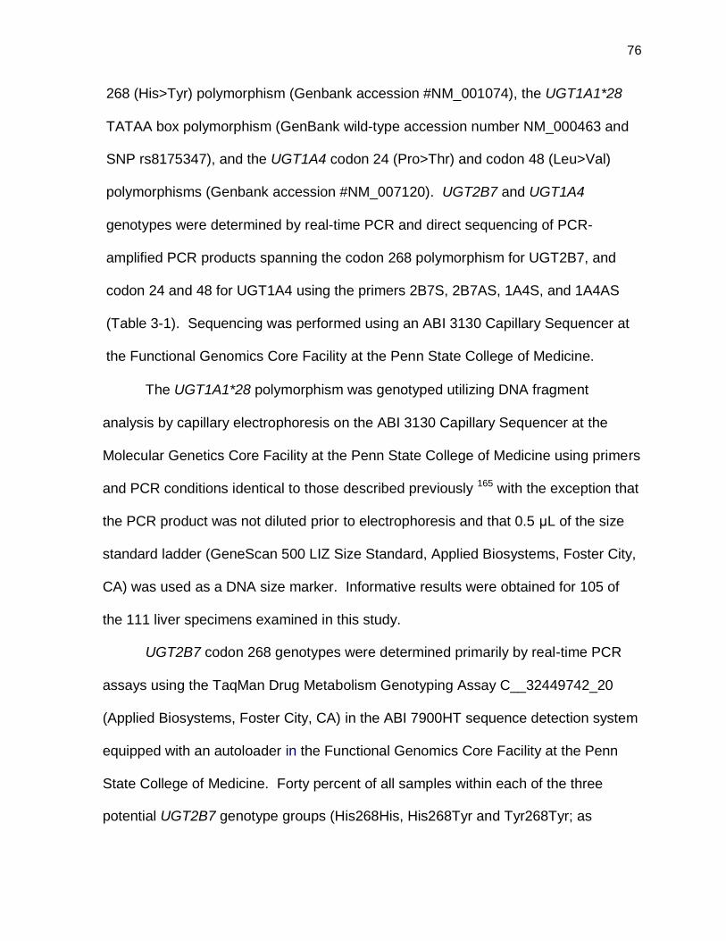
76
268 (His>Tyr) polymorphism (Genbank accession #NM_001074), the UGT1A1*28
TATAA box polymorphism (GenBank wild-type accession number NM_000463 and
SNP rs8175347), and the UGT1A4 codon 24 (Pro>Thr) and codon 48 (Leu>Val)
polymorphisms (Genbank accession #NM_007120). UGT2B7 and UGT1A4
genotypes were determined by real-time PCR and direct sequencing of PCR-
amplified PCR products spanning the codon 268 polymorphism for UGT2B7, and
codon 24 and 48 for UGT1A4 using the primers 2B7S, 2B7AS, 1A4S, and 1A4AS
(Table 3-1). Sequencing was performed using an ABI 3130 Capillary Sequencer at
the Functional Genomics Core Facility at the Penn State College of Medicine.
The UGT1A1*28 polymorphism was genotyped utilizing DNA fragment
analysis by capillary electrophoresis on the ABI 3130 Capillary Sequencer at the
Molecular Genetics Core Facility at the Penn State College of Medicine using primers
and PCR conditions identical to those described previously 165 with the exception that
the PCR product was not diluted prior to electrophoresis and that 0.5 μL of the size
standard ladder (GeneScan 500 LIZ Size Standard, Applied Biosystems, Foster City,
CA) was used as a DNA size marker. Informative results were obtained for 105 of
the 111 liver specimens examined in this study.
UGT2B7 codon 268 genotypes were determined primarily by real-time PCR
assays using the TaqMan Drug Metabolism Genotyping Assay C__32449742_20
(Applied Biosystems, Foster City, CA) in the ABI 7900HT sequence detection system
equipped with an autoloader in the Functional Genomics Core Facility at the Penn
State College of Medicine. Forty percent of all samples within each of the three
potential UGT2B7 genotype groups (His268His, His268Tyr and Tyr268Tyr; as

77
Table 3-1. Primer sequences utilized for UGT genotyping. UGT Primer Name Primer Set Location*
1A4 1A4S 5'-GGCTTCTGCTGAGATGGCCAG-3' -13 to +8
1A4AS 5'-CCTTGAGTGTAGCCCAGCGT-3' +277 to +306
2B7
2B7S 5'-CTATAGTGCTTTACTTTGACTTTTGGTTCG-3' +642 to +670
2B7AS 5'-GCTAGAAAAGCAAAGAAGGGAAAAAATGATTAGTTATATCTGA-3' +1555 to +1597
2B7ScDNA 5'-TGCAGATGCTATTTTTCCCTGTA-3' +456 to +478
2B7AScDNA 5'-GAACCTTTTGTGGGATCTGGGCC-3' +984 to +1006
*relative to the ATG translational start site

78
identified by real-time PCR) were further confirmed by standard PCR and cycle
sequencing performed with the ABI 3130XL Capillary Sequencer at the Molecular
Genetics Core Facility at the Penn State College of Medicine. For those samples for
which real-time assays of UGT2B7 genotypes were inconclusive (n=13), PCR and
direct sequencing analysis was performed as described above. For eight of these
samples, both real-time and direct sequencing failed to provide informative results.
Direct sequencing of UGT2B7 cDNA was then performed successfully for these
samples using matching total RNA as template. Briefly, after reverse transcription
(RT)-PCR was performed using standard conditions with 5 g total liver RNA as
template and oligo(dT) as primer, PCR was performed using 2 L of cDNA as
template the primers 2B7ScDNA and 2B7AScDNA (Table 3-1). Direct sequencing of
these RT-PCR-amplified fragments was performed using the ABI 3130XL Capillary
Sequencer at the Molecular Genetics Core Facility at the Penn State College of
Medicine using the same primers used for PCR.
vi. UGT2B7 mRNA expression analysis. Matching total RNA was available
for expression analysis for 99 of the 111 liver specimens analyzed in this study. Five
ug of RNA was used for cDNA synthesis using standard reverse transcription
methods as described above. 20 ng of cDNA was used for expression analysis using
the ABI gene expression kit assay for UGT2B7 (Hs02556232_s1; Applied
Biosystems, Foster City, CA) with the ABI 7900HT detection system equipped with
an autoloader in the Functional Genomics Core Facility at the Penn State College of
Medicine. Expression assays were performed in triplicate with expression

79
normalized relative to the expression levels of the housekeeping gene PPIA within
the same samples.
vii. Immunoblot analyses. Breast tissue homogenate and microsome
protein expression were analyzed by immunoblot analyses. Protein was extracted
from fresh breast homogenate or microsomes and 100 µg were loaded onto
denaturing 8 percent SDS-PAGE gels. Proteins were transferred to a PVDF
membrane by a traditional wet transfer for 2 hours at 30 volts. Blots were blocked for
1 hr at room temperature with 5 percent milk in TBST and subsequently incubated in
primary antibody, also in 5 percent milk in TBST, overnight at 37ºC. HRP-conjugated
anti-rabbit or anti-mouse secondary antibodies were used at 1:5,000 for 1 hour at
room temperature. Blots were incubated with SuperSignal West Dura extended
duration chemiluminescence substrate (Pierce/Thermo Scientific, Pittsburgh, PA) for
5 min. Film was developed with a standard film processor. The blots were stripped
and probed for the loading control β-actin, as stated above. The concentrations of
the primary antibodies were as follows: UGT1A 1:5000, UGT2B 1:2500, and β-actin
1:2000.
viii. Statistical analysis. The Student's t-test (2-sided) was used for
comparing kinetic values of glucuronidation formation for HLM glucuronidation rates
stratified by UGT genotypes. The one-way ANOVA trend test was used to compare
HLM glucuronidation rates across multiple UGT genotypes. Kinetic constants were
determined using Graphpad Prism4 (GraphPad Prism, La Jolla, CA) software.

80
D. Results
i. Glucuronidation activities of HLM. To determine the rate of O- and N+-
glucuronidation of trans-4-OH-TAM and trans-endoxifen, glucuronidation activity
assays were performed for 111 independent HLM samples and analyzed by UPLC
and UPLC/MS/MS. The concentrations of trans-4-OH-TAM or trans-endoxifen
substrate used in the HLM glucuronidation activity assays were determined by kinetic
analyses of three randomly-chosen HLM specimens. The resulting KM’s were 4 μM
and 30 μM for trans-4-OH-TAM and trans-endoxifen, respectively (data not shown).
HLM glucuronidation activity assays, containing 4 μM trans-4-OH-TAM for 60
min, resulted in two major putative glucuronide peaks, the TAM-4-O-glucuronide and
the 4-OH-TAM-N+-glucuronide, which exhibited retention times of 1.76 and 3.35 min,
respectively, distinct from free trans-4-OH-TAM which eluted at 3.95 min (Figure 3-2,
panel A). HLM incubated with 30 μM trans-endoxifen for 30 min exhibited a single
major putative glucuronide peak observed at a retention time of 1.95 min, which was
distinct from the endoxifen peak eluting at 3.90 min (Figure 3-2, panel B). All putative
glucuronide peaks eluted at retention times identical to previously characterized
TAM-glucuronide standards,72 and were confirmed by tandem MS (Figure 3-2, panels
C-D; the MS/MS patterns observed for trans-TAM-4-O-glucuronide and trans-4-OH-
TAM-N+-glucuronide are identical). For 4-OH-TAM glucuronidation assays, a third,
smaller peak eluting at a retention time of 2.02 min was confirmed to be cis-TAM-
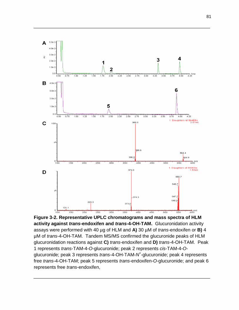
81
Figure 3-2. Representative UPLC chromatograms and mass spectra of HLM
activity against trans-endoxifen and trans-4-OH-TAM. Glucuronidation activity
assays were performed with 40 µg of HLM and A) 30 µM of trans-endoxifen or B) 4
µM of trans-4-OH-TAM. Tandem MS/MS confirmed the glucuronide peaks of HLM
glucuronidation reactions against C) trans-endoxifen and D) trans-4-OH-TAM. Peak
1 represents trans-TAM-4-O-glucuronide; peak 2 represents cis-TAM-4-O-
glucuronide; peak 3 represents trans-4-OH-TAM-N+-glucuronide; peak 4 represents
free trans-4-OH-TAM; peak 5 represents trans-endoxifen-O-glucuronide; and peak 6
represents free trans-endoxifen,

82
4-O-glucuronide, likely formed due to spontaneous interconversion between the trans
and cis 4-OH-TAM isomers. Similar to that observed in previous studies for three
HLM specimens,149 N-glucuronidation was not observed in any of the glucuronidation
activity assays of trans-endoxifen by the 111 HLM examined in the present analysis.
The mean rate of formation ± the standard deviation of TAM-4-O-glucuronide, 4-OH-
TAM-N+-glucuronide and endoxifen-O-glucuronide in HLM were 141 + 45, 175 + 52
and 168 + 66 pmol•min-1•mg-1, respectively. The minimum and maximum
glucuronide formation in all HLM ranged from 4.5-, 10-, and 17-fold for TAM-4-O-
glucuronide, 4-OH-TAM-N+-glucuronide, and endoxifen-O- glucuronide, respectively.
The range of the ratio of TAM-4-O-glucuronide : 4-OH-TAM-N+-glucuronide in the
HLM samples was 8.0-fold.
ii. UGT genotype and glucuronidation phenotype analysis. Previous
studies indicated that the polymorphic UGT2B7268Tyr results in a significant reduction
in the glucuronidation of trans-endoxifen and trans-4-OH-TAM in UGT2B7268Tyr over-
expressing cell lines, as compared to the wild-type UGT2B7268His cell line.150 To
examine the effect of UGT genotype on HLM formation of trans-endoxifen- and trans-
TAM-4-O-glucuronides, HLM glucuronidation rates (n = 111) were stratified by UGT
genotype.
Stratification of HLM O-glucuronidation activities by UGT2B7 codon 268
genotype resulted in a significant 27% decrease in the O-glucuronidation of trans-
endoxifen in HLM that were homozygous for the variant UGT2B7268Tyr genotype (p =
0.002; n = 28), as compared to HLM homozygous for the wild-type
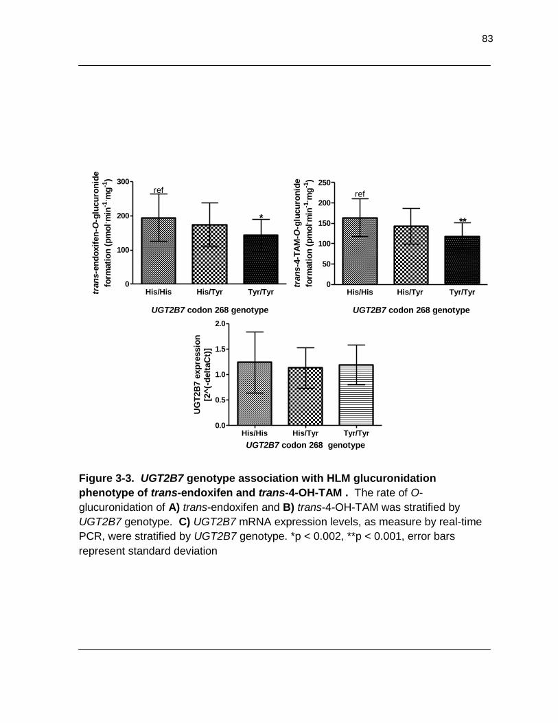
83
Figure 3-3. UGT2B7 genotype association with HLM glucuronidation
phenotype of trans-endoxifen and trans-4-OH-TAM . The rate of O-
glucuronidation of A) trans-endoxifen and B) trans-4-OH-TAM was stratified by
UGT2B7 genotype. C) UGT2B7 mRNA expression levels, as measure by real-time
PCR, were stratified by UGT2B7 genotype. *p < 0.002, **p < 0.001, error bars
represent standard deviation
His/His His/Tyr Tyr/Tyr0
100
200
300
*
ref
UGT2B7 codon 268 genotype
tran
s- e
nd
oxif
en
-O-g
lucu
ron
ide
form
ati
on
(p
mo
l. min
-1. m
g-1
)
His/His His/Tyr Tyr/Tyr0
50
100
150
200
250
**
ref
UGT2B7 codon 268 genotype
tran
s-4
-TA
M-O
-glu
cu
ron
ide
form
ati
on
(p
mo
l. min
-1. m
g-1
)
His/His His/Tyr Tyr/Tyr0.0
0.5
1.0
1.5
2.0
UGT2B7 codon 268 genotype
UG
T2B
7 e
xp
ressio
n
[2^
(-d
elt
aC
t)]
A B
C

84
UGT2B7268His genotype (n = 25; Figure 3-3, panel A). In addition, a significant trend
of decreasing O-glucuronidation of trans-endoxifen was observed in HLM with
increasing numbers of the UGT2B7268Tyr allele (p=0.009). Similarly, a 13% decrease
in TAM-4-O-glucuronide formation in HLM heterozygous for the polymorphic
UGT2B7268Tyr genotype (p = 0.059; n = 28) and a significant 28% decrease in TAM-4-
O-glucuronide formation in HLM homozygous for the UGT2B7268Tyr genotype (p <
0.001), as compared to HLM homozygous for the wild-type UGT2B7268His genotype
(Figure 3-3, panel B). A significant 17% decrease in TAM-4-O-glucuronide formation
was observed in HLM heterozygous for the UGT2B7268Tyr genotype (p = 0.01), as
compared to HLM homozygous for the variant UGT2B7268Tyr genotype. A significant
trend of decreasing O-glucuronidation of trans-4-OH-TAM was observed in HLM with
increasing numbers of the UGT2B7268Tyr allele (p<0.001).
A previous study reported the single nucleotide polymorphism (SNP) of a
thymidine to a cytosine located within the UGT2B7 promoter at -161, relative to the
ATG translational start site, that is in complete linkage disequilibrium with the SNP
that results in the UGT2B7268Tyr allele.166 Similarly, the SNP at -161 was in 100
percent linkage disequilibrium with the UGT2B7268Tyr allele in the present population.
UGT2B7 mRNA expression was analyzed to examine the possibility that UGT2B7
expression levels due to the -161 SNP were influencing the UGT2B7 genotype-
phenotype relationship. The mean level of UGT2B7 expression was similar in liver
specimens within each of the UGT2B7 genotype groups, with specimens
homozygous for the wild-type UGT2B7268His genotype exhibiting a 4 percent higher

85
level of expression than liver specimens homozygous for the variant UGT2B7268Tyr
genotype (Figure 3-3, panel C).
A previous report demonstrated that UGT1A4 performs N+-glucuronidation of
4-OH-TAM, and that the polymorphic UGT1A424Pro/48Val exhibited increased
glucuronidation activity.44 HLM glucuronidation of trans-TAM-4-O-glucuronide was
stratified by UGT1A4 genotype. Surprisingly, no significant associations were
observed for the variant UGT1A424Pro/48Val, as well as the variant UGT1A424Thr/48Leu,
and HLM N+-glucuronidation activity against 4-OH-TAM (Figure 3-4, panels A and B).
Although UGT1A1 was not as active as UGTs 1A4 and 2B7 against trans-
endoxifen and trans-4-OH-TAM, due to its high level of expression in liver,
glucuronidation rates of HLM were also stratified by UGT1A1*28 genotype. Non-
significant decreases in O-glucuronidation activity of 14 and 11% were observed
against the trans isomers of 4-OH-TAM and endoxifen, respectively, in HLM
homozygous for the polymorphic UGT1A1*28 genotype, as compared to HLM
homozygous for the wild-type UGT1A1*1 genotype (Figure 3-5, panels C and D).
This decrease remained non-significant when combining HLM that were either
homozygous polymorphic for UGT1A1*28 or heterozygous polymorphic for
UGT1A1*28 genotypes (data not shown).
iii. Glucuronidation activity of breast tissue. In addition to UGT2B7,
UGT1A8 was also found to have high activity against trans-endoxifen and trans-4-
OH-TAM.149 Both enzymes are also expressed in breast tissue.91, 162 Therefore, to
determine the feasibility of a genotype-phenotype analysis in breast tissue, the
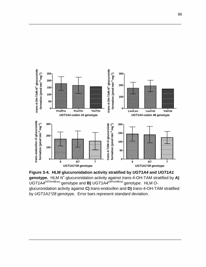
86
Figure 3-4. HLM glucuronidation activity stratified by UGT1A4 and UGT1A1
genotype. HLM N+-glucuronidation activity against trans-4-OH-TAM stratified by A)
UGT1A424Thr/48Leu genotype and B) UGT1A424Pro/48Val genotype. HLM O-
glucuronidation activity against C) trans-endoxifen and D) trans-4-OH-TAM stratified
by UGT1A1*28 genotype. Error bars represent standard deviation.
Leu/Leu Leu/Val Val/Val0
100
200
300
UGT1A4 codon 48 genotype
tran
s-4
-OH
-TA
M-N
+-g
lucu
ron
ide
form
ati
on
(p
mo
l. min
-1. m
g-1
)Pro/Pro Pro/Thr Thr/Thr
0
50
100
150
200
250
UGT1A4 codon 24 genotype
tran
s-4
-OH
-TA
M-N
+-g
lucu
ron
ide
form
ati
on
(p
mo
l. min
-1. m
g-1
)
6 6/7 70
100
200
300
UGT1A1*28 genotype
tran
s-e
nd
oxif
en
-O-g
lucu
ron
ide
form
ati
on
(p
mo
l. min
-1. m
g-1
)
6 6/7 70
50
100
150
200
UGT1A1*28 genotype
tran
s-4
-TA
M-O
-glu
cu
ron
ide
form
ati
on
(p
mo
l. min
-1. m
g-1
)
B A
C D
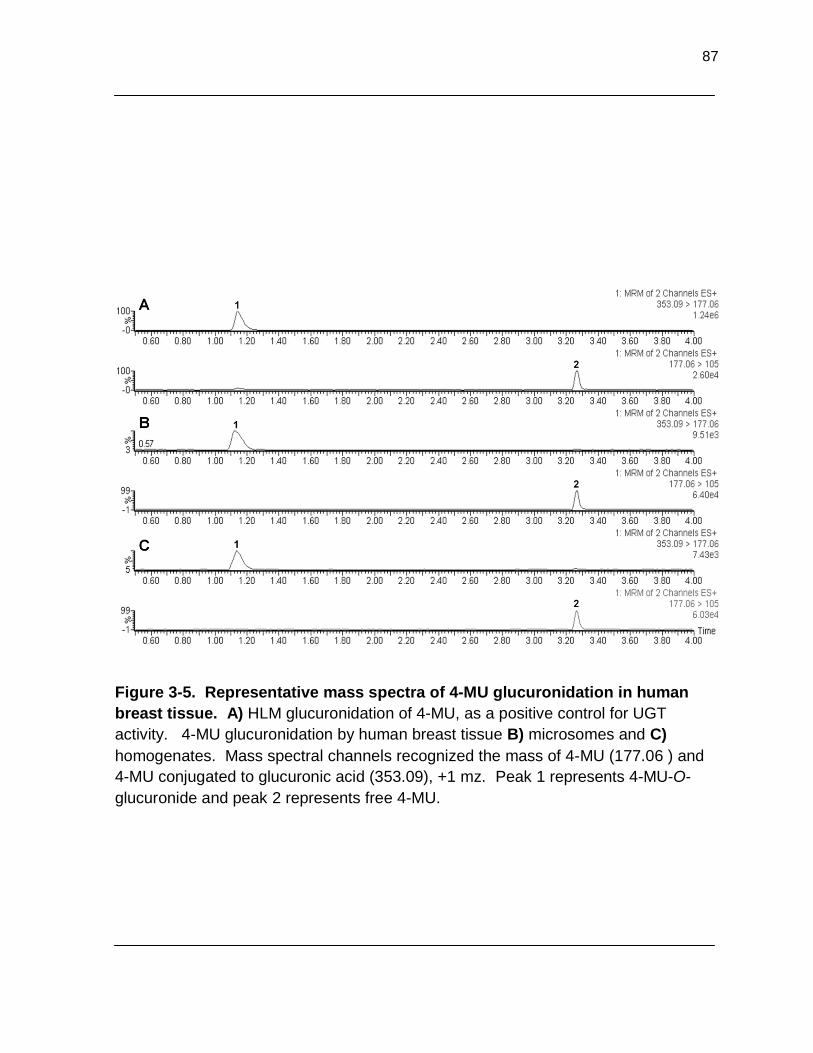
87
Figure 3-5. Representative mass spectra of 4-MU glucuronidation in human
breast tissue. A) HLM glucuronidation of 4-MU, as a positive control for UGT
activity. 4-MU glucuronidation by human breast tissue B) microsomes and C)
homogenates. Mass spectral channels recognized the mass of 4-MU (177.06 ) and
4-MU conjugated to glucuronic acid (353.09), +1 mz. Peak 1 represents 4-MU-O-
glucuronide and peak 2 represents free 4-MU.
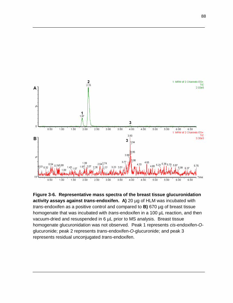
88
Figure 3-6. Representative mass spectra of the breast tissue glucuronidation
activity assays against trans-endoxifen. A) 20 µg of HLM was incubated with
trans-endoxifen as a positive control and compared to B) 670 µg of breast tissue
homogenate that was incubated with trans-endoxifen in a 100 µL reaction, and then
vacuum-dried and resuspended in 6 µL prior to MS analysis. Breast tissue
homogenate glucuronidation was not observed. Peak 1 represents cis-endoxifen-O-
glucuronide; peak 2 represents trans-endoxifen-O-glucuronide; and peak 3
represents residual unconjugated trans-endoxifen.

89
glucuronidation activity of breast tissue homogenates and microsomes were
examined. Both breast homogenates and microsomes exhibited glucuronidation
activity against 4-MU, a marker of UGT activity (Figure 3-5, panels B and C).
However, glucuronidation activity by multiple specimens against both trans-endoxifen
(Figure 3-6, panel B) and trans-4-OH-TAM (Figure 3-6, panel C) was not observed,
despite pre-concentrating the samples by 10-fold prior to analysis.
iv. Immunoblot analysis of breast tissue. To ensure that UGT protein was
present in breast homogenates and microsomes, immunoblot analysis was
performed. UGT1A and UGT2B family members were observed in immunoblots of
breast tissue homogenate and microsomes and the immunoreactive species were
similar to the UGT1A1 and UGT2B7 positive control standards (Figure 3-7). A small
difference in size is not unusual due to post-translational modifications, such as
glycosylation, that occurs to UGT enzymes stably expressed in cell lines. The UGT
standards are purified recombinant enzymes and are not expected to have post-
translational modifications. Due to the absence of glucuronidation activity in multiple
breast homogenate and microsomal specimens, further analyses were not pursued.

90
Figure 3-7. Immunoblot analysis of UGT protein expression in human breast
tissue. Protein was extracted from human breast tissue homogenate and
microsomes and 75 µg of total protein was analyzed for A) UGT1A family member
protein expression in purified UGT1A1 standard (250 ng of UGT protein), breast
homogenate (BH) specimen 1, BH specimen 2, HLM as a positive control, HK293 as
a negative control, and breast microsome (BM) specimen 2. B) UGT2B family
member expression was examined in purified UGT2B7 standard (250 ng of UGT
protein), BH specimen 1, BH specimen 2, HLM, and BM specimen 2.

91
E. Discussion
The present study examines the potential role of UGT polymorphisms in the
metabolism of the trans isomers of 4-OH-TAM and endoxifen, the major active
metabolites of tamoxifen. Several UGTs were previously shown to be active against
these metabolites, with UGT2B7 being the most active hepatic UGT. As UGT2B7
expression has been detected in a variety of tissues including the liver, the
gastrointestinal tract, and the breast,152, 162, 166-169 variations in UGT2B7 function or
expression could potentially significantly impact individual response to drugs or
chemotherapeutic agents. The data presented in this study demonstrate that O-
glucuronidation of both trans-endoxifen and trans-4-OH-TAM in HLM was
significantly associated with UGT2B7 genotype, with lower activities correlated with
specimens that were hetero- or homozygous for the UGT2B7268Tyr allele. These data
were consistent with the observation that HEK293 cells that stably expressed the
UGT2B7268Tyr variant exhibited lower glucuronidation activity against both TAM
metabolites, as compared to cells stably expressing wild-type UGT2B7268His. These
results are also consistent with a study showing a functional role for this
polymorphism against other substrates, including tobacco carcinogen metabolites like
4-(methylnitrosamino)-1-(3-pyridyl)-1-butanol (NNAL).164
Previous studies have observed a SNP located at -161(T>C) relative to the
ATG translational start site in the promoter of UGT2B7 that is in complete linkage
disequilibrium with the SNP at UGT2B7 codon 268.170 The SNP at -161 was in
complete linkage disequilibibium in the present study as well. Unlike a previous
report that found this SNP to alter UGT2B7 expression and alter morphine

92
metabolism,170 in the present study, UGT2B7 genotype was not associated with a
change in UGT2B7 mRNA expression, as there was less than a 4 percent difference
in expression levels between HLM homozygous for the wild-type UGT2B7268His
versus the polymorphic UGT2B7268Tyr genotypes. These data suggest that the
decrease in O-glucuronidation activity against TAM metabolites in HLM associated
with the UGT2B7 codon 268 polymorphism is indeed due to functional changes
within the UGT2B7 enzyme.
In addition to UGT2B7, polymorphisms in UGTs 1A1 and 1A4, which were
previously shown to be active against 4-OH-TAM and endoxifen,44, 149 were examined
for their effect on HLM glucuronidation activity. A microsatellite (TA)-repeat
polymorphism present in the TATAA box of the UGT1A1 promoter has been linked to
lower UGT1A1 expression171-172 and lower activity against a variety of endogenous
and exogenous substrates, including bilirubin,142, 171, 173 carcinogens such as
metabolites of benzo(a)pyrene,171 and chemotherapeutic agents such as SN-38, the
major, active metabolite of irinotecan.174-175 The non-statistically significant trend of
decreased glucuronidation activity against TAM metabolites that was observed in
HLMs from subjects with one or more UGT1A1*28 alleles is consistent with previous
studies indicating that UGT1A1 exhibits between 4.4 and 5.4-fold lower overall
activity against these substrates in vitro than UGT2B7,149
and may therefore play a
less significant role in TAM metabolism.
The N-glucuronidation of TAM and 4-OH-TAM was previously shown to be
performed exclusively by UGT1A4.44, 176 Although a previous study demonstrated
that the UGT1A4 codon 48 polymorphism was linked to small, but significant,
93
alterations in N+-glucuronidation activity against TAM and 4-OH-TAM in vitro,44 a
significant difference was not observed in HLM against trans-4-OH-TAM in the
present study. This may be due to the relatively low number of HLM specimens that
were heterozygous (n=13) or homozygous (n=1) for the UGT1A448Val variant in this
study. An absent association between the UGT1A4 codon 24 polymorphism and
HLM N+-glucuronidation phenotypes of trans-4-OH-TAM glucuronidation is consistent
with previous data that demonstrated that wild-type UGT1A424Pro and variant
UGT1A424Val exhibited similar enzyme activities against this substrate.44
The extra-hepatic UGTs 1A10 and 1A8 exhibited the highest levels of activity
in vitro against the trans isomers of 4-OH-TAM and endoxifen in previous studies.149
However, the polymorphic UGT1A10139Lys is observed at very low prevalence in the
population; less than 1 percent in Caucasian and Asians, and only up to 4 percent in
blacks (Table 2-5).157 Therefore, the functional effect of the UGT1A10 polymorphism
would be difficult to examine due to the low prevalence. In contrast, UGT1A8 has a
polymorphism at codons 173 and 277 that have allelic prevalences of up to 40
percent and 2.5 percent, respectively, in Caucasians (Table 2-5).91, 93, 158 In addition,
the previous observations that UGT1A8 is highly active against TAM metabolites and
is well-expressed in the breast 91, 177 suggests that, like the UGT2B7 codon 268
polymorphism, the UGT1A8 polymorphism may be important in individual response
to TAM.
Human breast tissue was procured for the preparation of homogenate and
microsomes for screening of glucuronidation activity. Glucuronidation activity by
these specimens was not detected for trans-endoxifen or trans-4-OH-TAM. If

94
glucuronidation activity against trans-endoxifen and trans-4-OH-TAM were observed,
a genotype-phenotype study of breast tissue UGT genotype and glucuronidation
phenotype would have been performed. UGT1A and UGT2B family immunoreactive
proteins were detected in both breast tissue homogenate and microsomes,
suggesting that the UGTs are present in the present breast tissue specimens. This is
consistent with previous studies that demonstrated the presence of UGT2B7 protein
in breast tissue by immunohistochemistry and immunoblot analyses.91, 162 In
addition, 4-MU glucuronidation by breast homogenate and microsomes was
observed in the present study. A majority of the UGTs are highly active against 4-
MU, making it a favorable marker of UGT activity. Similarly, a published report
demonstrated UGT2B7 activity in normal breast tissue against 4-hydroxy-estrone (4-
OH-estrone) that was reduced in invasive breast tumor tissue.162 However, breast
tissue homogenate and microsomes did not exhibit glucuronidation activity against
trans-endoxifen or trans-4-OH-TAM. Multiple protein concentrations, as well as
concentrating the glucuronidation reaction mixtures prior to analysis, did not yield
positive results.
The previous report by Gestl et al162 that demonstrated UGT2B7 activity in
breast tissue detailed a careful dissection of adipose tissue from breast tissue
specimens. Visible adipose tissue was also dissected from the breast tissue
specimens utilized in the present study; however, during the centrifugation steps for
preparing microsomal fractions, a fatty layer was observed. Although this layer was
avoided during preparation, it is possible that enough lipids were remaining to
interfere with the glucuronidation activity assays. Both trans-endoxifen and trans-4-
95
OH-TAM are lipophilic and to a greater extent than 4-MU or 4-OH-estrone. It is
possible that the TAM metabolites were “trapped” by the excess lipids, preventing
their glucuronidation by UGTs present in the breast homogenate and microsomes.
Alternatively, the breast tissue specimens used in this study may have simply had
very low levels of UGT1A8 and UGT2B7 enzyme activity. In this case, TAM
metabolism catalyzed in the liver is even more important because it would be the
major site of endoxifen and 4-OH-TAM inactivation by glucuronidation.
The results of the HLM UGT2B7 genotype and trans-endoxifen and trans-4-
OH-TAM glucuronidation phenotype analysis examined in the present study are
consistent with that observed previously for functional polymorphisms in the CYP2D6
gene. Decreased levels of endoxifen were observed in the serum of TAM-treated
women following stratification by the CYP2D6 deletion genotype.50 Similarly, a
reduction in endoxifen plasma levels was also observed when CYP2D6 inhibitors
were co-administered with TAM.69 The CYP2D6*4 deletion allele has been
associated with time until breast cancer recurrence, relapse-free survival, disease-
free survival, and overall survival in patients treated with TAM.50, 69, 159 In addition,
patients with the CYP2D6*4 genotype report few, if any, occurrences of hot flashes.50
These data suggest an important role for endoxifen in TAM therapeutic efficacy.
Despite a strong correlation between CYP2D6 genotype and serum levels of
endoxifen in patients treated with TAM, large variability in circulating endoxifen levels
are still observed after controlling for CYP2D6 genotype.50, 69 The evidence
presented in the present study suggests that inter-individual differences in TAM
glucuronidation pathways may help explain this variability.
96
In summary, this study indicates that genetic variants in UGTs that are highly
active against TAM metabolites significantly alter TAM metabolism in HLM and,
potentially, TAM elimination in TAM-treated individuals. Similar to that described
above for CYP2D6, this could potentially affect overall patient response to TAM.
Additional studies examining the effect of UGT2B7 genotypes in women undergoing
TAM therapy, such as plasma TAM metabolite levels and overall patient response to
TAM, will be required to further examine the role of UGT polymorphisms on the
therapeutic efficacy of TAM.

97
Chapter 4
Src over-expression and UGT2B7 enzyme activity against the major, active metabolites of tamoxifen
98
A. Abstract
Src, a non-receptor tyrosine kinase, has been shown to be upregulated in
several cancer cell types, including breast cancer. UGT2B7 is the predominant
hepatic UGT that glucuronidates the major, active metabolites of the anti-breast
cancer drug TAM, trans-endoxifen and trans-4-OH-TAM. Previous studies in HLM
specimens have shown that heter- or homozygotes with the UGT2B7*2
(UGT2B7268Tyr) allele have significantly decreased rates of glucuronidating trans-
endoxifen and trans-4-OH-TAM. In addition, recent reports indicate that UGT2B7
enzyme activity is altered by phosphorylation at Tyr236 and/or Tyr438 by Src. The
goal of the present study was to determine if tyrosine kinase status within the cell
influences the wild-type UGT2B7268His or the variant UGT2B7268Tyr enzyme activity
against trans-endoxifen and trans-4-OH-TAM, in vitro. HEK293 cell lines stably
expressing UGT2B7268His or UGT2B7268Tyr were transfected with c-Src or v-Src and
kinetic analyses were performed. Wild-type UGT2B7268His cells over-expressing c-
Src or v-Src exhibited a significant 3- and 5-fold reduction, respectively, in the
glucuronidation of trans-endoxifen, as compared to the parent UGT2B7268His cell line
(p ≤ 0.009). Similarly, a 5- and 8-fold reduction in the glucuronidation of trans-4-OH-
TAM was observed. However, no significant difference in glucuronidation capacity
was observed in the c-Src or v-Src over-expressing polymorphism-bearing
UGT2B7268Tyr cell lines, as compared to the parent variant UGT2B7268Tyr cell line (p ≤
0.0002).
Decreased glucuronidation activity of HLM against TAM metabolites has been
observed previously for the UGT2B7268Tyr polymorphic enzyme (Chapter 3). Src
99
over-expression caused a significant decrease in wild-type UGT2B7268His enzyme
activity against several substrates, including the major, active metabolites of TAM,
trans-endoxifen and trans-4-OH-TAM, and the activity is reduced to the level
observed for polymorphic UGT2B7268Tyr enzyme. These results suggest that Src
protein expression status combined with UGT2B7*2 genotype may play an important
role in the metabolism of TAM and ultimately, patient response to TAM therapy.

100
B. Introduction
i. Phosphorylation by Src. Phosphorylation is a post-translational
modification whereby a γ-phosphoryl moiety is transferred from ATP to the amino
acids Ser, Thr, or Tyr of a protein,178 that is catalyzed by serine/threonine kinases or
tyrosine kinases, respectively. Src is a non-receptor tyrosine kinase that
phosphorylates a variety of protein substrates, including those of well studied
pathways, such as focal adhesion kinase179 (FAK) and Crk-and Src-associated
substrate180 (CAS), both involved in important focal adhesion pathways. In addition
to cytoplasmic and plasma membrane-bound substrates, Src has been observed to
phosphorylate protein targets located within the nucleus, such as Sam68,181-182 an
RNA-binding protein that is implicated in the G1/S transition.183 This interaction may
be important in regulating mitosis-related events during cell division.184
Cellular-Src (c-Src) is a proto-oncogene and can become an oncogene. c-Src
is important in a variety of normal cellular processes, such as differentiation,
proliferation, cell division,124-125 survival,126 cell adhesion, cell motility,126-128
morphology, and bone remodeling and reabsorption.124, 129-130 Aberrant Src gene
expression can cause dysregulation of the cell cycle and is associated with cancer.
Increased protein and/or activity levels of c-Src have been observed in human
neoplasms such as breast,185 colon,186 and pancreatic cancers.187 Interestingly, cells
that acquire TAM resistance exhibit increased levels of Src and there is an
association in ER-positive breast cancer patients of activated Src in the cytoplasm of
their breast tumor and reduced survival time with endocrine therapy. MCF7 cells, a
breast cancer cell line that over-expresses active Src, develop a greater migratory
101
and invasive behavior and their growth is not inhibited when treated with TAM. When
these cells are treated with a Src specific inhibitor, their morphology changes so that
the cells appear to regain their cell-to-cell contacts and both migratory and invasive
behavior is decreased.139
ii. Phosphorylation of UGT2B7 by Src. A recent study suggested that
UGT2B7 is phosphorylated by Src.111 A database search of UGT2B7 identified three
putative protein kinase C (PKC) sites, located at Thr123, Ser132, and Ser437 and
two tyrosine kinase (TK) sites, located at Tyr236 and Tyr438. Curcumin, which is
both a PKC and a Src inhibitor, decreased UGT2B7 activity without affecting PKC-
site phosphorylation. In addition, site-directed mutations of the PKC sites did not
alter enzyme activity, suggesting that PKC site phosphorylation was not important for
UGT2B7 activity. However, site mutations at either or both TK sites resulted in
decreased enzyme activity. Transient co-transfection of UGT2B7 with c-Src or v-Src
caused a 1.5-fold increase in UGT2B7 activity against 4-OH-estrone, an endogenous
substrate for UGT2B7.111 These data suggest that UGT2B7 is phosphorylated by Src
and that phosphorylation alters enzyme activity.
iii. Hypothesis. The evidence presented above for 4-OH-estrone led to the
hypothesis that UGT2B7 enzyme activity altered by Src-mediated phosphorylation
affects the glucuronidation activity of UGT2B7 against the substrates trans-endoxifen
and trans-4-OH-TAM. In addition, the highly prevalent UGT2B7268Tyr polymorphism
(Table 2-5), which adds a novel Tyr residue to UGT2B7, if coupled with
phosphorylation by Src, may cause additional complexities in the glucuronidation
activity of these substrates.

102
C. Materials and methods
i. Chemicals and materials. trans-4-OH-TAM (98% pure), UDPGA,
alamethicin, and bovine serum albumin (for immunoblot) were purchased from
Sigma-Aldrich (St. Louis, MO). trans-endoxifen was purchased from Toronto
Research Chemicals (North York, Ontario, Canada). HPLC-grade ammonium
acetate, acetonitrile and peptide synthesis grade triethylamine were purchased from
Fisher Scientific (Pittsburgh, PA). Dulbecco’s modified Eagles medium, Dulbecco’s
phosphate-buffered saline (minus CaCl2 and MgCl2), fetal bovine serum (for tissue
culture), penicillin-streptomycin and geneticin (G418) were obtained from Gibco
(Grand Island, NY). The Platinum® Pfx DNA polymerase and the
pcDNA6.2/V5/GW/D-TOPO mammalian expression vector were obtained from
Invitrogen (Carlsbad, CA). The BCA protein assay kit was purchased from Pierce
(Rockford, IL), while the QIAEX II gel extraction kit was purchased from Qiagen
(Valencia, CA). The anti-UGT2B7 antibody was purchased from Millipore (Woburn,
MA), the anti-activate Src antibody (specific for Src without phosphorylation at amino
acid 529) was purchased from Invitrogen, the anti-β-actin antibody was purchased
from Sigma-Aldrich (St. Louis, MO), and the anti-calnexin, anti-Src, and anti-
phospho-tyrosine antibodies were purchased from Cell Signaling Technology
(Danvers, MA). All other chemicals used were procured from Fisher Scientific
(Pittsburgh, PA) unless otherwise specified.
ii. Src over-expressing cell lines. The HEK293 cell lines stably expressing
the wild-type UGT2B7268His
and variant UGT2B7268Tyr
used in this study have been
described previously.44, 164, 188 pOTB7 non-mammalian expression vectors containing
103
c-Src and v-Src were purchased from Open Biosystems (Huntsville, AL) and the Src
transcripts were copied by PCR using the primers cSrcS, cSrcAS, vSrcS, and
vSrcAS (Table 4-1). In order to ligate the Src PCR products into the vector, the lead
sequence CACC was added to the products by PCR utilizing the same anti-sense
primers and the sense primers cSrcCACC and vSrcCACC (Table 4-1). The Src PCR
products, led by CACC at the 5’ end, were ligated into the pcDNA6.2/V5/GW/D-
TOPO vector containing blasticidin resistance, for selection of the Src-containing
vector (Figure 4-1). All Src cDNA sequences were confirmed by dideoxy sequencing
prior to transfection by Lipofectamine into the HEK293 cell lines stably expressing
UGT2B7268His or UGT2B7268Tyr. Cells were grown in Dulbecco’s Modified Eagle’s
medium supplemented with 10 percent fetal bovine serum, 100 U/mL penicillin and
maintained in 350 g/mL G418, for selection of the UGT-containing vector, and 9
µg/mL blasticidin, for selection of the c-Src- or v-Src-containing vector, in a
humidified incubator in an atmosphere of 5 percent CO2. Cells were grown to 80
percent confluence prior to the preparation of cell homogenates as described in
Chapter 2 of this dissertation. For a subset of glucuronidation activity assays, 200
µM protein tyrosine phosphatase inhibitor, sodium orthovanadate, was added to the
cell pellets of UGT2B7268His cells and UGT2B7268His cells over-expressing c-Src or v-
Src prior to the homogenate preparation. Total homogenate protein concentrations
were measured using the BCA protein assay. UGT2B7 protein levels were
determined by immunoblot analysis, as previously described.44 Relative UGT2B7
protein levels were expressed as the mean of three independent experiments, and all
activity assays were normalized relative to UGT2B7 protein expression.

104
Table 4-1. Primer sequences utilized for the cloning of c-Src and v-Src.
Gene Primer Name Primer Set Location
c-Src
cSrcS 5'-ACGCCAGAGCTCCTGAGAAGATGTCAG-3' -20 to +7 cSrcAS 5'-AGTCCCCACAGGCCCAT-3' +1375 to +1391 cSrcCACC 5'-CACCACGCCAGAGCTCCTGAGAAGATGTCAG-3' 5' PCR product
v-Src
vSrcS 5'-CAGGACCATGGGTAGCAACAAGA-3' -7 to +16
vSrcAS 5'-CCCGCCTGTGCCTAGAGGTTC-3' +1602 to +1622
vSrcCACC 5'-CACCCAGGACCATGGGTAGCAACAAGA-3' 5' PCR product
*relative to the ATG translational start site

105
Figure 4-1. Diagram of the pcDNA vector for stable expression of c-Src and v-
Src in the UGT2B7-HEK293 cell line. The pcDNA6.2/V5/GW/D-TOPO vector
manufactured by Invitrogen (Carlsbad, CA) contains an ampicillin resistance gene for
bacterial selection and a blasticidin gene for mammalian cell selection. A CMV
promoter is located upstream of the PCR product insertion site. c-Src and v-Src with
the lead CACC sequence were ligated to the vector, resulting in a 451 or 437 of
translatable codons. This figure was modified from Invitrogen.

106
iii. Glucuronidation assays. The glucuronidation activities of homogenates
from HEK293 cell lines stably expressing UGT2B7268His or UGT2B7268Tyr and either c-
Src or v-Src against trans-4-OH-TAM, trans-endoxifen, and 4-OH-estrone were
performed as previously described.44, 149 Following an initial incubation of cell
homogenate protein (15-500 g) with alamethicin (50 g/mg protein) for 15 min in an
ice bath, glucuronidation reactions were performed in a final reaction volume of 25 L
at 37C in 50 mM Tris-HCl (pH 7.4), 10 mM MgCl2, 4 mM UDPGA, and 8 to 536 M
trans-endoxifen, 1 to 172 M trans-4-OH-TAM, or 2-140 µM 4-OH-estrone for 15 min
to 2 hours. Negative controls were incubated simultaneously with either ethanol
(instead of TAM metabolites) or DMSO (instead of 4-OH-estrone). The reactions
were terminated by the addition of 25 l of cold acetonitrile on ice and, following a 10
min incubation on ice, the reactions were centrifuged for 10 min at 4C at 16,100 g
and the supernatant was collected. All glucuronidation assays were performed in
triplicate.
Glucuronidation activity assays were analyzed by a Waters (Milford, MA)
AQUITY UPLC consisting of a binary gradient pump, an autosampler maintained at
4ºC, and a UV detector operated at 254 nm for the TAM metabolites and 280 nm for
4-OH-estrone. Samples were injected onto an AQUITY UPLC BEH C18 1.7 μM,
2.1X100 mm column (Waters) with the following gradient elution conditions: starting
with 74% buffer A (0.01mol/L NH4Ac, pH 5.0)/26% acetonitrile for the TAM
metabolites or 86% buffer A/14% buffer B for 4-OH-estrone for 2 min, with a
subsequent linear gradient to 75% acetonitrile over 2 min. The elution flow rate was
0.5 mL/min and 5-7 μL of the reactions were injected for all assays. The formation of

107
the O- glucuronides of 4-OH-TAM, endoxifen and 4-OH-estrone were quantified by
UPLC based on the ratio of the glucuronide versus the free parent compound.
Glucuronidation assays without TAM metabolites or 4-OH-estrone were regularly
analyzed as negative controls for glucuronidation activity.
iv. Src inhibitor treatment. UGT2B7268His cells and UGT2B7268His cells over-
expressing c-Src or v-Src were treated with 0, 0.5, 1, 10, 50, 100, or 150 µM of Src
inhibitor-1 (Sigma-Aldrich, St. Louis, MO) for 1 hour and incubated at 37ºC prior to
collection and homogenization. Src inhibitor-1 was dissolved in DMSO and cells
were treated with equal volumes of Src inhibitor-1 and/or DMSO. Cell homogenates
were utilized in glucuronidation assays as described above.
v. Immunoblot analyses. Protein expression of Src, active Src, phospho-
tyrosine, β-actin, and calnexin were analyzed by immunoblot analyses. Protein was
extracted from fresh homogenate of UGT2B7 cell lines and UGT2B7 cell lines over-
expressing c-Src and v-Src and 75 or 100 µg were loaded onto denaturing 8 percent
SDS-PAGE gels. For the immunoblots of Src and active Src, the proteins were
transferred to a PVDF membrane by the iBlot system (Invitrogen) using program 3 for
4 min and 30 sec. For the anti-phospho-tyrosine blots, the proteins were transferred
to a PVDF membrane by a traditional wet transfer for 2 hours at 30 volts. Blots were
blocked for 1 hr at room temperature with 5 percent milk or bovine serum albumin
(phospho-tyrosine only) in TBST and subsequently incubated in primary antibody in
either 5 percent milk or fetal bovine serum (phospho-tyrosine only) in TBST overnight
at 37ºC. HRP-conjugated secondary antibodies were used at 1:5,000 for 1 hour at
room temperature. Blots were incubated with SuperSignal West Dura extended

108
duration chemiluminescence substrate (Pierce/Thermo Scientific, Pittsburgh, PA) for
5 min. Film was developed with a standard film processor. The blots were stripped
and probed for the loading controls β-actin and/or calnexin, as stated above. The
concentrations of the primary antibodies were as follows: Src 1:1000, activated Src
2.5 µg/µL, phospho-tyrosine 1:2000, β-actin 1:5000, and calnexin 1:2000.
vi. Statistical analysis. The Student's t-test (2-sided) was performed to
compare kinetic values and glucuronidation rates. The one-way ANOVA trend test
was performed to compare glucuronidation rates across multiple variables. Kinetic
constants were determined using Graphpad Prism4 (GraphPad Software, La Jolla,
CA) software.

109
D. Results
i. Over-expression of Src in UGT2B7 stably expressing cell lines. To
determine the effect of Src over-expression on UGT2B7 enzyme activity, cell lines
that stably expressed UGT2B7268His or UGT2B7268Tyr were transfected with either c-
Src or v-Src. Immunoblot analysis indicated that c-Src and v-Src were successfully
over-expressed in wild-type UGT2B7268His or variant UGT2B7268Tyr expressing
HEK293 cell lines (Figure 4-2, panel A). Analysis by ImageJ software indicated that
in both UGT2B7268His and UGT2B7268Tyr cell lines, c-Src and v-Src were expressed
about 2- and 200-fold, respectively, over the levels in the original UGT2B7 cell lines
(Figure 4-2, panel B). Active Src, which is phosphorylated at 419Tyr but not at
Tyr529, is clearly present in the UGT2B7 cell lines over-expressing v-Src; however,
no active c-Src was detected in the c-Src over-expressing and the original
UGT2B7268His or UGT2B7268Tyr cell lines (Figure 4-2, panel C).
ii. Kinetic analyses of UGT2B7 and Src over-expressing cell lines
against trans-4-OH-TAM, trans-endoxifen, and 4-OH-estrone. Several other
reports demonstrated that UGT2B7 is the most important hepatic-expressed UGT in
the glucuronidation of active TAM metabolites.149-150 A previous report indicated that
transient co-expression of Src and UGT2B7 in COS-1 cells resulted in a 1.5-fold
increase in UGT2B7 activity against 4-OH-estrone.111 To determine if over-
expression of Src alters UGT2B7 glucuronidation activity against trans-endoxifen and
trans-4-OH-TAM, in vitro kinetic analyses of the original UGT2B7268His and
UGT2B7268Tyr
cell lines, and those that also over-express c-Src or v-Src, were
performed (Table 4-1).
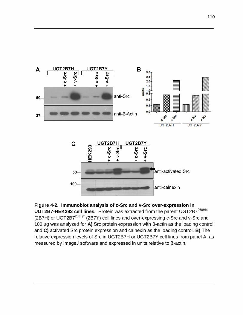
110
Figure 4-2. Immunoblot analysis of c-Src and v-Src over-expression in
UGT2B7-HEK293 cell lines. Protein was extracted from the parent UGT2B7268His
(2B7H) or UGT2B7268Tyr (2B7Y) cell lines and over-expressing c-Src and v-Src and
100 µg was analyzed for A) Src protein expression with β-actin as the loading control
and C) activated Src protein expression and calnexin as the loading control. B) The
relative expression levels of Src in UGT2B7H or UGT2B7Y cell lines from panel A, as
measured by ImageJ software and expressed in units relative to β-actin.

111
Table 4-2. Kinetic analyses of the glucuronidation of trans-endoxifen, trans-4-OH-TAM, and 4-OH-estrone by
Src over-expressing UGT2B7268His cell lines. 15 or 500 µg of cell homogenate proteins were incubated with varying
concentrations of trans-endoxifen (8-536 µM), trans-4-OH-TAM (1-172 µM), and 4-OH-estrone (2-140 µM) for 15 to 60
min and analyzed by UPLC. Kinetic analyses were performed by GraphPad Prism software and experiments were
performed in triplicate. *p ≤ 0.005, **p ≤ 0.02, †p ≤ 0.05.
trans-endoxifen
trans-4-OH-TAM
cell line Vmax Km Vmax/Km Vmax Km Vmax/Km
(pmol/min/µg) (µmol/L) (µL/min/µg) (pmol/min/µg) (µmol/L) (µL/min/µg)
2B7H 0.240 ± 0.065 31.3 ± 11.4 0.008 ± 0.002
1.38 ± 0.682 57.3 ± 28.4 0.024 ± 0.004
2B7H+ c-Src 0.147 ± 0.044 97.9 ± 73.7 0.002 ± 0.0008*
0.362 ± 0.409 93.2 ± 95.4 0.004 ± 0.002*
2B7H+ v-Src 0.115 ± 0.037† 80.2 ± 9.08* 0.001 ± 0.0006*
0.239 ± 0.101† 34.5 ± 2.70 0.007 ± 0.002**
4-OH-estrone
cell line
Vmax Km Vmax/Km
(pmol/min/µg) (µmol/L) (µL/min/µg)
2B7H 589 ± 161 15.9 ± 5.49 37.8 ± 3.89
2B7H + c-Src 195 ± 35.4** 16.2 ± 4.22 12.5 ± 3.76*
2B7H + v-Src 222 ± 27.2** 17.3 ± 3.16 13.4 ± 3.83*

112
Contrary to the findings of Mitra et al,111 the present study found that 4-OH-
estrone glucuronidation was significantly decreased by 3-fold in the homogenates of
UGT2B7268His cell lines over-expressing c-Src or v-Src, as compared to the original
UGT2B7268His homogenate (p ≤ 0.005), due to a significant 3-fold decrease in Vmax
(p ≤ 0.02; Figure 4-3, row A, column III). Similarly, UGT2B7268His homogenates over-
expressing c-Src or v-Src exhibited significant 4 - and 8-fold decreases, respectively,
in overall enzyme activity against trans-endoxifen (p ≤ 0.005; Figure 4-3, row A,
column I). This was due to an approximate 2-fold decrease in Vmax in the
homogenates of UGT2B7268His cell lines over-expressing c-Src and a significant 2-
fold decrease in Vmax (p ≤ 0.05) and a 2.5- to 3-fold increase in KM (p ≤ 0.005),
respectively, in the homogenates of cell lines over-expressing v-Src, as compared to
the original UGT2B7268His cell line. A significant 6- and 3.4-fold reduction in
UGT2B7268His glucuronidating activity was observed in the c-Src and v-Src over-
expressing cell line homogenates against trans-4-OH-TAM (p ≤ 0.005 and 0.02,
respectively), due to a 4- and 6-fold decrease in Vmax, as compared to the original cell
line (Figure 4-3, row A, column II).
As discussed in Chapter 3 of this dissertation, hetero- and homozygosity at the
UGT2B7*2 allele caused a significant reduction in individual HLM glucuronidation
activities against trans-4-OH-TAM and trans-endoxifen.150
To determine if variant
UGT2B7268Tyr phosphorylation by Src alters enzyme activity against the active TAM
metabolites, kinetic analyses were performed in the homogenates of cell lines stably
expressing both the variant UGT2B7268Tyr and c-Src or v-Src (Table 4-2; Figure 4-4,
row B, columns I-III). Interestingly, no significant difference was observed in the

113
Figure 4-3. Lineweaver-Burk graphs of the glucuronidation of trans-endoxifen, trans-4-OH-TAM, and 4-OH-estrone by UGT2B7 cells over-expressing Src. 15 or 500 µg of A) wild-type UGT2B7268Hi or B) the variant UGT2B7268Tyr stably expressing cell lines were incubated with varying concentrations of trans-endoxifen (column I), trans-4-OH-TAM (column II), or 4-OH-estrone (column III) for 15 min to 1 hr at 37ºC. The black lines with boxes represents the parent cell line, the red lines with triangles represents the cell lines over-expressing c-Src, and the green lines with up-side down triangles represents the cell lines over-expressing v-Src. Kinetic analyses were performed by GraphPad Prism software and are summarized in Tables 4-2 and 4-3. The error bars represent the standard deviation of three independent experiments.
A
B
I II III
0.05 0.10 0.15
100
200
300
1/trans-endoxifen (M)
1/t
rans-e
ndoxi
fen g
luc.
form
ation
(pm
ol m
in-1
g-1
)
0.0 0.2 0.4 0.60
100
200
300
1/trans-4-OH-TAM (M)
1/t
ran
s-4
-OH
-TA
M g
luc.
form
ation
(pm
ol m
in-1
g-1
)
0.2 0.4 0.6
0.05
0.10
0.15
1/4-OH-estrone (M)
1/4
-OH
-est
rone g
luc.
form
ation
(pm
ol
min
-1
g-1
)
0.05 0.10 0.15
50
100
150
1/trans-endoxifen (M)
1/t
ran
s-e
ndoxi
fen g
luc.
form
atio
n (p
mol m
in-1g
-1)
0.1 0.2 0.3
50
100
150
1/t
ran
s-4
-OH
-TA
M g
luc.
form
atio
n (p
mol m
in-1
g-1
)
1/trans-4-OH-TAM (M)
0.2 0.4 0.6
0.05
0.10
0.15
1/4
-OH
-est
rone g
luc.
form
ation
(pm
ol m
in-1g
-1)
1/4-OH-estrone (M)

114
Table 4-3. Kinetic analyses of the glucuronidation of trans-endoxifen, trans-4-OH-TAM, and 4-OH-estrone by
variant UGT2B7268Tyr cell lines over-expressing Src. 15 or 500 µg of cell homogenate protein was incubated with
varying concentrations of trans-endoxifen (8-268 µM), trans-4-OH-TAM (4-516 µM), and 4-OH-estrone (2-279 µM) for 15
to 60 min and analyzed by UPLC. Kinetic analyses were performed by GraphPad Prism software and experiments were
performed in triplicate.
trans-endoxifen
trans-4-OH-TAM
cell line Vmax Km Vmax/Km Vmax Km Vmax/Km
(pmol/min/µg) (µmol/L) (µL/min/µg) (pmol/min/µg) (µmol/L) (µL/min/µg)
2B7Y 0.372 ± 0.352 47.7 ± 23.0 0.014 ± 0.019
0.693 ± 0.523 78.1 ± 44.9 0.008 ± 0.002
2B7Y + c-Src 0.201 ± 0.088 102 ± 71.9 0.002 ± 0.001
0.738 ± 0.402 90.9 ± 30.2 0.008 ± 0.002
2B7Y + v-Src 0.256 ± 0.134 134 ± 149 0.003 ± 0.002
0.973 ± 0.685 134 ± 132 0.009 ± 0.003
4-OH-estrone
Vmax Km Vmax/Km
cell line (pmol/min/µg) (µmol/L) (µL/min/µg)
2B7Y 1204 ± 452 30.3 ± 12.9 40.2 ± 1.98
2B7Y + c-Src 795 ± 126 23.6 ± 6.10 35.3 ± 10.3
2B7Y + v-Src 1347 ± 126 29.6 ± 8.15 47.3 ± 9.21

glucuronidation activities of the homogenates of all three cell lines bearing
polymorphic UGT2B7 against trans-endoxifen, trans-4-OH-TAM, and 4-OH-estrone.
Although up to a 7-fold decrease in UGT2B7 glucuronidation activity is observed
against trans-endoxifen, it is not consistent with the trend observed against trans-4-
OH-TAM and 4-OH-estrone, and is most likely due to the large standard deviation of
the Vmax/Km of the parent variant UGT2B7 cell line activity (Table 4-3). This finding
suggests that the amino acid change at codon 268 alters the effect of Src on
UGT2B7 enzyme activity, as compared to the observations in the wild-type cell lines
over-expressing c-Src or v-Src.
iii. Treatment of UGT2B7 and Src over-expressing cell lines with Src
inhibitor-1. To further demonstrate that over-expression of c-Src or v-Src alters
UGT2B7 enzyme activity, UGT2B7268His cells and UGT2B7268His cells over-expressing
c-Src or v-Src were treated with 0, 0.5, 1, or 10 µM of Src inhibitor-1. A non-
significant decrease in glucuronidation activity against all substrates was observed
with increasing concentrations of Src inhibitor-1 treatment in the original
UGT2B7268His cell line, which is contrary to the expected result (data not shown).
However, Src inhibitor-1 treatment of UGT2B7268His cells over-expressing c-Src or v-
Src did not alter the protein expression of phospho-tyrosines, nor did it significantly
alter UGT2B7 enzyme activity against trans-endoxifen, trans-4-OH-TAM, or 4-OH-
estrone (data not shown). Due to the over-expression of c-Src and v-Src, the
concentrations of Src inhibitor-1 may not have been great enough to inhibit Src
activity. Therefore, cells were subsequently treated with 0, 50, 100, and 150 µM of
Src inhibitor-1. Immunoblot analyses indicated that the increased concentrations of

116
Src inhibitor-1 were toxic to the cells, which resulted in a decrease in the protein
expression of phospho-tyrosines, β-actin, and calnexin (data not shown). In addition,
at 100 and 150 µM of Src inhibitor-1, the anti-calnexin antibody recognized two
distinct bands, indicating that protein degradation had occurred. Due to cellular
toxicity, UGT2B7 enzyme glucuronidation activity could not be analyzed.

117
E. Discussion
Src is an important proto-oncogene that plays a critical role in a variety of
normal cellular processes such as differentiation, proliferation, cell division,124-125
survival,126 cell adhesion, cell motility,24, 126-128 morphology, and bone remodeling and
reabsorption.124, 129-130 Interestingly, increased c-Src expression has been observed
in human neoplasms such as breast, colon, gastric, lung, pancreatic, neural, and
ovarian cancers.123 Cell lines that express high levels of activated Src become more
invasive in vivo and are associated with metastases in animal models. In addition,
breast cancer exhibits increased Src activity as compared to normal issue.
Interestingly, cells that acquire TAM resistance exhibit increased levels of Src and
there is an association in ER-positive patients of activated Src in the cytoplasm of
their breast tumor and reduced survival time with endocrine therapy. MCF-7 cells
that over-express active Src develop a greater migratory and invasive behavior and
their growth is not inhibited when treated with TAM. When these cells are treated
with a Src specific inhibitor, their morphology changes so that the cells appear to
regain their cell-to-cell contacts and both migratory and invasive behavior is
decreased.139
A previous study demonstrated that UGT2B7 has the highest overall enzyme
activity of all the hepatic UGTs against both trans-endoxifen and trans-4-OH-TAM, in
vitro.149 In addition, individual HLM specimens that were hetero- or homozygous for
the UGT2B7*2 allele exhibited a significant reduction in the glucuronidation of both
trans-endoxifen and trans-4-OH-TAM.150 Therefore, women with increasing numbers
of the UGT2B7*2 allele may have increased levels of circulating trans-endoxifen and

118
trans-4-OH-TAM, which may alter their overall response to TAM therapy. These
findings suggest that UGT2B7 and the UGT2B7268Tyr variant play an important role in
TAM metabolism and are an important factor in patients undergoing TAM therapy.
Recent studies have indicated that UGT2B7 is phosphorylated by Src, which
results in altered UGT2B7 enzyme activity.110-111 Due to the importance of UGT2B7
in the metabolism of TAM, the finding that UGT2B7 is phosphorylated could have
important implications in women administered TAM. Src is often expressed at high
levels in breast tumors185 and UGT2B7 is also expressed in breast tissue.91
Interestingly, Mitra et al. found that Src can co-localize with UGT2B7111 and that
affinity purified UGT2B7 via a His tag incorporated greater amounts of radio-labeled
ATP when incubated with Src, as compared to incubations without Src,110 suggesting
that the phosphorylation of UGT2B7 by Src is feasible. Therefore, the goal of this
study was to determine if the stable transfection of Src into the wild-type UGT2B7 cell
line altered enzyme activity against either trans-endoxifen or trans-4-OH-TAM, in
vitro. Additionally, the His-to-Tyr amino acid change in the variant UGT2B7 isoform
represents a potential additional phosphorylation site, which could create additional
complexities in the TAM metabolism pathway. A second goal of the present study
was to determine if the stable transfection of Src in the polymorphic UGT2B7268Tyr cell
line resulted in additional alteration in enzyme activity, beyond what the amino acid
change causes.
UGT2B7268His and UGT2B7268Tyr stably expressing HEK293 cells were
transfected with c-Src or v-Src cDNA, resulting in over-expression of these kinase
isoforms. UGT2B7 cells over-expressing v-Src clearly showed an increase in
119
activated Src protein expression, indicating that additional Src in the active
conformation was available for phosphorylating targets and potentially simulating an
oncogenic transformation. UGT2B7 expression remained the same following
transfection with c-Src or v-Src (data not shown).
Kinetic analyses found that increased expression of either c-Src or v-Src in
UGT2B7268His cells caused a significant decrease in glucuronidation activity against
trans-endoxifen and trans-4-OH-TAM. Interestingly, the reduction in UGT2B7 activity
was incremental for trans-endoxifen and trans-4-OH-TAM, where the v-Src over-
expressing cell line had lower activity than the cell line over-expressing c-Src. This is
consistent with reports in the literature that v-Src activity is greater than that of c-
Src.122 This finding could impact TAM metabolism, as a decrease in UGT2B7 activity
would result in less glucuronidation of trans-endoxifen and trans-4-OH-TAM, thereby
producing higher circulating levels of active TAM metabolites. CYP2D6 variant
genotypes that result in a decrease in endoxifen plasma levels69 are associated with
an impaired clinical response to TAM and fewer side effects.50, 159 Additional studies
are needed to determine if phosphorylation of UGT2B7 by Src affects the circulating
levels of trans-endoxifen and trans-4-OH-TAM, in vivo.
A major limitation of the kinetic analyses was the small peaks observed by
UPLC corresponding to the endoxifen- or 4-OH-TAM-glucuronide conjugates,
particularly in the cell lines over-expressing Src. This difficulty led to the greater than
ideal standard deviations reported in the kinetic analyses summaries (Tables 4-2 and
4-3). Despite the broad standard deviations, the difference between the wild-type
UGT2B7 parent cell line and the cell lines over-expressing c-Src and v-Src were

120
highly significant and ranged from 3- to 8-fold. This large difference, combined with
the statistical significance, supports the trend of the data and indicates that if the
standard deviations were narrower, the same trend would most likely be observed.
Interestingly, the glucuronidation activity against 4-OH-estrone was also
significantly decreased, in contrast to the finding of Mitra et al. where 4-OH-estrone
glucuronidation was increased when c-Src or v-Src were over-expressed.111 There
are several methodological differences that might account for these discrepant
findings. The previous study reported percent of glucuronidation activity, which was
performed at one substrate concentration, whereas the present study reported the
kinetic constants that involved a concentration range of each substrate that would
encompass the KM. In addition, the previous study incubated the 4-OH-estrone
glucuronidation reactions for 2 hours. Based on the high reactivity of UGT2B7268His to
its endogenous substrate 4-OH-estrone, the present study utilized 15 min incubations
as the optimal glucuronidation reaction time for accurate results. In addition, the cell
line system used in the present study was the human cell line HEK293 for stable
expression of the proteins of interest. In contrast, the previous study used African
green monkey COS-1 cells and transient co-transfections were performed for both
the expression of UGT2B7 and the over-expression of c-Src and v-Src. Interestingly,
murine SYF-/- cells, which do not endogenously express Src or the Src family
members Yes or Fyn, exhibited a decrease in glucuronidation activity against 4-OH-
estrone when transfected with both UGT2B7 and c-Src, as compared to SYF-/- cells
only transfected with UGT2B7.110 This indicates that cell type may play a role in the
effect phosphorylation has on UGT2B7 enzyme activity. A human cell line is

121
probably a more accurate depiction of what may occur in vivo, as it is the most
physiologically similar to humans, unlike cell lines derived from other species.
However, we must acknowledge that all of these experiments (including our own)
represent artificial cellular expression systems.
The variant UGT2B7268Tyr exhibited decreased glucuronidation activity against
trans-endoxifen and trans-4-OH-TAM and an increase in glucuronidation activity
against 4-OH-estrone, in a similar pattern to what was previously described in
Chapter 3 of this dissertation and elsewhere. 91, 150 Interestingly, the increased
expression of c-Src or v-Src in the variant UGT2B7268Tyr cell line did not significantly
alter overall enzyme activity against trans-endoxifen, trans-4-OH-TAM, and 4-OH-
estrone. Although there was a trend of increasing Km with the variant UGT2B7 cell
lines over-expressing c-Src and v-Src against trans-endoxifen and trans-4-OH-TAM,
it was not significant and was not sufficient to significantly alter the Vmax/Km. In
addition, the UGT2B7268His cell lines over-expressing c-Src or v-Src had similar levels
of overall UGT2B7 enzyme activity as the UGT2B7268Tyr variant cell line, or lower, as
in the case of 4-OH-estrone. Potentially, the His-to-Tyr amino acid change, which
results in an additional phosphorylation site, prevented an additive decrease in
glucuronidation activity due to both the variant amino acid and phosphorylation by
Src. Alternatively, the amino acid change may alter protein folding in such a way that
it may cause no change in enzyme activity when phosphorylated at Tyr236 and/or
Tyr438 or altered folding may prevent the phosphorylation of Tyr 268. Additional
studies are needed to determine if Tyr268 is phosphorylated and to further

122
investigate why c-Src or v-Src over-expression does not alter UGT2B7268Tyr enzyme
activity against these substrates.
To further test the theory that the decrease in glucuronidation activity seen in
the c-Src and v-Src over-expressing cell lines is due to the phosphorylation of
UGT2B7 by Src, the cell lines were treated with a Src-specific inhibitor, Src inhibitor-
1. If Src phosphorylation altered UGT2B7 activity, increasing levels of
glucuronidation activity should be observed with increasing concentrations of Src
inhibitor-1 in the c-Src and v-Src over-expressing UGT2B7 cells, to about the level of
the original UGT2B7 cell line. However, treatment with 0, 0.5, 1, and 10 µM of Src
inhibitor-1 did not change the glucuronidation activity of UGT2B7 against trans-
endoxifen, trans-4-OH-TAM, or 4-OH-estrone in the UGT2B7 cell lines over-
expressing c-Src or v-Src. Treatment of the parent UGT2B7268His cell line with 10 µM
of Src inhibitor-1 did cause a decrease in the intensity and the number of bands in an
immunoblot analysis for phospho-tyrosine (data not shown). These data suggest that
Src inhibitor-1 is able to enter the cell and inhibit Src mediated tyrosine
phosphorylation. Failure to see the decrease in glucuronidation activity observed in
Src inhibitor treated UGT2B7268His cells over-expressing v-Src may be a result of
insufficient concentrations of the inhibitor due to the high levels of activated Src in the
v-Src over-expressing cells. Therefore, cells were treated with greater
concentrations with 0, 50, 100, and 150 µM of Src inhibitor-1. However, immunoblot
analyses indicate that these concentrations caused toxicity in the cells, as indicated
by the decrease in band intensity in the anti-phospho-tyrosine, anti-β-actin, and anti-
calnexin antibodies. Additional studies should be performed evaluating intermediate

123
concentrations of Src inhibitor-1 or using another Src-specific inhibitor, such as PP1
or PP2 (discussed further in Chapter 5).
The data in the present study suggests that the hypothesis that Src directly
interacts with UGT2B7 resulting in a phosphorylation event is false. Therefore, other
mechanisms that can cause a decrease in UGT2B7 activity following over-expression
of Src must be considered. Src is involved in a multitude of signaling pathways and it
is conceivable that one of these pathways may interact with the UGTs, in particular,
UGT2B7.
Src signaling results in the activation of other phosphorylating kinases, such
as phosphoinositide 3-kinase (PI3K) and protein kinase C (PKC). Interestingly, PKC
has been implicated in the phosphorylation of UT1A family members, although
additional studies are needed to confirm this finding. UGT2B7 has putative PKC
phosphorylation sites, although evidence provided by Mitra et al suggests that PKC is
not involved in UGT2B7 phosphorylation. It is possible that over-expression of Src
activates a greater number of PKC or PI3K enzymes than is typical, resulting in
increased phosphorylation events of UGT2B7.
The limitation of the theory that UGT2B7 is phosphorylated by a downstream
substrate of Src is the localization of UGT2B7. The UGTs, including UGT2B7, are
localized to the endoplasmic reticulum (EPR) membrane, with a majority of the
enzyme located within the lumen. In the artificial environment of cell homogenates,
the majority of the UGT2B7 enzyme is probably located within the lumen of a micelle.
Although the antibiotic alamethicin is used to create pores in the micelles, it is
unlikely that PKC or PI3K are able to reach the putative phosphorylation sites located

124
on UGT2B7. In contrast, Src has been observed to localize to the perinuclear
membrane, which is continuous with the EPR, and membrane encased vesicles,131,
133-135 lending feasibility to the co-localization of UGT2B7 and Src.
An alternative mechanism as to the cause of the decrease in UGT2B7
enzyme activity when Src is over-expressed is the phosphorylation of multidrug
resistance proteins by downstream Src substrates, leading to an increase in the
efflux of endoxifen and 4-OH-TAM from the cell prior to glucuronidation. Multidrug
resistance protein (MDRP) is phosphorylated by PKC, causing an increase in
transport activity.189 In addition, the multidrug resistant-associated protein, P-
glycoprotein, appears to also be phosphorylated by PKC. Interestingly, multidrug
resistant cell lines that are treated with a PKC activator cause an increase in P-
glycoprotein phosphorylation190-194 as well as reduced drug accumulation190, 195-196
and drug resistance.195-196 The opposite occurs when multidrug resistant cells are
treated with a PKC inhibitor.190, 192, 197 A recent study has observed that endoxifen is
a substrate for P-glycoprotein transport198 and based on structure and activity
similarities, it can be hypothesized that 4-OH-TAM is also transported by P-
glycoprotein.
In the cell, many multidrug resistance proteins are localized to the plasma
membrane. In the artificial environment of cell homogenate, multidrug resistance
proteins are most likely located in the micelle membrane. Endoxifen and 4-OH-TAM
diffuse into the micelle, where they can be glucuronidated. However, in the case of
this theory, the drug transport activity of multidrug resistant-associated proteins, such
as P-glycoprotein, are increased due to phosphorylation by PKC and endoxifen and

125
4-OH-TAM are instead transported out of the micelle before glucuronidation by
UGT2B7 can occur. The rapid efflux of endoxifen and 4-OH-TAM could explain the
reduced Vmax/Km observed in UGT2B7 cell lines over-expressing Src, as compared to
the parent cell line. In addition, this theory is also consistent with the observation of
the small endoxifen and 4-OH-TAM glucuronide conjugate peaks observed by UPLC.
The conclusion of Src downstream signaling, including following the activation
of PI3K and PKC, typically involves transcriptional regulation. An obvious
mechanism of decreased UGT2B7 activity is inhibition of UGT2B7 transcription and,
ultimately, a decrease in the number of UGT2B7 enzymes available for
glucuronidation of endoxifen and 4-OH-TAM. However, preliminary data in the
present study found UGT2B7 protein levels to be similar in the cell lines over-
expressing Src, as compared to the parent UGT2B7 cell line. This suggests that
protein levels are not affected by the over-expression of Src and other mechanisms,
such as those suggested above, are involved. However, confirmation of the protein
levels should be pursued in future studies, as well as determining UGT2B7 mRNA
level, to further rule out the mechanism of decreased protein levels.
In conclusion, over-expression of Src reduces stably-expressed wild-type
UGT2B7 enzyme activity to the level of the variant UGT2B7 activity against trans-
endoxifen, trans-4-OH-TAM, and 4-OH-estrone. However, the mechanism for this
remains uncharacterized and it is likely that the initial hypothesis of the present study
is false. Other mechanisms involving indirect activity of Src are possible, including
phosphorylation of downstream Src substrates and the upregulation of MDR
pathways. Additional studies are clearly required to determine the mechanism
126
behind the effect of Src over-expression on UGT2B7 activity and to assess potential
clinical implications.

127
Chapter 5
Final considerations and clinical implications

A. Final conclusions
The evidence presented in this dissertation provides additional knowledge
regarding the metabolism of tamoxifen (TAM) and specifically, how the
pharmacogenetics of the UDP-glucuronosyltransferase (UGT) family of phase II
metabolizing enzymes cause inter-individual differences in TAM metabolism (Figure
5-1). UGTs 1A8, 1A10, and 2B7 were identified as the most active UGTs against a
major, active metabolite of TAM, trans-endoxifen, in HEK293 cells individually stably
expressing the UGTs (Chapter 2). Interestingly, UGTs 1A8 and 1A10 are exclusively
extra-hepatic, while UGT2B7 is expressed in the liver, as well as other tissues (Table
1-1).113, 155 In addition, UGTs1A8 and 2B7 protein have been detected in breast
tissue,91, 162 the target tissue of TAM treatment. The present dissertation also
demonstrated that UGT2B7 genotype is associated with the glucuronidation
phenotype of human liver microsomes (HLM) against both trans-endoxifen and trans-
4-hydroxy(OH)-TAM (Chapter 3). HLM specimens that were hetero- or homozygous
for the polymorphic UGT2B7268Tyr allele exhibited a significant decrease in the
glucuronidation of trans-endoxifen and trans-4-OH-TAM. Human breast tissue
homogenates and microsomes did not glucuronidate the TAM metabolites. However,
these samples did glucuronidate 4-methylumbeliliferone (4-MU), a positive control,
demonstrating that the samples maintained their integrity during processing and
preparation. Finally, the effect of over-expression of Src in UGT2B7 cells on the
glucuronidation of trans-endoxifen and trans-4-OH-TAM was examined (Chapter 4).
Stable over-expression of Src in the wild-type UGT2B7 cells resulted in a significant
decrease in the glucuronidation of both TAM metabolites, similar to the level

129
Figure 5-1. Illustration highlighting the location of the UGTs most active
against TAM metabolites. TAM (blue arrows) is ingested orally and absorbed in the
small intestine, where UGTs 1A8 and 1A10 are expressed. However, probably only
a minimal amount of endoxifen or 4-OH-TAM is glucuronidated at this point because
TAM must first be metabolized by the CYP2D6 and/or CYP3A4/5. Following
absorption, TAM and its metabolites travel to the liver, where UGT2B7and CYP2D6
are expressed. Following metabolism in the liver, TAM and its metabolites (blue
arrows) travel systemically, including to the target tissue of TAM, the breast, where
UGTs 1A8 and 2B7 are expressed. Research presented in this dissertation found
that polymorphic UGT1A8 and 2B7 glucuronidated trans-endoxifen and trans-4-OH-
TAM at a reduced rate, as compared to their wild-type counterparts. A decreased
rate of glucuronidation would increase the concentration of circulating endoxifen and
4-OH-TAM, potentially affecting clinical response and acquired TAM resistance.

130
observed in cell lines stably expressing the polymorphic UGT2B7268Tyr. Interestingly,
over-expression of Src in the variant UGT2B7268Tyr cell line did not alter
glucuronidation activity, possibly due to the presence of the polymorphic Tyr residue.
The additional Tyr residue may be phosphorylated by Src or may alter protein folding
in such a way that it prevents the phosphorylation of the other Tyr residues.
B. Future directions
i. Chapter 4. Chapter 4 of this dissertation presented evidence that over-
expression of c-Src or v-Src in wild-type UGT2B7 stably expressing cell lines resulted
in a significant decrease in the glucuronidation of trans-endoxifen and trans-4-OH-
TAM. However, there are additional experiments that can be performed to enhance
the findings of this project.
In the present dissertation, UGT2B7 cells over-expressing Src were treated
with Src inhibitor-1 to inhibit Src activity, with the expectation that UGT2B7
glucuronidation of trans-endoxifen and trans-4-OH-TAM in these cells would regain
glucuronidation activity similar to the levels observed for the original UGT2B7 cell
line. However, initial treatment concentrations were too low to cause Src inhibition in
the cell lines over-expressing Src and higher concentrations resulted in cell toxicity.
This indicates that the therapeutic window is narrow and a more careful evaluation of
Src inhibitor-1 treatment is necessary. Therefore, a future experiment involving
treatment with intermediate levels of Src inhibitor-1, or other Src inhibitors such as
PP1 or PP2, should be performed.
Regardless of the Src inhibitor, the first step undertaken should be a dose-
response experiment in which the concentration of the inhibitor is varied. Immunoblot

131
analyses of phospho-tyrosine and β-actin protein expression should be performed to
determine if Src inhibition was successful without causing cell toxicity. A time-course
analysis should be performed to determine the optimal amount of time cells should
be treated with the Src inhibitor. The final experiment would combine four optimal
concentrations with the optimal time point which should provide a dose-dependent
increase in Src inhibition as determined by immunoblot analyses of phospho-tyrosine
and β-actin protein expression. If the immunoblots are positive, glucuronidation
activity assays should then be performed to determine the effect of Src inhibition on
UGT2B7 enzyme activity. If the difference in UGT2B7 enzyme activity is related to
Src function, the UGT2B7 cells over-expressing c-Src or v-Src treated with the
highest concentration of Src inhibitor-1 should exhibit similar UGT2B7 enzyme
activity as the original UGT2B7 cell line. If the data demonstrate no alteration in
UGT2B7 enzyme activity or a decrease in enzyme activity, then the possibility that
Src over-expression is not directly causing the observed decrease in UGT2B7
glucuronidation activity against trans-endoxifen and trans-4-OH-TAM would have to
be addressed. Other mechanisms, such as protein interactions, downstream
effectors, and other signaling pathways tied to Src, must be examined.
Another interesting future direction is the characterization of the putative
Src phosphorylation sites located on both the wild-type and polymorphic UGT2B7
enzyme. This study would determine the exact location(s) of tyrosine
phosphorylation on the wild-type UGT2B7 enzyme and if the location(s) are altered in
the polymorphic UGT2B7 enzyme. Additionally, this study would provide further
information regarding whether UGT2B7 is phosphorylated by a tyrosine kinase.

132
Following cell collection and protein extraction that is protected by the phosphatase
inhibitor sodium orthovanadate, proteins can be visualized by denaturing SDS-PAGE
gels stained with coomassie-blue. The putative UGT2B7 band, as illustrated by
band size and similarity to the location of the UGT2B7 pure protein standard band,
would be excised, purified, and digested by trypsin into short peptides. The short
peptides would be sequenced by mass spectrometry.199
ii. in vivo studies. The data in the present dissertation should be
confirmed by studies performed in vivo. A detailed pharmacokinetics study based on
CYP2D6 and UGT2B7 genotypes should be performed. Specifically, women
undergoing TAM therapy should be recruited for a study that would measure plasma
levels of endoxifen and 4-OH-TAM, as well as document clinical outcomes, reported
adverse events, and acquired TAM resistance. In addition, their respective CYP2D6
and UGT2B7 genotypes in breast tissue should be examined in order to form a
correlation between genotypes and protein levels of enzymes versus clinical outcome
and resistance.
C. Clinical implications
i. Acquired tamoxifen resistance. Acquired TAM resistance will
eventually occur for many women treated with TAM,58 often due to unknown
mechanisms. However, some studies have suggested that multidrug resistance
(MDR) pathways may play an important role in TAM resistance.58-59 MDR is a
phenomenon whereby a tumor acquires resistance to multiple anticancer drugs due
to increased activity or expression of drug transporters, enabling the tumor cells to

133
remove drugs at an increased rate. This mechanism protects the tumor from the
toxic effects of the drug. MDR can be accomplished by induction of the efflux
transporters by the particular anticancer drug. Alternatively, drug treatment has been
observed to result in over-expression62 and/or mutations that alter substrate
specificity in transporter genes associated with MDR.63-64
MDR causes challenges in the treatment of many diseases, including cancer.
A classic example of MDR occurs with breast cancer treatment with the
chemotherapy, doxorubicin (DOX). A major mechanism for DOX resistance is the
over-expression of P-glycoprotein, causing increased drug efflux, resulting in a
reduction in the amount of DOX accumulated in the cell.200 The low level of DOX
within the cancer cell is not able to cause toxicity, resulting in decreased tumor
response to DOX treatment.
ii. Multidrug resistance pathways in tamoxifen resistance. Alterations the
MDR pathways have been associated with acquired TAM resistance due to the
increased expression of proteins that provide transport and efflux functions to cancer
cells. For example, the multidrug resistance-associated protein (MRP) is expressed
at higher levels in TAM resistant MCF-7 cells, as compared to TAM sensitive MCF-7
cells.66 Expression of multidrug resistant protein 8 (MRP8, or more commonly
ABCC11) is also increased in TAM resistant MCF-7 cells. Interestingly, treatment of
MCF-7 cells with E2 reduced ABCC11 mRNA expression, which was reversed when
the cells were treated with TAM.67 Implications of MDR in TAM resistance have been
observed in vivo; the variant ABCC11 genotype has been associated with longer
recurrence-free survival in breast cancer patients treated with TAM.68

134
A recent study identified endoxifen as a substrate for P-glycoprotein. The
hepatic disposition of endoxifen in mice appeared to not be affected in P-
glycoprotein-deficient mice, but endoxifen accumulation was observed to be
significantly reduced in brain tissue when P-glycoprotein was present. The authors
suggest that high P-glycoprotein expression, such as at the blood-brain barrier, or
over-expression, such as in a breast tumor, could lead to an important reduction in
endoxifen accumulation (Figure 5-2).198 Although additional studies are needed to
determine the effect of P-glycoprotein and MDR in regards to endoxifen and 4-OH-
TAM, these data provide the feasibility of an important connection between the two.
Acquired TAM resistance due to MDR is an example of a major mechanism of
TAM resistance—low TAM concentrations within tumor cells. Interestingly, breast
cancer patients treated with TAM that have a CYP2D6 genotype resulting in poor
conversion of TAM to endoxifen and 4-OH-TAM are more likely to acquire TAM
resistance.50, 201 The potential mechanism is that low levels of active TAM
metabolites are not able to elicit a strong clinical response, but instead cause TAM
resistance, possibly by induction of MDR pathways.
iii. UGT2B7 genotype and MDR of tamoxifen. In breast cancer patients
that are wild-type for UGT2B7, the endoxifen and 4-OH-TAM that diffuses into tumor
cells are efficiently glucuronidated. Endoxifen and 4-OH-TAM are inactivated by
glucuronidation. This low level of active drug within the tumor cells may cause the
tumor to acquire TAM resistance by inducing MDR pathways, similar to the
mechanism of DOX resistance. In contrast, if a breast cancer patient has a variant
genotype for UGT2B7, then their glucuronidation activity against endoxifen and 4-

135
OH-TAM is decreased, allowing greater concentrations of active drug to accumulate
in the tumor cells. In this scenario, the TAM metabolites are able to elicit their anti-
estrogenic effect before MDR induction occurs, preventing cell growth. Therefore,
genotyping breast cancer patients prior to determining their course of treatment is
potentially important to prevent acquired TAM resistance. Women who are wild-type
for UGT2B7 may benefit more from an alternative treatment, such as with aromatase
inhibitors (AI).
iv. A personalized medicine approach to tamoxifen. The work presented
in this dissertation has important implications in the pharmacogenetics of TAM.
Although the findings presented must be validated in vivo, the data have the potential
to play an important role in a clinician’s breast cancer treatment design, such as
determining which breast cancer therapeutic to prescribe, the dose to be
administered, and patient counseling on potential adverse events. The findings
presented in this dissertation support a personalized medicine approach to breast
cancer treatment.
v. Aromatase inhibitors compared to tamoxifen. In addition to TAM, AI
are also frequently used in the treatment of estrogen receptor (ER)-positive breast
cancer. AIs inhibit CYP19A1, also referred to as aromatase, which converts
androstenedione to estrone and testosterone to 17β-estradiol (E2) in tissues other
than the ovaries. The third generation of AIs includes anastrozole, exemestane, and
letrozole. Recent clinical trials indicated that AIs may be more efficacious than TAM
in outcomes such as disease-free survival, time to recurrence, and time to distant
recurrence.202 However, a modeling analysis compared data from the BIG 1-98 trial,

136
Figure 5-2. P-glycoprotein transport of doxorubicin and endoxifen. In MDR
tumor cells, low concentrations of doxorubicin (DOX) or endoxifen potentially induce
the over-expression of P-glycoprotein. DOX or endoxifen enter the cell by diffusion
through the plasma membrane and are rapidly removed from the cell by P-
glycoprotein, preventing the therapeutic effect of the drug. This figure was modified
from Sawant, R.203

137
which included patients that were not genotyped for CYP2D6, and patients from the
North Central Cancer Treatment Group (NCCTG) trial that were genotyped for
CYP2D6 by Goetz et. al.50, 204 The modeling data suggested that TAM monotherapy
in a population that was homozygous for wild-type CYP2D6 genotype demonstrated
equal efficacy to AI monotherapy. In contrast, clinical trials comparing TAM to AIs
were performed in populations that were not genotyped for CYP2D6 and were
unselected,204 which may account for the discrepancies in outcomes. Clearly, a
study involving actual patients whose dosing is selected based on genotype is
needed to address the discrepancy. In addition, a recent study reported that
adjusting TAM dose based on CYP2D6 genotype improved the plasma levels of
endoxifen in patients. Women who were genotyped to be intermediate (reduced
CYP2D6 activity) or poor metabolizers (no CYP2D6 activity) of TAM were given 40
mg of TAM, instead of the standard 20 mg, which was given to the women who
where genotyped as extensive TAM metabolizers (wild-type CYP2D6 activity).160
Interestingly, AIs are metabolized by different phase I and phase II enzymes
than TAM. Oxidation of anastrozole is performed predominantly by CYP3A4/5 and
N-glucuronidation is performed by UGT1A4.205-206 O-glucuronidation of anastrozole
is not possible due to the absence of oxygen in its chemical structure (Figure 5-3).
Exemestane is also oxidized by CYP3A4, but 17-keto reduction seems to be the
major mode of metabolism and forms the major metabolite, 17-dihydroexemestane.
This metabolite is thought to be more active than the parent drug exemestane.207
Glucuronidation of 17-dihydroexemestane is predominantly performed by the hepatic
UGT2B17, but the exclusively extra-hepatic UGTs 1A10 and 1A8 are also

138
Figure 5-3. Chemical structures of TAM metabolites and AIs. The major, active
metabolites of TAM, endoxifen and 4-OH-TAM, as well as the aromatase inhibitors
(AI) letrozole, anastrozole, exemestane, and exemestane’s active metabolite, 17-
dihydroexemestane, are illustrated.

139
involved.208 CYPs 2A6 and 3A4 metabolize letrozole to inactive metabolites. Direct
glucuronidation of letrozole does not occur209 and investigations of the specific UGTs
involved in the glucuronidation of its metabolites have not been reported. Although
CYP3A4 demethylates TAM and 4-OH-TAM to form N-desmethyl-TAM and
endoxifen,38 respectively, CYP3A4 has not been associated with plasma levels of
TAM metabolites.50 In contrast, CYP2D6, which hydroxylates TAM and N-desmethyl-
TAM to form 4-OH-TAM and endoxifen,38 respectively, has been correlated with
endoxifen plasma levels.69-70, 159
vi. CYP2D6 extensive metabolizers. Individuals who have a CYP2D6
genotype that codes for the wild-type enzyme, or an isoform with similar enzyme
activity, are referred to as extensive metabolizers of TAM. The wild-type isoform of
CYP2D6 catalyzes the hydroxylation of TAM and N-desmethyl-TAM to the major,
more active metabolites, 4-OH-TAM and endoxifen, respectively (Figure 1-1).38-40
Studies suggest that endoxifen and 4-OH-TAM are the predominant species that
elicit a therapeutic response. Therefore, higher circulating levels of endoxifen and 4-
OH-TAM may result in a more positive clinical response to TAM therapy, albeit with
the potential for greater or more severe adverse events.49-50 Extensive metabolizers
of TAM are expected to have greater circulating levels of endoxifen and 4-OH-
TAM.69-70, 159 A patient that is an extensive metabolizer of TAM and homozygous or
heterozygous for the polymorphic UGT2B7268Tyr allele is expected to have the highest
circulating levels of endoxifen and 4-OH-TAM because of the extensive conversion
from TAM. In addition, these patients would also exhibit the highest levels of
endoxifen and 4-OH-TAM accumulation in tumor cells because of their reduced

140
inactivation due to a reduced rate of glucuronidation. Therefore, these patients
should be prescribed TAM for their anti-breast cancer therapy (Table 5-1) because
the highest amount of the active drug is available to the tumor and there is a lower
risk of acquired TAM resistance due to a low rate of inactivation within the tumor cell.
vii. CYP2D6 intermediate metabolizers. Individuals who have a CYP2D6
genotype that codes for a CYP2D6 enzyme with reduced activity are referred to as
intermediate metabolizers of TAM. These individuals experience less conversion of
TAM to endoxifen and 4-OH-TAM by CYP2D6 than individuals who have a wild-type
CYP2D6 enzyme and therefore lower levels of circulating endoxifen and 4-OH-
TAM.50, 69-70, 159 If the patient is an intermediate metabolizer, but is homozygous for
the variant UGT2B7268Tyr
allele, then TAM should still be considered for therapy
because relatively high levels of active drug would accumulate in the tumor cells.
The reduced rate of glucuronidation may result in high enough levels of endoxifen
and 4- OH-TAM for therapeutic efficacy and low risk of acquired TAM resistance. In
contrast, if an intermediate metabolizer of TAM is hetero- or homozygous for the wild-
type UGT2B7268His allele, then AIs may be a better choice for anti-breast cancer
therapy (Table 5-1). The higher level of glucuronidation activity may result in low
levels of endoxifen and 4-OH-TAM that would limit a clinical response and potentially
lead to induction of MDR pathways and ultimately, acquired TAM resistance.
vi. CYP2D6 poor metabolizers. Individuals with a CYP2D6 genotype that
results in an inactive CYP2D6 enzyme are only able to convert small amounts of
TAM to endoxifen and 4-OH-TAM, due to other CYP450s. Very low levels of

141
Table 5-1. Proposed clinical matrix for estrogen
receptor-positive breast cancer treatment.
Genotypes Treatment
CYP2D6 extensive
UGT2B7His TAM
TAM
UGT2B7Tyr
TAM
TAM
CYP2D6 intermed.
UGT2B7His AI
AI
UGT2B7Tyr
TAM
TAM
CYP2D6 poor
UGT2B7His AI
AI
UGT2B7Tyr
AI
AI

142
circulating endoxifen and 4-OH-TAM levels are observed and have been associated
with poor clinical outcomes. 50, 69-70, 159 In this case, the UGT2B7 genotype is
irrelevant, because very little endoxifen or 4-OH-TAM is present. Therefore,
individuals that are poor metabolizers of TAM should not receive TAM and AIs should
be prescribed instead (Table 5-1).
viii. Endoxifen as the parent drug. The importance of endoxifen as a major,
active metabolite of TAM has led to the active development of endoxifen as a breast
cancer therapeutic. Researchers at Jina Pharmaceuticals (Libertyville, IL) have
described the pharmacokinetics of endoxifen in 32 human subjects and
demonstrated that up to 4 mg of oral endoxifen is rapidly absorbed and systemically
available.210 This small study established the safety and feasibility of endoxifen as an
oral therapeutic in humans, but larger clinical trials are required to better determine
safety and efficacy. If larger clinical trials demonstrate positive results, administration
of endoxifen would eliminate the need for CYP2D6 genotyping. However, the UGTs
involved in endoxifen glucuronidation will remain important to the inactivation and
elimination of endoxifen.
D. Conclusion
The research presented in this dissertation provides improved understanding
of the pharmacogenetics of TAM. Genotyping patients for the enzymes involved in
both TAM and AI metabolism is important for personalized medicine and has the
potential to improve outcomes and prevent acquired resistance by administering the

143
optimal therapy for each individual. Although additional studies are required, this
work supports a personalized medicine approach to TAM therapy.

144
References
1. Altekruse SF KC, Krapcho M, Neyman N, Aminou R, Waldron W, Ruhl J, Howlader N, Tatalovich Z, Cho H, Manotto A, Eisner MP, Lewis DR, Cronin K, Chen HS, Feuer EJ, Stinchcomb DG, Edwards BK (eds.). SEER Cancer Statistics Review, 1975-2007. National Cancer Institute. 2010. http://seer.cancer.gov/csr/1975_2007/. Accessed 1/26/2011.
2. Hanahan D, Weinberg RA. The hallmarks of cancer. Cell. Jan 7 2000;100(1):57-70.
3. Pitot HC. Fundamentals of Oncology. 4 ed. New York: Marcel Dekker, Inc.; 2002.
4. Friedewald WF, Rous P. The Initiating and Promoting Elements in Tumor Production : An Analysis of the Effects of Tar, Benzpyrene, and Methylcholanthrene on Rabbit Skin. J Exp Med. Aug 1 1944;80(2):101-126.
5. Johnson MD, Zuo H, Lee KH, et al. Pharmacological characterization of 4-hydroxy-N-desmethyl tamoxifen, a novel active metabolite of tamoxifen. Breast Cancer Res Treat. May 2004;85(2):151-159.
6. Smith SA, Easton DF, Evans DG, Ponder BA. Allele losses in the region 17q12-21 in familial breast and ovarian cancer involve the wild-type chromosome. Nat Genet. Oct 1992;2(2):128-131.
7. Davis DL, Telang NT, Osborne MP, Bradlow HL. Medical hypothesis: bifunctional genetic-hormonal pathways to breast cancer. Environ Health Perspect. Apr 1997;105 Suppl 3:571-576.
8. Soto AM, Sonnenschein C. The role of estrogens on the proliferation of human breast tumor cells (MCF-7). J Steroid Biochem. Jul 1985;23(1):87-94.
9. Telang NT, Suto A, Wong GY, Osborne MP, Bradlow HL. Induction by estrogen metabolite 16 alpha-hydroxyestrone of genotoxic damage and aberrant proliferation in mouse mammary epithelial cells. J Natl Cancer Inst. Apr 15 1992;84(8):634-638.
10. Dees C, Garrett S, Henley D, Travis C. Effects of 60-Hz fields, estradiol and xenoestrogens on human breast cancer cells. Radiat Res. Oct 1996;146(4):444-452.
11. Guillemette C, Belanger A, Lepine J. Metabolic inactivation of estrogens in breast tissue by UDP-glucuronosyltransferase enzymes: an overview. Breast Cancer Res. 2004;6(6):246-254.
12. Key TJ, Wang DY, Brown JB, et al. A prospective study of urinary oestrogen excretion and breast cancer risk. Br J Cancer. Jun 1996;73(12):1615-1619.
13. Jancova P, Anzenbacher P, Anzenbacherova E. Phase II drug metabolizing enzymes. Biomed Pap Med Fac Univ Palacky Olomouc Czech Repub. Jun 2010;154(2):103-116.
14. Ingelman-Sundberg M, Sim SC, Gomez A, Rodriguez-Antona C. Influence of cytochrome P450 polymorphisms on drug therapies: pharmacogenetic, pharmacoepigenetic and clinical aspects. Pharmacol Ther. Dec 2007;116(3):496-526.

145
15. Glatt H. Sulfotransferases in the bioactivation of xenobiotics. Chem Biol Interact. Dec 1 2000;129(1-2):141-170.
16. Tukey RH, Strassburg CP. Human UDP-glucuronosyltransferases: metabolism, expression, and disease. Annu Rev Pharmacol Toxicol. 2000;40:581-616.
17. Wells PG, Mackenzie PI, Chowdhury JR, et al. Glucuronidation and the UDP-glucuronosyltransferases in health and disease. Drug Metab Dispos. Mar 2004;32(3):281-290.
18. Beekman JM, Allan GF, Tsai SY, Tsai MJ, O'Malley BW. Transcriptional activation by the estrogen receptor requires a conformational change in the ligand binding domain. Mol Endocrinol. Oct 1993;7(10):1266-1274.
19. Kraus WL, Weis KE, Katzenellenbogen BS. Inhibitory cross-talk between steroid hormone receptors: differential targeting of estrogen receptor in the repression of its transcriptional activity by agonist- and antagonist-occupied progestin receptors. Mol Cell Biol. Apr 1995;15(4):1847-1857.
20. Klein-Hitpass L, Schorpp M, Wagner U, Ryffel GU. An estrogen-responsive element derived from the 5' flanking region of the Xenopus vitellogenin A2 gene functions in transfected human cells. Cell. Sep 26 1986;46(7):1053-1061.
21. Halachmi S, Marden E, Martin G, MacKay H, Abbondanza C, Brown M. Estrogen receptor-associated proteins: possible mediators of hormone-induced transcription. Science. Jun 3 1994;264(5164):1455-1458.
22. Chen JD, Evans RM. A transcriptional co-repressor that interacts with nuclear hormone receptors. Nature. Oct 5 1995;377(6548):454-457.
23. Yong M, Schwartz SM, Atkinson C, et al. Associations between polymorphisms in glucuronidation and sulfation enzymes and sex steroid concentrations in premenopausal women in the United States. J Steroid Biochem Mol Biol. Mar 2011;124(1-2):10-18.
24. Tamoxifen for early breast cancer: an overview of the randomised trials. Early Breast Cancer Trialists' Collaborative Group. Lancet. May 16 1998;351(9114):1451-1467.
25. Bratherton DG, Brown CH, Buchanan R, et al. A comparison of two doses of tamoxifen (Nolvadex) in postmenopausal women with advanced breast cancer: 10 mg bd versus 20 mg bd. Br J Cancer. Aug 1984;50(2):199-205.
26. Jordan VC. Metabolites of tamoxifen in animals and man: identification, pharmacology, and significance. Breast Cancer Res Treat. 1982;2(2):123-138.
27. Lien EA, Solheim E, Kvinnsland S, Ueland PM. Identification of 4-hydroxy-N-desmethyltamoxifen as a metabolite of tamoxifen in human bile. Cancer Res. Apr 15 1988;48(8):2304-2308.
28. Langan Fahey SM JV, Fritz NF, Robinson SP, Waters D, Tormey DC. Clinical pharmacology and endocrinology of long-term tamoxifen therapy. . In: Jordan VC, ed. Long-term tamoxifen treatment for breast cancer. Madison: University of Wisconsin Press; 1994:27-56.
29. Osborne CK. Tamoxifen in the treatment of breast cancer. N Engl J Med. Nov 26 1998;339(22):1609-1618.

146
30. Senkus-Konefka E, Konefka T, Jassem J. The effects of tamoxifen on the female genital tract. Cancer Treat Rev. May 2004;30(3):291-301.
31. Cuzick J, Powles T, Veronesi U, et al. Overview of the main outcomes in breast-cancer prevention trials. Lancet. Jan 25 2003;361(9354):296-300.
32. Horwitz KB, Jackson TA, Bain DL, Richer JK, Takimoto GS, Tung L. Nuclear receptor coactivators and corepressors. Mol Endocrinol. Oct 1996;10(10):1167-1177.
33. Barkhem T, Carlsson B, Nilsson Y, Enmark E, Gustafsson J, Nilsson S. Differential response of estrogen receptor alpha and estrogen receptor beta to partial estrogen agonists/antagonists. Mol Pharmacol. Jul 1998;54(1):105-112.
34. Borgna JL, Scali J. Differential interactions of estrogens and antiestrogens at the 17 beta-hydroxy or counterpart function with the estrogen receptor. Eur J Biochem. Aug 1 1991;199(3):575-585.
35. McDonnell DP, Clemm DL, Hermann T, Goldman ME, Pike JW. Analysis of estrogen receptor function in vitro reveals three distinct classes of antiestrogens. Mol Endocrinol. Jun 1995;9(6):659-669.
36. McInerney EM, Weis KE, Sun J, Mosselman S, Katzenellenbogen BS. Transcription activation by the human estrogen receptor subtype beta (ER beta) studied with ER beta and ER alpha receptor chimeras. Endocrinology. Nov 1998;139(11):4513-4522.
37. Grese TA, Sluka JP, Bryant HU, et al. Molecular determinants of tissue selectivity in estrogen receptor modulators. Proc Natl Acad Sci U S A. Dec 9 1997;94(25):14105-14110.
38. Desta Z, Ward BA, Soukhova NV, Flockhart DA. Comprehensive evaluation of tamoxifen sequential biotransformation by the human cytochrome P450 system in vitro: prominent roles for CYP3A and CYP2D6. J Pharmacol Exp Ther. Sep 2004;310(3):1062-1075.
39. Crewe HK, Notley LM, Wunsch RM, Lennard MS, Gillam EM. Metabolism of tamoxifen by recombinant human cytochrome P450 enzymes: formation of the 4-hydroxy, 4'-hydroxy and N-desmethyl metabolites and isomerization of trans-4-hydroxytamoxifen. Drug Metab Dispos. Aug 2002;30(8):869-874.
40. Dehal SS, Kupfer D. CYP2D6 catalyzes tamoxifen 4-hydroxylation in human liver. Cancer Res. Aug 15 1997;57(16):3402-3406.
41. Mani C, Hodgson E, Kupfer D. Metabolism of the antimammary cancer antiestrogenic agent tamoxifen. II. Flavin-containing monooxygenase-mediated N-oxidation. Drug Metab Dispos. Jul-Aug 1993;21(4):657-661.
42. Parte P, Kupfer D. Oxidation of tamoxifen by human flavin-containing monooxygenase (FMO) 1 and FMO3 to tamoxifen-N-oxide and its novel reduction back to tamoxifen by human cytochromes P450 and hemoglobin. Drug Metab Dispos. Oct 2005;33(10):1446-1452.
43. Kaku T, Ogura K, Nishiyama T, Ohnuma T, Muro K, Hiratsuka A. Quaternary ammonium-linked glucuronidation of tamoxifen by human liver microsomes and UDP-glucuronosyltransferase 1A4. Biochem Pharmacol. Jun 1 2004;67(11):2093-2102.

147
44. Sun D, Chen G, Dellinger RW, Duncan K, Fang JL, Lazarus P. Characterization of tamoxifen and 4-hydroxytamoxifen glucuronidation by human UGT1A4 variants. Breast Cancer Res. 2006;8(4):R50.
45. Malet C, Spritzer P, Cumins C, Guillaumin D, Mauvais-Jarvis P, Kuttenn F. Effect of 4-hydroxytamoxifen isomers on growth and ultrastructural aspects of normal human breast epithelial (HBE) cells in culture. J Steroid Biochem Mol Biol. Nov 2002;82(4-5):289-296.
46. Williams ML, Lennard MS, Martin IJ, Tucker GT. Interindividual variation in the isomerization of 4-hydroxytamoxifen by human liver microsomes: involvement of cytochromes P450. Carcinogenesis. Dec 1994;15(12):2733-2738.
47. Lien EA, Solheim E, Lea OA, Lundgren S, Kvinnsland S, Ueland PM. Distribution of 4-hydroxy-N-desmethyltamoxifen and other tamoxifen metabolites in human biological fluids during tamoxifen treatment. Cancer Res. Apr 15 1989;49(8):2175-2183.
48. Poon GK, Chui YC, McCague R, et al. Analysis of phase I and phase II metabolites of tamoxifen in breast cancer patients. Drug Metab Dispos. Nov-Dec 1993;21(6):1119-1124.
49. Borgna JL, Rochefort H. Hydroxylated metabolites of tamoxifen are formed in vivo and bound to estrogen receptor in target tissues. J Biol Chem. Jan 25 1981;256(2):859-868.
50. Goetz MP, Rae JM, Suman VJ, et al. Pharmacogenetics of tamoxifen biotransformation is associated with clinical outcomes of efficacy and hot flashes. J Clin Oncol. Dec 20 2005;23(36):9312-9318.
51. Lim YC, Desta Z, Flockhart DA, Skaar TC. Endoxifen (4-hydroxy-N-desmethyl-tamoxifen) has anti-estrogenic effects in breast cancer cells with potency similar to 4-hydroxy-tamoxifen. Cancer Chemother Pharmacol. May 2005;55(5):471-478.
52. Lim YC, Li L, Desta Z, et al. Endoxifen, a secondary metabolite of tamoxifen, and 4-OH-tamoxifen induce similar changes in global gene expression patterns in MCF-7 breast cancer cells. J Pharmacol Exp Ther. Aug 2006;318(2):503-512.
53. Lee KH, Ward BA, Desta Z, Flockhart DA, Jones DR. Quantification of tamoxifen and three metabolites in plasma by high-performance liquid chromatography with fluorescence detection: application to a clinical trial. J Chromatogr B Analyt Technol Biomed Life Sci. Jul 5 2003;791(1-2):245-253.
54. Katzenellenbogen BS, Norman MJ, Eckert RL, Peltz SW, Mangel WF. Bioactivities, estrogen receptor interactions, and plasminogen activator-inducing activities of tamoxifen and hydroxy-tamoxifen isomers in MCF-7 human breast cancer cells. Cancer Res. Jan 1984;44(1):112-119.
55. Robertson DW, Katzenellenbogen JA, Long DJ, Rorke EA, Katzenellenbogen BS. Tamoxifen antiestrogens. A comparison of the activity, pharmacokinetics, and metabolic activation of the cis and trans isomers of tamoxifen. J Steroid Biochem. Jan 1982;16(1):1-13.
56. Murphy CS, Langan-Fahey SM, McCague R, Jordan VC. Structure-function relationships of hydroxylated metabolites of tamoxifen that control the

148
proliferation of estrogen-responsive T47D breast cancer cells in vitro. Mol Pharmacol. Nov 1990;38(5):737-743.
57. Lazarus P, Blevins-Primeau AS, Zheng Y, Sun D. Potential role of UGT pharmacogenetics in cancer treatment and prevention: focus on tamoxifen. Ann N Y Acad Sci. Feb 2009;1155:99-111.
58. Clarke R, Leonessa F, Welch JN, Skaar TC. Cellular and molecular pharmacology of antiestrogen action and resistance. Pharmacol Rev. Mar 2001;53(1):25-71.
59. Osborne CK, Coronado E, Allred DC, Wiebe V, DeGregorio M. Acquired tamoxifen resistance: correlation with reduced breast tumor levels of tamoxifen and isomerization of trans-4-hydroxytamoxifen. J Natl Cancer Inst. Oct 16 1991;83(20):1477-1482.
60. Osborne CK, Wiebe VJ, McGuire WL, Ciocca DR, DeGregorio MW. Tamoxifen and the isomers of 4-hydroxytamoxifen in tamoxifen-resistant tumors from breast cancer patients. J Clin Oncol. Feb 1992;10(2):304-310.
61. Osborne CK, Jarman M, McCague R, Coronado EB, Hilsenbeck SG, Wakeling AE. The importance of tamoxifen metabolism in tamoxifen-stimulated breast tumor growth. Cancer Chemother Pharmacol. 1994;34(2):89-95.
62. Bram EE, Ifergan I, Grimberg M, Lemke K, Skladanowski A, Assaraf YG. C421 allele-specific ABCG2 gene amplification confers resistance to the antitumor triazoloacridone C-1305 in human lung cancer cells. Biochem Pharmacol. Jun 30 2007;74(1):41-53.
63. Honjo Y, Hrycyna CA, Yan QW, et al. Acquired mutations in the MXR/BCRP/ABCP gene alter substrate specificity in MXR/BCRP/ABCP-overexpressing cells. Cancer Res. Sep 15 2001;61(18):6635-6639.
64. Robey RW, Honjo Y, Morisaki K, et al. Mutations at amino-acid 482 in the ABCG2 gene affect substrate and antagonist specificity. Br J Cancer. Nov 17 2003;89(10):1971-1978.
65. Vlaming ML, Pala Z, van Esch A, et al. Functionally overlapping roles of Abcg2 (Bcrp1) and Abcc2 (Mrp2) in the elimination of methotrexate and its main toxic metabolite 7-hydroxymethotrexate in vivo. Clin Cancer Res. May 1 2009;15(9):3084-3093.
66. Choi HK, Yang JW, Roh SH, Han CY, Kang KW. Induction of multidrug resistance associated protein 2 in tamoxifen-resistant breast cancer cells. Endocr Relat Cancer. Jun 2007;14(2):293-303.
67. Honorat M, Mesnier A, Vendrell J, et al. ABCC11 expression is regulated by estrogen in MCF7 cells, correlated with estrogen receptor alpha expression in postmenopausal breast tumors and overexpressed in tamoxifen-resistant breast cancer cells. Endocr Relat Cancer. Mar 2008;15(1):125-138.
68. Kiyotani K, Mushiroda T, Imamura CK, et al. Significant effect of polymorphisms in CYP2D6 and ABCC2 on clinical outcomes of adjuvant tamoxifen therapy for breast cancer patients. J Clin Oncol. Mar 10 2010;28(8):1287-1293.
69. Borges S, Desta Z, Li L, et al. Quantitative effect of CYP2D6 genotype and inhibitors on tamoxifen metabolism: implication for optimization of breast cancer treatment. Clin Pharmacol Ther. Jul 2006;80(1):61-74.

149
70. Murdter TE, Schroth W, Bacchus-Gerybadze L, et al. Activity levels of tamoxifen metabolites at the estrogen receptor and the impact of genetic polymorphisms of phase I and II enzymes on their concentration levels in plasma. Clin Pharmacol Ther. May 2011;89(5):708-717.
71. Jin Y, Desta Z, Stearns V, et al. CYP2D6 genotype, antidepressant use, and tamoxifen metabolism during adjuvant breast cancer treatment. J Natl Cancer Inst. Jan 5 2005;97(1):30-39.
72. Zheng Y, Sun D, Sharma AK, Chen G, Amin S, Lazarus P. Elimination of antiestrogenic effects of active tamoxifen metabolites by glucuronidation. Drug Metab Dispos. Oct 2007;35(10):1942-1948.
73. Li D, Fournel-Gigleux S, Barre L, et al. Identification of aspartic acid and histidine residues mediating the reaction mechanism and the substrate specificity of the human UDP-glucuronosyltransferases 1A. J Biol Chem. Dec 14 2007;282(50):36514-36524.
74. Mackenzie PI. UGT family nomenclature question. In: Blevins-Primeau AS, ed2011:1.
75. Bock KW. Functions and transcriptional regulation of adult human hepatic UDP-glucuronosyl-transferases (UGTs): mechanisms responsible for interindividual variation of UGT levels. Biochem Pharmacol. Sep 15 2010;80(6):771-777.
76. Levesque E, Girard H, Journault K, Lepine J, Guillemette C. Regulation of the UGT1A1 bilirubin-conjugating pathway: role of a new splicing event at the UGT1A locus. Hepatology. Jan 2007;45(1):128-138.
77. Girard H, Levesque E, Bellemare J, Journault K, Caillier B, Guillemette C. Genetic diversity at the UGT1 locus is amplified by a novel 3' alternative splicing mechanism leading to nine additional UGT1A proteins that act as regulators of glucuronidation activity. Pharmacogenet Genomics. Dec 2007;17(12):1077-1089.
78. Bellemare J, Rouleau M, Harvey M, Guillemette C. Modulation of the human glucuronosyltransferase UGT1A pathway by splice isoform polypeptides is mediated through protein-protein interactions. J Biol Chem. Feb 5 2010;285(6):3600-3607.
79. Bellemare J, Rouleau M, Harvey M, Tetu B, Guillemette C. Alternative-splicing forms of the major phase II conjugating UGT1A gene negatively regulate glucuronidation in human carcinoma cell lines. Pharmacogenomics J. Oct 2010;10(5):431-441.
80. Bushey RT, Chen G, Blevins-Primeau AS, Krzeminski J, Amin S, Lazarus P. Characterization of UDP-glucuronosyltransferase 2A1 (UGT2A1) variants and their potential role in tobacco carcinogenesis. Pharmacogenet Genomics. Feb 2011;21(2):55-65.
81. Jedlitschky G, Cassidy AJ, Sales M, Pratt N, Burchell B. Cloning and characterization of a novel human olfactory UDP-glucuronosyltransferase. Biochem J. Jun 15 1999;340 ( Pt 3):837-843.
82. Sten T, Bichlmaier I, Kuuranne T, Leinonen A, Yli-Kauhaluoma J, Finel M. UDP-glucuronosyltransferases (UGTs) 2B7 and UGT2B17 display converse specificity in testosterone and epitestosterone glucuronidation, whereas

150
UGT2A1 conjugates both androgens similarly. Drug Metab Dispos. Feb 2009;37(2):417-423.
83. Sneitz N, Court MH, Zhang X, et al. Human UDP-glucuronosyltransferase UGT2A2: cDNA construction, expression, and functional characterization in comparison with UGT2A1 and UGT2A3. Pharmacogenet Genomics. Oct 24 2009.
84. Levesque E, Menard V, Laverdiere I, et al. Extensive splicing of transcripts encoding the bile acid-conjugating enzyme UGT2B4 modulates glucuronidation. Pharmacogenet Genomics. Mar 2010;20(3):195-210.
85. Mackenzie PI, Rogers A, Treloar J, Jorgensen BR, Miners JO, Meech R. Identification of UDP glycosyltransferase 3A1 as a UDP N-acetylglucosaminyltransferase. J Biol Chem. Dec 26 2008;283(52):36205-36210.
86. MacKenzie PI, Rogers A, Elliot DJ, et al. The novel UDP glycosyltransferase 3A2: cloning, catalytic properties, and tissue distribution. Mol Pharmacol. Mar 2011;79(3):472-478.
87. Meech R, Mackenzie PI. UGT3A: novel UDP-glycosyltransferases of the UGT superfamily. Drug Metab Rev. Feb 2010;42(1):45-54.
88. Bosio A, Binczek E, Le Beau MM, Fernald AA, Stoffel W. The human gene CGT encoding the UDP-galactose ceramide galactosyl transferase (cerebroside synthase): cloning, characterization, and assignment to human chromosome 4, band q26. Genomics. May 15 1996;34(1):69-75.
89. Wiener D, Doerge DR, Fang JL, Upadhyaya P, Lazarus P. Characterization of N-glucuronidation of the lung carcinogen 4-(methylnitrosamino)-1-(3-pyridyl)-1-butanol (NNAL) in human liver: importance of UDP-glucuronosyltransferase 1A4. Drug Metab Dispos. Jan 2004;32(1):72-79.
90. Coffman BL, Rios GR, King CD, Tephly TR. Human UGT2B7 catalyzes morphine glucuronidation. Drug Metab Dispos. Jan 1997;25(1):1-4.
91. Thibaudeau J, Lepine J, Tojcic J, et al. Characterization of common UGT1A8, UGT1A9, and UGT2B7 variants with different capacities to inactivate mutagenic 4-hydroxylated metabolites of estradiol and estrone. Cancer Res. Jan 1 2006;66(1):125-133.
92. Innocenti F, Iyer L, Ramirez J, Green MD, Ratain MJ. Epirubicin glucuronidation is catalyzed by human UDP-glucuronosyltransferase 2B7. Drug Metab Dispos. May 2001;29(5):686-692.
93. Bernard O, Tojcic J, Journault K, Perusse L, Guillemette C. Influence of nonsynonymous polymorphisms of UGT1A8 and UGT2B7 metabolizing enzymes on the formation of phenolic and acyl glucuronides of mycophenolic acid. Drug Metab Dispos. Sep 2006;34(9):1539-1545.
94. Bourne P, Murray-Rust J, Lakey JH. Protein-nucleic acid interactions. Folding and binding. Curr Opin Struct Biol. Feb 2001;11(1):9-10.
95. Patana AS, Kurkela M, Goldman A, Finel M. The human UDP-glucuronosyltransferase: identification of key residues within the nucleotide-sugar binding site. Mol Pharmacol. Sep 2007;72(3):604-611.
96. Miley MJ, Zielinska AK, Keenan JE, Bratton SM, Radominska-Pandya A, Redinbo MR. Crystal structure of the cofactor-binding domain of the human
151
phase II drug-metabolism enzyme UDP-glucuronosyltransferase 2B7. J Mol Biol. Jun 1 2007;369(2):498-511.
97. Magdalou J, Fournel-Gigleux S, Ouzzine M. Insights on membrane topology and structure/function of UDP-glucuronosyltransferases. Drug Metab Rev. Feb 2010;42(1):159-166.
98. Ouzzine M, Magdalou J, Burchell B, Fournel-Gigleux S. An internal signal sequence mediates the targeting and retention of the human UDP-glucuronosyltransferase 1A6 to the endoplasmic reticulum. J Biol Chem. Oct 29 1999;274(44):31401-31409.
99. Levesque E, Turgeon D, Carrier JS, Montminy V, Beaulieu M, Belanger A. Isolation and characterization of the UGT2B28 cDNA encoding a novel human steroid conjugating UDP-glucuronosyltransferase. Biochemistry. Apr 3 2001;40(13):3869-3881.
100. Bock KW, Kohle C. Topological aspects of oligomeric UDP-glucuronosyltransferases in endoplasmic reticulum membranes: advances and open questions. Biochem Pharmacol. May 1 2009;77(9):1458-1465.
101. Kobayashi T, Sleeman JE, Coughtrie MW, Burchell B. Molecular and functional characterization of microsomal UDP-glucuronic acid uptake by members of the nucleotide sugar transporter (NST) family. Biochem J. Dec 1 2006;400(2):281-289.
102. Muraoka M, Kawakita M, Ishida N. Molecular characterization of human UDP-glucuronic acid/UDP-N-acetylgalactosamine transporter, a novel nucleotide sugar transporter with dual substrate specificity. FEBS Lett. Apr 20 2001;495(1-2):87-93.
103. Izukawa T, Nakajima M, Fujiwara R, et al. Quantitative analysis of UDP-glucuronosyltransferase (UGT) 1A and UGT2B expression levels in human livers. Drug Metab Dispos. Aug 2009;37(8):1759-1768.
104. Fujiwara R, Nakajima M, Oda S, et al. Interactions between human UDP-glucuronosyltransferase (UGT) 2B7 and UGT1A enzymes. J Pharm Sci. Jan 2010;99(1):442-454.
105. Fujiwara R, Nakajima M, Yamanaka H, Katoh M, Yokoi T. Interactions between human UGT1A1, UGT1A4, and UGT1A6 affect their enzymatic activities. Drug Metab Dispos. Oct 2007;35(10):1781-1787.
106. Ghosh SS, Sappal BS, Kalpana GV, Lee SW, Chowdhury JR, Chowdhury NR. Homodimerization of human bilirubin-uridine-diphosphoglucuronate glucuronosyltransferase-1 (UGT1A1) and its functional implications. J Biol Chem. Nov 9 2001;276(45):42108-42115.
107. Operana TN, Tukey RH. Oligomerization of the UDP-glucuronosyltransferase 1A proteins: homo- and heterodimerization analysis by fluorescence resonance energy transfer and co-immunoprecipitation. J Biol Chem. Feb 16 2007;282(7):4821-4829.
108. Basu NK, Kole L, Basu M, Chakraborty K, Mitra PS, Owens IS. The major chemical-detoxifying system of UDP-glucuronosyltransferases requires regulated phosphorylation supported by protein kinase C. J Biol Chem. Aug 22 2008;283(34):23048-23061.

152
109. Basu NK, Kovarova M, Garza A, et al. Phosphorylation of a UDP-glucuronosyltransferase regulates substrate specificity. Proc Natl Acad Sci U S A. May 3 2005;102(18):6285-6290.
110. Mitra PS, Basu NK, Basu M, Chakraborty S, Saha T, Owens IS. Regulated phosphorylation of a major UDP-glucuronosyltransferase isozyme by tyrosine kinases dictates endogenous substrate selection for detoxification. J Biol Chem. Jan 14 2011;286(2):1639-1648.
111. Mitra PS, Basu NK, Owens IS. Src supports UDP-glucuronosyltransferase-2B7 detoxification of catechol estrogens associated with breast cancer. Biochem Biophys Res Commun. May 15 2009;382(4):651-656.
112. Li YQ, Prentice DA, Howard ML, Mashford ML, Desmond PV. Bilirubin and bile acids may modulate their own metabolism via regulating uridine diphosphate-glucuronosyltransferase expression in the rat. J Gastroenterol Hepatol. Aug 2000;15(8):865-870.
113. Nakamura A, Nakajima M, Yamanaka H, Fujiwara R, Yokoi T. Expression of UGT1A and UGT2B mRNA in human normal tissues and various cell lines. Drug Metab Dispos. Aug 2008;36(8):1461-1464.
114. Verreault M, Kaeding J, Caron P, et al. Regulation of endobiotics glucuronidation by ligand-activated transcription factors: physiological function and therapeutic potential. Drug Metab Rev. Feb 2010;42(1):110-122.
115. Togawa H, Shinkai S, Mizutani T. Induction of human UGT1A1 by bilirubin through AhR dependent pathway. Drug Metab Lett. Dec 2008;2(4):231-237.
116. Kanou M, Usui T, Ueyama H, Sato H, Ohkubo I, Mizutani T. Stimulation of transcriptional expression of human UDP-glucuronosyltransferase 1A1 by dexamethasone. Mol Biol Rep. Sep 2004;31(3):151-158.
117. Sugatani J, Yamakawa K, Tonda E, et al. The induction of human UDP-glucuronosyltransferase 1A1 mediated through a distal enhancer module by flavonoids and xenobiotics. Biochem Pharmacol. Mar 1 2004;67(5):989-1000.
118. Iwuchukwu OF, Tallarida RJ, Nagar S. Resveratrol in combination with other dietary polyphenols concomitantly enhances antiproliferation and UGT1A1 induction in Caco-2 cells. Life Sci. Jun 6 2011;88(23-24):1047-1054.
119. Svehlikova V, Wang S, Jakubikova J, Williamson G, Mithen R, Bao Y. Interactions between sulforaphane and apigenin in the induction of UGT1A1 and GSTA1 in CaCo-2 cells. Carcinogenesis. Sep 2004;25(9):1629-1637.
120. Galijatovic A, Otake Y, Walle UK, Walle T. Induction of UDP-glucuronosyltransferase UGT1A1 by the flavonoid chrysin in Caco-2 cells--potential role in carcinogen bioinactivation. Pharm Res. Mar 2001;18(3):374-379.
121. Erichsen TJ, Ehmer U, Kalthoff S, et al. Genetic variability of aryl hydrocarbon receptor (AhR)-mediated regulation of the human UDP glucuronosyltransferase (UGT) 1A4 gene. Toxicol Appl Pharmacol. Jul 15 2008;230(2):252-260.
122. Bjorge JD, Jakymiw A, Fujita DJ. Selected glimpses into the activation and function of Src kinase. Oncogene. Nov 20 2000;19(49):5620-5635.
123. Roskoski R, Jr. Src protein-tyrosine kinase structure and regulation. Biochem Biophys Res Commun. Nov 26 2004;324(4):1155-1164.

153
124. Aleshin A, Finn RS. SRC: a century of science brought to the clinic. Neoplasia. Aug 2010;12(8):599-607.
125. Roche S, Fumagalli S, Courtneidge SA. Requirement for Src family protein tyrosine kinases in G2 for fibroblast cell division. Science. Sep 15 1995;269(5230):1567-1569.
126. Yeatman TJ. A renaissance for SRC. Nat Rev Cancer. Jun 2004;4(6):470-480. 127. Carragher NO, Westhoff MA, Fincham VJ, Schaller MD, Frame MC. A novel
role for FAK as a protease-targeting adaptor protein: regulation by p42 ERK and Src. Curr Biol. Aug 19 2003;13(16):1442-1450.
128. Carragher NO, Westhoff MA, Riley D, et al. v-Src-induced modulation of the calpain-calpastatin proteolytic system regulates transformation. Mol Cell Biol. Jan 2002;22(1):257-269.
129. Horne WC, Neff L, Chatterjee D, Lomri A, Levy JB, Baron R. Osteoclasts express high levels of pp60c-src in association with intracellular membranes. J Cell Biol. Nov 1992;119(4):1003-1013.
130. Lowe C, Yoneda T, Boyce BF, Chen H, Mundy GR, Soriano P. Osteopetrosis in Src-deficient mice is due to an autonomous defect of osteoclasts. Proc Natl Acad Sci U S A. May 15 1993;90(10):4485-4489.
131. Courtneidge SA, Levinson AD, Bishop JM. The protein encoded by the transforming gene of avian sarcoma virus (pp60src) and a homologous protein in normal cells (pp60proto-src) are associated with the plasma membrane. Proc Natl Acad Sci U S A. Jul 1980;77(7):3783-3787.
132. David-Pfeuty T, Nouvian-Dooghe Y. Immunolocalization of the cellular src protein in interphase and mitotic NIH c-src overexpresser cells. J Cell Biol. Dec 1990;111(6 Pt 2):3097-3116.
133. Kaplan KB, Swedlow JR, Varmus HE, Morgan DO. Association of p60c-src with endosomal membranes in mammalian fibroblasts. J Cell Biol. Jul 1992;118(2):321-333.
134. Resh MD, Erikson RL. Characterization of pp60src phosphorylation in vitro in Rous sarcoma virus-transformed cell membranes. Mol Cell Biol. May 1985;5(5):916-922.
135. Willingham MC, Jay G, Pastan I. Localization of the ASV src gene product to the plasma membrane of transformed cells by electron microscopic immunocytochemistry. Cell. Sep 1979;18(1):125-134.
136. Gonzalez L, Agullo-Ortuno MT, Garcia-Martinez JM, et al. Role of c-Src in human MCF7 breast cancer cell tumorigenesis. J Biol Chem. Jul 28 2006;281(30):20851-20864.
137. Myoui A, Nishimura R, Williams PJ, et al. C-SRC tyrosine kinase activity is associated with tumor colonization in bone and lung in an animal model of human breast cancer metastasis. Cancer Res. Aug 15 2003;63(16):5028-5033.
138. Verbeek BS, Vroom TM, Adriaansen-Slot SS, et al. c-Src protein expression is increased in human breast cancer. An immunohistochemical and biochemical analysis. J Pathol. Dec 1996;180(4):383-388.

154
139. Morgan L, Gee J, Pumford S, et al. Elevated Src kinase activity attenuates Tamoxifen response in vitro and is associated with poor prognosis clinically. Cancer Biol Ther. Aug 2009;8(16):1550-1558.
140. Abe Y, Fujiwara R, Oda S, Yokoi T, Nakajima M. Interpretation of the Effects of Protein Kinase C Inhibitors on Human UDP-glucuronosyltransferase 1A (UGT1A) Proteins in cellulo. Drug Metab Pharmacokinet. Jun 30 2011;26(3):256-265.
141. Bosma PJ, Seppen J, Goldhoorn B, et al. Bilirubin UDP-glucuronosyltransferase 1 is the only relevant bilirubin glucuronidating isoform in man. J Biol Chem. Jul 8 1994;269(27):17960-17964.
142. Bosma PJ, Chowdhury JR, Bakker C, et al. The genetic basis of the reduced expression of bilirubin UDP-glucuronosyltransferase 1 in Gilbert's syndrome. N Engl J Med. Nov 2 1995;333(18):1171-1175.
143. Strassburg CP, Lankisch TO, Manns MP, Ehmer U. Family 1 uridine-5'-diphosphate glucuronosyltransferases (UGT1A): from Gilbert's syndrome to genetic organization and variability. Arch Toxicol. Jul 2008;82(7):415-433.
144. Beutler E, Gelbart T, Demina A. Racial variability in the UDP-glucuronosyltransferase 1 (UGT1A1) promoter: a balanced polymorphism for regulation of bilirubin metabolism? Proc Natl Acad Sci U S A. Jul 7 1998;95(14):8170-8174.
145. Iolascon A, Faienza MF, Centra M, Storelli S, Zelante L, Savoia A. (TA)8 allele in the UGT1A1 gene promoter of a Caucasian with Gilbert's syndrome. Haematologica. Feb 1999;84(2):106-109.
146. Nagar S, Blanchard RL. Pharmacogenetics of uridine diphosphoglucuronosyltransferase (UGT) 1A family members and its role in patient response to irinotecan. Drug Metab Rev. 2006;38(3):393-409.
147. Balliet RM, Chen G, Gallagher CJ, Dellinger RW, Sun D, Lazarus P. Characterization of UGTs active against SAHA and association between SAHA glucuronidation activity phenotype with UGT genotype. Cancer Res. Apr 1 2009;69(7):2981-2989.
148. Gallagher CJ, Balliet RM, Sun D, Chen G, Lazarus P. Sex differences in UDP-glucuronosyltransferase 2B17 expression and activity. Drug Metab Dispos. Dec 2010;38(12):2204-2209.
149. Sun D, Sharma AK, Dellinger RW, et al. Glucuronidation of active tamoxifen metabolites by the human UDP-glucuronosyltransferases (UGTs). Drug Metab Dispos. Jul 30 2007.
150. Blevins-Primeau AS, Sun D, Chen G, et al. Functional significance of UDP-glucuronosyltransferase variants in the metabolism of active tamoxifen metabolites. Cancer Res. Mar 1 2009;69(5):1892-1900.
151. Dellinger RW, Fang JL, Chen G, Weinberg R, Lazarus P. Importance of UDP-glucuronosyltransferase 1A10 (UGT1A10) in the detoxification of polycyclic aromatic hydrocarbons: decreased glucuronidative activity of the UGT1A10139Lys isoform. Drug Metab Dispos. Jun 2006;34(6):943-949.
152. Ren Q, Murphy SE, Zheng Z, Lazarus P. O-Glucuronidation of the lung carcinogen 4-(methylnitrosamino)-1- (3-pyridyl)-1-butanol (NNAL) by human
155
UDP-glucuronosyltransferases 2B7 and 1A9. Drug Metab Dispos. Nov 2000;28(11):1352-1360.
153. Sun D, Sharma AK, Dellinger RW, et al. Glucuronidation of active tamoxifen metabolites by the human UDP glucuronosyltransferases. Drug Metab Dispos. Nov 2007;35(11):2006-2014.
154. Lepine J, Bernard O, Plante M, et al. Specificity and regioselectivity of the conjugation of estradiol, estrone, and their catecholestrogen and methoxyestrogen metabolites by human uridine diphospho-glucuronosyltransferases expressed in endometrium. J Clin Endocrinol Metab. Oct 2004;89(10):5222-5232.
155. Ohno S, Nakajin S. Determination of mRNA expression of human UDP-glucuronosyltransferases and application for localization in various human tissues by real-time reverse transcriptase-polymerase chain reaction. Drug Metab Dispos. Jan 2009;37(1):32-40.
156. Bhasker CR, McKinnon W, Stone A, et al. Genetic polymorphism of UDP-glucuronosyltransferase 2B7 (UGT2B7) at amino acid 268: ethnic diversity of alleles and potential clinical significance. Pharmacogenetics. Nov 2000;10(8):679-685.
157. Elahi A, Bendaly J, Zheng Z, et al. Detection of UGT1A10 polymorphisms and their association with orolaryngeal carcinoma risk. Cancer. Aug 15 2003;98(4):872-880.
158. Huang YH, Galijatovic A, Nguyen N, et al. Identification and functional characterization of UDP-glucuronosyltransferases UGT1A8*1, UGT1A8*2 and UGT1A8*3. Pharmacogenetics. Jun 2002;12(4):287-297.
159. Goetz MP, Knox SK, Suman VJ, et al. The impact of cytochrome P450 2D6 metabolism in women receiving adjuvant tamoxifen. Breast Cancer Res Treat. Jan 2007;101(1):113-121.
160. Irvin WJ, Jr., Walko CM, Weck KE, et al. Genotype-Guided Tamoxifen Dosing Increases Active Metabolite Exposure in Women With Reduced CYP2D6 Metabolism: A Multicenter Study. J Clin Oncol. Jul 18 2011.
161. Ray T. FDA Panel Leans Toward Including CYP2D6 Dx in Tamoxifen Lable, But is Split on Language. Pharmacogenetics Reporter. 2006. http://www.genomeweb.com/dxpgx/fda-panel-leans-toward-including-cyp2d6-dx-tamoxifen-label-split-language. Accessed August 2, 2011.
162. Gestl SA, Green MD, Shearer DA, Frauenhoffer E, Tephly TR, Weisz J. Expression of UGT2B7, a UDP-glucuronosyltransferase implicated in the metabolism of 4-hydroxyestrone and all-trans retinoic acid, in normal human breast parenchyma and in invasive and in situ breast cancers. Am J Pathol. Apr 2002;160(4):1467-1479.
163. Lazarus P, Zheng Y, Aaron Runkle E, Muscat JE, Wiener D. Genotype-phenotype correlation between the polymorphic UGT2B17 gene deletion and NNAL glucuronidation activities in human liver microsomes. Pharmacogenet Genomics. Nov 2005;15(11):769-778.
164. Wiener D, Fang JL, Dossett N, Lazarus P. Correlation between UDP-glucuronosyltransferase genotypes and 4-(methylnitrosamino)-1-(3-pyridyl)-1-
156
butanone glucuronidation phenotype in human liver microsomes. Cancer Res. Feb 1 2004;64(3):1190-1196.
165. Huang CK, Dulau A, Su-Rick CJ, Pan Q. Validation of rapid polymerase chain reaction-based detection of all length polymorphisms in the UGT 1A1 gene promoter. Diagn Mol Pathol. Mar 2007;16(1):50-53.
166. Turgeon D, Carrier JS, Levesque E, Hum DW, Belanger A. Relative enzymatic activity, protein stability, and tissue distribution of human steroid-metabolizing UGT2B subfamily members. Endocrinology. Feb 2001;142(2):778-787.
167. Nakamura A, Nakajima M, Yamanaka H, Fujiwara R, Yokoi T. Expression of UGT1A and UGT2B mRNA in human normal tissues and various cell lines. Drug Metab Dispos. May 14 2008.
168. Strassburg CP, Strassburg A, Nguyen N, Li Q, Manns MP, Tukey RH. Regulation and function of family 1 and family 2 UDP-glucuronosyltransferase genes (UGT1A, UGT2B) in human oesophagus. Biochem J. Mar 1 1999;338 ( Pt 2):489-498.
169. Zheng Z, Fang JL, Lazarus P. Glucuronidation: an important mechanism for detoxification of benzo[a]pyrene metabolites in aerodigestive tract tissues. Drug Metab Dispos. Apr 2002;30(4):397-403.
170. Innocenti F, Liu W, Fackenthal D, et al. Single nucleotide polymorphism discovery and functional assessment of variation in the UDP-glucuronosyltransferase 2B7 gene. Pharmacogenet Genomics. Aug 2008;18(8):683-697.
171. Fang JL, Lazarus P. Correlation between the UDP-glucuronosyltransferase (UGT1A1) TATAA box polymorphism and carcinogen detoxification phenotype: significantly decreased glucuronidating activity against benzo(a)pyrene-7,8-dihydrodiol(-) in liver microsomes from subjects with the UGT1A1*28 variant. Cancer Epidemiol Biomarkers Prev. Jan 2004;13(1):102-109.
172. Hsieh TY, Shiu TY, Huang SM, et al. Molecular pathogenesis of Gilbert's syndrome: decreased TATA-binding protein binding affinity of UGT1A1 gene promoter. Pharmacogenet Genomics. Apr 2007;17(4):229-236.
173. Monaghan G, Ryan M, Seddon R, Hume R, Burchell B. Genetic variation in bilirubin UPD-glucuronosyltransferase gene promoter and Gilbert's syndrome. Lancet. Mar 2 1996;347(9001):578-581.
174. Iyer L, King CD, Whitington PF, et al. Genetic predisposition to the metabolism of irinotecan (CPT-11). Role of uridine diphosphate glucuronosyltransferase isoform 1A1 in the glucuronidation of its active metabolite (SN-38) in human liver microsomes. J Clin Invest. Feb 15 1998;101(4):847-854.
175. Iyer L, Hall D, Das S, et al. Phenotype-genotype correlation of in vitro SN-38 (active metabolite of irinotecan) and bilirubin glucuronidation in human liver tissue with UGT1A1 promoter polymorphism. Clin Pharmacol Ther. May 1999;65(5):576-582.
176. Ogura K, Ishikawa Y, Kaku T, et al. Quaternary ammonium-linked glucuronidation of trans-4-hydroxytamoxifen, an active metabolite of tamoxifen, by human liver microsomes and UDP-glucuronosyltransferase 1A4. Biochem Pharmacol. Apr 28 2006;71(9):1358-1369.

157
177. Lehmann L, Wagner J. Gene expression of 17beta-estradiol-metabolizing isozymes: comparison of normal human mammary gland to normal human liver and to cultured human breast adenocarcinoma cells. Adv Exp Med Biol. 2008;617:617-624.
178. al-Obeidi FA, Wu JJ, Lam KS. Protein tyrosine kinases: structure, substrate specificity, and drug discovery. Biopolymers. 1998;47(3):197-223.
179. Lipfert L, Haimovich B, Schaller MD, Cobb BS, Parsons JT, Brugge JS. Integrin-dependent phosphorylation and activation of the protein tyrosine kinase pp125FAK in platelets. J Cell Biol. Nov 1992;119(4):905-912.
180. Sakai R, Iwamatsu A, Hirano N, et al. A novel signaling molecule, p130, forms stable complexes in vivo with v-Crk and v-Src in a tyrosine phosphorylation-dependent manner. EMBO J. Aug 15 1994;13(16):3748-3756.
181. Fumagalli S, Totty NF, Hsuan JJ, Courtneidge SA. A target for Src in mitosis. Nature. Apr 28 1994;368(6474):871-874.
182. Taylor SJ, Shalloway D. An RNA-binding protein associated with Src through its SH2 and SH3 domains in mitosis. Nature. Apr 28 1994;368(6474):867-871.
183. Barlat I, Maurier F, Duchesne M, Guitard E, Tocque B, Schweighoffer F. A role for Sam68 in cell cycle progression antagonized by a spliced variant within the KH domain. J Biol Chem. Feb 7 1997;272(6):3129-3132.
184. Pillay I, Nakano H, Sharma SV. Radicicol inhibits tyrosine phosphorylation of the mitotic Src substrate Sam68 and retards subsequent exit from mitosis of Src-transformed cells. Cell Growth Differ. Nov 1996;7(11):1487-1499.
185. Jacobs C, Rubsamen H. Expression of pp60c-src protein kinase in adult and fetal human tissue: high activities in some sarcomas and mammary carcinomas. Cancer Res. Apr 1983;43(4):1696-1702.
186. Cartwright CA, Coad CA, Egbert BM. Elevated c-Src tyrosine kinase activity in premalignant epithelia of ulcerative colitis. J Clin Invest. Feb 1994;93(2):509-515.
187. Lutz MP, Esser IB, Flossmann-Kast BB, et al. Overexpression and activation of the tyrosine kinase Src in human pancreatic carcinoma. Biochem Biophys Res Commun. Feb 13 1998;243(2):503-508.
188. Coffman BL, King CD, Rios GR, Tephly TR. The glucuronidation of opioids, other xenobiotics, and androgens by human UGT2B7Y(268) and UGT2B7H(268). Drug Metab Dispos. Jan 1998;26(1):73-77.
189. Ma L, Krishnamachary N, Center MS. Phosphorylation of the multidrug resistance associated protein gene encoded protein P190. Biochemistry. Mar 14 1995;34(10):3338-3343.
190. Bates SE, Lee JS, Dickstein B, Spolyar M, Fojo AT. Differential modulation of P-glycoprotein transport by protein kinase inhibition. Biochemistry. Sep 7 1993;32(35):9156-9164.
191. Blobe GC, Sachs CW, Khan WA, et al. Selective regulation of expression of protein kinase C (PKC) isoenzymes in multidrug-resistant MCF-7 cells. Functional significance of enhanced expression of PKC alpha. J Biol Chem. Jan 5 1993;268(1):658-664.

158
192. Chambers TC, McAvoy EM, Jacobs JW, Eilon G. Protein kinase C phosphorylates P-glycoprotein in multidrug resistant human KB carcinoma cells. J Biol Chem. May 5 1990;265(13):7679-7686.
193. Chambers TC, Raynor RL, Kuo JF. Multidrug-resistant human KB carcinoma cells are highly resistant to the protein phosphatase inhibitors okadaic acid and calyculin A. Analysis of potential mechanisms involved in toxin resistance. Int J Cancer. Jan 21 1993;53(2):323-327.
194. Hamada H, Hagiwara K, Nakajima T, Tsuruo T. Phosphorylation of the Mr 170,000 to 180,000 glycoprotein specific to multidrug-resistant tumor cells: effects of verapamil, trifluoperazine, and phorbol esters. Cancer Res. Jun 1 1987;47(11):2860-2865.
195. Dong ZY, Ward NE, Fan D, Gupta KP, O'Brian CA. In vitro model for intrinsic drug resistance: effects of protein kinase C activators on the chemosensitivity of cultured human colon cancer cells. Mol Pharmacol. Apr 1991;39(4):563-569.
196. Fine RL, Patel J, Chabner BA. Phorbol esters induce multidrug resistance in human breast cancer cells. Proc Natl Acad Sci U S A. Jan 1988;85(2):582-586.
197. Miyamoto K, Inoko K, Wakusawa S, et al. Inhibition of multidrug resistance by a new staurosporine derivative, NA-382, in vitro and in vivo. Cancer Res. Apr 1 1993;53(7):1555-1559.
198. Teft WA, Mansell SE, Kim RB. Endoxifen, the active metabolite of tamoxifen, is a substrate of the efflux transporter P-glycoprotein (multidrug resistance 1). Drug Metab Dispos. Mar 2011;39(3):558-562.
199. Shou W, Verma R, Annan RS, et al. Mapping phosphorylation sites in proteins by mass spectrometry. Methods Enzymol. 2002;351:279-296.
200. Slovak ML, Hoeltge GA, Dalton WS, Trent JM. Pharmacological and biological evidence for differing mechanisms of doxorubicin resistance in two human tumor cell lines. Cancer Res. May 15 1988;48(10):2793-2797.
201. Schroth W, Goetz MP, Hamann U, et al. Association between CYP2D6 polymorphisms and outcomes among women with early stage breast cancer treated with tamoxifen. JAMA. Oct 7 2009;302(13):1429-1436.
202. Bria E, Carlini P, Cuppone F, Vaccaro V, Milella M, Cognetti F. Early recurrence risk: aromatase inhibitors versus tamoxifen. Expert Rev Anticancer Ther. Aug 2010;10(8):1239-1253.
203. Sawant R. Lipid-Modified Polyethyleimine: A Versatile Platform for Enhanced Gene Delivery and SiRNA-Mediated Silencing. Pilot Project [image]. image. Available at: http://www.northeastern.edu/ccne/research/pilot-project/. Accessed 9/28/2011, 2011.
204. Punglia RS, Burstein HJ, Winer EP, Weeks JC. Pharmacogenomic variation of CYP2D6 and the choice of optimal adjuvant endocrine therapy for postmenopausal breast cancer: a modeling analysis. J Natl Cancer Inst. May 7 2008;100(9):642-648.
205. Lazarus P, Sun D. Potential role of UGT pharmacogenetics in cancer treatment and prevention: focus on tamoxifen and aromatase inhibitors. Drug Metab Rev. Feb 2010;42(1):182-194.

159
206. Kamdem LK, Liu Y, Stearns V, et al. In vitro and in vivo oxidative metabolism and glucuronidation of anastrozole. Br J Clin Pharmacol. Dec 2010;70(6):854-869.
207. Buzzetti F, Di Salle E, Longo A, Briatico G. Synthesis and aromatase inhibition by potential metabolites of exemestane (6-methylenandrosta-1,4-diene-3,17-dione). Steroids. Nov 1993;58(11):527-532.
208. Sun D, Chen G, Dellinger RW, Sharma AK, Lazarus P. Characterization of 17-dihydroexemestane glucuronidation: potential role of the UGT2B17 deletion in exemestane pharmacogenetics. Pharmacogenet Genomics. Oct 2010;20(10):575-585.
209. Sioufi A, Gauducheau N, Pineau V, et al. Absolute bioavailability of letrozole in healthy postmenopausal women. Biopharm Drug Dispos. Dec 1997;18(9):779-789.
210. Ahmad A, Shahabuddin S, Sheikh S, et al. Endoxifen, a new cornerstone of breast cancer therapy: demonstration of safety, tolerability, and systemic bioavailability in healthy human subjects. Clin Pharmacol Ther. Dec 2010;88(6):814-817.

VITA Andrea S. Blevins Primeau
EDUCATION
Pennsylvania State University, College of Medicine
Department of Pharmacology
Ph.D. in Integrative Biosciences 2005-2011
Pennsylvania State University, Capital Campus M.B.A. 2007-2009
Lebanon Valley College B.S. in Biology 2001-2005
MEMBERSHIPS
American Association of Cancer Research
American Medical Writers Association
AWARDS
Graduate Assistantship, Penn State University, College of Medicine 2005 – 2011
Doctoral Research Fellowship Incentive Award, Penn State University 2006
Mary E. Graham Scholarship, Biology Dept of Lebanon Valley College 2002 – 2005
Presidential Vickroy Scholarship, Lebanon Valley College 2001 – 2005
PUBLICATIONS (N=7)
Blevins-Primeau AS, Sun D, Chen G, Sharma AK, Gallagher CJ, Amin S, Lazarus P. Functional significance of UDP-glucuronosyltransferase variants in the metabolism of active tamoxifen metabolites. Cancer Res. 2009 Mar 1;69(5):1892-900.
Lazarus P, Blevins-Primeau AS, Zheng Y, Sun D. Potential role of UGT pharmacogenetics in cancer treatment and prevention: focus on tamoxifen. Ann N Y Acad Sci. 2009 Feb;1155:99-111.
Chen G, Blevins-Primeau AS, Dellinger RW, Muscat JE, Lazarus P. Glucuronidation of nicotine and cotinine by UGT2B10: loss of function by the UGT2B10 Codon 67 (Asp>Tyr) polymorphism. Cancer Res. 2007 Oct 1;67(19):9024-9.
Bushey RT, Chen G, Blevins-Primeau AS, Krzeminski J, Amin S, Lazarus P.
Characterization of UDP-glucuronosyltransferase 2A1 (UGT2A1) variants and their potential role in tobacco carcinogenesis. Pharmacogen Genomics. 2011;21(2): 55-65.
Dellinger RW, Chen G, Blevins-Primeau AS, Krzeminski J, Amin S, Lazarus P.
Glucuronidation of PhIP and N-OH-PhIP by UDP-glucuronosyltransferase 1A10.
Carcinogenesis. 2007 Nov;28(11):2412-8.
Sun D, Sharma AK, Dellinger RW, Blevins-Primeau AS, Balliet RM, Chen G, Boyiri T,
Amin S, Lazarus P. Glucuronidation of active tamoxifen metabolites by the human UDP
glucuronosyltransferases. Drug Metab Dispos. 2007 Nov;35(11):2006-14.