identification of a sphingolipid a-glucuronosyltransferase that is … · identification of a...
TRANSCRIPT

Identification of a Sphingolipid a-GlucuronosyltransferaseThat Is Essential for Pollen Function in ArabidopsisC W OPEN
Emilie A. Rennie,a,b,c Berit Ebert,a,b Godfrey P. Miles,d Rebecca E. Cahoon,e Katy M. Christiansen,a,b
Solomon Stonebloom,a,b Hoda Khatab,e David Twell,f Christopher J. Petzold,a,b Paul D. Adams,a,b,g
Paul Dupree,d Joshua L. Heazlewood,a,b Edgar B. Cahoon,e and Henrik Vibe Schellera,b,c,1
a Joint BioEnergy Institute, Emeryville, California 94608b Physical Biosciences Division, Lawrence Berkeley National Laboratory, Berkeley, California 94720cDepartment of Plant and Microbial Biology, University of California, Berkeley, California 94720dDepartment of Biochemistry, University of Cambridge, Cambridge CB2 1QW, United KingdomeCenter for Plant Science Innovation and Department of Biochemistry, University of Nebraska, Lincoln, Nebraska 68588f Department of Biology, University of Leicester, Leicester LE1 7RH, United KingdomgDepartment of Bioengineering, University of California, Berkeley, California 94720
Glycosyl inositol phosphorylceramide (GIPC) sphingolipids are a major class of lipids in fungi, protozoans, and plants. GIPCs areabundant in the plasma membrane in plants, comprising around a quarter of the total lipids in these membranes. Plant GIPCscontain unique glycan decorations that include a conserved glucuronic acid (GlcA) residue and various additional sugars; however,no proteins responsible for glycosylating GIPCs have been identified to date. Here, we show that the Arabidopsis thaliana proteinINOSITOL PHOSPHORYLCERAMIDE GLUCURONOSYLTRANSFERASE1 (IPUT1) transfers GlcA from UDP-GlcA to GIPCs. Todemonstrate IPUT1 activity, we introduced the IPUT1 gene together with genes for a UDP-glucose dehydrogenase fromArabidopsis and a human UDP-GlcA transporter into a yeast mutant deficient in the endogenous inositol phosphorylceramide (IPC)mannosyltransferase. In this engineered yeast strain, IPUT1 transferred GlcA to IPC. Overexpression or silencing of IPUT1 inNicotiana benthamiana resulted in an increase or a decrease, respectively, in IPC glucuronosyltransferase activity in vitro. Plants inwhich IPUT1 was silenced accumulated IPC, the immediate precursor, as well as ceramides and glucosylceramides. Plantsoverexpressing IPUT1 showed an increased content of GIPCs. Mutations in IPUT1 are not transmitted through pollen, indicatingthat these sphingolipids are essential in plants.
INTRODUCTION
Glycosyl inositol phosphorylceramide (GIPC) sphingolipids area major class of lipids in fungi, protozoans, and plants. Theselipids consist of a phosphorylceramide backbone linked to aninositol and additional sugar residues. In plants, GIPCs are highlyglycosylated and thus have limited solubility in typical lipid ex-traction solvents (Sperling and Heinz, 2003); as a result, they havenot received as much attention as other major types of lipids.However, recent reports indicate that they make up 25 to 50% ofthe plasma and tonoplast membranes (Sperling et al., 2005;Markham et al., 2006, 2013) and that they are involved in manyessential processes, including pathogen defense (Wang et al.,2008; Mortimer et al., 2013), symbiosis (Perotto et al., 1995), andmembrane organization such as formation of lipid rafts (Borneret al., 2005).
Sphingolipid biosynthesis has been discussed in several recentreviews (Zäuner et al., 2010; Markham et al., 2013). In brief, syn-thesis begins with the condensation of serine and palmitoyl-CoAby the enzyme serine palmitoyltransferase to form the long-chainsphingobase (LCB) 3-ketosphinganine (Dietrich et al., 2008). Muta-tions in genes encoding subunits of serine palmitoyltransferase arepollen lethal, indicating that sphingolipids are essential in plants(Chen et al., 2006; Kimberlin et al., 2013). 3-Ketosphinganine re-ductase then converts 3-ketosphinganine to sphinganine (Chaoet al., 2011), which can be modified by hydroxylation and un-saturation to create up to nine different LCBs with variations instructure. Free LCBs can be converted to ceramide by ceramidesynthases, also known as Lag 1 Homolog (LOH) enzymes, whichacylate LCBs with fatty acids. Different LOH enzymes acylate trihy-droxy and dihydroxy LCBs, implicating these enzymes in control ofsphingolipid flux through different biosynthetic pathways (Markhamet al., 2011; Ternes et al., 2011). Ceramides may then be phos-phorylated by ceramide kinases such as ACCELERATED CELLDEATH5 (Liang et al., 2003), glucosylated by glucosylceramidesynthases to synthesize glucosylceramides (Hillig et al., 2003), orsubstituted with inositol phosphate by inositol phosphorylceramide(IPC) synthases (IPCSs) (Wang et al., 2008). IPC may then be gly-cosylated by glycosyltransferases in the Golgi to produce GIPCs.Identification of genes encoding sphingolipid biosynthetic
proteins has implicated sphingolipids in many important processesincluding ion transport (Chao et al., 2011), endomembrane
1Address correspondence to [email protected] author responsible for distribution of materials integral to the findingspresented in this article in accordance with the policy described in theInstructions for Authors (www.plantcell.org) is: Henrik Vibe Scheller([email protected]).C Some figures in this article are displayed in color online but in black andwhite in the print edition.W Online version contains Web-only data.OPENArticles can be viewed online without a subscription.www.plantcell.org/cgi/doi/10.1105/tpc.114.129171
The Plant Cell, Vol. 26: 3314–3325, August 2014, www.plantcell.org ã 2014 American Society of Plant Biologists. All rights reserved.

trafficking (Markham et al., 2011), programmed cell death (Lianget al., 2003), cold tolerance (Chen et al., 2012), stomatal closure(Coursol et al., 2005), and pollen development (Chen et al.,2006). However, little information is available about the rolesof sphingolipid glycosylation. Identifying glycosyltransferasesinvolved in GIPC synthesis is challenging because whileIPC structure is largely conserved, glycosylation patterns varymore widely between kingdoms. Plant GIPCs contain acore a(1,4)-linked GlcA that can be modified by addition of(N-acetyl)glucosamine, mannose, galactose, arabinose, and fu-cose residues (Carter et al., 1969; Kaul and Lester, 1978; Sperlingand Heinz, 2003; Buré et al., 2011; Cacas et al., 2013; Mortimeret al., 2013). These glycan structures are unique to plants; thus,finding the enzymes responsible for synthesizing the glycosylportion of these glycolipids cannot rely on homology, and noenzymes have yet been identified in this pathway. Only oneprotein involved in GIPC glycosylation has been identified:GOLGI-LOCALIZED NUCLEOTIDE SUGAR TRANSPORTER1,a transporter that supplies GDP-mannose to the Golgi lumen formannosylation of GIPCs (Mortimer et al., 2013). gonst1 GIPCshave reduced mannose content, and mutant plants are dwarfedand develop spontaneous lesions on leaves.
We focused our investigation on an Arabidopsis thalianaprotein that we have named INOSITOL PHOSPHORYLCER-AMIDE GLUCURONOSYLTRANSFERASE1 (IPUT1) based onthe results described in this report. IPUT1 is a member of Gly-cosyltransferase Family 8 and was formerly named PLANT
GLYCOGENIN-LIKE STARCH INITIATION PROTEIN6 because itis distantly related to glycogenin and has been suggested tocatalyze the initiation of starch synthesis (Chatterjee et al.,2005). However, several of lines of evidence led us to suspectthat IPUT1 is actually an IPC glucuronosyltransferase. First,IPUT1 is closely related to both the GLUCURONIC ACID SUB-STITUTION OF XYLAN (GUX) proteins, which transfer GlcA ontothe cell wall polymer xylan (Mortimer et al., 2010; Oikawa et al.,2010; Rennie et al., 2012), and the Galactinol Synthase (GolS)proteins, which transfer galactose onto inositol to synthesizegalactinol (Taji et al., 2002). We reasoned that IPUT1 might usethe same substrate, UDP-GlcA, as the GUX proteins while usingthe same acceptor, inositol (in the form of inositol phosphor-ylceramide), as the GolS proteins (Figure 1; Supplemental DataSet 1). Second, IPUT1 belongs to a glycosyltransferase familythat transfers sugars with a retaining mechanism resulting ina-linked sugars, consistent with the a conformation of the GlcAin GIPCs. Finally, both fluorescent protein fusions and proteomicstudies have shown that IPUT1 is localized to the Golgi appa-ratus (Dunkley et al., 2004; Parsons et al., 2012; Rennie et al.,2012), where glycosylation of sphingolipids is expected to occur(Markham et al., 2013).We used several approaches to investigate the role of IPUT1
in GIPC synthesis. Our results show that IPUT1 does in factpossess IPC glucuronosyltransferase activity and that expres-sion of IPUT1 is correlated with this activity in plants. In addition,we found that IPUT1 is required in Arabidopsis, highlighting
Figure 1. Phylogenetic Relationship of the GolS, GUX, and IPUT1 Proteins.
IPUT1 is related to both the GolS and GUX proteins, which transfer sugars with a retaining mechanism that creates a glycosidic linkages. IPUT1 usesthe same substrate, UDP-GlcA, as the GUX proteins, while using the same acceptor, inositol (Ins) or inositol phosphorylceramide, as the GolS proteins(dashed lines). Likelihood values are given at nodes.
Sphingolipid a-Glucuronosyltransferase 3315

the importance of these abundant, yet poorly understood,sphingolipids.
RESULTS
Introducing the Plant-Type Sphingolipid Pathwayinto Yeast Confirms That IPUT1 Is an InositolPhosphorylceramide Glucuronosyltransferase
Because GIPCs from Saccharomyces cerevisiae do not containGlcA, we used this system to test whether IPUT1 can transferGlcA onto IPC. The major GIPC species in yeast are man-nosyl inositol phosphorylceramide (MIPC) and mannosyl-
diinositolphosphorylceramide (Obeid et al., 2002); however, theyeast mutant sur1D is defective in the mannosyltransferase thattransfers mannose to IPC to synthesize MIPC and therefore ac-cumulates IPC (Beeler et al., 1997), the predicted acceptor sub-strate for IPUT1. Because S. cerevisiae does not naturallysynthesize UDP-GlcA, the predicted donor substrate for IPUT1, weintroduced the UDP-glucose dehydrogenase gene UGD2 fromArabidopsis (Klinghammer and Tenhaken, 2007) into the sur1Dmutant yeast strain to produce this substrate. We also introducedthe human UDP-GlcA transporter, hUGTrel7, to ensure that UDP-GlcA is present in the Golgi lumen (Muraoka et al., 2001). Thisstrategy is outlined in Figure 2A. Lysates from sur1D yeast ex-pressing IPUT1, UGD2, and hUGTrel7 were analyzed by immu-noblotting to ensure that proteins were expressed (Figure 2B).
Figure 2. Production of GlcA-IPC in Yeast.
(A) Diagram of strategy for expressing proteins to produce GlcA-IPC. UDP-Glucose Dehydrogenase 2 (UGD2), human UDP-Galactose TransporterRelative 7 (hUGTrel7), and IPUT1 were transformed into sur1D yeast. Cross indicates deletion of sur1.(B) Immunoblot showing expression of heterologous proteins in yeast with empty vectors (1) or vectors driving expression of UGD2, hUGTrel7, andIPUT1 (2).(C) Mass spectrometry analysis of lipids from yeast transformed with control (empty) or UGD2, hUGTrel7, and IPUT1 vectors. Major IPC species at m/z952.7 and 968.7 containing trihydroxy C18 saturated long-chain sphingobases acylated with mono- or dihydroxy C26 saturated fatty acids, respectively,and corresponding GlcA-IPC species at m/z 1128.6 and 1144.6 are indicated.(D) Tandem mass spectrometry confirming identification of the GlcA-IPC peak at m/z 1128.6. Expected m/z for each fragment ion is indicated in thediagram.[See online article for color version of this figure.]
3316 The Plant Cell

Lipids from sur1D yeast expressing UGD2, hUGTrel7, andIPUT1 were extracted and analyzed by mass spectrometry (Figure2C). The major IPC species in these yeast contained trihydroxyC18 saturated (t18:0) LCBs acylated to mono- or dihydroxyC26 fatty acids, with [M-H]2 peaks at m/z 952.7 and 968.7, re-spectively (Guan and Wenk, 2006) (Figure 2C). These IPC speciesare relatively well characterized in yeast and are referred to asIPC-C and IPC-D (Dunn et al., 1998). In wild-type yeast, additionalpeaks at m/z 1114.8 and 1130.8 represent MIPC-C and MIPC-Dwith the addition of a mannose residue (162 atomic mass units)(Guan and Wenk, 2006). However, these peaks were reduced insur1D yeast as expected, and when UGD2, hUGTrel7, and IPUT1were expressed, peaks appeared at m/z 1128.6 and 1144.6 thatrepresent the addition of a GlcA residue (176 atomic mass units)(Figure 2C). Tandem mass spectrometry confirmed the identity ofthe compound giving rise to the peak at m/z 1128.6 as GlcA-IPC-C with a C26 fatty acid (Figure 2D). Because the compoundrepresented by the peak at m/z 1144.6 appeared to be presentin only a small amount, we were not able to collect tandemmass spectrometry data on this compound; however, becauseit was present only in yeast expressing both UDG2 and IPUT1(Supplemental Figure 1), it is very likely that this compound isGlcA-IPC-D containing a C26 fatty acid.
Lipids from transformed yeast were also visualized by thin-layerchromatography (TLC). Several bands representing polar lipids in-cluding IPC were visible near the solvent front, and expression ofIPUT1 led to the appearance of an additional lipid band (arrowhead,Figure 3A). The slower migration of this band compared with IPC inthe solvent system used is consistent with the addition of a chargedgroup such as GlcA (Kaul and Lester, 1975), and when the bandwas excised and analyzed with mass spectrometry it was de-termined to contain the peak at m/z 1128.6 representing GlcA-IPC.
As additional controls, sur1D yeast were transformed withall combinations of either empty vectors or vectors containingUGD2, hUGTrel7, and/or IPUT1 cDNA. Both UGD2 and IPUT1were necessary for production of GlcA-IPC (SupplementalFigure 1). The requirement for UGD2 indicates that the uronicacid in the compound with m/z 1128.6 is GlcA and that UDP-GlcA is the substrate used in glucuronosylation. GlcA-IPC pro-duction was more pronounced when the UDP-GlcA transporterhUGTrel7 was expressed. The detectable GlcA-IPC productionin the absence of hUGTrel7 indicates that there is backgroundtransport of UDP-GlcA into the Golgi lumen in yeast; this isconsistent with previous observations (Muraoka et al., 2001).
Overexpressing IPUT1 Leads to Increased IPCGlucuronosyltransferase Activity, While Silencing IPUT1Leads to Decreased Activity in Nicotiana benthamiana
We developed an enzyme assay for IPC glucuronosyltransferaseactivity in microsomes from N. benthamiana. Although the ac-ceptor IPC is not commercially available, it is present in plantmicrosomal fractions (Bromley et al., 2003). Microsomes wereincubated with UDP-[14C]GlcA to allow [14C]GlcA to be trans-ferred onto endogenous IPC, and the reaction was then stoppedand lipids were extracted and separated by TLC. The position ofthe radiolabeled lipids was visualized using a phosphor imager(Figure 3A). Radiolabel was incorporated into several bands;
however, only one band was resistant to mild alkaline hydrolysis,which cleaves ester-linked glycerolipid but not amide-linkedsphingolipid fatty acids (Pinto et al., 1992). In addition, this bandmigrated to the same position as the GlcA-IPC produced intransgenic yeast, which was used as a standard. The band wastherefore designated as [14C]GlcA-IPC.To test whether IPUT1 is responsible for the production of
[14C]GlcA-IPC in planta, we transiently expressed both IPUT1-YFP-HA and GUX1-YFP-HA fusion proteins in N. benthamianaleaves by infiltration with Agrobacterium tumefaciens. GUX1 isa glucuronosyltransferase that acts on the cell wall polymerxylan and was used as a control to determine whether IPCglucuronosyltransferase activity was specific to IPUT1. Both
Figure 3. Assay for IPC Glucuronosyltransferase Activity in N. benthamiana.
(A) TLC of lipids extracted from sur1D yeast with empty vectors (1) orvectors with UGD2, hUGTrel7, and IPUT1 (2). GlcA-IPC is indicated bythe arrowhead. Radiolabeled lipids from N. benthamiana microsomesincubated with UDP-[14C]GlcA followed by mock (3) or mild alkalinehydrolysis treatment (4) were developed on the same TLC plate.(B) Incorporation of radiolabel into GlcA-IPC bands produced from mi-crosomes prepared from N. benthamiana plants expressing p19 (control),overexpressing GUX1 (GUX-OE) or IPUT1 (IPUT1-OE), or plants silencingthe control gene GUS (GUS-S) or IPUT1 (IPUT1-S). *, Significantly dif-ferent from p19 (t test, P < 0.05); #, significantly different from GUS-S(t test, P < 0.05).(C) Immunoblot of microsomal proteins from N. benthamiana plantsexpressing p19 or overexpressing GUX1 (GUX-OE) or IPUT1 (IPUT1-OE).(D) Expression levels of N. benthamiana IPUT1 homologs H1, H2, and H3in IPUT1-silenced plants. All transcript levels were normalized to ACT2,UBI, and EF1a. Values shown are normalized to the transcript level ofIPUT1-H1 in GUS-silenced plants. All values are the mean 6 SE of threebiological replicates.[See online article for color version of this figure.]
Sphingolipid a-Glucuronosyltransferase 3317
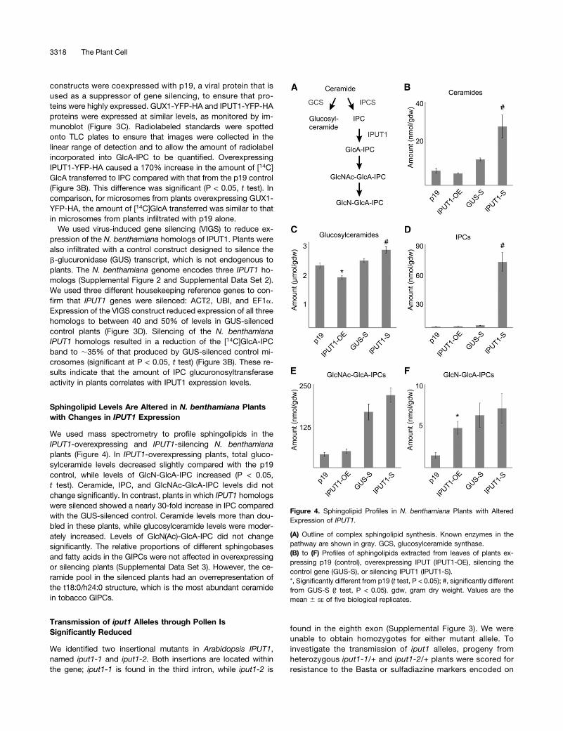
constructs were coexpressed with p19, a viral protein that isused as a suppressor of gene silencing, to ensure that pro-teins were highly expressed. GUX1-YFP-HA and IPUT1-YFP-HAproteins were expressed at similar levels, as monitored by im-munoblot (Figure 3C). Radiolabeled standards were spottedonto TLC plates to ensure that images were collected in thelinear range of detection and to allow the amount of radiolabelincorporated into GlcA-IPC to be quantified. OverexpressingIPUT1-YFP-HA caused a 170% increase in the amount of [14C]GlcA transferred to IPC compared with that from the p19 control(Figure 3B). This difference was significant (P < 0.05, t test). Incomparison, for microsomes from plants overexpressing GUX1-YFP-HA, the amount of [14C]GlcA transferred was similar to thatin microsomes from plants infiltrated with p19 alone.
We used virus-induced gene silencing (VIGS) to reduce ex-pression of the N. benthamiana homologs of IPUT1. Plants werealso infiltrated with a control construct designed to silence theb-glucuronidase (GUS) transcript, which is not endogenous toplants. The N. benthamiana genome encodes three IPUT1 ho-mologs (Supplemental Figure 2 and Supplemental Data Set 2).We used three different housekeeping reference genes to con-firm that IPUT1 genes were silenced: ACT2, UBI, and EF1a.Expression of the VIGS construct reduced expression of all threehomologs to between 40 and 50% of levels in GUS-silencedcontrol plants (Figure 3D). Silencing of the N. benthamianaIPUT1 homologs resulted in a reduction of the [14C]GlcA-IPCband to ;35% of that produced by GUS-silenced control mi-crosomes (significant at P < 0.05, t test) (Figure 3B). These re-sults indicate that the amount of IPC glucuronosyltransferaseactivity in plants correlates with IPUT1 expression levels.
Sphingolipid Levels Are Altered in N. benthamiana Plantswith Changes in IPUT1 Expression
We used mass spectrometry to profile sphingolipids in theIPUT1-overexpressing and IPUT1-silencing N. benthamianaplants (Figure 4). In IPUT1-overexpressing plants, total gluco-sylceramide levels decreased slightly compared with the p19control, while levels of GlcN-GlcA-IPC increased (P < 0.05,t test). Ceramide, IPC, and GlcNAc-GlcA-IPC levels did notchange significantly. In contrast, plants in which IPUT1 homologswere silenced showed a nearly 30-fold increase in IPC comparedwith the GUS-silenced control. Ceramide levels more than dou-bled in these plants, while glucosylceramide levels were moder-ately increased. Levels of GlcN(Ac)-GlcA-IPC did not changesignificantly. The relative proportions of different sphingobasesand fatty acids in the GIPCs were not affected in overexpressingor silencing plants (Supplemental Data Set 3). However, the ce-ramide pool in the silenced plants had an overrepresentation ofthe t18:0/h24:0 structure, which is the most abundant ceramidein tobacco GIPCs.
Transmission of iput1 Alleles through Pollen IsSignificantly Reduced
We identified two insertional mutants in Arabidopsis IPUT1,named iput1-1 and iput1-2. Both insertions are located withinthe gene; iput1-1 is found in the third intron, while iput1-2 is
found in the eighth exon (Supplemental Figure 3). We wereunable to obtain homozygotes for either mutant allele. Toinvestigate the transmission of iput1 alleles, progeny fromheterozygous iput1-1/+ and iput1-2/+ plants were scored forresistance to the Basta or sulfadiazine markers encoded on
Figure 4. Sphingolipid Profiles in N. benthamiana Plants with AlteredExpression of IPUT1.
(A) Outline of complex sphingolipid synthesis. Known enzymes in thepathway are shown in gray. GCS, glucosylceramide synthase.(B) to (F) Profiles of sphingolipids extracted from leaves of plants ex-pressing p19 (control), overexpressing IPUT (IPUT1-OE), silencing thecontrol gene (GUS-S), or silencing IPUT1 (IPUT1-S).*, Significantly different from p19 (t test, P < 0.05); #, significantly differentfrom GUS-S (t test, P < 0.05). gdw, gram dry weight. Values are themean 6 SE of five biological replicates.
3318 The Plant Cell

T-DNA insertions. Self-fertilization of iput1-1/+ and iput1-2/+plants resulted in an ;1:1 ratio of resistant to susceptibleprogeny, indicating a deficiency in transmission through eitherthe male or female gametophyte (Table 1). Reciprocal crosseswith wild-type plants showed that while transmission of iput1alleles through female gametophytes was not significantly af-fected, transmission through pollen was significantly reduced(P < 0.01, x2 test).
We tested expression of IPUT1 by quantitative PCR and foundthat it is expressed throughout pollen development (Figure 5A).These results indicate that IPUT1 could be required for normalpollen development. However, examination of pollen from bothiput1-1/+ and iput1-2/+ plants showed no difference fromwild-type pollen grains when viewed under a light microscope(Figures 5B and 5C), indicating that there are no gross mor-phological pollen defects. Staining with 49,6-diamidino-2-phenylindole (DAPI) (Figures 5D and 5E) or fluorescein diacetate(Figures 5F and 5G) further indicated that pollen nuclei developnormally and that pollen grains are viable. Pollen germinationrates were only slightly lower in both iput1-1/+and iput1-2/+than in wild-type plants, indicating that iput1 pollen is able togerminate (Table 2), and iput1 pollen tubes appeared to grownormally (Figures 5H and 5I). Tube growth rates of pollen fromiput1-1/+ and iput1-2/+ plants, based on measurement of tubelength 6-8 h after germination, were only slightly lower thanthose of pollen from wild-type plants (Supplemental Figure 5).Thus, the defect in transmission through the male gametophyteis not due to improper pollen development or inability of pollentubes to germinate and grow.
Quantitative PCR indicated that IPUT1 is expressed in allArabidopsis tissues and at all stages tested (SupplementalFigure 5). This ubiquitous expression is similar to that of othersphingolipid biosynthetic proteins (Chen et al., 2006; Tsegayeet al., 2007) and is expected for a protein required for synthesisof a major lipid class.
DISCUSSION
In this study, we provide evidence showing that IPUT1 is aninositol phosphorylceramide glucuronosyltransferase. The ex-pression of IPUT1 in yeast led to the production of GlcA-IPC,indicating that IPUT1 is capable of transferring a GlcA residue
Table 1. Transmission Efficiency of iput1 Alleles
Parent Genotype Self-Fertilized TEFemale TEMale
iput1-1/+ 0.98:1 (672) 95% (455) 1% (527)iput1-2/+ 0.93:1 (349) 92% (349) 2% (519)
Transmission efficiency of the iput1 T-DNA alleles was determined usingthe ratio of Basta-resistant to Basta-sensitive progeny (iput1-1/+) or theratio of Sulfadiazine-resistant to Sulfadiazine-sensitive progeny (iput1-2/+)after self-fertilization and after performing reciprocal crosses with wild-type plants. Self-fertilized, ratio of number of progeny with T-DNA insertionto number of progeny without T-DNA insertion. TE, number of progenywith T-DNA insertion/number of progeny without T-DNA insertion 3 100.Total numbers of seedlings scored are indicated in parentheses. Pooleddata represented in this table are from multiple generations.
Figure 5. iput1 Pollen Develops Normally.
(A) RT-PCR of IPUT1 and Histone H3 (control) in the uninucleate mi-crospore (UNM), bicellular pollen (BCP), tricellular pollen (TCP), andmature pollen grains (MPG). RT-PCRs used pooled spores from twobiological replicates.(B), (D), (F), and (H) Pollen from wild-type plants.(C), (E), (G), and (I) Pollen from iput1-1/+ plants. Phenotype of wild typeand iput1 pollen is indistinguishable.Light microscopy of mature pollen grains ([A] and [B]); fluorescencemicroscopy after DAPI staining ([D] and [E]) and fluorescein diacetatestaining ([F] and [G]). Arrows indicate the diffusely stained vegetativenucleus; arrowheads indicate the densely stained sperm cell nuclei. Lightmicroscopy of germinated pollen tubes, 6 h after germination ([H] and[I]). Bars = 10 mm in (B) to (F) and 100 mm in (G) to (I).
Sphingolipid a-Glucuronosyltransferase 3319

from UDP-GlcA onto IPC in yeast. In addition, assays usingmicrosomes from N. benthamiana plants transiently over-expressing IPUT1 confirmed that IPUT1 is an active IPC:glu-curonosyltransferase in plants. Plants in which IPUT1 homologswere silenced showed a decrease in IPC:glucuronosyltransfer-ase activity.
Plants produce several different ceramide species with var-iations in long-chain sphingobase and fatty acid structure. Themakeup of ceramides differs between sphingolipid pools suchas free ceramides, ceramide phosphates, glucosylceramides,and GIPCs (Markham et al., 2006). We therefore undertooka sphingolipidomic approach to determine how changes inIPUT1 expression affected various sphingolipid pools. AlteringIPUT1 expression in N. benthamiana led to changes in sphin-golipids including ceramides, glucosylceramides, IPC, and theglycosylated sphingolipids GlcN-GlcA-IPC and GlcNAc-GlcA-IPC (Figure 4). Most notably, silencing the N. benthamiana ho-mologs of IPUT1 caused a nearly 30-fold increase in levels ofIPC, the substrate for IPUT1. Given that production of IPC byIPCS is the first committed step in GIPC synthesis, it is notsurprising that this large increase in IPC levels was accompa-nied by changes in other sphingolipid pools. Ceramide levelswere also significantly increased in IPUT1-silenced plants,probably because the accumulation of IPC prevented ceramidefrom being utilized by IPCS. More detailed analysis of the ce-ramide pool supports the notion that the major ceramidestructure in the IPCs and GIPCs we examined was t18:0/h24:0,and this ceramide was preferentially enriched in IPUT1-silencedplants (Supplemental Figure 3). When the flux toward GIPCs wasdecreased, ceramide precursors may instead have been re-directed toward glucosylceramide synthesis. The small increasein glucosylceramide levels in IPUT1-silenced plants is consistentwith this scenario. Conversely, glucosylceramide levels de-creased in IPUT1-overexpressing plants, indicating that sphin-golipids that may have otherwise been used in glucosylceramidesynthesis were instead used to synthesize GIPCs.
Due to the lack of available acceptors, we were not ableto investigate the in vitro acceptor specificity of IPUT1 indetail; however, expression of IPUT1 in yeast caused glucur-onosylation of IPCs containing at least two different ceramidespecies (Figure 2B), indicating that IPUT1 is capable of some-what relaxed acceptor specificity at least with respect to ce-ramide fatty acid hydroxylation and length. Further investigationof IPUT1 specificity, as well as of ceramide specificities for thethree IPCS orthologs that catalyze the committed step of GIPCsynthesis (Mina et al., 2010), may help clarify how GIPC syn-thesis is controlled.
IPUT1 is essential in Arabidopsis. We identified two allelicmutants in IPUT1. iput1 alleles were transmitted through pollenat 1 to 2% of wild-type levels, indicating a role for IPUT1 inpollen function; however, iput1 pollen and pollen tubes ap-peared to develop normally (Figure 5). Although germination andtube growth rates were decreased slightly in pollen from iput1-1/+and iput1-2/+ plants (Table 2; Supplemental Figure 4), thedecreases were not large enough to indicate that iput1 pollenare unable to germinate or grow properly or to explain why only1 to 2% of iput1 alleles were transmitted through pollen. Theseresults indicate that the major defect in iput1 pollen is man-ifested during tube guidance or ovule fertilization. Several sce-narios could explain how GIPCs are involved in these processes.Many plasma membrane-localized transporters and signalingcomponents are required for tube guidance and fertilization andare located only at the pollen tube tips, indicating that precisespatial organization of the membrane is necessary for theseprocesses (Cheung and Wu, 2008). Lipid rafts have been sug-gested to play a role in positioning membrane proteins duringpollen tube growth (Lalanne et al., 2004; Liu et al., 2009), andthere is some evidence that GIPCs are enriched in lipid rafts(Borner et al., 2005). Lipid rafts may also be directly involved insignal transduction, for example, by organizing proteins such asreceptors in microenvironments that allow specific receptorproteins to interact (Simons and Toomre, 2000) or by regulatingreceptors allosterically (Coskun et al., 2011). It is possible thatmembrane composition influences how pollen tubes perceivesignaling molecules produced by maternal tissues. Further workwith IPUT1 may help to determine more precisely how GIPCsare involved in pollen tube guidance and fertilization.IPUT1 function is not limited to pollen. We found that IPUT1 is
expressed at all developmental stages tested (Figure 5). IPUT1has also been found in Golgi proteomes from several studiesusing tissue cultures, indicating that it is expressed under cellculture conditions (Dunkley et al., 2004; Parsons et al., 2012).This widespread expression is not unexpected, since recentreports have indicated that GIPCs are a major component ofplant plasma membranes (Sperling et al., 2005; Markham et al.,2006). Sphingolipids are also concentrated in the outer leaflet ofthe membrane, indicating that they are subject to additionallevels of organization (Markham et al., 2013). It is becoming in-creasingly evident that GIPCs are quantitatively important andprobably have functions in the secretory system and plasmamembrane that have not yet been determined.GIPCs have also recently been implicated in salicylic acid-
dependent signaling and the hypersensitive response duringdefense against pathogens. inositol phosphorylceramide syn-thase1 (ipcs1) mutants expressing the resistance gene RPW8have elevated salicylic acid levels and form hypersensitive le-sions in the absence of pathogens (Wang et al., 2008). Thismutant is defective in one of the three IPCS genes in Arabi-dopsis and accumulates ceramide, leading to the suggestionthat this precursor activates a signaling network that leads to theformation of hypersensitive lesions. Sphingolipid signaling iswell characterized in animals, where ceramides are bioactivelipids that can induce apoptosis (Hannun and Obeid, 2008).Similarly, golgi-localized nucleotide sugar transporter1 (gonst1)mutants are defective in GIPC mannosylation and show
Table 2. Germination Rate of Pollen from iput1/+ Heterozygotes
Parent Genotype Pollen Germination Rate
Col-0 74.6% (2372)iput1-1/+ 71.5% (3078)iput1-2/+ 70.0% (3427)
Pollen germination rates of iput1 heterozygotes. Total numbers of pollengrains scored are indicated in parentheses.
3320 The Plant Cell
comparable spontaneous hypersensitive lesions and increasedsalicylic acid (Mortimer et al., 2013). However, gonst1 mutantsdid not have increased ceramide levels, indicating that GIPCmannosylation itself, rather than levels of GIPC precursors, isnecessary for regulating the hypersensitive response (Mortimeret al., 2013). The mechanisms by which sphingolipids participatein the hypersensitive response are thus very unclear. Futurework with iput1 mutants may provide an opportunity to dissecthow GIPC glycosylation contributes to changes in varioussphingolipid pools and how glycan structures are involved inresponse to pathogens.
METHODS
Phylogenetic Analysis
Protein sequences were obtained from The Arabidopsis InformationResource (www.arabidopsis.org) or the Sol Genomics Network (http://solgenomics.net/) and were aligned using MUSCLE 3.7 (Edgar, 2004).Phylogenetic trees were built using PhyML 3.0 (Guindon et al., 2010) andviewed using FigTree version 1.3.1 (http://tree.bio.ed.ac.uk/software/figtree/).Bootstrap values shown at nodes were calculated using 1000 replicates.
Cloning and Construction of Expression Vectors
Sequences of all primers used in this study can be found in SupplementalTable 1. Arabidopsis thaliana clones were amplified using mixed organand developmental cDNA libraries as template. The human cDNAhUGTrel7 was obtained from the CCSB Human Orfeome Collection(Thermo Scientific). Entry clones were constructed using Gatewaytechnology (Invitrogen) via BP reaction in pDONR223. The reverse primerscontained no stop codon to enable C-terminal fusions. Gene-specificprimers were used to amplify cDNA, and PCR products were thenreamplified with the Gateway attB-specific primers before the BP re-combination reaction. All entry clones were verified by restriction analysisand sequencing. Gateway expression vectors were constructed via LRreaction. The binary vector pEarleyGate101, which contains a 35S pro-moter and a C-terminal YFP-HA tag, was used for expression in Nicotianabenthamiana (Earley et al., 2006). Yeast expression vectors used werea Gateway-enabled pRS423 with a HIS3 marker (Sikorski and Hieter,1989) for hUGTrel7 and pDRf1-GW with URA3 or LEU2 markers (Loquéet al., 2007) for UGD2 and IPUT1 cDNA. The sur1D knockout yeast strain(MATa Strain YPL057C) was obtained from the Yeast Knockout Collection(Thermo Scientific). For N. benthamiana clones, cDNA from leaves wasused as template. The binary vectors pTRV RNA1 and pTRV RNA2 wereused for VIGS (Liu et al., 2002). A 400-bp cDNA fragment with sequencesimilarity to all three IPUT1 homologs was amplified and ligated into themultiple cloning site of pTRV2 using EcoRI and XbaI restriction sites.
Electrospray Ionization Mass Spectrometry
Yeast were grown in appropriate media, pelleted at 2000g, and lipids wereextracted three times in 5 volumes of 1-butanol. The extracts werecombined, partitioned once against 0.5 volume water to remove salts andproteins, and dried. Dried lipids were resuspended in chloroform/methanol/[4M ammoniumhydroxide plus 1.8M ammonium acetate] (9:7:2 v/v/v) priorto analysis by mass spectrometry. Samples were analyzed by infusion at20 mL/min using a Q TRAP LC/MS/MS system (AB SCIEX) equipped witha TurboIonSpray ion source. The QTRAP system was operated in negativeion mode using the enhanced MS (EMS) for MS and enhanced product ion(EPI) for tandem mass spectrometry and a scan rate of 1000 (D/s). Thenumber of scans to sumwas set to 2, the scanmodewas set to profile, and
a dynamic fill time was selected. In EMSmode, the systemwas set to scanfrom 800 to 1300 m/z for 0.5 s, whereas for EPI a range of 50 to 1200 m/zwas employed and a collision energy of 280 eV was used. The ion sprayvoltage was set at 24200 V, source temperature (TEM) at 350°C and ionsource gas GS1 was 20 and GS2 was set to 7. All data were collected andanalyzed using Analyst 1.5.2 (AB SCIEX).
TLC
Pelleted yeast were extracted in chloroform/methanol/[4 M ammoniumhydroxide plus 1.8 M ammonium acetate] (9:7:2 v/v/v) at 40°C andcentrifuged to separate the organic and aqueous phases. The aqueousphase was cooled to 220°C and then centrifuged again. The bottomphase was collected, dried, spotted onto silica gel TLC plates, and de-veloped in the same solvent used for extraction.
Plant Material and Transient Expression
Three- to four-week-oldN. benthamiana ‘Domin’ plants were infiltratedwithAgrobacterium tumefaciens. Agrobacterium strain C58-1 pGV3850 car-rying the appropriate vectors was grown to log phase, pelleted at 3500g,and resuspended in infiltrationmedium consisting of 10mMMES-KOH, pH5.6, 10mMMgCl2, and 200 mM acetosyringone before being infiltrated intothe abaxial surfaces of leaves. Agrobacterium carrying the vector with thegene of interest wasmixed 1:1 with a strain carrying a plasmid with the p19gene from Tomato bushy stunt virus (Voinnet et al., 2003). The final opticaldensity at 600 nm (OD600) for each strain was 1.0. As much of each leaf aspossible (;95%)was infiltrated. The expression of genes fused to YFPwasverified by monitoring YFP fluorescence with a Leica D4000B epifluo-rescence microscope and all cells in infiltrated areas were shown to expressprotein. Entire leaves were harvested 4 d after infiltration.
VIGS
VIGS was performed essentially as described (Burch-Smith et al., 2004).Two-week-old plants were used for VIGS experiments. Agrobacteriumstrains carrying the IPUT1-TRV RNA2 or GUS-TRV RNA2 vectors wereresuspended in infiltration medium as above and mixed 1:1 with Agro-bacterium carrying the TRV RNA1 vector for a final OD600 of;0.4 for eachstrain. Cultures were infiltrated into the abaxial surfaces of leaves. Si-lenced leaves were harvested 10 d after infiltration. Spread of silencingwasmonitored by infiltrating neighboring plants with a construct designedto silence phytoene desaturase, causing photobleaching.
Microsome Preparation
All microsome preparation steps took place at 4°C. Leaf tissue was groundin buffer containing 50 mM HEPES-KOH, pH 7.0, 400 mM sucrose, 1 mMphenylmethanesulfonyl fluoride, 1% (w/v) polyvinylpolypyrrolidone, andprotease inhibitors (Roche Complete protease inhibitor tablets used asinstructed by the manufacturer). The homogenate was filtered through twolayers ofMiracloth (EMDMillipore) and centrifuged at 3000g for 10min, andthe supernatant was then centrifuged at 50,000g for 1 h. The pellet wasresuspended in 50 mM HEPES-KOH, pH 7.0, and 400 mM sucrose. Mi-crosomes were used immediately or frozen in liquid nitrogen and stored at280°C. No significant loss of activity was detected after freezing.
IPC Glucuronosyltransferase Assay
Assay reactions included 50 mM HEPES-KOH, pH 7.0, 5 mM MnCl2,1 mMDTT, 400mM sucrose, 100 mgN. benthamianamicrosomal proteins,and 4.4 mMUDP-[14C]GlcA (1480 Bq; MP Biomedicals) in a total volume of50 mL. Reactions were mixed and incubated at 21°C for 1 h, and the re-actionwas then stopped by the addition of 300mL 1-butanol. The butanolic
Sphingolipid a-Glucuronosyltransferase 3321

phasewas partitioned against 300 mL water, and then an additional 300 mL1-butanol was added and the partition was repeated. The butanolic phaseswere combined, dried, spotted onto silica gel TLC plates (Sigma-Aldrich),and developed in chloroform/methanol/[4 M ammonium hydroxide plus1.8 M ammonium acetate] (9:7:2 v/v/v). Plates were exposed to phosphorscreens for 2 to 5 d before imaging with a Typhoon 8600 Variable Modephosphor imager. Band intensities were quantified using ImageJ softwarefollowing instructions given in the user guide (http://imagej.nih.gov/ij/index.html) (Schneider et al., 2012). Mild alkaline hydrolysis was performed byincubating samples in methanolic 0.1 M potassium hydroxide for 1 h at21°C, neutralizing with acetic acid, and drying the samples. After thistreatment, samples were resuspended in butanol and partitioned againstwater to remove salts.
Detection of Proteins by Immunoblot
Yeast proteins were extracted from yeast as described (Kushnirov, 2000).N. benthamiana microsomal proteins were washed twice with 80% ac-etone to remove residual membranes before analysis. Proteins wereresolved by SDS-PAGE on 7% to 15% gradient gels and blotted ontonitrocellulose membranes (GE Healthcare). Yeast protein blots wereprobed with a 1:10,000 dilution of an antibody raised in rabbit against theattB2 Gateway site (Invitrogen) (Eudes et al., 2011) followed by a 1:20,000dilution of goat anti-rabbit IgG conjugated to horseradish peroxidase(Sigma-Aldrich). N. benthamiana protein blots were probed with a 1:1000dilution of rat monoclonal anti-HA antibody (Sigma-Aldrich) followed bya 1:15,000 dilution of goat anti-rat IgG conjugated to horseradish per-oxidase (Sigma-Aldrich). Chemiluminescence was detected using ECLPlus detection reagent (GE Healthcare). Blots were imaged using aChemiDoc-It 600 Imaging System (UVP).
Expression Analysis
For quantitative PCR, RNA was extracted from Arabidopsis or N. ben-thamiana tissue using an RNeasy Plant Mini Kit (Qiagen). One microgramof RNA was used as template in a reverse transcriptase reaction using aniScript cDNA synthesis kit (Bio-Rad). Real-time PCR was done withAbsolute SYBR Green ROX Mix (ABgene) on a StepOnePlus Real-timePCR system (Applied Biosystems) according to conditions describedearlier (Czechowski et al., 2005) using StepOne 2.0 software (AppliedBiosystems). Primers for Arabidopsis quantitative PCR reference geneswere taken from (Valdivia et al., 2013). Quantitative PCR data werequantified using the comparative Ct method (Schmittgen and Livak, 2008)using geometric averaging of the three reference genes as the commonreference (Vandesompele et al., 2002). For pollen expression analysis,RNA was extracted from pollen at different stages of development asdescribed (Honys and Twell, 2004). Samples of 750 ng of total RNA forpollen stages were reverse transcribed in a 20 mL reaction using Su-perscript II RNase H reverse transcriptase (Invitrogen) and an oligo(dT)primer as per the manufacturer’s instructions. For PCR amplification, 1 mLof a 103 diluted cDNA was used in a 25-mL reaction using Biotaq DNApolymerase (Bioline) and 12.5 pmol of each primer. PCR conditions wereas follows: 96°C for 1 min, 30 cycles of 96°C for 30 s, 55°C for 30 s, and72°C for 40 s followed by 5min at 72°C. Histone H3 (At4g40040) was usedas a control as described (Brownfield et al., 2009).
Sphingolipidomics
N. benthamiana leaf tissue was lyophilized and powderized using amortarand pestle. Sphingolipids were extracted from 30 mg of the powderizedtissue using isopropanol/hexane/water (55:20:25 v/v/v) followed by 33%methylamine treatment as previously described (Markham and Jaworski,2007) and resuspended in tetrahydrofuran/methanol/water (2:1:2 v/v/v)containing 0.1% formic acid. Sphingolipidswere analyzed using a Shimadzu
Prominence UPLC coupled with a QTRAP4000 mass spectrometer (ABSCIEX) as described (Kimberlin et al., 2013). Instrument potentials andchromatography conditions for the initial detection of N-acetyl-sugar-containing GIPCs were as for hexose(R1:OH)-glucuronic acid-inositol-phosphoceramides (GlcOH-GlcA-IPCs) described previously (Kimberlinet al., 2013). Multiple reaction monitoring (MRM) Q1/Q3 transitions todetect N-acetyl-sugar-containing GIPCs (GlcNAc-GlcA-IPCS) werecalculated by adding 41 mass units to the Q1 ion of previously describedGlcOH-GlcA-IPC MRM Q1/Q3 transitions (Markham and Jaworski, 2007).GlcN-GlcA-IPCMRMQ1/Q3 transitionswere calculated by subtracting onemass unit from the Q1 ion of GlcOH-GlcA-IPC species and are distin-guished from GlcOH-GlcA-IPCs by longer elution times. The retention timeand M+H mass of the IPCs were first confirmed by precursor ion scanningfor the t18:1/h24:0 backbone (precursors of m/z 664.6) combined withchromatographic separation of GIPCs as described (Kimberlin et al., 2013).Based on this information, MRM Q1/Q3 transitions for IPC detection werecalculated based on known structures and fragmentation patterns and aregiven in Supplemental Table 2.
Arabidopsis Mutant Isolation and Transmission
To determine transmission efficiency (TE), seeds (iput1-2) were sown onsolid medium (0.8% [w/v] agar) containing half-strength Murashige andSkoog (Murashige and Skoog, 1962) salts including vitamins (Sigma-Aldrich) and sucrose (1% (w/v) with Sulfadiazine (4-amino-N-2-pyr-imidinylbenzene-sulfonamide; Sigma-Aldrich) at 5 mg/L. Seeds (iput1-1)were sown in soil (John Innes No. 1) in a growth room (20°C, 100 mmol s21
m22, 16-h-light/8-h-dark, 60% humidity). Three-week-old plants weresprayed with BASTA at a concentration of 75 mg/L. TE through male andfemale gametophytes was determined by crossing iput1/+ heterozygoteswith wild-type plants and scoring resistance of progeny to the appropriateselection as described (Howden et al., 1998). TE was calculated as(number of resistant seedlings/number of susceptible seedlings) 3 100.
Microscopy Analyses
Analysis of nuclear DNA in mature pollen with DAPI was performed aspreviously described (Park et al., 1998). Fluorescein diacetate staining tomonitor pollen viability was performed by incubating freshly isolatedpollen in 0.3 Mmannitol solution containing 2 mgmL fluorescein diacetateas described (Goubet et al., 2003).
Pollen Germination
Pollen were collected and germinated as described (Boavida andMcCormick, 2007). Liquid germination media consisted of 0.01% boricacid, 5 mM CaCl2, 5 mM KCl, 1 mM MgSO4, and 10% sucrose at pH 7.5.Pollen was allowed to germinate at 22°C for 6 to 8 h, and then imageswere taken using a Leica D4000B epifluorescence microscope coupledwith a Leica DC500 camera.
Accession Numbers
Sequence data from this article can be found at The Arabidopsis In-formation Resource under accession number At5g18480 (IPUT1). Theaccession numbers of the Arabidopsis mutants used in this study areiput1-1 (SAIL.532.E01-N822575) and iput1-2 (GABI-KAT 856G03).
Supplemental Data
The following materials are available in the online version of this article.
Supplemental Figure 1. Production of GlcA-IPC in sur1D YeastExpressing UGD2, hUGTrel7, and IPUT1.
3322 The Plant Cell

Supplemental Figure 2. Homologs of GUX, PGSIP, and IPUT1Proteins in N. benthamiana.
Supplemental Figure 3. Diagram of Insertions in iput1-1 and iput1-2Alleles.
Supplemental Figure 4. Tube Growth Rate of Pollen from iput1-1/+and iput1-2/+ Arabidopsis Plants.
Supplemental Figure 5. Expression of IPUT1 in Different ArabidopsisTissues.
Supplemental Table 1. List of Primers Used in This Study.
Supplemental Table 2. Parameters for MRM Detection of IPCs inPositive Ion Mode.
Supplemental Data Set 1. Alignments Used to Generate thePhylogeny Presented in Figure 1.
Supplemental Data Set 2. Alignments Used to Generate thePhylogeny Presented in Supplemental Figure 2.
Supplemental Data Set 3. Summary of Sphingolipidomic Data onIPUT1-Overexpressing and IPUT1-Silencing N. benthamiana Plants.
ACKNOWLEDGMENTS
We thank Sherry Chan for technical assistance and Tsan-Yiu Chiu forproviding vectors. This work was supported by the U.S. Departmentof Energy, Office of Science, Office of Biological and EnvironmentalResearch, through Contract DE-AC02-05CH11231 between LawrenceBerkeley National Lab and the U.S. Department of Energy and byNational Science Foundation Graduate Research Fellowship ProgramGrant DGE 1106400 (E.A.R.) and National Science Foundation GrantMCB 115850 (E.B.C.).
AUTHOR CONTRIBUTIONS
E.A.R., B.E., G.P.M., S.S., K.M.C., H.K., D.T., C.J.P., P.D.A., P.D., J.L.H.,and H.V.S. designed research. E.A.R., B.E., G.P.M., R.E.C., S.S., K.M.C.,E.B.C., and H.K. performed research. E.A.R., B.E., G.P.M., R.E.C., E.B.C.,and H.V.S. analyzed data. E.A.R. and H.V.S. wrote the article.
Received June 20, 2014; revised June 20, 2014; accepted July 22, 2014;published August 8, 2014.
REFERENCES
Beeler, T.J., Fu, D., Rivera, J., Monaghan, E., Gable, K., and Dunn,T.M. (1997). SUR1 (CSG1/BCL21), a gene necessary for growth ofSaccharomyces cerevisiae in the presence of high Ca2+ concentrationsat 37 ° C, is required for mannosylation of inositolphosphorylceramide.Mol. Gen. Genet. 255: 570–579.
Boavida, L.C., and McCormick, S. (2007). Technical advance:temperature as a determinant factor for increased and reproduciblein vitro pollen germination in Arabidopsis thaliana. Plant J. 52:570–582.
Borner, G.H.H., Sherrier, D.J., Weimar, T., Michaelson, L.V.,Hawkins, N.D., Macaskill, A., Napier, J.A., Beale, M.H., Lilley,K.S., and Dupree, P. (2005). Analysis of detergent-resistant membranesin Arabidopsis. Evidence for plasma membrane lipid rafts. Plant Physiol.137: 104–116.
Bromley, P.E., Li, Y.O., Murphy, S.M., Sumner, C.M., and Lynch,D.V. (2003). Complex sphingolipid synthesis in plants: characterizationof inositolphosphorylceramide synthase activity in bean microsomes.Arch. Biochem. Biophys. 417: 219–226.
Brownfield, L., Hafidh, S., Borg, M., Sidorova, A., Mori, T., andTwell, D. (2009). A plant germline-specific integrator of spermspecification and cell cycle progression. PLoS Genet. 5: e1000430.
Burch-Smith, T.M., Anderson, J.C., Martin, G.B., and Dinesh-Kumar, S.P. (2004). Applications and advantages of virus-inducedgene silencing for gene function studies in plants. Plant J. 39:734–746.
Buré, C., Cacas, J.-L., Wang, F., Gaudin, K., Domergue, F.,Mongrand, S., and Schmitter, J.-M. (2011). Fast screening of highlyglycosylated plant sphingolipids by tandem mass spectrometry. RapidCommun. Mass Spectrom. 25: 3131–3145.
Cacas, J.-L., Buré, C., Furt, F., Maalouf, J.-P., Badoc, A., Cluzet, S.,Schmitter, J.-M., Antajan, E., and Mongrand, S. (2013). Biochemicalsurvey of the polar head of plant glycosylinositolphosphoceramidesunravels broad diversity. Phytochemistry 96: 191–200.
Carter, H.E., Strobach, D.R., and Hawthorne, J.N. (1969).Biochemistry of the sphingolipids. 18. Complete structure oftetrasaccharide phytoglycolipid. Biochemistry 8: 383–388.
Chao, D.-Y., et al. (2011). Sphingolipids in the root play an importantrole in regulating the leaf ionome in Arabidopsis thaliana. Plant Cell23: 1061–1081.
Chatterjee, M., Berbezy, P., Vyas, D., Coates, S., and Barsby, T.(2005). Reduced expression of a protein homologous to glycogeninleads to reduction of starch content in Arabidopsis leaves. PlantSci. 168: 501–509.
Chen, M., Markham, J.E., and Cahoon, E.B. (2012). Sphingolipid D8unsaturation is important for glucosylceramide biosynthesis andlow-temperature performance in Arabidopsis. Plant J. 69: 769–781.
Chen, M., Han, G., Dietrich, C.R., Dunn, T.M., and Cahoon, E.B.(2006). The essential nature of sphingolipids in plants as revealed bythe functional identification and characterization of the ArabidopsisLCB1 subunit of serine palmitoyltransferase. Plant Cell 18: 3576–3593.
Cheung, A.Y., and Wu, H.M. (2008). Structural and signalingnetworks for the polar cell growth machinery in pollen tubes. Annu.Rev. Plant Biol. 59: 547–572.
Coskun, Ü., Grzybek, M., Drechsel, D., and Simons, K. (2011).Regulation of human EGF receptor by lipids. Proc. Natl. Acad. Sci.USA 108: 9044–9048.
Coursol, S., Le Stunff, H., Lynch, D.V., Gilroy, S., Assmann, S.M.,and Spiegel, S. (2005). Arabidopsis sphingosine kinase and theeffects of phytosphingosine-1-phosphate on stomatal aperture.Plant Physiol. 137: 724–737.
Czechowski, T., Stitt, M., Altmann, T., Udvardi, M.K., and Scheible,W.-R. (2005). Genome-wide identification and testing of superiorreference genes for transcript normalization in Arabidopsis. PlantPhysiol. 139: 5–17.
Dietrich, C.R., Han, G., Chen, M., Berg, R.H., Dunn, T.M., andCahoon, E.B. (2008). Loss-of-function mutations and inducibleRNAi suppression of Arabidopsis LCB2 genes reveal the critical roleof sphingolipids in gametophytic and sporophytic cell viability. PlantJ. 54: 284–298.
Dunkley, T.P.J., Watson, R., Griffin, J.L., Dupree, P., and Lilley,K.S. (2004). Localization of organelle proteins by isotope tagging(LOPIT). Mol. Cell. Proteomics 3: 1128–1134.
Dunn, T.M., Haak, D., Monaghan, E., and Beeler, T.J. (1998).Synthesis of monohydroxylated inositolphosphorylceramide (IPC-C)in Saccharomyces cerevisiae requires Scs7p, a protein with both a
Sphingolipid a-Glucuronosyltransferase 3323

cytochrome b5-like domain and a hydroxylase/desaturase domain.Yeast 14: 311–321.
Earley, K.W., Haag, J.R., Pontes, O., Opper, K., Juehne, T., Song,K., and Pikaard, C.S. (2006). Gateway-compatible vectors for plantfunctional genomics and proteomics. Plant J. 45: 616–629.
Edgar, R.C. (2004). MUSCLE: multiple sequence alignment with highaccuracy and high throughput. Nucleic Acids Res. 32: 1792–1797.
Eudes, A., Baidoo, E.E., Yang, F., Burd, H., Hadi, M.Z., Collins,F.W., Keasling, J.D., and Loqué, D. (2011). Production of tranilast[N-(39,49-dimethoxycinnamoyl)-anthranilic acid] and its analogs inyeast Saccharomyces cerevisiae. Appl. Microbiol. Biotechnol. 89:989–1000.
Goubet, F., Misrahi, A., Park, S.K., Zhang, Z., Twell, D., andDupree, P. (2003). AtCSLA7, a cellulose synthase-like putativeglycosyltransferase, is important for pollen tube growth andembryogenesis in Arabidopsis. Plant Physiol. 131: 547–557.
Guan, X.L., and Wenk, M.R. (2006). Mass spectrometry-basedprofiling of phospholipids and sphingolipids in extracts fromSaccharomyces cerevisiae. Yeast 23: 465–477.
Guindon, S., Dufayard, J.-F., Lefort, V., Anisimova, M., Hordijk, W.,and Gascuel, O. (2010). New algorithms and methods to estimatemaximum-likelihood phylogenies: assessing the performance ofPhyML 3.0. Syst. Biol. 59: 307–321.
Hannun, Y.A., and Obeid, L.M. (2008). Principles of bioactive lipidsignalling: lessons from sphingolipids. Nat. Rev. Mol. Cell Biol. 9:139–150.
Hillig, I., Leipelt, M., Ott, C., Zähringer, U., Warnecke, D., andHeinz, E. (2003). Formation of glucosylceramide and sterol glucoside bya UDP-glucose-dependent glucosylceramide synthase from cottonexpressed in Pichia pastoris. FEBS Lett. 553: 365–369.
Honys, D., and Twell, D. (2004). Transcriptome analysis of haploidmale gametophyte development in Arabidopsis. Genome Biol. 5:R85.
Howden, R., Park, S.K., Moore, J.M., Orme, J., Grossniklaus, U.,and Twell, D. (1998). Selection of T-DNA-tagged male and femalegametophytic mutants by segregation distortion in Arabidopsis.Genetics 149: 621–631.
Kaul, K., and Lester, R.L. (1975). Characterization of inositol-containingphosphosphingolipids from tobacco leaves: isolation and identificationof two novel, major lipids: N-acetylglucosamidoglucuronidoinositolphosphorylceramide and glucosamidoglucuronidoinositol phos-phorylceramide. Plant Physiol. 55: 120–129.
Kaul, K., and Lester, R.L. (1978). Isolation of six novelphosphoinositol-containing sphingolipids from tobacco leaves.Biochemistry 17: 3569–3575.
Kimberlin, A.N., Majumder, S., Han, G., Chen, M., Cahoon, R.E.,Stone, J.M., Dunn, T.M., and Cahoon, E.B. (2013). Arabidopsis56-amino acid serine palmitoyltransferase-interacting proteinsstimulate sphingolipid synthesis, are essential, and affect mycotoxinsensitivity. Plant Cell 25: 4627–4639.
Klinghammer, M., and Tenhaken, R. (2007). Genome-wide analysisof the UDP-glucose dehydrogenase gene family in Arabidopsis,a key enzyme for matrix polysaccharides in cell walls. J. Exp. Bot.58: 3609–3621.
Kushnirov, V.V. (2000). Rapid and reliable protein extraction fromyeast. Yeast 16: 857–860.
Lalanne, E., Honys, D., Johnson, A., Borner, G.H.H., Lilley, K.S.,Dupree, P., Grossniklaus, U., and Twell, D. (2004). SETH1 andSETH2, two components of the glycosylphosphatidylinositol anchorbiosynthetic pathway, are required for pollen germination and tubegrowth in Arabidopsis. Plant Cell 16: 229–240.
Liang, H., Yao, N., Song, J.T., Luo, S., Lu, H., and Greenberg, J.T.(2003). Ceramides modulate programmed cell death in plants.Genes Dev. 17: 2636–2641.
Liu, P., Li, R.-L., Zhang, L., Wang, Q.-L., Niehaus, K., Baluška, F.,�Samaj, J., and Lin, J.-X. (2009). Lipid microdomain polarization isrequired for NADPH oxidase-dependent ROS signaling in Piceameyeri pollen tube tip growth. Plant J. 60: 303–313.
Liu, Y., Schiff, M., Marathe, R., and Dinesh-Kumar, S.P. (2002).Tobacco Rar1, EDS1 and NPR1/NIM1 like genes are required for N-mediated resistance to tobacco mosaic virus. Plant J. 30: 415–429.
Loqué, D., Lalonde, S., Looger, L.L., von Wiren, N., and Frommer, W.(2007). A cytosolic trans-activation domain essential for ammoniumuptake. Nature 446: 195–198.
Markham, J.E., and Jaworski, J.G. (2007). Rapid measurement ofsphingolipids from Arabidopsis thaliana by reversed-phase high-performance liquid chromatography coupled to electrospray ionizationtandem mass spectrometry. Rapid Commun. Mass Spectrom. 21:1304–1314.
Markham, J.E., Li, J., Cahoon, E.B., and Jaworski, J.G. (2006).Separation and identification of major plant sphingolipid classesfrom leaves. J. Biol. Chem. 281: 22684–22694.
Markham, J.E., Lynch, D.V., Napier, J.A., Dunn, T.M., and Cahoon,E.B. (2013). Plant sphingolipids: function follows form. Curr. Opin.Plant Biol. 16: 350–357.
Markham, J.E., Molino, D., Gissot, L., Bellec, Y., Hématy, K.,Marion, J., Belcram, K., Palauqui, J.-C., Satiat-Jeunemaître, B.,and Faure, J.D. (2011). Sphingolipids containing very-long-chainfatty acids define a secretory pathway for specific polar plasmamembrane protein targeting in Arabidopsis. Plant Cell 23: 2362–2378.
Mortimer, J.C., Miles, G.P., Brown, D.M., Zhang, Z., Segura, M.P.,Weimar, T., Yu, X., Seffen, K.A., Stephens, E., Turner, S.R., andDupree, P. (2010). Absence of branches from xylan in Arabidopsisgux mutants reveals potential for simplification of lignocellulosicbiomass. Proc. Natl. Acad. Sci. USA 107: 17409–17414.
Mina, J.G., Okada, Y., Wansadhipathi-Kannangara, N.K., Pratt, S.,Shams-Eldin, H., Schwarz, R.T., Steel, P.G., Fawcett, T., andDenny, P.W. (2010). Functional analyses of differentially expressedisoforms of the Arabidopsis inositol phosphorylceramide synthase.Plant Mol. Biol. 73: 399–407.
Mortimer, J.C., et al. (2013). Abnormal glycosphingolipid mannosylationtriggers salicylic acid-mediated responses in Arabidopsis. Plant Cell 25:1881–1894.
Muraoka, M., Kawakita, M., and Ishida, N. (2001). Molecular characterizationof human UDP-glucuronic acid/UDP-N-acetylgalactosamine transporter,a novel nucleotide sugar transporter with dual substrate specificity. FEBSLett. 495: 87–93.
Murashige, T., and Skoog, F. (1962). A revised medium for rapidgrowth and bio assays with tobacco tissue cultures. Physiol. Plant.15: 473–497.
Obeid, L.M., Okamoto, Y., and Mao, C. (2002). Yeast sphingolipids:metabolism and biology. Biochim. Biophys. Acta 1585: 163–171.
Oikawa, A., Joshi, H.J., Rennie, E.A., Ebert, B., Manisseri, C.,Heazlewood, J.L., and Scheller, H.V. (2010). An integrative approachto the identification of Arabidopsis and rice genes involved in xylan andsecondary wall development. PLoS ONE 5: e15481.
Park, S.K., Howden, R., and Twell, D. (1998). The Arabidopsisthaliana gametophytic mutation gemini pollen1 disrupts microsporepolarity, division asymmetry and pollen cell fate. Development 125:3789–3799.
Parsons, H.T., et al. (2012). Isolation and proteomic characterizationof the Arabidopsis Golgi defines functional and novel componentsinvolved in plant cell wall biosynthesis. Plant Physiol. 159: 12–26.
3324 The Plant Cell

Perotto, S., Donovan, N., Drobak, B.K., and Brewin, N.J. (1995).Differential expression of a glycosyl inositol phospholipid antigenon the peribacteroid membrane during pea nodule development.Mol. Plant Microbe Interact. 8: 560–568.
Pinto, W.J., Srinivasan, B., Shepherd, S., Schmidt, A., Dickson,R.C., and Lester, R.L. (1992). Sphingolipid long-chain-baseauxotrophs of Saccharomyces cerevisiae: genetics, physiology, anda method for their selection. J. Bacteriol. 174: 2565–2574.
Rennie, E.A., Hansen, S.F., Baidoo, E.E.K., Hadi, M.Z., Keasling,J.D., and Scheller, H.V. (2012). Three members of the Arabidopsisglycosyltransferase family 8 are xylan glucuronosyltransferases.Plant Physiol. 159: 1408–1417.
Schmittgen, T.D., and Livak, K.J. (2008). Analyzing real-time PCRdata by the comparative C(T) method. Nat. Protoc. 3: 1101–1108.
Schneider, C.A., Rasband, W.S., and Eliceiri, K.W. (2012). NIHImage to ImageJ: 25 years of image analysis. Nat. Methods 9: 671–675.
Sikorski, R.S., and Hieter, P. (1989). A system of shuttle vectors andyeast host strains designed for efficient manipulation of DNA inSaccharomyces cerevisiae. Genetics 122: 19–27.
Simons, K., and Toomre, D. (2000). Lipid rafts and signaltransduction. Nat. Rev. Mol. Cell Biol. 1: 31–39.
Sperling, P., and Heinz, E. (2003). Plant sphingolipids: structuraldiversity, biosynthesis, first genes and functions. Biochim. Biophys.Acta 1632: 1–15.
Sperling, P., Franke, S., Lüthje, S., and Heinz, E. (2005). Areglucocerebrosides the predominant sphingolipids in plant plasmamembranes? Plant Physiol. Biochem. 43: 1031–1038.
Taji, T., Ohsumi, C., Iuchi, S., Seki, M., Kasuga, M., Kobayashi, M.,Yamaguchi-Shinozaki, K., and Shinozaki, K. (2002). Important
roles of drought- and cold-inducible genes for galactinol synthasein stress tolerance in Arabidopsis thaliana. Plant J. 29: 417–426.
Ternes, P., Feussner, K., Werner, S., Lerche, J., Iven, T., Heilmann,I., Riezman, H., and Feussner, I. (2011). Disruption of the ceramidesynthase LOH1 causes spontaneous cell death in Arabidopsisthaliana. New Phytol. 192: 841–854.
Tsegaye, Y., Richardson, C.G., Bravo, J.E., Mulcahy, B.J., Lynch, D.V.,Markham, J.E., Jaworski, J.G., Chen, M., Cahoon, E.B., and Dunn,T.M. (2007). Arabidopsis mutants lacking long chain base phosphatelyase are fumonisin-sensitive and accumulate trihydroxy-18:1 longchain base phosphate. J. Biol. Chem. 282: 28195–28206.
Valdivia, E.R., Herrera, M.T., Gianzo, C., Fidalgo, J., Revilla, G.,Zarra, I., and Sampedro, J. (2013). Regulation of secondary wallsynthesis and cell death by NAC transcription factors in themonocot Brachypodium distachyon. J. Exp. Bot. 64: 1333–1343.
Vandesompele, J., De Preter, K., Pattyn, F., Poppe, B., Van Roy, N.,De Paepe, A., and Speleman, F. (2002). Accurate normalization ofreal-time quantitative RT-PCR data by geometric averaging ofmultiple internal control genes. Genome Biol. 3: H0034.
Voinnet, O., Rivas, S., Mestre, P., and Baulcombe, D. (2003). Anenhanced transient expression system in plants based on suppressionof gene silencing by the p19 protein of tomato bushy stunt virus. PlantJ. 33: 949–956.
Wang, W., et al. (2008). An inositolphosphorylceramide synthase isinvolved in regulation of plant programmed cell death associatedwith defense in Arabidopsis. Plant Cell 20: 3163–3179.
Zäuner, S., Ternes, P., and Warnecke, D. (2010). Biosynthesis ofsphingolipids in plants (and some of their functions). In Sphingoli-pids as Signaling and Regulatory Molecules, C. Chalfant and M.Poeta, eds (New York: Springer), pp. 249–263.
Sphingolipid a-Glucuronosyltransferase 3325

DOI 10.1105/tpc.114.129171; originally published online August 8, 2014; 2014;26;3314-3325Plant Cell
Heazlewood, Edgar B. Cahoon and Henrik Vibe SchellerStonebloom, Hoda Khatab, David Twell, Christopher J. Petzold, Paul D. Adams, Paul Dupree, Joshua L.
Emilie A. Rennie, Berit Ebert, Godfrey P. Miles, Rebecca E. Cahoon, Katy M. Christiansen, SolomonArabidopsis
-Glucuronosyltransferase That Is Essential for Pollen Function inαIdentification of a Sphingolipid
This information is current as of August 20, 2018
Supplemental Data /content/suppl/2014/07/23/tpc.114.129171.DC1.html
References /content/26/8/3314.full.html#ref-list-1
This article cites 69 articles, 24 of which can be accessed free at:
Permissions https://www.copyright.com/ccc/openurl.do?sid=pd_hw1532298X&issn=1532298X&WT.mc_id=pd_hw1532298X
eTOCs http://www.plantcell.org/cgi/alerts/ctmain
Sign up for eTOCs at:
CiteTrack Alerts http://www.plantcell.org/cgi/alerts/ctmain
Sign up for CiteTrack Alerts at:
Subscription Information http://www.aspb.org/publications/subscriptions.cfm
is available at:Plant Physiology and The Plant CellSubscription Information for
ADVANCING THE SCIENCE OF PLANT BIOLOGY © American Society of Plant Biologists