interpositional arthroplasty using first carpo … was restricted in flexion, circumduction, and...
TRANSCRIPT
Received 10/18/2016 Review began 10/28/2016 Review ended 10/28/2016 Published 11/03/2016
© Copyright 2016Kapoor et al. This is an open accessarticle distributed under the terms ofthe Creative Commons AttributionLicense CC-BY 3.0., which permitsunrestricted use, distribution, andreproduction in any medium,provided the original author andsource are credited.
First Carpo-metacarpal Joint Arthritis:Interpositional Arthroplasty usingTrapeziumChirag Kapoor , Ankur Kansagra , Maulik Jhaveri , Aditya Merh , Paresh Golwala
1. Orthopaedics, Sumandeep Vidyapeeth, Vadodara, Gujarat
Corresponding author: Chirag Kapoor, [email protected] Disclosures can be found in Additional Information at the end of the article
AbstractThumb pain secondary to arthritis at the basal joint of the thumb is a common condition,especially in women, and can be quite disabling. An accurate diagnosis can be readily madefrom the history and examination. Reconstructive procedures for each stage of the disease areaimed at relieving pain and restoring thumb motion and strength. There are a number ofmethods available to treat this condition both conservatively and surgically with variablesuccess rates. We present a case of a middle-aged female with first carpo-metacarpal (CMC)joint arthritis in whom we have tried a new technique in which the trapezium is excised,crushed, put in a sponge covering and then inserted back in the void created after excision. Atthe one-year follow-up, the patient was pain-free and had full range of thumb movement.
Categories: Pain Management, Orthopedics, Epidemiology/Public HealthKeywords: carpo-metacarpal, arthritis, trapezium
IntroductionCarpo-metacarpal (CMC) joint arthritis is a prevalent condition affecting up to 10% of middle-aged women [1]. The basal joint of the thumb consists of four trapezial articulations: thetrapeziometacarpal (TM), trapeziotrapezoid, scaphotrapezial (ST), and trapezium-indexmetacarpal articulations. Only the TM and ST joints lie along the longitudinal compression axisof the thumb. North and Eaton have observed that radiographic disease most commonly affectsthese two joints [2]. There are many treatment options available with variable results.
Conservative treatment is considered in early stages of the disease while advanced stages haveto be dealt with surgically. We present a case of a middle-aged female with symptomatic latestage first CMC joint arthritis who was treated surgically by a new technique, with an excellentoutcome. Informed consent was obtained from the patient for this study.
Case PresentationThe patient was a 55-year-old female with a six-month history of pain in the base of the rightthumb. The pain was insidious at onset, gradually progressing, non-radiating, and it increasedon activity. She had a weak grip and had pain when she made a pinching movement. She had noother comorbidities or associated conditions like diabetes or rheumatoid arthritis.
On examination, there was tenderness and swelling at the base of the right thumb without anysigns of inflammation. The thumb CMC grind test was positive. The thumb active range of
1 1 1 1 1
Open Access CaseReport DOI: 10.7759/cureus.861
How to cite this articleKapoor C, Kansagra A, Jhaveri M, et al. (November 03, 2016) First Carpo-metacarpal Joint Arthritis:Interpositional Arthroplasty using Trapezium. Cureus 8(11): e861. DOI 10.7759/cureus.861

movement was restricted in flexion, circumduction, and opposition as compared to the normalthumb. Further passive movement was possible but was painful. Metacarpo-phalangeal (MCP)joint hyperextension instability was present as compared to the contralateral side. There wasno associated neurovascular deficit with normal sensations and motor power of the thumb.
Plain radiographs revealed degeneration of the first trapeziometacarpal joint, i.e. first CMCjoint, with irregular and sclerosed joint margins and a few osteophytes on the radial aspect ofthe thumb base (Figure 1).
FIGURE 1: Pre-operative radiograph (antero-posterior andoblique views)Image showing arthritic first metacarpal-trapezium joint.
After obtaining a written informed consent from the patient, she was submitted to surgery. Adorsal longitudinal incision was taken centring over the CMC joint till the thenar eminence,with care taken to protect the superficial branches of the radial nerve. The trapezium wasexposed and excised in toto. The degenerated cartilage at the base of the first metacarpal wasshaved off. The excised trapezium was crushed to small pieces and put inside a wet spongepiece which completely engulfed it (Figure 2). This sponge with the bone pieces was put in thecavity that was created after the excision of the trapezium (Figure 2).
2016 Kapoor et al. Cureus 8(11): e861. DOI 10.7759/cureus.861 2 of 6

FIGURE 2: Operative techniqueLeft: trapezium crushed and put inside a sponge covering.
Right: trapezium graft put inside the cavity created after trapeziectomy.
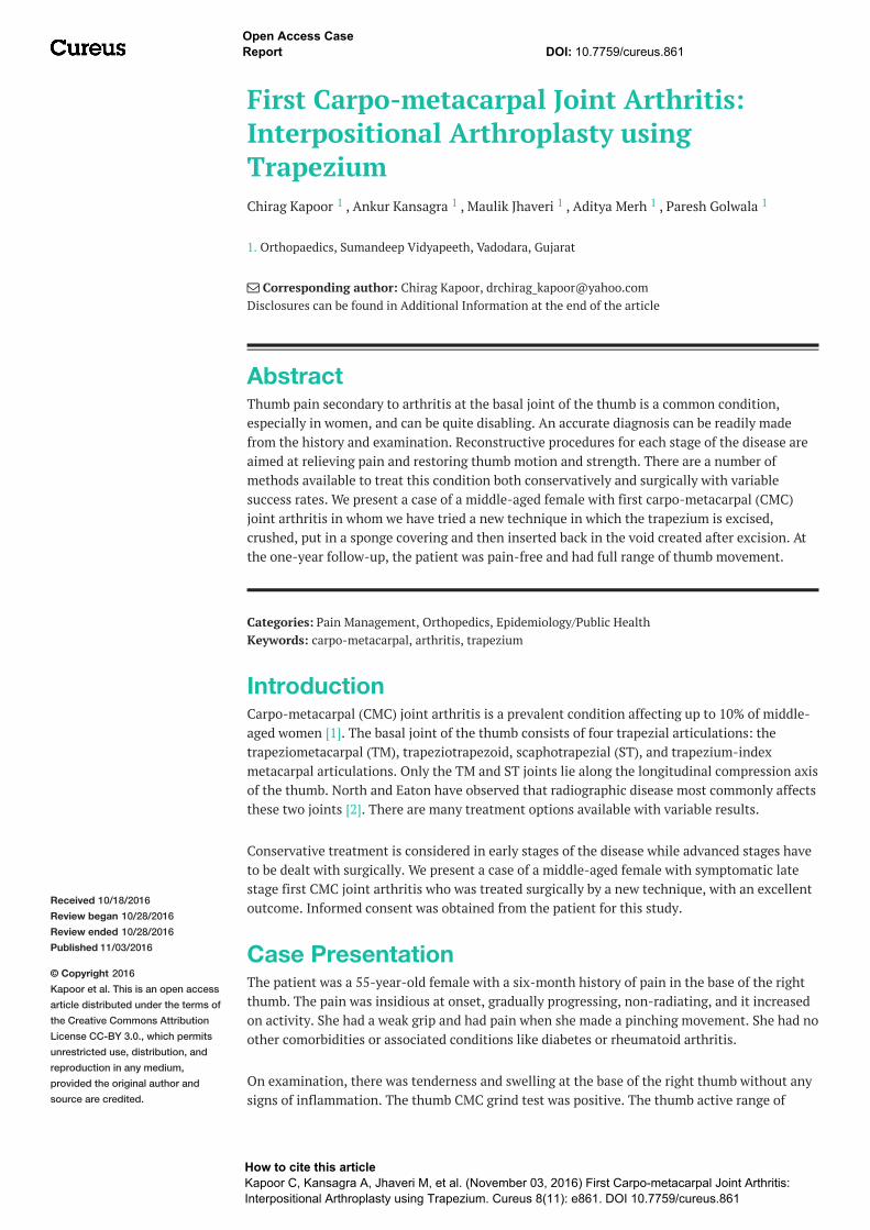
A 2 mm K-wire was passed starting from the base of the first metacarpal through the spongepiece and transfixed into the scaphoid (Figures 3-4). This was done as a support to the joint forpostoperative immobilization, along with a below-elbow scaphoid cast for six weeks. The K-wire and cast were then removed, and she was started with thumb range of movement exercises(active and passive) for two months.
FIGURE 3: Intraoperative imagesImage showing K-wire transfixed in metacarpal and scaphoid.
2016 Kapoor et al. Cureus 8(11): e861. DOI 10.7759/cureus.861 3 of 6
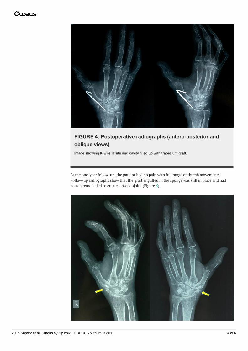
FIGURE 4: Postoperative radiographs (antero-posterior andoblique views)Image showing K-wire in situ and cavity filled up with trapezium graft.
At the one-year follow-up, the patient had no pain with full range of thumb movements.Follow-up radiographs show that the graft engulfed in the sponge was still in place and hadgotten remodelled to create a pseudojoint (Figure 5).
2016 Kapoor et al. Cureus 8(11): e861. DOI 10.7759/cureus.861 4 of 6

FIGURE 5: Follow-up radiograph at one yearImage showing the trapezium that had gotten remodelled to create a pseudojoint.
DiscussionCarpo-metacarpal (CMC) osteoarthritis, also known as trapezio-metacarpal osteoarthritis orosteoarthritis at the base of the thumb is a reparative joint disease affecting the first carpo-metacarpal joint [3]. This joint is formed by the trapezium bone of the wrist and the firstmetacarpal bone of the thumb. Because of its relative instability, this joint is a frequent site forosteoarthritis [1].
It is believed that laxity of the ligaments especially the palmar beak ligament, surrounding thefirst CMC joint is the main cause of arthritis [4]. This instability causes misalignment of thejoint bones, which will then rub against each other, which causes wearing of the cushioningcartilage of the joint surfaces, resulting in damage of the joint [5].
It is commonly seen in obese females after menopause, as was also seen in our case [6].Tenderness is usually well localised over the joint, and this can be reproduced with thumb andfinger pressure applied directly over the affected joint. Crepitus evident on examination implieserosion of the articular cartilage [7]. All these features were consistent with our patient.
There are numerous conservative treatment options which include application of splints andslings, analgesics and injection of steroids, but these can be used in early stages of the diseaseonly and in less symptomatic patients [2]. For advanced stage arthritis, various surgicalprocedures like excision of the trapezium (trapeziectomy) with ligament reconstruction, with orwithout tendon interposition arthroplasty have been described [8-9].
Recently, some surgeons have started doing trapezial excision alone referred to as 'haematomaarthroplasty [10] and have had favourable short term results, although loss of trapezial heightwith subsequent scaphoid impingement is a feared long-term consequence which may affectlong-term outcome. In an attempt to prevent this collapse, several alternatives to the simpletrapeziectomy have been popularized. These include interposing autogenous or alloplastictissue between the carpals and the base of the metacarpal, reconstructing the supportingligaments with various tendons and CMC arthrodesis. Arthrodesis has the disadvantage oflimitation of joint movement, and ligament reconstruction requires technical expertise withsurgical morbidities.
Our method of reconstruction includes removal of the trapezium and crushing it and thenengulfing it in a sponge covering. Our hypothesis is that this will allow for a new pseudojoint tobe formed between the trapezium and the base of the first metacarpal without causing it to fusebecause of the sponge covering, thus retaining movement at the joint. Also, this will avoid theneed of ligament reconstruction and interposition, thus reducing morbidity. Moreover, fillingthe gap with this graft is preferable in terms of function, stability, and position of the thumb asit avoids the complications such as shortening or subluxation of the thumb.
ConclusionsThere are many surgical methods to treat this condition, but all are associated with a fewdemerits. We feel that our method of treatment has no associated comorbidities and requiresless technical expertise, but at the same time offers excellent results. So we feel this innovative
2016 Kapoor et al. Cureus 8(11): e861. DOI 10.7759/cureus.861 5 of 6

technique is a worthwhile approach for treating CMC joint arthritis.
Additional InformationDisclosuresHuman subjects: Consent was obtained by all participants in this study. Conflicts of interest:In compliance with the ICMJE uniform disclosure form, all authors declare the following:Payment/services info: All authors have declared that no financial support was received fromany organization for the submitted work. Financial relationships: All authors have declaredthat they have no financial relationships at present or within the previous three years with anyorganizations that might have an interest in the submitted work. Other relationships: Allauthors have declared that there are no other relationships or activities that could appear tohave influenced the submitted work.
References1. Armstrong A, Hunter JB, Davis TR: The prevalence of degenerative arthritis of the base of the
thumb in post-menopausal women. J Hand Surg Br. 1994, 19:340–341.2. North ER, Eaton RG: Degenerative joint disease of the trapezium: a comparative radiographic
and anatomic study. J Hand Surg Am. 1983, 8:160–166.3. Pellegrini VD Jr: Osteoarthritis of the trapeziometacarpal joint: the pathophysiology of
articular cartilage degeneration. II. Articular wear patterns in the osteoarthritic joint. J HandSurg Am. 1991, 16:975–982.
4. Doerschuk SH, Hicks DG, Chinchilli VM, Pellegrini VD: Histopathology of the palmar beakligament in trapeziometacarpal osteoarthritis. J Hand Surg Am. 1999, 24:496–504.10.1053/jhsu.1999.0496
5. Kihara H: Anatomical study of the normal and degenerative articular surfaces on the firstcarpometacarpal joint. [Article in Japanese]. Nihon Seikeigeka Gakkai Zasshi. 1992, 66:228–239.
6. Dahaghin S, Bierma-Zeinstra SM, Koes BW, et al.: Do metabolic factors add to the effect ofoverweight on hand osteoarthritis? The Rotterdam Study. Ann Rheum Dis. 2007, 66:916–920.10.1136/ard.2005.045724
7. Pomerance JF: Painful basal joint arthritis of the thumb. Part I: anatomy, pathophysiology,and diagnosis. Am J Orthop (Belle Mead NJ). 1995, 24:401–408.
8. Ghavami A, Oishi SN: Thumb trapeziometacarpal arthritis: treatment with ligamentreconstruction tendon interposition arthroplasty. Plast Reconstr Surg. 2006, 117:116e-28e.10.1097/01.prs.0000214652.31293.23
9. Van Heest AE, Kallemeier P: Thumb carpal metacarpal arthritis . J Am Acad Orthop Surg.2008, 16:140–151.
10. Jones NF, Maser BM: Treatment of arthritis of the trapeziometacarpal joint withtrapeziectomy and hematoma arthroplasty. Hand Clin. 2001, 17:237–43.
2016 Kapoor et al. Cureus 8(11): e861. DOI 10.7759/cureus.861 6 of 6