integrative analysis of hif binding and transactivation ... · integrative analysis of hif binding...
TRANSCRIPT

Integrative analysis of HIF binding andtransactivation reveals its role in maintaining histonemethylation homeostasisXiaobo Xiaa, Madeleine E. Lemieuxa, Wei Lib,c,1, Jason S. Carrolld,2, Myles Brownd, X. Shirley Liub,c,and Andrew L. Kunga,e,3
Departments of aPediatric Oncology, bBiostatistics and Computational Biology, and dMedical Oncology, Dana–Farber Cancer Institute, 44 Binney Street, andHarvard Medical School, Boston, MA 02115; cHarvard School of Public Health, 677 Huntington Avenue, Boston, MA 02115; and eDivision of PediatricHematology/Oncology, Children’s Hospital Boston, 300 Longwood Avenue, Boston, MA 02115
Edited by Steven L. McKnight, University of Texas Southwestern Medical Center, Dallas, TX, and approved January 23, 2009 (received for reviewOctober 9, 2008)
Adaptation to hypoxia is mediated through a coordinated transcrip-tional response driven largely by hypoxia-inducible factor 1 (HIF-1).We used ChIP-chip and gene expression profiling to identify directtargets of HIF-1 transactivation on a genome-wide scale. Severalhundred direct HIF-1 targets were identified and, as expected, werehighly enriched for proteins that facilitate metabolic adaptation tohypoxia. Surprisingly, there was also striking enrichment for thefamily of 2-oxoglutarate dioxygenases, including the jumonji-domainhistone demethylases. We demonstrate that these histone demethy-lases are direct HIF targets, and their up-regulation helps maintainepigenetic homeostasis under hypoxic conditions. These results sug-gest that the coordinated increase in expression of several oxygen-dependent enzymes by HIF may help compensate for decreased levelsof oxygen under conditions of cellular hypoxia.
ChIP-chip � hypoxia � jumonji protein � dioxygenase � epigenetics
Adequate oxygenation is essential for normal physiology andfunctioning of all cells. In response to decreased oxygen
tension, a coordinated transcriptional response is activated tomaintain cellular homeostasis. This transcriptional program ismediated, at least in part, by activation of the heterodimerictranscription factors hypoxia-inducible factor 1 and 2 (HIF-1 andHIF-2). The HIFs are essential for several physiological processes,including normal development, erythropoietin (EPO) production,and wound healing. Unfortunately, HIF signaling also contributesto the pathophysiology of tumors by facilitating metabolic adapta-tion and by promoting angiogenesis, invasion, and metastasis (1).
The activity of HIF-1 and HIF-2 are controlled at multiple levels.The primary point of regulation is at the level of abundance of the�-subunits. Under normal oxygen (normoxic) conditions, HIF-1�and HIF-2� are hydroxylated by the HIF prolyl hydroxylasesEGLN1–3, which target the proteins for binding to the von Hippel–Lindau (vHL) ubiquitin E3 ligase complex and rapid proteosomaldegradation (2–6). As oxygen levels drop, prolyl-hydroxylationdecreases, resulting in accumulation of HIF �-subunits and het-erodimerization with ARNT (HIF-1�). As a consequence, HIFheterodimer levels are nominal under physiologic oxygen levels andincrease exponentially with decreasing oxygen tension (7). HIFheterodimers then recruit transcriptional coactivator complexes (8,9), and transactivate target genes containing the cognate hypoxia-response element (HRE) (1).
The direct transcriptional targets of HIF-1 play important roles infacilitating both short-term and long-term adaptation to hypoxia (1).Metabolic homeostasis is achieved by shifting from oxidative phos-phorylation to anaerobic glycolysis through increased expression of theglycolytic enzymes and glucose transporters, inhibition of the TCAcycle, and induction of pH-regulating systems. A second HIF-1-mediatedprogramincreasesoxygendeliveryby inducingvasodilatation,increased vascular permeability, enhanced erythropoiesis and angio-genesis. Specific sites of HIF-1 binding have been validated within the
promoter or enhancers of �50 genes (10, 11). Alignment of thesequences encompassing these well-characterized functional HREs(transcriptionally activated by hypoxia) has revealed a consensus HIF-1-binding motif (the core HRE) of 5�-RCGTG-3� (R � A or G).
Because the core HRE is too promiscuous to accurately predictbinding a priori, we used ChIP-chip to define HIF-1 chromatinbinding on a genome-wide level. We integrated these results withgene expression profiling to interrogate mechanisms regulatinghypoxia-induced gene expression and to more comprehensivelyidentify direct targets of HIF-1 transactivation. We found that thefamily of 2-oxoglutarate-dependent dioxygenases are coordinatelytargeted by HIF, and up-regulated expression helps maintain globallevels of histone methylation under hypoxic conditions.
ResultsIdentification of HIF-1-Binding Sites by ChIP-chip. To identify HIF-1-binding sites across the genome, HepG2 cells were grown underboth normoxic (ambient) and hypoxic (0.5% O2) conditions, andChIP was performed by using a HIF-1� polyclonal antibody withoutappreciable cross-reactivity to HIF-2� (Fig. S1A). ChIP and inputDNA were hybridized to Affymetrix GeneChip Human Tiling 2.0RArray Sets, consisting of probes covering the entire nonrepetitivehuman genome at 35-bp resolution. The model-based analysis fortiling arrays (MAT) algorithm (12) was used to identify probe signalpeaks comparing triplicate biological replicates of HIF-1 ChIPDNA to their matched inputs by using an initial P value cutoff of1E�5. Consistent with the fact that HIF-1� protein levels increasedramatically in hypoxic compared with normoxic samples (Fig.S1B), 91% of the putative HIF-1-binding sites were characterizedby a positive peak call only under hypoxic conditions (e.g., ENO1and INSIG2; Fig. 1A). However, because HIF-1� is not entirelyabsent in normoxia, peaks in which probe intensities were signifi-cantly increased under hypoxic conditions by comparison withnormoxia were also retained (e.g., JMJD1A and ZNF292; Fig. 1A).A minority of peaks (7%) had peak intensities that did not increasein hypoxic compared with normoxic samples (e.g., ADI1 andPHF12; Fig. 1A), and these were suspected to be nonspecific.
Author contributions: X.X., M.E.L., J.S.C., M.B., and A.L.K. designed research; X.X. per-formed research; X.X., M.E.L., W.L., J.S.C., X.S.L., and A.L.K. contributed new reagents/analytic tools; X.X., M.E.L., W.L., M.B., X.S.L., and A.L.K. analyzed data; and X.X., M.E.L., andA.L.K. wrote the paper.
The authors declare no conflict of interest.
This article is a PNAS Direct Submission.
1Present address: Division of Biostatistics, Dan L. Duncan Cancer Center, Department of Mo-lecular and Cellular Biology, Baylor College of Medicine, One Baylor Plaza, Houston, TX 77030.
2Present address: Cancer Research U.K., Cambridge Research Institute, Li Ka Shing Centre,Robinson Way, Cambridge CB2 0RE, United Kingdom.
3To whom correspondence should be addressed. E-mail: andrew�[email protected].
This article contains supporting information online at www.pnas.org/cgi/content/full/0810067106/DCSupplemental.
4260–4265 � PNAS � March 17, 2009 � vol. 106 � no. 11 www.pnas.org�cgi�doi�10.1073�pnas.0810067106
Dow
nloa
ded
by g
uest
on
Janu
ary
3, 2
020
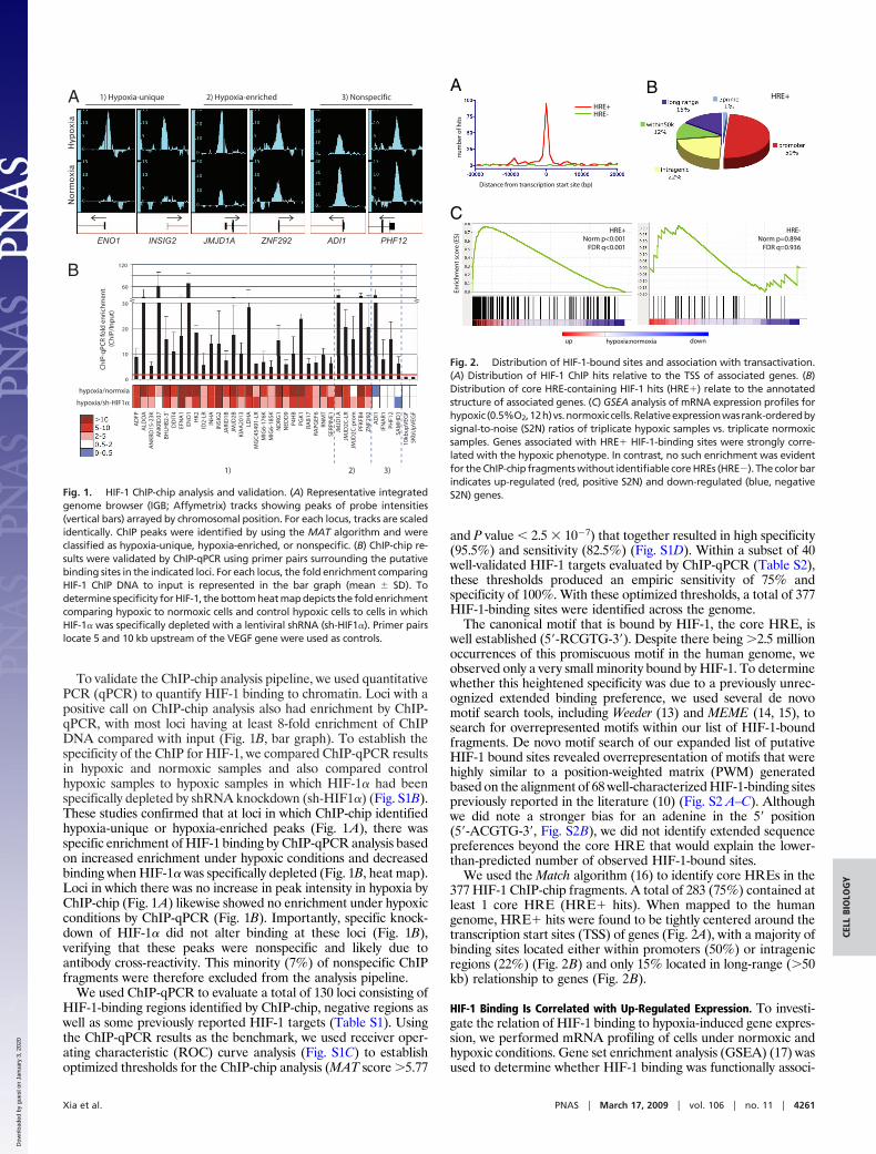
To validate the ChIP-chip analysis pipeline, we used quantitativePCR (qPCR) to quantify HIF-1 binding to chromatin. Loci with apositive call on ChIP-chip analysis also had enrichment by ChIP-qPCR, with most loci having at least 8-fold enrichment of ChIPDNA compared with input (Fig. 1B, bar graph). To establish thespecificity of the ChIP for HIF-1, we compared ChIP-qPCR resultsin hypoxic and normoxic samples and also compared controlhypoxic samples to hypoxic samples in which HIF-1� had beenspecifically depleted by shRNA knockdown (sh-HIF1�) (Fig. S1B).These studies confirmed that at loci in which ChIP-chip identifiedhypoxia-unique or hypoxia-enriched peaks (Fig. 1A), there wasspecific enrichment of HIF-1 binding by ChIP-qPCR analysis basedon increased enrichment under hypoxic conditions and decreasedbinding when HIF-1� was specifically depleted (Fig. 1B, heat map).Loci in which there was no increase in peak intensity in hypoxia byChIP-chip (Fig. 1A) likewise showed no enrichment under hypoxicconditions by ChIP-qPCR (Fig. 1B). Importantly, specific knock-down of HIF-1� did not alter binding at these loci (Fig. 1B),verifying that these peaks were nonspecific and likely due toantibody cross-reactivity. This minority (7%) of nonspecific ChIPfragments were therefore excluded from the analysis pipeline.
We used ChIP-qPCR to evaluate a total of 130 loci consisting ofHIF-1-binding regions identified by ChIP-chip, negative regions aswell as some previously reported HIF-1 targets (Table S1). Usingthe ChIP-qPCR results as the benchmark, we used receiver oper-ating characteristic (ROC) curve analysis (Fig. S1C) to establishoptimized thresholds for the ChIP-chip analysis (MAT score �5.77
and P value � 2.5 � 10�7) that together resulted in high specificity(95.5%) and sensitivity (82.5%) (Fig. S1D). Within a subset of 40well-validated HIF-1 targets evaluated by ChIP-qPCR (Table S2),these thresholds produced an empiric sensitivity of 75% andspecificity of 100%. With these optimized thresholds, a total of 377HIF-1-binding sites were identified across the genome.
The canonical motif that is bound by HIF-1, the core HRE, iswell established (5�-RCGTG-3�). Despite there being �2.5 millionoccurrences of this promiscuous motif in the human genome, weobserved only a very small minority bound by HIF-1. To determinewhether this heightened specificity was due to a previously unrec-ognized extended binding preference, we used several de novomotif search tools, including Weeder (13) and MEME (14, 15), tosearch for overrepresented motifs within our list of HIF-1-boundfragments. De novo motif search of our expanded list of putativeHIF-1 bound sites revealed overrepresentation of motifs that werehighly similar to a position-weighted matrix (PWM) generatedbased on the alignment of 68 well-characterized HIF-1-binding sitespreviously reported in the literature (10) (Fig. S2 A–C). Althoughwe did note a stronger bias for an adenine in the 5� position(5�-ACGTG-3�, Fig. S2B), we did not identify extended sequencepreferences beyond the core HRE that would explain the lower-than-predicted number of observed HIF-1-bound sites.
We used the Match algorithm (16) to identify core HREs in the377 HIF-1 ChIP-chip fragments. A total of 283 (75%) contained atleast 1 core HRE (HRE� hits). When mapped to the humangenome, HRE� hits were found to be tightly centered around thetranscription start sites (TSS) of genes (Fig. 2A), with a majority ofbinding sites located either within promoters (50%) or intragenicregions (22%) (Fig. 2B) and only 15% located in long-range (�50kb) relationship to genes (Fig. 2B).
HIF-1 Binding Is Correlated with Up-Regulated Expression. To investi-gate the relation of HIF-1 binding to hypoxia-induced gene expres-sion, we performed mRNA profiling of cells under normoxic andhypoxic conditions. Gene set enrichment analysis (GSEA) (17) wasused to determine whether HIF-1 binding was functionally associ-
B
A
Hypoxia
Normoxia
ENO1
1) Hypoxia-unique 2) Hypoxia-enriched 3) Nonspecific
← → → → ← ←
A
DFP
ALD
OA
AN
KR
D15
-23K
A
NK
RD
37
B
HLH
B2-
3'
DD
IT4
EF
NA
1
EN
O1
HK
2
I
D2-
LR
IN
HA
IN
SIG
2
J
AR
ID1B
JM
JD2B
K
IAA
2013
L
DH
A M
GC
4549
1-LR
MIG
6-17
6K
M
IG6-
185K
ND
RG
1
N
EDD
9
P4H
B
PG
K1
R
AB
17
RA
PGEF
6
RN
MT
S
ERPI
NE1
JM
JD1A
JM
JD2C
-LR
J
MJD
2C-p
rom
PFK
FB4
ZN
F292
AD
I1
I
FNA
R1
PH
F12
SAM
HD
1
10k
bu
pV
EGF
5K
bU
pV
EGF
1)
Ch
IP-q
PCR
fold
en
rich
men
t (C
hIP
/In
pu
t)
120
60
30
20
10
0
hypoxia/normxia
hypoxia/sh-HIF1α
2) 3)
INSIG2 JMJD1A ZNF292 ADI1 PHF12
Fig. 1. HIF-1 ChIP-chip analysis and validation. (A) Representative integratedgenome browser (IGB; Affymetrix) tracks showing peaks of probe intensities(vertical bars) arrayed by chromosomal position. For each locus, tracks are scaledidentically. ChIP peaks were identified by using the MAT algorithm and wereclassified as hypoxia-unique, hypoxia-enriched, or nonspecific. (B) ChIP-chip re-sults were validated by ChIP-qPCR using primer pairs surrounding the putativebinding sites in the indicated loci. For each locus, the fold enrichment comparingHIF-1 ChIP DNA to input is represented in the bar graph (mean SD). Todetermine specificity for HIF-1, the bottom heat map depicts the fold enrichmentcomparing hypoxic to normoxic cells and control hypoxic cells to cells in whichHIF-1� was specifically depleted with a lentiviral shRNA (sh-HIF1�). Primer pairslocate 5 and 10 kb upstream of the VEGF gene were used as controls.
A B
C
HRE+
Enri
chm
ent
sco
re (E
S)
HRE+Norm p<0.001 FDR q<0.001
hypoxia:normoxiaup down
HRE-Norm p=0.894 FDR q=0.936
HRE+HRE-
Distance from transcription start site (bp)
nu
mb
er o
f hit
s
Fig. 2. Distribution of HIF-1-bound sites and association with transactivation.(A) Distribution of HIF-1 ChIP hits relative to the TSS of associated genes. (B)Distribution of core HRE-containing HIF-1 hits (HRE�) relate to the annotatedstructure of associated genes. (C) GSEA analysis of mRNA expression profiles forhypoxic (0.5%O2,12h)vs.normoxiccells.Relativeexpressionwasrank-orderedbysignal-to-noise (S2N) ratios of triplicate hypoxic samples vs. triplicate normoxicsamples. Genes associated with HRE� HIF-1-binding sites were strongly corre-lated with the hypoxic phenotype. In contrast, no such enrichment was evidentfor the ChIP-chip fragments without identifiable core HREs (HRE�). The color barindicates up-regulated (red, positive S2N) and down-regulated (blue, negativeS2N) genes.
Xia et al. PNAS � March 17, 2009 � vol. 106 � no. 11 � 4261
CELL
BIO
LOG
Y
Dow
nloa
ded
by g
uest
on
Janu
ary
3, 2
020
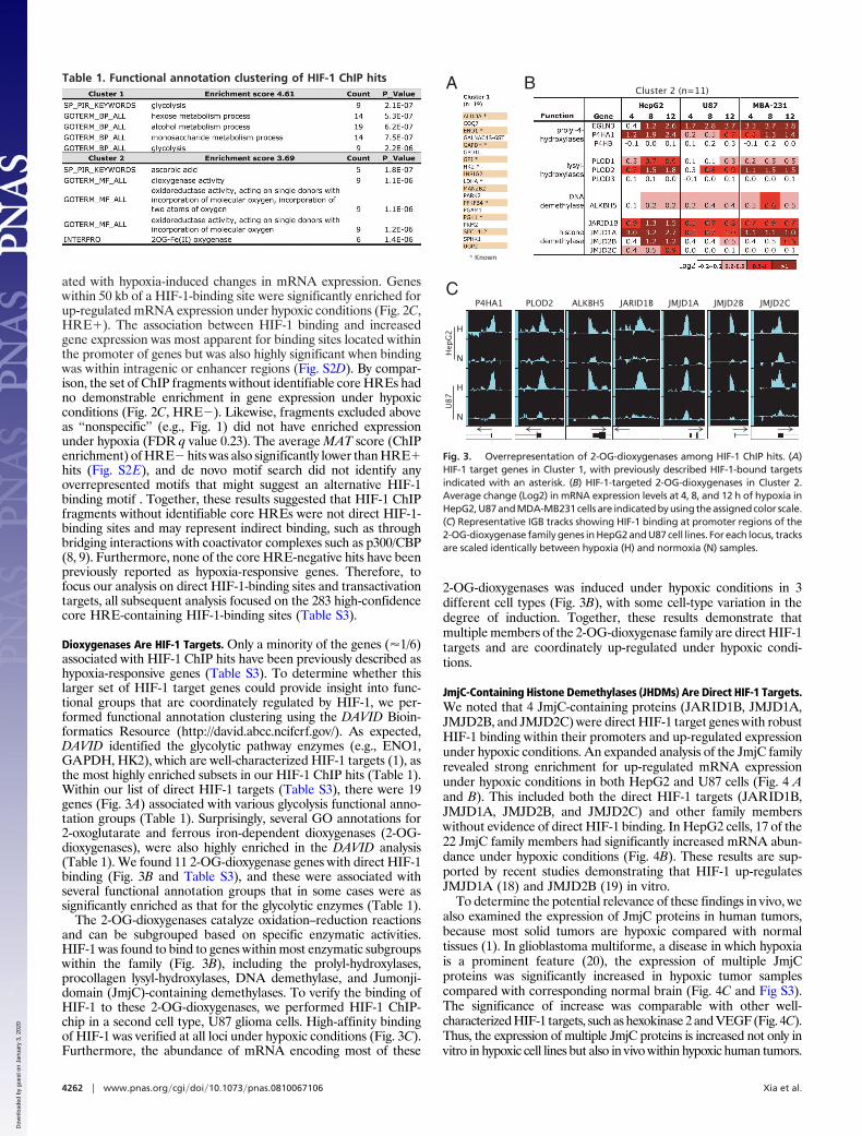
ated with hypoxia-induced changes in mRNA expression. Geneswithin 50 kb of a HIF-1-binding site were significantly enriched forup-regulated mRNA expression under hypoxic conditions (Fig. 2C,HRE�). The association between HIF-1 binding and increasedgene expression was most apparent for binding sites located withinthe promoter of genes but was also highly significant when bindingwas within intragenic or enhancer regions (Fig. S2D). By compar-ison, the set of ChIP fragments without identifiable core HREs hadno demonstrable enrichment in gene expression under hypoxicconditions (Fig. 2C, HRE�). Likewise, fragments excluded aboveas ‘‘nonspecific’’ (e.g., Fig. 1) did not have enriched expressionunder hypoxia (FDR q value 0.23). The average MAT score (ChIPenrichment) of HRE� hits was also significantly lower than HRE�hits (Fig. S2E), and de novo motif search did not identify anyoverrepresented motifs that might suggest an alternative HIF-1binding motif . Together, these results suggested that HIF-1 ChIPfragments without identifiable core HREs were not direct HIF-1-binding sites and may represent indirect binding, such as throughbridging interactions with coactivator complexes such as p300/CBP(8, 9). Furthermore, none of the core HRE-negative hits have beenpreviously reported as hypoxia-responsive genes. Therefore, tofocus our analysis on direct HIF-1-binding sites and transactivationtargets, all subsequent analysis focused on the 283 high-confidencecore HRE-containing HIF-1-binding sites (Table S3).
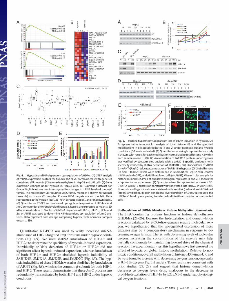
Dioxygenases Are HIF-1 Targets. Only a minority of the genes (�1/6)associated with HIF-1 ChIP hits have been previously described ashypoxia-responsive genes (Table S3). To determine whether thislarger set of HIF-1 target genes could provide insight into func-tional groups that are coordinately regulated by HIF-1, we per-formed functional annotation clustering using the DAVID Bioin-formatics Resource (http://david.abcc.ncifcrf.gov/). As expected,DAVID identified the glycolytic pathway enzymes (e.g., ENO1,GAPDH, HK2), which are well-characterized HIF-1 targets (1), asthe most highly enriched subsets in our HIF-1 ChIP hits (Table 1).Within our list of direct HIF-1 targets (Table S3), there were 19genes (Fig. 3A) associated with various glycolysis functional anno-tation groups (Table 1). Surprisingly, several GO annotations for2-oxoglutarate and ferrous iron-dependent dioxygenases (2-OG-dioxygenases), were also highly enriched in the DAVID analysis(Table 1). We found 11 2-OG-dioxygenase genes with direct HIF-1binding (Fig. 3B and Table S3), and these were associated withseveral functional annotation groups that in some cases were assignificantly enriched as that for the glycolytic enzymes (Table 1).
The 2-OG-dioxygenases catalyze oxidation–reduction reactionsand can be subgrouped based on specific enzymatic activities.HIF-1 was found to bind to genes within most enzymatic subgroupswithin the family (Fig. 3B), including the prolyl-hydroxylases,procollagen lysyl-hydroxylases, DNA demethylase, and Jumonji-domain (JmjC)-containing demethylases. To verify the binding ofHIF-1 to these 2-OG-dioxygenases, we performed HIF-1 ChIP-chip in a second cell type, U87 glioma cells. High-affinity bindingof HIF-1 was verified at all loci under hypoxic conditions (Fig. 3C).Furthermore, the abundance of mRNA encoding most of these
2-OG-dioxygenases was induced under hypoxic conditions in 3different cell types (Fig. 3B), with some cell-type variation in thedegree of induction. Together, these results demonstrate thatmultiple members of the 2-OG-dioxygenase family are direct HIF-1targets and are coordinately up-regulated under hypoxic condi-tions.
JmjC-Containing Histone Demethylases (JHDMs) Are Direct HIF-1 Targets.We noted that 4 JmjC-containing proteins (JARID1B, JMJD1A,JMJD2B, and JMJD2C) were direct HIF-1 target genes with robustHIF-1 binding within their promoters and up-regulated expressionunder hypoxic conditions. An expanded analysis of the JmjC familyrevealed strong enrichment for up-regulated mRNA expressionunder hypoxic conditions in both HepG2 and U87 cells (Fig. 4 Aand B). This included both the direct HIF-1 targets (JARID1B,JMJD1A, JMJD2B, and JMJD2C) and other family memberswithout evidence of direct HIF-1 binding. In HepG2 cells, 17 of the22 JmjC family members had significantly increased mRNA abun-dance under hypoxic conditions (Fig. 4B). These results are sup-ported by recent studies demonstrating that HIF-1 up-regulatesJMJD1A (18) and JMJD2B (19) in vitro.
To determine the potential relevance of these findings in vivo, wealso examined the expression of JmjC proteins in human tumors,because most solid tumors are hypoxic compared with normaltissues (1). In glioblastoma multiforme, a disease in which hypoxiais a prominent feature (20), the expression of multiple JmjCproteins was significantly increased in hypoxic tumor samplescompared with corresponding normal brain (Fig. 4C and Fig S3).The significance of increase was comparable with other well-characterized HIF-1 targets, such as hexokinase 2 and VEGF (Fig. 4C).Thus, the expression of multiple JmjC proteins is increased not only invitro in hypoxic cell lines but also in vivo within hypoxic human tumors.
Table 1. Functional annotation clustering of HIF-1 ChIP hits A
* Known
B
C JARID1B JMJD1A JMJD2B JMJD2CALKBH5P4HA1 PLOD2
Hep
G2
U87
H
N
H
N← →← → → →←
Cluster 2 (n=11)
Fig. 3. Overrepresentation of 2-OG-dioxygenases among HIF-1 ChIP hits. (A)HIF-1 target genes in Cluster 1, with previously described HIF-1-bound targetsindicated with an asterisk. (B) HIF-1-targeted 2-OG-dioxygenases in Cluster 2.Average change (Log2) in mRNA expression levels at 4, 8, and 12 h of hypoxia inHepG2,U87andMDA-MB231cellsare indicatedbyusingtheassignedcolor scale.(C) Representative IGB tracks showing HIF-1 binding at promoter regions of the2-OG-dioxygenase family genes in HepG2 and U87 cell lines. For each locus, tracksare scaled identically between hypoxia (H) and normoxia (N) samples.
4262 � www.pnas.org�cgi�doi�10.1073�pnas.0810067106 Xia et al.
Dow
nloa
ded
by g
uest
on
Janu
ary
3, 2
020
Quantitative RT-PCR was used to verify increased mRNAabundance of HIF-1-targeted JmjC proteins under hypoxic condi-tions (Fig. 4D). We used shRNA knockdown of HIF-1� andHIF-2� to determine the specificity of hypoxia-induced expression.Individually, shRNA depletion of HIF-1� or HIF-2� did notsignificant affect hypoxia-induced expression, whereas knockdownof both HIF-1� and HIF-2� abolished hypoxia inducibility ofJARID1B, JMJD1A, JMJD2B, and JMJD2C (Fig. 4E). The hyp-oxia inducibility of these JHDMs was also abolished by knockdownof ARNT (Fig. 4E), which is the common �-subunit for both HIF-1and HIF-2. These results demonstrate that these JmjC proteins areredundantly transactivated by both HIF-1 and HIF-2 under hypoxicconditions.
Up-Regulation of JHDMs Maintains Histone Methylation Homeostasis.The JmjC-containing proteins function as histone demethylases(JHDMs) (21–26). Because the hydroxylation and demethylationreactions catalyzed by 2-OG-dioxygenases require molecular oxy-gen, we hypothesized that the up-regulated expression of theseenzymes may be a compensatory mechanism in response to de-creasing oxygen tension. That is, with decreasing levels of molecularoxygen, increasing the concentration of the enzyme may helppartially compensate by maintaining forward drive of the chemicalreaction. To experimentally test this hypothesis, we first assessed theeffect of hypoxia on global histone methylation. Relative to nor-moxic conditions, overall methylation of histone H3 lysines 4, 9, and36 were found to increase with decreasing oxygen tension, especiallyat 0.5–1% oxygen (Fig. 5 A and B). These results are consistent withprior studies (27, 28) and suggest that histone demethylationdecreases as oxygen levels drop, analogous to the decrease inprolyl-hydroxylation of HIF-1� by EGLN1–3 under subphysiologi-cal oxygen tensions.
A HepG2
Norm p<0.001 FDR q=0.005
U87Norm p=0.035 FDR q=0.218
hypoxia:normoxiaup down
C
BEn
richm
ent S
core
(ES)
KIAA1718/JHDM1DJMJD1A/JHDM2AJMJD2BJMJD3JARID1B/PLU-1PTDSR/JMJD6JMJD1C/JHDM2CJMJD2CJARID2FBXL10/JHDM1BUTXJARID1C/SMCXJMJD1B/JHDM2BJARID1A/RBP2PHF8JARID1D/SMCYFBXL11/JHDM1APHF2UTYJMJD2DJMJD2AHR
1 2 3 1 2 3
Hyp
oxia
Nor
mox
ia
D E
Enric
hmen
t Sco
re (E
S)
N T N T N T N T N T N T N T N T N T N T N T N T N T N T N T
Log2
Inte
nsity
HK2
VEG
FA LOX CP
HIG
2
PLO
D1
RIT1
GBE
1
JMJD
1A
JMJD
2A
JMJD
2B
JMJD
6
JARI
D1A
JMJD
1C
JMJD
4
GSE4290 N=23 T=77
Fig. 4. Hypoxia- and HIF-dependent up-regulation of JHDMs. (A) GSEA analysisof mRNA expression profiles for hypoxic (12 h) vs. normoxic cells with gene setcontainingallknownJmjChistonedemethylases inHepG2andU87cells. (B)Geneexpression changes under hypoxia in HepG2 cells. (C) Expression dataset forGrade IV glioblastoma was interrogated for changes in mRNA levels of the JmjCfamily. The most highly up-regulated JmjC family member is shown for normaltissue (N) vs. tumor (T) samples. Known HIF-1 targets are on the left. Datarepresented as the median (bar), 25–75th percentiles (box), and range (whiskers).(D) Quantitative RT-PCR verification of up-regulated expression of HIF-1-boundJmjC genes under different levels of hypoxia. Results are expressed as mean SDafter normalization to �-actin. (E) shRNA depletion of HIF-1�, HIF-2�, HIF1� and2�, or ARNT was used to determine HIF-dependent up-regulation of JmjC pro-teins. Data represent fold change comparing hypoxic with normoxic samples(mean SD).
A B
1 2 1 2 1 2 1 2 1 2N 5% 2% 1% 0.5% N 5% 2% 1% 0.5%
TNRA-hs2GpeH
H3
H3K4me3
1 2 1 2 1 2 1 2 1 2
D
E
C
fold
cha
nge
(hyp
oxia
/nor
mox
ia)
H3k4me3
fold
cha
nge
by w
este
rn-b
lot
(
hypo
xia/
norm
oxia
)
JARID1B
β-actin
HIF-1a
N 5% 2% 1% 0.5% N 5% 2% 1% 0.5% O2 HepG2sh
-GFP
N 1% N 1%sh
-JARID
1B
sh-ARNT
1 2 1 2 1 2 1 2 1 2N 5% 2% 1% 0.5%
HepG2
H3
H3K9me2
H3K36me3
F JARID1B H3K4me3 DAPI
Hyp
oxia
Nor
mox
ia
Fig. 5. Histone hypermethylations from loss of JHDM induction in hypoxia. (A)A representative immunoblot analysis of total histone H3 and the specifiedmodifications in biological replicates (1 and 2) under normoxic (N) and hypoxicconditions (O2 levels indicated). (B) Quantitation of a single representative studyis shown,withresults foreachmodificationnormalizedtototalhistoneH3withineach sample (mean SD). (C) Accumulation of JARID1B protein under hypoxiawas verified by Western blot analysis with a JARID1B-specific antibody, withspecificity verified by shRNA depletion of JARID1B (Left). Knockdown of ARNT(sh-ARNT) (Right) reducesaccumulationofJARID1Binhypoxia. (D)GlobalhistoneH3 and H3K4me3 levels were determined in unmodified HepG2 cells, controlshRNAcells (sh-GFP),andARNTdepletedcells (sh-ARNT).Westernblotanalysis forhistone H3 and H3K4me3 of duplicate biological replicates (1 and 2) is shown fora representative experiment. (E) Quantitated results represented as mean SD.(F) A HA-JARID1B expression construct was transfected into HepG2 sh-ARNT cells.Normoxic and hypoxic cells were stained with anti-HA (red) and anti-H3K4me3(green) antibodies. In both conditions, overexpression of JARID1B reduced theH3K4me3 level by comparing transfected cells (with arrows) to nontransfectedcells.
Xia et al. PNAS � March 17, 2009 � vol. 106 � no. 11 � 4263
CELL
BIO
LOG
Y
Dow
nloa
ded
by g
uest
on
Janu
ary
3, 2
020
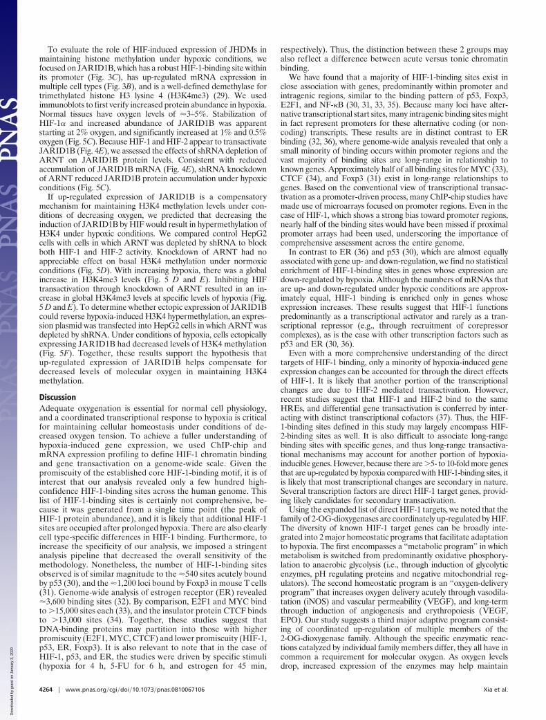
To evaluate the role of HIF-induced expression of JHDMs inmaintaining histone methylation under hypoxic conditions, wefocused on JARID1B, which has a robust HIF-1-binding site withinits promoter (Fig. 3C), has up-regulated mRNA expression inmultiple cell types (Fig. 3B), and is a well-defined demethylase fortrimethylated histone H3 lysine 4 (H3K4me3) (29). We usedimmunoblots to first verify increased protein abundance in hypoxia.Normal tissues have oxygen levels of �3–5%. Stabilization ofHIF-1� and increased abundance of JARID1B was apparentstarting at 2% oxygen, and significantly increased at 1% and 0.5%oxygen (Fig. 5C). Because HIF-1 and HIF-2 appear to transactivateJARID1B (Fig. 4E), we assessed the effects of shRNA depletion ofARNT on JARID1B protein levels. Consistent with reducedaccumulation of JARID1B mRNA (Fig. 4E), shRNA knockdownof ARNT reduced JARID1B protein accumulation under hypoxicconditions (Fig. 5C).
If up-regulated expression of JARID1B is a compensatorymechanism for maintaining H3K4 methylation levels under con-ditions of decreasing oxygen, we predicted that decreasing theinduction of JARID1B by HIF would result in hypermethylation ofH3K4 under hypoxic conditions. We compared control HepG2cells with cells in which ARNT was depleted by shRNA to blockboth HIF-1 and HIF-2 activity. Knockdown of ARNT had noappreciable effect on basal H3K4 methylation under normoxicconditions (Fig. 5D). With increasing hypoxia, there was a globalincrease in H3K4me3 levels (Fig. 5 D and E). Inhibiting HIFtransactivation through knockdown of ARNT resulted in an in-crease in global H3K4me3 levels at specific levels of hypoxia (Fig.5 D and E). To determine whether ectopic expression of JARID1Bcould reverse hypoxia-induced H3K4 hypermethylation, an expres-sion plasmid was transfected into HepG2 cells in which ARNT wasdepleted by shRNA. Under conditions of hypoxia, cells ectopicallyexpressing JARID1B had decreased levels of H3K4 methylation(Fig. 5F). Together, these results support the hypothesis thatup-regulated expression of JARID1B helps compensate fordecreased levels of molecular oxygen in maintaining H3K4methylation.
DiscussionAdequate oxygenation is essential for normal cell physiology,and a coordinated transcriptional response to hypoxia is criticalfor maintaining cellular homeostasis under conditions of de-creased oxygen tension. To achieve a fuller understanding ofhypoxia-induced gene expression, we used ChIP-chip andmRNA expression profiling to define HIF-1 chromatin bindingand gene transactivation on a genome-wide scale. Given thepromiscuity of the established core HIF-1-binding motif, it is ofinterest that our analysis revealed only a few hundred high-confidence HIF-1-binding sites across the human genome. Thislist of HIF-1-binding sites is certainly not comprehensive, be-cause it was generated from a single time point (the peak ofHIF-1 protein abundance), and it is likely that additional HIF-1sites are occupied after prolonged hypoxia. There are also clearlycell type-specific differences in HIF-1 binding. Furthermore, toincrease the specificity of our analysis, we imposed a stringentanalysis pipeline that decreased the overall sensitivity of themethodology. Nonetheless, the number of HIF-1-binding sitesobserved is of similar magnitude to the �540 sites acutely boundby p53 (30), and the �1,200 loci bound by Foxp3 in mouse T cells(31). Genome-wide analysis of estrogen receptor (ER) revealed�3,600 binding sites (32). By comparison, E2F1 and MYC bindto �15,000 sites each (33), and the insulator protein CTCF bindsto �13,000 sites (34). Together, these studies suggest thatDNA-binding proteins may partition into those with higherpromiscuity (E2F1, MYC, CTCF) and lower promiscuity (HIF-1,p53, ER, Foxp3). It is also relevant to note that in the case ofHIF-1, p53, and ER, the studies were driven by specific stimuli(hypoxia for 4 h, 5-FU for 6 h, and estrogen for 45 min,
respectively). Thus, the distinction between these 2 groups mayalso reflect a difference between acute versus tonic chromatinbinding.
We have found that a majority of HIF-1-binding sites exist inclose association with genes, predominantly within promoter andintragenic regions, similar to the binding pattern of p53, Foxp3,E2F1, and NF-�B (30, 31, 33, 35). Because many loci have alter-native transcriptional start sites, many intragenic binding sites mightin fact represent promoters for these alternative coding (or non-coding) transcripts. These results are in distinct contrast to ERbinding (32, 36), where genome-wide analysis revealed that only asmall minority of binding occurs within promoter regions and thevast majority of binding sites are long-range in relationship toknown genes. Approximately half of all binding sites for MYC (33),CTCF (34), and Foxp3 (31) exist in long-range relationships togenes. Based on the conventional view of transcriptional transac-tivation as a promoter-driven process, many ChIP-chip studies havemade use of microarrays focused on promoter regions. Even in thecase of HIF-1, which shows a strong bias toward promoter regions,nearly half of the binding sites would have been missed if proximalpromoter arrays had been used, underscoring the importance ofcomprehensive assessment across the entire genome.
In contrast to ER (36) and p53 (30), which are almost equallyassociated with gene up- and down-regulation, we find no statisticalenrichment of HIF-1-binding sites in genes whose expression aredown-regulated by hypoxia. Although the numbers of mRNAs thatare up- and down-regulated under hypoxic conditions are approx-imately equal, HIF-1 binding is enriched only in genes whoseexpression increases. These results suggest that HIF-1 functionspredominantly as a transcriptional activator and rarely as a tran-scriptional repressor (e.g., through recruitment of corepressorcomplexes), as is the case with other transcription factors such asp53 and ER (30, 36).
Even with a more comprehensive understanding of the directtargets of HIF-1 binding, only a minority of hypoxia-induced geneexpression changes can be accounted for through the direct effectsof HIF-1. It is likely that another portion of the transcriptionalchanges are due to HIF-2 mediated transactivation. However,recent studies suggest that HIF-1 and HIF-2 bind to the sameHREs, and differential gene transactivation is conferred by inter-acting with distinct transcriptional cofactors (37). Thus, the HIF-1-binding sites defined in this study may largely encompass HIF-2-binding sites as well. It is also difficult to associate long-rangebinding sites with specific genes, and thus long-range transactiva-tional mechanisms may account for another portion of hypoxia-inducible genes. However, because there are �5- to 10-fold more genesthat are up-regulated by hypoxia compared with HIF-1-binding sites, itis likely that most transcriptional changes are secondary in nature.Several transcription factors are direct HIF-1 target genes, provid-ing likely candidates for secondary transactivation.
Using the expanded list of direct HIF-1 targets, we noted that thefamily of 2-OG-dioxygenases are coordinately up-regulated by HIF.The diversity of known HIF-1 target genes can be broadly inte-grated into 2 major homeostatic programs that facilitate adaptationto hypoxia. The first encompasses a ‘‘metabolic program’’ in whichmetabolism is switched from predominantly oxidative phosphory-lation to anaerobic glycolysis (i.e., through induction of glycolyticenzymes, pH regulating proteins and negative mitochondrial reg-ulators). The second homeostatic program is an ‘‘oxygen-deliveryprogram’’ that increases oxygen delivery acutely through vasodila-tation (iNOS) and vascular permeability (VEGF), and long-termthrough induction of angiogenesis and erythropoiesis (VEGF,EPO). Our study suggests a third major adaptive program consist-ing of coordinated up-regulation of multiple members of the2-OG-dioxygenase family. Although the specific enzymatic reac-tions catalyzed by individual family members differ, they all have incommon a requirement for molecular oxygen. As oxygen levelsdrop, increased expression of the enzymes may help maintain
4264 � www.pnas.org�cgi�doi�10.1073�pnas.0810067106 Xia et al.
Dow
nloa
ded
by g
uest
on
Janu
ary
3, 2
020

forward drive of critical hydroxylation and demethylation reactions.For example, the striking enrichment for JHDMs suggests thatcoordinated up-regulation of these enzymes counterbalances de-creased levels of oxygen to maintain global histone methylationpatterns. Together, these results suggest that in the face of hypoxia,cellular homeostasis is maintained in part through a third majoradaptive program—a ‘‘dioxygenase homeostasis program’’—thathelps maintain the forward drive of certain critical oxygen-dependent dioxygenases.
MethodsDetailed experimental methods are contained in SI Materials and Methods.
ChIP-chip. HepG2 and U87 cells (ATCC) were cultured under normoxic (ambient)or hypoxic (0.5%O2, 4 h) conditions. HIF-1� ChIP-chip was performed with aHIF-1� pAb (Table S4) as previously described (32). HepG2 HIF-1� ChIPed DNAreplicates and inputs were amplified and hybridized onto the Affymetrix Gene-Chip Human Tiling 2.0R Array Set. U87 HIF-1 ChIP samples were hybridized ontoAffymetrix GeneChip Human Promoter 1.0R Array. The MAT algorithm (12) wasused to identify peaks of probe intensity (‘‘hits’’). ChIP hits were associated withRefSeq genes from the University of California Santa Cruz (UCSC) RefGene tablefor HG18 based on chromosomal position.
qPCR Validation of ChIP Hits. Quantitative PCR primers were designed againstregions of interest and also 2 negative control regions (5 and 10 kb upstream ofthe VEGF gene). All primer sequences are specified in Table S1. HIF-1-specificbinding was defined as �2-fold enrichment when HIF-1 ChIP and matched inputsamples were compared, and a �2-fold greater binding in hypoxic samplesrelative to either normoxic cells or hypoxic HIF-1� knockdown (sh-HIF1�) cells.
Identification of Putative Core HREs and Motif Search. A PWM was generated from68 reported human functional HRE sequences (10). We used Match (16) to locate
sites in our ChIP hits matching this PWM, using a core score cutoff of 0.9 andmatrix score of 0.85.
mRNA Expression Profiling. HepG2, U87, and MDA-MB231 cells were collectedunder normoxic conditions (0 h) and after 4, 8, and 12 h of hypoxia (0.5% O2).Triplicates were hybridize to Affymetrix HG-U133Plus2 arrays. For analysis ofhuman tumor material, a dataset for Grade IV glioblastoma multiforme (38) wasdownloaded from the National Center for Biotechnology Information.
GSEA and Functional Annotation Clustering. We created gene sets containing allgenes that could be associated with a ChIP hit (HRE� hits or HRE� hits, within 50kb). These sets along with a gene set containing all known JHDMs were added toa file of gene sets (c5.mf.v2.5.symbols.gmt) downloaded from the GSEA web siteat the Broad Institute (www.broad.mit.edu/). We used the command line versionof GSEA2.0 with gene set permutation to derive significance, with signal-to-noiseas the distance metric and maximum expression to collapse probe sets to genes.For functional annotation clustering, the gene sets containing all genes associ-ated with a ChIP hit (with 50 kb) were uploaded onto the David Go Annotationsite (http://david.abcc.ncifcrf.gov).
Western Blot Analyses and Immunofluorescence. Histones were isolated by astandard acid extraction protocol and standardized amounts of protein werefractionated by SDS/PAGE followed by Western blot analysis. All antibodies usedare specified in Table S3. Densitometric quantitation of each histone modifica-tion was normalized to total histone H3 to correct for loading. HepG2-shARNTcells were transfected with a JARID1B expression plasmid pCS2 � 3HA-JARID1B (agift from Yang Shi, Harvard Medical School). Cells were incubated for 24 h undereither normoxic or hypoxic (0.5%O2) conditions, fixed, and stained with anti-HAmAb and H3K4me3 pAb.
ACKNOWLEDGMENTS. We thank Yang Shi and Xiaodong Li (Harvard MedicalSchool) for providing the JARID1B expression plasmid and protocols and Chris Fry(Cell Signaling Technology) for JARID1B antibody. This work was supported bythe Sidney Kimmel Foundation, American Cancer Society, National Institutes ofHealth, and the DFCI-Novartis Drug Discovery Program (A.L.K.).
1. Semenza GL (2003) Targeting HIF-1 for cancer therapy. Nat Rev Cancer 3:721–732.2. BruickRK,McKnightSL (2001)Aconservedfamilyofprolyl-4-hydroxylases thatmodifyHIF.
Science 294:1337–1340.3. Huang LE, Gu J, Schau M, Bunn HF (1998) Regulation of hypoxia-inducible factor 1alpha
is mediated by an O2-dependent degradation domain via the ubiquitin-proteasomepathway. Proc Natl Acad Sci USA 95:7987–7992.
4. Ivan M, et al. (2001) HIFalpha targeted for VHL-mediated destruction by proline hydroxy-lation: Implications for O2 sensing Science 292:464–468.
5. Jaakkola P, et al. (2001) Targeting of HIF-alpha to the von Hippel-Lindau ubiquitylationcomplex by O2-regulated prolyl hydroxylation. Science 292:468–472.
6. Kallio PJ, Wilson WJ, O’Brien S, Makino Y, Poellinger L (1999) Regulation of the hypoxia-inducible transcription factor 1alpha by the ubiquitin-proteasome pathway. J Biol Chem274:6519–6525.
7. Jiang BH, Semenza GL, Bauer C, Marti HH (1996) Hypoxia-inducible factor 1 levels varyexponentiallyoveraphysiologically relevant rangeofO2tension. AmJPhysiol271:C1172–C1180.
8. AranyZ,etal. (1996)Anessential role forp300/CBP inthecellular responsetohypoxia. ProcNatl Acad Sci USA 93:12969–12973.
9. Ema M, et al. (1999) Molecular mechanisms of transcription activation by HLF andHIF1alpha in response to hypoxia: Their stabilization and redox signal-induced interactionwith CBP/p300 EMBO J 18:1905–1914.
10. Wenger RH, Stiehl DP, Camenisch G (2005) Integration of oxygen signaling at the consen-sus HRE Sci STKE 2005:re12.
11. Hirota K, Semenza GL (2006) Regulation of angiogenesis by hypoxia-inducible factor 1 CritRev Oncol Hematol 59:15–26.
12. Johnson WE, et al. (2006) Model-based analysis of tiling-arrays for ChIP-chip. Proc NatlAcad Sci USA 103:12457–12462.
13. Pavesi G, et al. (2006) MoD Tools: Regulatory motif discovery in nucleotide sequences fromco-regulated or homologous genes. Nucleic Acids Res 34:W566–W570.
14. Bailey TL, Elkan C (1994) Fitting a mixture model by expectation maximization to discovermotifs in biopolymers. Proc Int Conf Intell Syst Mol Biol ISMB 2:28–36.
15. Bailey TL, Williams N, Misleh C, Li WW (2006) MEME: Discovering and analyzing DNA andprotein sequence motifs. Nucleic Acids Res 34:W369–W373.
16. Kel AE, et al. (2003) MATCH: A tool for searching transcription factor binding sites in DNAsequences. Nucleic Acids Res 31:3576–3579.
17. Subramanian A, et al. (2005) Gene set enrichment analysis: A knowledge-based approachfor interpreting genome-wide expression profiles. Proc Natl Acad Sci USA 102:15545–15550.
18. Wellmann S, et al. (2008) Hypoxia upregulates the histone demethylase JMJD1A via HIF-1Biochem Biophys Res Commun 372:892–897, 2008.
19. Pollard PJ, et al. (2008) Regulation of Jumonji-domain containing histone demethylases byhypoxia inducible factor (HIF) 1-alpha. Biochem J 416:387–394.
20. Brat DJ, Mapstone TB (2003) Malignant glioma physiology: Cellular response to hypoxiaand its role in tumor progression. Ann Intern Med 138:659–668.
21. Cloos PA, et al. (2006) The putative oncogene GASC1 demethylates tri- and dimethylatedlysine 9 on histone H3. Nature 442:307–311.
22. Fodor BD, et al. (2006) Jmjd2b antagonizes H3K9 trimethylation at pericentric hetero-chromatin in mammalian cells. Genes Dev 20:1557–1562.
23. Takeuchi T, Watanabe Y, Takano-Shimizu T, Kondo S (2006) Roles of jumonji and jumonjifamily genes in chromatin regulation and development. Dev Dyn 235:2449–2459.
24. Whetstine JR, et al. (2006) Reversal of histone lysine trimethylation by the JMJD2 family ofhistone demethylases. Cell 125:467–481.
25. Wissmann M, et al. (2007) Cooperative demethylation by JMJD2C and LSD1 promotesandrogen receptor-dependent gene expression. Nat Cell Biol 9:347–353.
26. Yamane K, et al. (2006) JHDM2A, a JmjC-Containing H3K9 demethylase, Facilitates Tran-scription Activation by Androgen Receptor. Cell 125:483–495.
27. Chen H, Yan Y, Davidson TL, Shinkai Y, Costa M (2006) Hypoxic stress induces dimethylatedhistone H3 lysine 9 through histone methyltransferase G9a in mammalian cells. Cancer Res66:9009–9016.
28. Johnson AB, Denko N, Barton MC (2008) Hypoxia induces a novel signature of chromatinmodifications and global repression of transcription. Mutat Res 640:174–179.
29. Yamane K, et al. (2007) PLU-1 is an H3K4 demethylase involved in transcriptional repres-sion and breast cancer cell proliferation. Mol Cell 25:801–812.
30. Wei CL, et al. (2006) A global map of p53 transcription-factor binding sites in the humangenome. Cell 124:207–219.
31. Zheng Y, et al. (2007) Genome-wide analysis of Foxp3 target genes in developing andmature regulatory T cells. Nature 445:936–940.
32. Carroll JS, et al. (2005) Chromosome-wide mapping of estrogen receptor binding revealslong-range regulation requiring the forkhead protein FoxA1. Cell 122:33–43.
33. Bieda M, Xu X, Singer MA, Green R, Farnham PJ (2006) Unbiased location analysis ofE2F1-bindingsites suggestsawidespreadroleforE2F1 inthehumangenome.GenomeRes16:595–605.
34. Kim TH, et al. (2007) Analysis of the vertebrate insulator protein CTCF-binding sites in thehuman genome. Cell 128:1231–1245.
35. Martone R, et al. (2003) Distribution of NF-�B-binding sites across human chromosome 22.Proc Natl Acad Sci USA 100:12247–12252.
36. Carroll JS,etal. (2006)Genome-wideanalysisofestrogenreceptorbindingsites.NatGenet38:1289–1297.
37. Hu CJ, Sataur A, Wang L, Chen H, Simon MC (2007) The N-terminal transactivation domainconfers target gene specificity of hypoxia inducible factors HIF-1{alpha} and HIF-2{alpha}.Mol Biol Cell 18:4528–4542.
38. Sun L, et al. (2006) Neuronal and glioma-derived stem cell factor induces angiogenesiswithin the brain. Cancer Cell 9:287–300.
Xia et al. PNAS � March 17, 2009 � vol. 106 � no. 11 � 4265
CELL
BIO
LOG
Y
Dow
nloa
ded
by g
uest
on
Janu
ary
3, 2
020