influenza virus-induced lung inflammation was modulated … · scale bar: 100 mm. inflammation ......
TRANSCRIPT

Title Influenza Virus-Induced Lung Inflammation Was Modulated byCigarette Smoke Exposure in Mice
Author(s) Han, Y; Ling, MT; Mao, H; Zheng, J; Liu, M; Lam, KT; Liu, Y; Tu,W; Lau, YL
Citation PLoS One, 2014, v. 9 n. 1, p. e86166
Issued Date 2014
URL http://hdl.handle.net/10722/194976
Rights Creative Commons: Attribution 3.0 Hong Kong License

Influenza Virus-Induced Lung Inflammation WasModulated by Cigarette Smoke Exposure in MiceYan Han1, Man To Ling1, Huawei Mao1, Jian Zheng1, Ming Liu2, Kwok Tai Lam1, Yuan Liu1, Wenwei Tu1*,
Yu-Lung Lau1*
1 Department of Paediatrics and Adolescent Medicine, Li Ka Shing Faculty of Medicine, University of Hong Kong, Hong Kong Special Administrative Region, People’s
Republic of China, 2 State Key Laboratory of Respiratory Diseases, Guangzhou Institute of Respiratory Diseases, First Affiliated Hospital, Guangzhou Medical College,
Guangzhou, People’s Republic of China
Abstract
Although smokers have increased susceptibility and severity of seasonal influenza virus infection, there is no report aboutthe risk of 2009 pandemic H1N1 (pdmH1N1) or avian H9N2 (H9N2/G1) virus infection in smokers. In our study, we usedmouse model to investigate the effect of cigarette smoke on pdmH1N1 or H9N2 virus infection. Mice were exposed tocigarette smoke for 21 days and then infected with pdmH1N1 or H9N2 virus. Control mice were exposed to air in parallel.We found that cigarette smoke exposure alone significantly upregulated the lung inflammation. Such prior cigarette smokeexposure significantly reduced the disease severity of subsequent pdmH1N1 or H9N2 virus infection. For pdmH1N1infection, cigarette smoke exposed mice had significantly lower mortality than the control mice, possibly due to thesignificantly decreased production of inflammatory cytokines and chemokines. Similarly, after H9N2 infection, cigarettesmoke exposed mice displayed significantly less weight loss, which might be attributed to lower cytokines and chemokinesproduction, less macrophages, neutrophils, CD4+ and CD8+ T cells infiltration and reduced lung damage compared to thecontrol mice. To further investigate the underlying mechanism, we used nicotine to mimic the effect of cigarette smokeboth in vitro and in vivo. Pre-treating the primary human macrophages with nicotine for 72 h significantly decreased theirexpression of cytokines and chemokines after pdmH1N1 or H9N2 infection. The mice subcutaneously and continuouslytreated with nicotine displayed significantly less weight loss and lower inflammatory response than the control mice uponpdmH1N1 or H9N2 infection. Moreover, a7 nicotinic acetylcholine receptor knockout mice had more body weight loss thanwild-type mice after cigarette smoke exposure and H9N2 infection. Our study provided the first evidence that thepathogenicity of both pdmH1N1 and H9N2 viruses was alleviated in cigarette smoke exposed mice, which might partially beattributed to the immunosuppressive effect of nicotine.
Citation: Han Y, Ling MT, Mao H, Zheng J, Liu M, et al. (2014) Influenza Virus-Induced Lung Inflammation Was Modulated by Cigarette Smoke Exposure inMice. PLoS ONE 9(1): e86166. doi:10.1371/journal.pone.0086166
Editor: Dominik Hartl, University of Tubingen, Germany
Received August 2, 2013; Accepted December 6, 2013; Published January 21, 2014
Copyright: � 2014 Han et al. This is an open-access article distributed under the terms of the Creative Commons Attribution License, which permits unrestricteduse, distribution, and reproduction in any medium, provided the original author and source are credited.
Funding: This work was supported by the Area of Excellence program on Influenza, the University Grants Committee of the Hong Kong SAR, China (AoE/M-12/06); General Research Fund, Research Grants Council of Hong Kong; a Commission Grant from the Research Fund for the Control of Infectious Diseases (RFCID) ofthe Health, Welfare and Food Bureau of the Hong Kong SAR Government (2009-2014, Lab-11); Edward Sai-Kim Hotung Paediatric Education and Research Fund.The funders had no role in study design, data collection and analysis, decision to publish, or preparation of the manuscript.
Competing Interests: The authors have declared that no competing interests exist.
* E-mail: [email protected] (Y. Lau); [email protected] (WT)
Introduction
Influenza A viruses cause regular outbreaks worldwide with
significant morbidity and mortality in humans and animals [1,2].
In 2009, a new swine-origin pandemic H1N1 (pdmH1N1) rapidly
spread throughout the world [3]. By 2012, 284,500 deaths were
associated with pdmH1N1, majority of which occurred in people
younger than 65 years and most of them occurred in Africa and
Southeast Asia [4]. H9N2 avian influenza A virus (H9N2/G1) has
caused recurrent human infections in Asia since 1998 [5]. Until
2003, 8 cases of H9N2 infection have been reported in China, all
with mild influenza-like symptoms and recovered [6,7]. The new
pandemic outbreak of pdmH1N1 and continuous sporadic reports
of human infection with H9N2 highlight the ever-existing threat of
influenza A virus infection on public health [1,7,8].
Cigarette smoke has been considered the main risk factor for
morbidity and mortality related to cardiovascular disease, cancer,
and chronic obstructive pulmonary disease [9,10]. Although the
adverse effect of cigarette smoke on human health is well
documented, there is evidence showing that cigarette smoke
might decrease the incidence of some inflammatory and neuro-
degenerative diseases, including ulcerative colitis, endometriosis,
pigeon breeders’ disease, sarcoidosis and Parkinson’s disease [11].
Mainstream cigarette smoke produces almost 5,000 chemicals,
among which, nicotine is mainly responsible for initiation and
maintenance of tobacco dependence [12,13]. Nicotine has
immune-suppressive effect by binding to a7 nicotinic acetylcholine
receptor (a7 nAChR) and hence activating cholinergic anti-
inflammatory pathway [14].
Current epidemiological investigations showed that smokers are
at increased risk of influenza virus infection compared with non-
smokers [15–19], which might be due to the immunosuppressive
effect of cigarette smoke. It was reported that such immunologic
abnormalities in smokers could be resolved within 6 weeks after
cigarette smoke cessation [9]. However, all these studies were on
the seasonal influenza (A/H3N2, A/H1N1 and Type B) viruses.
PLOS ONE | www.plosone.org 1 January 2014 | Volume 9 | Issue 1 | e86166
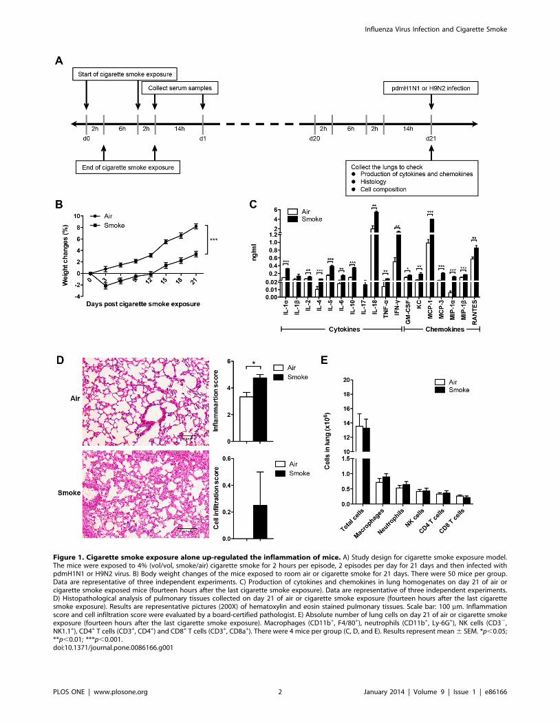
Figure 1. Cigarette smoke exposure alone up-regulated the inflammation of mice. A) Study design for cigarette smoke exposure model.The mice were exposed to 4% (vol/vol, smoke/air) cigarette smoke for 2 hours per episode, 2 episodes per day for 21 days and then infected withpdmH1N1 or H9N2 virus. B) Body weight changes of the mice exposed to room air or cigarette smoke for 21 days. There were 50 mice per group.Data are representative of three independent experiments. C) Production of cytokines and chemokines in lung homogenates on day 21 of air orcigarette smoke exposed mice (fourteen hours after the last cigarette smoke exposure). Data are representative of three independent experiments.D) Histopathological analysis of pulmonary tissues collected on day 21 of air or cigarette smoke exposure (fourteen hours after the last cigarettesmoke exposure). Results are representative pictures (200X) of hematoxylin and eosin stained pulmonary tissues. Scale bar: 100 mm. Inflammationscore and cell infiltration score were evaluated by a board-certified pathologist. E) Absolute number of lung cells on day 21 of air or cigarette smokeexposure (fourteen hours after the last cigarette smoke exposure). Macrophages (CD11b+, F4/80+), neutrophils (CD11b+, Ly-6G+), NK cells (CD32,NK1.1+), CD4+ T cells (CD3+, CD4+) and CD8+ T cells (CD3+, CD8a+). There were 4 mice per group (C, D, and E). Results represent mean 6 SEM. *p,0.05;**p,0.01; ***p,0.001.doi:10.1371/journal.pone.0086166.g001
Influenza Virus Infection and Cigarette Smoke
PLOS ONE | www.plosone.org 2 January 2014 | Volume 9 | Issue 1 | e86166

Until now, there is no report related to the susceptibility and
severity of pdmH1N1 or avian H9N2 influenza virus infection in
smokers. As pdmH1N1 and H9N2 can infect mice [20] and mice
are widely used to examine the influenza pathology [21], we chose
a mouse model to investigate the risk of pdmH1N1 and H9N2
infection in cigarette smoke exposed mice. We hypothesized that
the disease severity of pdmH1N1 and H9N2 infection would be
exacerbated by cigarette smoke exposure, and we also used
nicotine to mimic the effect of cigarette smoke.
Materials and Methods
Ethics StatementWritten consent for the use of buffy coat for research purposes
was obtained from the donors by the Hong Kong Red Cross Blood
Transfusion Services at the time of blood donation. The use of
buffy coat for this experiment was approved by the Institutional
Review Board of the University of Hong Kong/Hospital Authority
Hong Kong West Cluster (IRB reference number: UW 07–390).
All manipulations on animal work in this manuscript were
performed in compliance with ‘‘Code of Practice for Care and Use
of Animals for Experimental Purposes (2004)’’ and approved by
the Committee on the Use of Live Animals in Teaching and
Research (CULATR), the University of Hong Kong (reference
number: 2631-12 and 2819-12).
Influenza virus preparation and titrationInfluenza virus A/California/04/2009 (pdmH1N1) was grown
in embryonated chicken eggs. Influenza virus A/Quail/Hong
Kong/G1/97 (H9N2) was propagated in Madin-Darby canine
kidney (MDCK) cells with modified Eagle’s medium (Invitrogen,
Grand Island, NY, USA) containing 2 mg/L N-Tosyl-L-Phenyl-
alanine Chloromethyl Ketone-treated trypsin (Sigma, St. Louis,
MO, USA). MDCK cell line was purchased from ATCC
(American Type Culture Collection, Manassas, VA, USA) and
routinely maintained in our lab. Viruses were purified by
ultracentrifugation as described previously [20,22]. The virus titer
was determined by titration in MDCK cells, with daily observation
of cytopathogenic effect, and confirmed by hemagglutination
assay. The tissue culture infective dose affecting 50% of the
cultures (TCID50)/ml was calculated by the Reed-Muench
formula.
Smoking mouse model and virus infectionFemale C57B6/N mice of 6–8 weeks were obtained from
Laboratory Animal Unit, the University of Hong Kong; a7
nAChR knockout mice and wild-type mice were purchased from
the Jackson Laboratory. The mice were exposed to 4% (vol/vol,
smoke/air) cigarette smoke for 2 hours per episode, 2 episodes per
day with commercially available cigarette (Camel; filters, Japan
Tobacco INC) for 21 days (Fig. 1A) using the modified ventilated
cigarette smoke exposure chambers as described [23,24]. Control
mice were exposed only to fresh air (0%, vol/vol, smoke/air) in
another ventilated chamber with the same procedure. Four-
teen hours after the last cigarette smoke exposure, the mice were
anesthetized and inoculated intranasally with 30 ml of 56103
TCID50 pdmH1N1, or 105 TCID50 H9N2 virus. Simultaneously,
control mice were treated with PBS in the same way. The well-
being of animals was monitored three times daily after cigarette
smoke exposure and influenza virus infection. The criteria that we
used to monitor the well-being of the mice including appearance,
food and water intake, clinical signs (temperature, cardiac and
respiratory rates) and behavior (natural or provoked). The mice
were humanely euthanized when they showed any one of the
following termination criteria: body weight loss $30%, body
temperature 62uC, cardiac/respiratory rate 650%, and sign of
severe pneumonia (very weak and pre-comatose). All animal care
and experimental protocols were conducted in accordance with
the CULATR guidelines of the University of Hong Kong.
These results demonstrated that the peak mortality in the
control mice on day 5 post-pdmH1N1 infection might partially be
attributed to the hyper-cytokine response. This mortality was
significantly reduced by cigarette smoke exposure, which might be
due to the significantly decreased production of inflammatory
cytokines, as well as reduced pulmonary tissues pathology. The
immunosuppressive effect of cigarette smoke exposure was
independent of virus replication.
Quantitation of cotinineThe half-life of nicotine is very short and will be metabolized
into different metabolites. The average eradication half-life of
nicotine in mouse plasma is approximately 6–7 minutes and
cotinine is the major stable metabolite of nicotine [25]. Then
serum level of cotinine instead of nicotine was measured using
commercially available kit (Neogen, KY, USA) following the
manufacturer’s instructions. All measurements were performed in
duplicate.
Preparation of mouse lung homogenatesThe mouse lungs were harvested at the specific time points and
homogenized using a tissue homogenizer (Omni International,
Kennesaw, GA, USA) as previously described [20,26]. The
homogenates were centrifuged at 1500 g for 10 minutes at 4uC.
The supernatants were collected and separated into aliquots. Virus
titer was determined immediately and the remaining supernatants
were stored at 270uC for cytokines and chemokines detection.
Cytokines and chemokines determinationExpression levels of interleukin (IL) 1a, IL-1b, IL-2, IL-4, IL-5,
IL-6, IL-8, IL-10, IL-17, IL-18, tumor necrosis factor (TNF) a,
interferon (IFN) c, granulocyte-macrophage colony-stimulating
factor (GM-CSF), keratinocyte chemoattractant (KC), monocyte
chemotactic protein (MCP) 1, MCP-3, macrophage inflammatory
proteins (MIP) 1a, MIP-1b, monokine induced by interferon
gamma (MIG), regulated and normal T-cell expressed and
secreted (RANTES) in the lung homogenates or cell supernatants
were quantitatively determined by flow cytometry based immu-
noassay (eBioscience, San Diego, CA, USA) according to the
manufacturer’s protocol [20]. The samples were acquired on a
LSRII (BD, San Diego, CA, USA) and the amount (ng/ml) was
calculated by FlowCytomixTM Pro 3.0 software (eBioscience).
Pulmonary histopathologyThe lung tissues were fixed in 10% formalin, embedded in
paraffin, cut and stained with hematoxylin and eosin to analyze
inflammation-associated lung damage. Histopathologic inflamma-
tion score of lung tissues was evaluated by a board-certified
pathologist blinded to experimental design. Lung inflammatory
changes were graded using a semi-quantitative scoring system
based on the following parameters: peri-bronchiolar and bronchial
infiltrates, bronchiolar and bronchial luminal exudates, perivas-
cular infiltrates, parenchymal pneumonia, and edema, as previ-
ously described [20,27]. Each parameter was graded on a scale of
0–4 with 0, absent; 1, slight; 2, mild; 3, moderate; and 4, severe.
The total lung inflammation score was expressed as the sum of the
scores for each parameter. The degree of cell infiltration was
Influenza Virus Infection and Cigarette Smoke
PLOS ONE | www.plosone.org 3 January 2014 | Volume 9 | Issue 1 | e86166

independently scored of 0–3 with 0, no cells; 1, few cells; 2,
moderate influx of cells; and 3, extensive influx of cells [20,26].
To summarize, after H9N2 virus infection, cigarette smoke
exposed mice had less weight loss, possibly due to less cytokines
production, lower cellular infiltration and less lung damage
compared to the control mice.
Figure 2. Cigarette smoke exposure decreased the severity of pdmH1N1 infection in mice. The mice were exposed to room air orcigarette smoke for 21 days and then infected with pdmH1N1 virus. A) Survival curve of mice infected with pdmH1N1 virus. There were 6-9 mice pergroup. Data are representative of three independent experiments. B) Body weight changes of mice infected with pdmH1N1 virus. There were 6–9mice per group. Data are representative of three independent experiments. C) Lung virus titers of pdmH1N1 infected mice. There were 4–10 mice pergroup. Data are representative of two independent experiments. D) Absolute number of lung cells on day 5 of pdmH1N1 infection. Macrophages(CD11b+, F4/80+), neutrophils (CD11b+, Ly-6G+), NK cells (CD32, NK1.1+), CD4+ T cells (CD3+, CD4+) and CD8+ T cells (CD3+, CD8a+). There were 4–5mice per group. E) Histopathological analysis of pulmonary tissues collected on day 5 of pdmH1N1 infection. Results are representative pictures(200X) of hematoxylin and eosin stained pulmonary tissues. Scale bar: 100 mm. Inflammation score and cell infiltration score were evaluated by aboard-certified pathologist. There were 4–8 mice per group. Data are representative of two independent experiments. Results represent mean 6
SEM. *p,0.05 was determined by Log-rank (Mantel-Cox) test; #p,0.05, ##p,0.01; ###p,0.001 were tested by ANOVA of four groups.doi:10.1371/journal.pone.0086166.g002
Influenza Virus Infection and Cigarette Smoke
PLOS ONE | www.plosone.org 4 January 2014 | Volume 9 | Issue 1 | e86166

Lung cells isolation and flow cytometric analysisThe mouse lungs were cut into small pieces and incubated in
Iscove’s Modified Dulbecco’s Medium (Invitrogen) containing
1mg/ml collagenase D (Roche, Mannheim, Germany) and 20 U/
ml DNase I (Roche) at 37uC with shaking for 1–2 hours. After
treating with Ammonium-Chloride-Potassium lysing buffer and
Figure 3. Cigarette smoke exposure suppressed the production of cytokines at later time point of pdmH1N1 infection. The mice wereexposed to room air or cigarette smoke for 21 days and then infected with pdmH1N1 virus. The lungs were collected on day 1, 3 and 5 afterpdmH1N1 infection. A) The production of cytokines. B) The production of chemokines. Results represent mean 6 SEM of 4–10 mice per group. Dataare representative of two independent experiments. #p,0.05, ##p,0.01; ###p,0.001 were tested by ANOVA of four groups; *p,0.05; **p,0.01;***p,0.001 were determined by Tukey post hoc test.doi:10.1371/journal.pone.0086166.g003
Influenza Virus Infection and Cigarette Smoke
PLOS ONE | www.plosone.org 5 January 2014 | Volume 9 | Issue 1 | e86166

filtering by a 70 mm nylon mesh, 16106 cells were stained with a
combination of monoclonal antibodies (mAbs) of PE-Cy5-anti-
CD11b (M1/70, eBioscience), eFluor 450-anti-F4/80 (BM8,
eBioscience), and Alexa Fluor 700-anti-Ly-6G (RB6-8C5,
eBioscience); or a combination of mAbs of eFluor 450-anti-CD3
(17A2, eBioscience), FITC-anti-NK1.1 (PK136, BioLegend, San
Figure 4. Cigarette smoke exposure reduced the severity of H9N2 infection in mice. The mice were exposed to room air or cigarettesmoke for 21 days and then infected with H9N2 virus. A) Body weight changes of mice infected with H9N2 virus. There were 6–7 mice per group.Data are representative of three independent experiments. ***p,0.001 was compared between Air+H9N2 and Smoke+H9N2 and was examined bymultiple regression analysis adjusted for time. B) Lung virus titers of H9N2 infected mice. There were 4–7 mice per group. C) Absolute number of lungcells on day 9 of H9N2 infection. Macrophages (CD11b+, F4/80+), neutrophils (CD11b+, Ly-6G+), NK cells (CD32, NK1.1+), CD4+ T cells (CD3+, CD4+) andCD8+ T cells (CD3+, CD8a+). There were 4–5 mice per group. D) Histopathological analysis of pulmonary tissues collected on day 9 of H9N2 infection.Results are representative pictures (200X) of hematoxylin and eosin stained pulmonary tissues. Scale bar: 100 mm. Inflammation score and cellinfiltration score were evaluated by a board-certified pathologist. There were 4 mice per group. Results represent mean 6 SEM. ###p,0.001 wastested by ANOVA of four groups; *p,0.05; **p,0.01; ***p,0.001 were determined by Tukey post hoc test (C and D).doi:10.1371/journal.pone.0086166.g004
Influenza Virus Infection and Cigarette Smoke
PLOS ONE | www.plosone.org 6 January 2014 | Volume 9 | Issue 1 | e86166

Diego, CA, USA), PE-anti-CD4 (RM4-5, BD), and APC-anti-
CD8a (53-6.7, BD); or their relevant isotype-specific antibodies as
described previously [26,28]. Before running samples, counting
beads were added into the stained cells (Molecular Probes,
Carlsbad, CA, USA). All samples were acquired on LSR II and
analyzed by FlowJo software (TreeStar, Ashland, OR, USA).
Figure 5. Cigarette smoke exposure suppressed the production of cytokines and chemokines of H9N2 infected mice. The mice wereexposed to room air or cigarette smoke for 21 days and then infected with H9N2 virus. The lungs were collected on day 5, 9 and 14 after H9N2infection. A) The production of cytokines. B) The production of chemokines. Results represent mean 6 SEM of 4–7 mice per group. #p,0.05,##p,0.01; ###p,0.001 were tested by ANOVA of four groups; *p,0.05; **p,0.01; ***p,0.001 were determined by Tukey post hoc test.doi:10.1371/journal.pone.0086166.g005
Influenza Virus Infection and Cigarette Smoke
PLOS ONE | www.plosone.org 7 January 2014 | Volume 9 | Issue 1 | e86166

Nicotine treatment and virus infection of the cells.Peripheral blood mononuclear cells (PBMC) were isolated from
buffy coat of healthy donors (from Hong Kong Red Cross) by
LymphoprepTM (Fresenius Kabi Norge AS) gradient centrifuga-
tion. The research protocol was approved by the Institutional
Review Board of the University of Hong Kong/Hospital Authority
Hong Kong West Cluster. Primary human macrophages were
generated from PBMC as described [29]. Briefly, the monocytes
were cultured in RPMI 1640 medium (Invitrogen) supplemented
with 5% autologous serum and were seeded into the 24-well plates,
cultured for 14 days to differentiate into macrophages. The purity
of monocytes was examined by flow cytometry (LSRII) with anti-
CD14 monoclonal antibody (Invitrogen), and was consistently
over 90%. A549 cell line was purchased from ATCC and routinely
maintained in our lab. A549 cells were grown in the medium of
RPMI 1640 contains 10% fetal bovine serum. A549 cells are
cancer cells and are widely used as an in vitro model for type II
pulmonary epithelial cells [30]. Thus primary human macrophag-
es and A549 cells were used to examine the effect of nicotine on
pdmH1N1 or H9N2 virus infection. Primary human macrophages
and A549 cells were pre-treated with 10 mM nicotine (Sigma) or
PBS for 72 h with the medium changed every 12 h; and then
infected with pdmH1N1 or H9N2 (MOI = 2). At 24 h or 48 h post
virus infection, the supernatant was collected for cytokines and
chemokines measurement.
Nicotine in vivo studyFemale C57B6/N mice of 6–8 weeks were anesthetized and
implanted with Alzet (Cupertino, CA, USA) micro-osmotic pumps
(model 1004) subcutaneously, which provided nicotine at rate of
24 mg/kg/day [31]. Control mice were implanted with pumps
filled with sterile water. On day 22 of nicotine exposure, the mice
were anesthetized and inoculated intranasally with 30 ml of 56103
TCID50 pdmH1N1, or 105 TCID50 H9N2 viruses. After infection,
the mice were weighed and monitored every day. The pumps kept
deliver nicotine after pdmH1N1 or H9N2 virus infection. All
animal care and experimental protocols were conducted in
accordance with the CULATR guidelines of the University of
Hong Kong.
Statistical analysisData are expressed as mean 6 SEM. Statistical analysis was
performed by unpaired two-tailed Student’s t test or one-way
analysis of variance (ANOVA) followed by Tukey post hoc test in
Prism 5.0 software (GraphPad). The p value of the difference for
survival was determined by Log-rank (Mantel-Cox) test. The p
value of the difference for body weight change was examined by
multiple regression analysis adjusted for time. A p value ,0.05 was
considered significant.
Results
Cigarette smoke exposure alone decreased the bodyweight gain and elevated lung inflammation
Before investigating the impact of influenza virus infection in
cigarette smoke exposed mice, the influence of cigarette smoke
exposure alone on mice was determined. Cigarette smoke exposed
mice had significantly less body weight gain than the control mice
(Fig. 1B). Significantly higher production of IL-1a, IL-2, IL-4, IL-
5, IL-6, IL-10, IL-18, TNF-a, IFN-c, GM-CSF, KC, MCP-1,
MCP-3, MIP-1a, MIP-1b and RANTES were shown in cigarette
smoke exposed mice than that in control mice (Fig. 1C). Cigarette
smoke exposed mice had increased lung damage than control
mice, with thickening of intra-alveolar septa and hemorrhages.
Moreover, cigarette smoke exposure significantly increased the
inflammation score and mildly enhanced the cell infiltration score
(Fig. 1D). For the lung cell composition, there were no differences
for the number of total cells, macrophages, neutrophils, NK cells,
Figure 6. Mouse serum cotinine levels at different time points.A) Serum cotinine levels during 21 days of cigarette smoke exposure.The serum samples were collected on day 7, day 14 and day 21(immediately or 14 hours after cessation of cigarette smoke exposure).(n = 2 and n = 4 for air and smoke exposure group respectively). B)Serum cotinine levels of air or cigarette smoke exposed mice on day 1,day 5 and day 14 after pdmH1N1/H9N2 virus infection. (n = 2 and n = 4for air and smoke exposure group respectively). C) Serum cotinine levelsof sterile water or nicotine treated mice on day 0 and day 14 afterpdmH1N1/H9N2 virus infection. (n = 4 for both sterile water andnicotine treatment groups). Results represent mean 6 SEM.doi:10.1371/journal.pone.0086166.g006
Influenza Virus Infection and Cigarette Smoke
PLOS ONE | www.plosone.org 8 January 2014 | Volume 9 | Issue 1 | e86166

Figure 7. Nicotine suppressed the inflammatory response induced by pdmH1N1 infection in vitro and in vivo. A) Nicotine inhibited thepdmH1N1-induced production of cytokines and chemokines in primary human macrophages. Primary human macrophages were pre-treated with10 mM nicotine for 72 h, and then infected with pdmH1N1 virus. Data represent 4–5 independent experiments. ##p,0.01 and ###p,0.001 weretested by ANOVA of three groups; *p,0.05 and ***p,0.001 were determined by Tukey post hoc test. B) Body weight changes of nicotine treatedmice after pdmH1N1 virus infection. The mice were pre-treated with nicotine subcutaneously for 21 days and then infected by pdmH1N1 virus. Therewere 5 mice per group. Data are representative of two independent experiments. *p,0.05 was examined by multiple regression analysis adjusted fortime. C) Lung virus titers of nicotine treated mice at indicated time points after pdmH1N1 infection. There were 4 mice per group. D) Production ofcytokines and chemokines in the lung homogenates from nicotine treated mice after pdmH1N1 virus infection. There were 4–5 mice per group.*p,0.05 and **p,0.01 were performed by unpaired two-tailed Student’s t test. Results represent mean 6 SEM.doi:10.1371/journal.pone.0086166.g007
Influenza Virus Infection and Cigarette Smoke
PLOS ONE | www.plosone.org 9 January 2014 | Volume 9 | Issue 1 | e86166

Figure 8. Nicotine inhibited the inflammatory response induced by H9N2 infection in vitro and in vivo. A) Nicotine suppressed the H9N2-induced production of cytokines and chemokines in primary human macrophages. Primary human macrophages were pre-treated with 10 mMnicotine for 72 h, and then infected with H9N2 virus. Data represent 4–5 independent experiments. ##p,0.01 and ###p,0.001 were tested byANOVA of three groups; *p,0.05 was determined by Tukey post hoc test. B) Body weight changes of nicotine treated mice after H9N2 virus infection.The mice were pre-treated with nicotine subcutaneously for 21 days and then infected by H9N2 virus. There were 5 mice per group. Data arerepresentative of two independent experiments. ***p,0.001 was examined by multiple regression analysis adjusted for time. C) Lung virus titers ofnicotine treated mice at indicated time points after H9N2 infection. There were 4 mice per group. D) Production of cytokines and chemokines in thelung homogenates from nicotine treated mice after H9N2 virus infection. There were 4–5 mice per group. *p,0.05 and **p,0.01 were performed byunpaired two-tailed Student’s t test. Results represent mean 6 SEM.doi:10.1371/journal.pone.0086166.g008
Influenza Virus Infection and Cigarette Smoke
PLOS ONE | www.plosone.org 10 January 2014 | Volume 9 | Issue 1 | e86166

CD4+ T cells and CD8+ T cells between cigarette smoke exposed
mice and control mice (Fig. 1E).
Collectively, cigarette smoke exposure could induce less body
weight gain, which might be attributed to up-regulated production
of inflammatory cytokines, and increased lung damage compared
with the control mice. However, cigarette smoke exposure could
not change the lung cell composition.
Mortality rate of pdmH1N1 infection was decreased byprior cigarette smoke exposure
In order to investigate the role of upregulated inflammation
caused by the prior cigarette smoke exposure on the subsequent
influenza A virus infection, the mice were infected with pdmH1N1
virus after 21 days of cigarette smoke exposure. Cigarette smoke
exposure significantly decreased the mice mortality (Fig. 2A) but
had no effect on body weight changes (Fig. 2B) compared to air
exposure after pdmH1N1 infection. Because the mice began to die
on day 4, early time points of day 1, 3, and 5 after pdmH1N1
infection were selected for the following measurements. There was
no difference for lung virus titer at all time points between air and
cigarette smoke exposed mice upon pdmH1N1 infection (Fig. 2C).
After pdmH1N1 virus infection, cigarette smoke exposed mice had
significantly higher production of IL-1a, IL-1b, IL-2, IFN-c, KC,
RANTES on day 3; IL-5, IL-10 and MIP-1a both on day 1 and
day 3 in the lungs than the control mice. However on day 5, the
phenomenon was reversed, cigarette smoke exposed mice
produced significantly lower cytokines than the control mice,
including IL-1a, IL-1b, IL-4, IL-5, IL-6, IL-10, IL-17, IL-18,
TNF-a and IFN-c (Fig. 3). For lung cell composition, cigarette
smoke exposure could not affect the number of total cells,
macrophages, neutrophils, NK cells, CD4+ T cells and CD8+ T
cells in the lung on day 5 after pdmH1N1 infection (Fig. 2D). Less
lung damage was observed in cigarette smoke exposed mice on
day 5 after pdmH1N1 infection than the control mice; however,
the difference was not statistically significant (Fig. 2E).
Severity of H9N2 infection was reduced by prior cigarettesmoke exposure
To further investigate whether the immunosuppressive effect of
cigarette smoke exposure was independent of influenza subtypes,
we tested avian H9N2 virus. Cigarette smoke exposed mice
showed significantly lower body weight loss than the control mice
(Fig. 4A). There was no death from H9N2 infection; the mice were
sacrificed on day 5, 9 and 14 post-infection and the mouse lungs
were collected for the following measurements. There was no
difference for virus titer at all time points between air and cigarette
smoke exposed mice after H9N2 infection (Fig. 4B). For
inflammatory response, cigarette smoke exposed mice had
significantly lower production of IL-1a, IL-1b, IL-10, TNF-a,
IFN-c, KC, MCP-1, MCP-3 and MIP-1b on day 9, IL-6 and
RANTES on day 5 after H9N2 infection compared with the
control mice (Fig. 5). Cigarette smoke exposed mice displayed
significantly less number of macrophages, neutrophils, CD4+ T
cells and CD8+ T cells than the control mice on day 9 after H9N2
infection (Fig. 4C). Moreover, significantly less lung damage was
observed in cigarette smoke exposed mice than in control mice on
day 9 after H9N2 infection (Fig. 4D).
PdmH1N1-induced inflammatory response and diseaseseverity were suppressed by nicotine in vitro and in vivorespectively
The above data demonstrated that the severity of pdmH1N1 or
H9N2 infection was alleviated by cigarette smoke exposure via
decreasing the inflammatory response. The underlying mecha-
nisms were further investigated. Nicotine is one of the major
components in cigarette smoke but its half-life is short. The serum
concentration of cotinine, a major metabolite of nicotine, was
about 250 ng/ml, which was comparable to the serum cotinine
levels in cigarette smokers [32], immediately after cessation of
cigarette smoke exposure, and decreased to around 4 ng/ml at
14 hour after cessation of cigarette smoke exposure (Fig. 6A).
Moreover, the mean serum cotinine concentration was 0.93 ng/
ml, 0.22 ng/ml and 0.10 ng/ml on day 1, day 5 and day 14 post
pdmH1N1 or H9N2 virus infection respectively (Fig. 6B). This
data demonstrated that the effect of nicotine might be on-going
during the 14 days of pdmH1N1 or H9N2 virus infection. Then
we hypothesized that it is nicotine in cigarette smoke that might be
responsible for the immunosuppressive effect.
We firstly used in vitro method to test our hypothesis. In primary
human macrophages, pdmH1N1-induced production of TNF-a,
IL-8 and MIG was significantly inhibited by nicotine (Fig. 7A). In
the human lung alveolar epithelial cell line A549, nicotine could
decrease the production of MCP-1 at 24 h, IL-8 at 48 h post-
pdmH1N1 infection respectively (Fig. S1A). For the in vivo study,
we chose the dose of 24 mg/kg/day because the serum cotinine
concentration was maintained at around 70 ng/ml both on day 0
and day 14 after pdmH1N1 or H9N2 virus infection (Fig. 6C),
which was within the range of serum cotinine concentration of the
cigarette smoke exposure group (Fig. 6A). Our data demonstrated
that there was no death in both sterile water and nicotine delivery
groups after pdmH1N1 infection. However, nicotine treated mice
had significantly less body weight loss after pdmH1N1 infection
compared to control mice (Fig. 7B). There was no difference for
lung virus titer at indicated time points between nicotine treated
and control mice after pdmH1N1 infection (Fig. 7C). For the
inflammatory response, nicotine treated mice had significantly less
production of IL-6 on day 3, MCP-1 and RANTES on day 7 after
pdmH1N1 infection compared with control mice (Fig. 7D). In
summary, the inflammatory response and disease severity induced
by pdmH1N1 virus could be suppressed by nicotine.
H9N2-induced inflammatory response and diseaseseverity were inhibited by nicotine in vitro and in vivorespectively
We further investigated whether the immunosuppressive effect
of nicotine was independent of influenza subtypes and then tested
Figure 9. Cigarette smoke exposure resulted in increased bodyweight loss in a7 nAChR knockout mice as compared to wild-type mice after H9N2 virus infection. Wild-type and a7 nAChRknockout mice were exposed to cigarette smoke for 21 days and theninfected with H9N2 virus. The body weight changes were monitored for14 days. Results represent mean 6 SEM. N = 4 and n = 6 for a7 nAChRknockout and wild-type mice respectively. *p,0.05 was determined bymultiple regression analysis adjusted for time.doi:10.1371/journal.pone.0086166.g009
Influenza Virus Infection and Cigarette Smoke
PLOS ONE | www.plosone.org 11 January 2014 | Volume 9 | Issue 1 | e86166

H9N2 virus. H9N2 induced expression of TNF-a, IL-8, MIG and
RANTES was significantly suppressed by nicotine in primary
human macrophages (Fig. 8A). Nicotine could also suppress the
expression of IL-8 at 24 h post-H9N2 infection in A549 cells
(Fig. S1B). There was no mortality in both sterile water and
nicotine delivery groups after H9N2 infection. However, nicotine
treated mice had significantly less body weight loss after H9N2
infection compared to control mice (Fig. 8B). There was no
difference for lung virus titer at indicated time points between
nicotine treated and control mice after H9N2 infection (Fig. 8C).
For the inflammatory response, nicotine could suppress the
secretion of IL-1a, IL-4, IL-5, IL-10 and RANTES on day 6
and IL-17 on day 14 significantly after H9N2 infection compared
with control mice (Fig. 8D). Taken together, nicotine had
immunosuppressive effect on H9N2 infection.
Cigarette smoke exposure resulted in increased bodyweight loss in a7 nAChR knockout mice as compared towild-type mice after H9N2 virus infection
Nicotine exerts its immunosuppressive effect by binding to a7
nAChR and hence activating cholinergic anti-inflammatory
pathway [14]. In order to further test the hypothesis that it is
nicotine in cigarette smoke that might be responsible for the
immunosuppressive effect, we used a7 nAChR knockout mice and
wild-type mice to perform the mechanistic study. Wild-type and
a7 nAChR knockout mice were exposed to cigarette smoke for
21 days and then infected with H9N2 virus at fourteen hours after
the last cigarette smoke exposure. Our data demonstrated that
cigarette smoke exposure resulted in significantly increased body
weight loss in a7 nAChR knockout mice than in wild-type mice
after H9N2 virus infection (Fig. 9).
Discussion
The present study demonstrated that cigarette smoke exposure
alone up-regulated the mouse lung inflammation. The pathoge-
nicity caused by pdmH1N1 or H9N2 virus infection was alleviated
by such prior cigarette smoke exposure. The beneficial effect was
due to the immunosuppressive effect of cigarette smoke exposure
that down-regulated the influenza virus induced hyper-inflamma-
tory response. Indeed, the immunosuppressive effect of cigarette
smoke has been previously reported, including macrophages
extracted from smokers or smoke exposed mice have reduced
inflammation in response to LPS or poly I:C stimulation [33,34].
Furthermore, this study demonstrated that the immunosuppressive
effect of cigarette smoke was partially attributed to nicotine.
Our findings of the beneficial effect of cigarette smoke exposure
in pdmH1N1 and H9N2 virus infected mice are contrary to the
current epidemiological evidence that showed the mortality and
hospitalization rates were increased in smokers who had seasonal
influenza infection [15,16,19]. This discrepancy could be ex-
plained by both viral and host determinants. Both 2009
pdmH1N1 and H9N2 viruses, that crossed species from swine
and bird to human respectively, are more virulent than seasonal
influenza virus [35,36]. Therefore, epidemiological data regarding
the impact of cigarette smoke on seasonal influenza virus infection
might not be applicable to pdmH1N1 and H9N2 virus directly.
There is an urgent need to conduct epidemiological investigations
to fully understand the impact of smoking on the severity of
pdmH1N1 or H9N2 virus infection.
The interaction of cigarette smoke exposure and influenza virus
infection in mouse model is complex. Due to the differences in the
composition of cigarette smoke, the method and duration of smoke
exposure, the dose and subtypes of the influenza A virus, the
results of different animal studies might be contradictory. Gualano
et al., using BALB/C mice exposed to smoke with 9 Winfield Red
cigarettes per day for 4 days, and infected with H3N1 virus, found
that smoke exposure prior to influenza virus infection led to more
lung inflammation, higher viral burden and greater body weight
loss [37]. Another study, using C57BL/6 mice exposed to 1R3
cigarettes smoke for 3–5 months and infected with low dose or
high dose H1N1-A/FM/1/47, demonstrated that cigarette smoke
attenuated the inflammatory response to low dose virus infection,
while high dose infection resulted in higher inflammation and
significant morbidity [38]. Our study, using C57BL/6 mice,
ventilated cigarette smoke exposure system, 21 days of cigarette
smoke exposure and pdmH1N1 or H9N2 virus, demonstrated that
prior cigarette smoke exposure decreased the severity of influenza
virus infection by attenuating the hyper-inflammatory response.
Inflammation is a double-edged sword; adequate inflammation
is necessary for the development of immune response, clearance of
pathogens and recovery from tissue injuries, while excessive
inflammatory response would be life threatening. PdmH1N1 and
H9N2 influenza viruses could produce excessive inflammatory
cytokines compared with seasonal influenza virus [35,36], which
might contribute to the more severe disease. In support with these
observation, here, we also found that, comparing with cigarette
smoke exposed mice, control mice had hyper-inflammatory
response with higher production of several cytokines, including
IL-1a, IL-1b, IL-6, IL-10, TNF-a and IFN-c on day 5 of
pdmH1N1 and day 9 of H9N2 infection, coinciding with the mice
having peak mortality and body weight loss respectively. Other
studies also demonstrated the deleterious role of dysregulation of
cytokines. Enhanced secretion of IL-1a and IL-1b could lead to
acute pulmonary inflammatory pathology upon influenza A virus
infection in mouse model [39]. Elevated IL-6 levels are associated
with disease severity triggered by pdmH1N1 infection in mice
[40]. Anti-TNF-a agents could decrease lung inflammation and
prolong survival of A/PR/8 infected mice; reduce weight loss and
illness severity in mice upon influenza A X31 infection [41]. The
expression of IFN-c is significantly increased in pdmH1N1
infected patients [42]. The deleterious role of hyper-production
of IL-10 is supported by IL-10 deficiency mice having enhanced
survival upon high-dose influenza virus infection [43]. In our
study, cigarette smoke exposure significantly suppressed the
production of these inflammatory cytokines after pdmH1N1 and
H9N2 infection, leading to the decreased inflammatory response
in the lung. The suppressed inflammation can explain the lower
mortality after pdmH1N1 infection; less body weight loss, lung
injury and immune cells infiltration after H9N2 infection in
cigarette smoke exposed mice.
The cell types infiltrating into the lung were mostly neutrophils
and macrophages at early time points, with lymphocytes
predominant at later time points of infection in our study (data
not shown). The hyper-expression of chemokines, such as KC,
MCP-1, MCP-3 and MIP-1b in control mice might coordinate the
increased recruitment of neutrophils and macrophages on day 9
post-H9N2 infection. Moreover, the increased production of
RANTES on day 5, which is chemotactic for T cells, might
contribute to higher infiltration of CD4+ and CD8+ T cells in the
control mice at day 9 after H9N2 infection. IL-6, except for its role
in resolution of innate immunity, is also an important cytokine to
regulate the shift from innate immune response to adaptive
immune response and enhance the proliferation of T cells and
influenza-specific T memory cells [44,45]. Therefore, the signif-
icantly higher production of IL-6 on day 5 might contribute to the
significantly higher recruitment of CD4+ and CD8+ T cells in the
control mice at day 9 after H9N2 infection. The excessive number
Influenza Virus Infection and Cigarette Smoke
PLOS ONE | www.plosone.org 12 January 2014 | Volume 9 | Issue 1 | e86166

of macrophages, neutrophils, CD4+ and CD8+ T cells might lead
to deleterious lung immunopathology. By releasing the oxygen
radicals and proteolytic enzymes, such as neutrophil elastase and
matrix metalloproteinases-8 (MMP-8), MMP-9 and MMP-12,
neutrophils and macrophages could cause lung damage [46]. The
recruitment of macrophages in the lung could also contribute to
the alveolar epithelial cell destruction and apoptosis [47]. More
CD4+ T cells could induce more inflammatory cytokines, such as
IL-6, IL-10 and IFN-c, leading to more inflammatory response.
Furthermore, CD4+ T cells could directly lead to severe
immunopathology and tissue damage via a cytokine-independent
manner in influenza infection [48]. CD8+ CTLs could exacerbate
influenza viral pathology and induce mortality at high viral dose
infection [49]. Our data of significantly increased immune cells
number in the lung of control mice upon H9N2 infection
corroborated this observation. Cigarette smoke exposure signifi-
cantly improved this deleterious effect.
In our study, cigarette smoke had no effect on viral burden,
which is consistent with other investigations [50]. Some studies
however showed increased virus titer in cigarette smoke exposed
mice [37] and in smokers [51,52]. Interestingly, Robbins et al.
found that cigarette smoke exposure enhanced the viral burden in
low dose influenza A virus infection, but had no effect in high dose
influenza infection [38]. Taken together, these data indicate that
the immunosuppressive effect of cigarette smoke on influenza A
virus infection is independent of viral burden.
The immunosuppressive effect of nicotine has been previously
reported, including ameliorating inflammatory diseases, such as
ulcerative colitis [53,54] and cutaneous inflammation [55].
Transcutaneous nicotine administration could attenuate the
LPS-induced systemic inflammatory response in human subjects
[56]. Our in vitro and in vivo experiments confirmed such
immunosuppressive effect of nicotine. Other studies also demon-
strated that H1N1-A/PR/8/34-induced morbidity and mortality
were decreased by nicotine treatment in mice [57,58]. However,
they found that nicotine promoted influenza infection with
increased lung virus titer, which is contrary to our results that
nicotine could not affect lung virus replication. Together with our
study, these observations suggest that the immunosuppressive
effect of nicotine might be independent of virus replication.
For safety consideration, we could not expose the mice to
cigarette smoke after pdmH1N1 or H9N2 virus infection.
Although the half-life of nicotine is very short, the cotinine could
still be detectable on day 14 after pdmH1N1 or H9N2 virus
infection. This data demonstrated that the effect of nicotine might
be on-going during the 14 days of pdmH1N1 or H9N2 virus
infection.
Although we suggested that cigarette smoke exposure decreased
the severity of influenza A virus infection, we are not intent to
encourage people to smoke. The immunosuppressive effect of
cigarette smoke plays a protective role just in our mouse model,
when encountering highly virulent influenza virus infection that
induces hyper-reaction of inflammatory responses. However, the
immunosuppressive effect of cigarette smoke will be deleterious for
people suffering from seasonal influenza virus infection [15,16,19],
which produces appropriate inflammation that is necessary for the
generation of immune responses, elimination of pathogens and
recovery from tissue injuries [9,59].
Our study provides the first in vivo evidence that cigarette smoke,
mediated partially by nicotine, could alleviate the pathogenicity of
both pdmH1N1 and H9N2 viruses. Future epidemiological studies
of pdmH1N1/H9N2 infection in smokers would be important to
clarify the implications of our findings in humans.
Supporting Information
Figure S1 Nicotine suppressed the expression of che-mokines after pdmH1N1 and H9N2 infection in A549cells. A549 cells were pre-treated with 10 mM nicotine for 72 h,
and then infected with pdmH1N1 or H9N2 virus. A) Effect of
nicotine in pdmH1N1-induced inflammatory response. B) Impact
of nicotine on H9N2-induced inflammatory response. Data are
mean 6 SEM and represent 4 independent experiments. #p,0.05
and ###p,0.001 were tested by ANOVA of three groups;
**p,0.01 and ***p,0.001 were determined by Tukey post hoc
test.
(TIF)
Acknowledgments
We thank Dr. Judith Choi Wo Mak in the Department of Medicine in the
University of Hong Kong and Prof. Chi Hin Cho in the School of
Biomedical Sciences in the Chinese University of Hong Kong for
establishing the ventilated cigarette smoke exposure system. We also thank
University Postgraduate Studentship and University Postgraduate Fellow-
ship in The University of Hong Kong.
Author Contributions
Conceived and designed the experiments: YH HM WT Y. Lau. Performed
the experiments: YH MTL JZ KTL Y. Liu. Analyzed the data: YH HM
ML WT Y. Lau. Contributed reagents/materials/analysis tools: YH.
Wrote the paper: YH. Reviewed and critically revised the manuscript: WT
Y. Lau.
References
1. Itoh Y, Shinya K, Kiso M, Watanabe T, Sakoda Y, et al. (2009) In vitro and in
vivo characterization of new swine-origin H1N1 influenza viruses. Nature 460:
1021–1025.
2. Kuiken T, Riteau B, Fouchier RA, Rimmelzwaan GF (2012) Pathogenesis of
influenza virus infections: the good, the bad and the ugly. Curr Opin Virol 2:
276–286.
3. WHO (2009) Acute Respiratory Infections (Update September 2009).
4. Dawood FS, Iuliano AD, Reed C, Meltzer MI, Shay DK, et al. (2012) Estimated
global mortality associated with the first 12 months of 2009 pandemic influenza
A H1N1 virus circulation: a modelling study. Lancet Infect Dis 12: 687–695.
5. Butt KM, Smith GJ, Chen H, Zhang LJ, Leung YH, et al. (2005) Human
infection with an avian H9N2 influenza A virus in Hong Kong in 2003. J Clin
Microbiol 43: 5760–5767.
6. WHO (1999) 1999– Influenza A(H9N2) in Hong Kong Special Administrative
Region (SAR) and China – Update.
7. WHO (2003) Influenza A(H9N2) in Hong Kong Special Administrative Region
of China (SAR).
8. Khoufache K, Berri F, Nacken W, Vogel AB, Delenne M, et al. (2013) PAR1contributes to influenza A virus pathogenicity in mice. J Clin Invest 123: 206–
214.
9. Arcavi L, Benowitz NL (2004) Cigarette smoking and infection. Arch InternMed 164: 2206–2216.
10. Goncalves RB, Coletta RD, Silverio KG, Benevides L, Casati MZ, et al. (2011)Impact of smoking on inflammation: overview of molecular mechanisms.
Inflamm Res 60: 409–424.
11. Sopori M (2002) Effects of cigarette smoke on the immune system. Nat RevImmunol 2: 372–377.
12. Gardi C, Valacchi G (2012) Cigarette smoke and ozone effect on murineinflammatory responses. Ann N Y Acad Sci 1259: 104–111.
13. Jackson KJ, Walters CL, Miles MF, Martin BR, Damaj MI (2009)
Characterization of pharmacological and behavioral differences to nicotine inC57Bl/6 and DBA/2 mice. Neuropharmacology 57: 347–355.
14. Andersson U, Tracey KJ (2012) Reflex principles of immunological homeostasis.Annu Rev Immunol 30: 313–335.
15. Rogot E, Murray JL (1980) Smoking and causes of death among U.S. veterans:
16 years of observation. Public Health Rep 95: 213–222.
Influenza Virus Infection and Cigarette Smoke
PLOS ONE | www.plosone.org 13 January 2014 | Volume 9 | Issue 1 | e86166

16. Kark JD, Lebiush M (1981) Smoking and epidemic influenza-like illness in
female military recruits: a brief survey. Am J Public Health 71: 530–532.
17. Kark JD, Lebiush M, Rannon L (1982) Cigarette smoking as a risk factor for
epidemic a(h1n1) influenza in young men. N Engl J Med 307: 1042–1046.
18. Hanshaoworakul W, Simmerman JM, Narueponjirakul U, Sanasuttipun W,Shinde V, et al. (2009) Severe human influenza infections in Thailand:
oseltamivir treatment and risk factors for fatal outcome. PLoS One 4: e6051.
19. Wilson KM, Pier JC, Wesgate SC, Cohen JM, Blumkin AK (2013) Secondhand
tobacco smoke exposure and severity of influenza in hospitalized children.
J Pediatr 162: 16–21.
20. Ling MT, Tu W, Han Y, Mao H, Chong WP, et al. (2012) Mannose-binding
lectin contributes to deleterious inflammatory response in pandemic H1N1 andavian H9N2 infection. J Infect Dis 205: 44–53.
21. Doherty PC, Turner SJ, Webby RG, Thomas PG (2006) Influenza and the
challenge for immunology. Nat Immunol 7: 449–455.
22. Tu W, Mao H, Zheng J, Liu Y, Chiu SS, et al. (2010) Cytotoxic T lymphocytes
established by seasonal human influenza cross-react against 2009 pandemicH1N1 influenza virus. J Virol 84: 6527–6535.
23. Chan KH, Ho SP, Yeung SC, So WH, Cho CH, et al. (2009) Chinese green tea
ameliorates lung injury in cigarette smoke-exposed rats. Respir Med 103: 1746–1754.
24. Wong HP, Li ZJ, Shin VY, Tai EK, Wu WK, et al. (2009) Effects of cigarettesmoking and restraint stress on human colon tumor growth in mice. Digestion
80: 209–214.
25. Matta SG, Balfour DJ, Benowitz NL, Boyd RT, Buccafusco JJ, et al. (2007)
Guidelines on nicotine dose selection for in vivo research. Psychopharmacology
(Berl) 190: 269–319.
26. Tu W, Zheng J, Liu Y, Sia SF, Liu M, et al. (2011) The aminobisphosphonate
pamidronate controls influenza pathogenesis by expanding a gammadelta T cellpopulation in humanized mice. J Exp Med 208: 1511–1522.
27. Goodman AG, Fornek JL, Medigeshi GR, Perrone LA, Peng X, et al. (2009)
P58(IPK): a novel ‘‘CIHD’’ member of the host innate defense response againstpathogenic virus infection. PLoS Pathog 5: e1000438.
28. Zheng J, Liu Y, Qin G, Chan PL, Mao H, et al. (2009) Efficient induction andexpansion of human alloantigen-specific CD8 regulatory T cells from naive
precursors by CD40-activated B cells. J Immunol 183: 3742–3750.
29. Qin G, Liu Y, Zheng J, Ng IH, Xiang Z, et al. (2011) Type 1 responses ofhuman Vgamma9Vdelta2 T cells to influenza A viruses. J Virol 85: 10109–
10116.
30. Foster KA, Oster CG, Mayer MM, Avery ML, Audus KL (1998) Character-
ization of the A549 cell line as a type II pulmonary epithelial cell model for drug
metabolism. Exp Cell Res 243: 359–366.
31. Dickson PE, Rogers TD, Lester DB, Miller MM, Matta SG, et al. (2011)
Genotype-dependent effects of adolescent nicotine exposure on dopaminefunctional dynamics in the nucleus accumbens shell in male and female mice: a
potential mechanism underlying the gateway effect of nicotine. Psychopharma-cology (Berl) 215: 631–642.
32. Hukkanen J, Jacob P 3rd, Benowitz NL (2005) Metabolism and disposition
kinetics of nicotine. Pharmacol Rev 57: 79–115.
33. Chen H, Cowan MJ, Hasday JD, Vogel SN, Medvedev AE (2007) Tobacco
smoking inhibits expression of proinflammatory cytokines and activation of IL-1R-associated kinase, p38, and NF-kappaB in alveolar macrophages stimulated
with TLR2 and TLR4 agonists. J Immunol 179: 6097–6106.
34. Gaschler GJ, Zavitz CC, Bauer CM, Skrtic M, Lindahl M, et al. (2008) Cigarettesmoke exposure attenuates cytokine production by mouse alveolar macrophages.
Am J Respir Cell Mol Biol 38: 218–226.
35. Kang YM, Song BM, Lee JS, Kim HS, Seo SH (2011) Pandemic H1N1
influenza virus causes a stronger inflammatory response than seasonal H1N1
influenza virus in ferrets. Arch Virol 156: 759–767.
36. Zhou J, Law HK, Cheung CY, Ng IH, Peiris JS, et al. (2006) Differential
expression of chemokines and their receptors in adult and neonatal macrophagesinfected with human or avian influenza viruses. J Infect Dis 194: 61–70.
37. Gualano RC, Hansen MJ, Vlahos R, Jones JE, Park-Jones RA, et al. (2008)
Cigarette smoke worsens lung inflammation and impairs resolution of influenzainfection in mice. Respir Res 9: 53.
38. Robbins CS, Bauer CM, Vujicic N, Gaschler GJ, Lichty BD, et al. (2006)
Cigarette smoke impacts immune inflammatory responses to influenza in mice.Am J Respir Crit Care Med 174: 1342–1351.
39. Schmitz N, Kurrer M, Bachmann MF, Kopf M (2005) Interleukin-1 isresponsible for acute lung immunopathology but increases survival of respiratory
influenza virus infection. J Virol 79: 6441–6448.
40. Paquette SG, Banner D, Zhao Z, Fang Y, Huang SS, et al. (2012) Interleukin-6is a potential biomarker for severe pandemic H1N1 influenza A infection. PLoS
One 7: e38214.41. Darwish I, Mubareka S, Liles WC (2011) Immunomodulatory therapy for severe
influenza. Expert Rev Anti Infect Ther 9: 807–822.42. Liu Y, Chen H, Sun Y, Chen F (2012) Antiviral role of Toll-like receptors and
cytokines against the new 2009 H1N1 virus infection. Mol Biol Rep 39: 1163–
1172.43. McKinstry KK, Strutt TM, Buck A, Curtis JD, Dibble JP, et al. (2009) IL-10
deficiency unleashes an influenza-specific Th17 response and enhances survivalagainst high-dose challenge. J Immunol 182: 7353–7363.
44. Jones SA (2005) Directing transition from innate to acquired immunity: defining
a role for IL-6. J Immunol 175: 3463–3468.45. Longhi MP, Wright K, Lauder SN, Nowell MA, Jones GW, et al. (2008)
Interleukin-6 is crucial for recall of influenza-specific memory CD4 T cells. PLoSPathog 4: e1000006.
46. Brusselle GG, Joos GF, Bracke KR (2011) New insights into the immunology ofchronic obstructive pulmonary disease. Lancet 378: 1015–1026.
47. Herold S, Steinmueller M, von Wulffen W, Cakarova L, Pinto R, et al. (2008)
Lung epithelial apoptosis in influenza virus pneumonia: the role of macrophage-expressed TNF-related apoptosis-inducing ligand. J Exp Med 205: 3065–3077.
48. Damjanovic D, Small CL, Jeyananthan M, McCormick S, Xing Z (2012)Immunopathology in influenza virus infection: uncoupling the friend from foe.
Clin Immunol 144: 57–69.
49. Moskophidis D, Kioussis D (1998) Contribution of virus-specific CD8+ cytotoxicT cells to virus clearance or pathologic manifestations of influenza virus infection
in a T cell receptor transgenic mouse model. J Exp Med 188: 223–232.50. Bauer CM, Zavitz CC, Botelho FM, Lambert KN, Brown EG, et al. (2010)
Treating viral exacerbations of chronic obstructive pulmonary disease: insightsfrom a mouse model of cigarette smoke and H1N1 influenza infection. PLoS
One 5: e13251.
51. Jaspers I, Horvath KM, Zhang W, Brighton LE, Carson JL, et al. (2010)Reduced expression of IRF7 in nasal epithelial cells from smokers after infection
with influenza. Am J Respir Cell Mol Biol 43: 368–375.52. Noah TL, Zhou H, Monaco J, Horvath K, Herbst M, et al. (2011) Tobacco
smoke exposure and altered nasal responses to live attenuated influenza virus.
Environ Health Perspect 119: 78–83.53. Lunney PC, Leong RW (2012) Review article: Ulcerative colitis, smoking and
nicotine therapy. Aliment Pharmacol Ther 36: 997–1008.54. Guslandi M (1999) Nicotine treatment for ulcerative colitis. Br J Clin Pharmacol
48: 481–484.55. Misery L (2004) Nicotine effects on skin: are they positive or negative? Exp
Dermatol 13: 665–670.
56. Wittebole X, Hahm S, Coyle SM, Kumar A, Calvano SE, et al. (2007) Nicotineexposure alters in vivo human responses to endotoxin. Clin Exp Immunol 147:
28–34.57. Razani-Boroujerdi S, Singh SP, Knall C, Hahn FF, Pena-Philippides JC, et al.
(2004) Chronic nicotine inhibits inflammation and promotes influenza infection.
Cell Immunol 230: 1–9.58. Sopori ML, Kozak W, Savage SM, Geng Y, Soszynski D, et al. (1998) Effect of
nicotine on the immune system: possible regulation of immune responses bycentral and peripheral mechanisms. Psychoneuroendocrinology 23: 189–204.
59. Huttunen R, Heikkinen T, Syrjanen J (2011) Smoking and the outcome of
infection. J Intern Med 269: 258–269.
Influenza Virus Infection and Cigarette Smoke
PLOS ONE | www.plosone.org 14 January 2014 | Volume 9 | Issue 1 | e86166