b.m.c. randika wimalasiri - ousl home - the open ... inflammation 1.pdf · •chronic inflammation-...
TRANSCRIPT
B.M.C. Randika Wimalasiri

What is Inflammation?
A reaction of a living tissue & its micro-circulation to
a pathogenic insult.
A defense mechanism for survival .
Designated with ‘itis’
Ex: appendicitis
Inflammation Tissue reaction to injury
Definition - a protective response involving host cells, blood vessels, and proteins and other mediators that is intended to eliminate the initial cause of cell injury, as well as the necrotic cells and tissues resulting from the original insult, and to initiate the process of repair.
https://www.inkling.com/read/robbins-basic-pathology-kumar-abbas-aster-9th/chapter-2/overview-of-inflammation-and
Inflammatory Response
Complex response that involves:
circulatory (hemo-dyanamic) changes
changes in vessel wall permeability
response of white blood cells
release of soluble mediators
Protective Role of inflammation Although inflammation is a necessary process, it must
be controlled.
It serves to inform the individual that an area has been injured.
It restricts function to prevent further injury to the area.
Preparation for the healing process
means of removal or destruction of the offending agent
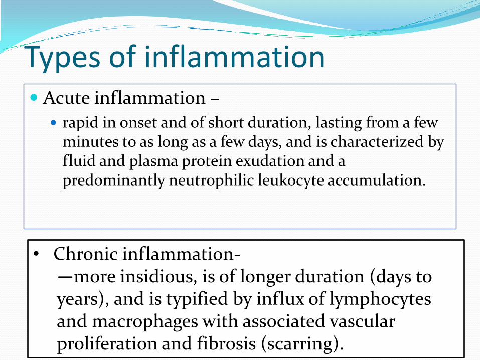
Types of inflammation Acute inflammation –
rapid in onset and of short duration, lasting from a few minutes to as long as a few days, and is characterized by fluid and plasma protein exudation and a predominantly neutrophilic leukocyte accumulation.
• Chronic inflammation-―more insidious, is of longer duration (days to years), and is typified by influx of lymphocytes and macrophages with associated vascular proliferation and fibrosis (scarring).

Acute inflammation Definitions
rapid tissue response to trauma or any other injurious agent
the local tissue response of living tissue to injury
the reaction of vascular tissue and other supportive elements of a tissue to an injury which results in formation of an exudates
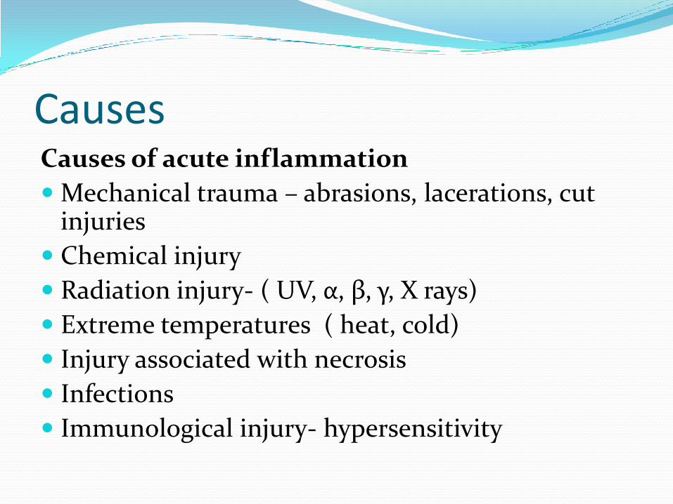
CausesCauses of acute inflammation
Mechanical trauma – abrasions, lacerations, cut injuries
Chemical injury
Radiation injury- ( UV, α, β, γ, X rays)
Extreme temperatures ( heat, cold)
Injury associated with necrosis
Infections
Immunological injury- hypersensitivity
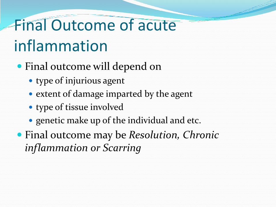
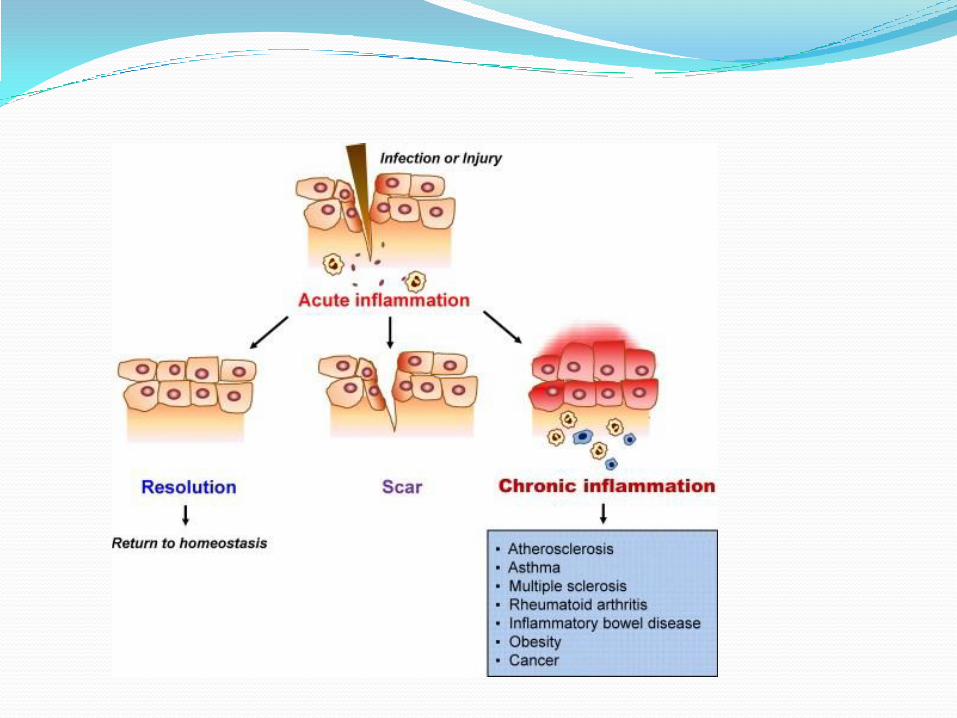
Final Outcome of acute inflammation Final outcome will depend on
type of injurious agent
extent of damage imparted by the agent
type of tissue involved
genetic make up of the individual and etc.
Final outcome may be Resolution, Chronic inflammation or Scarring
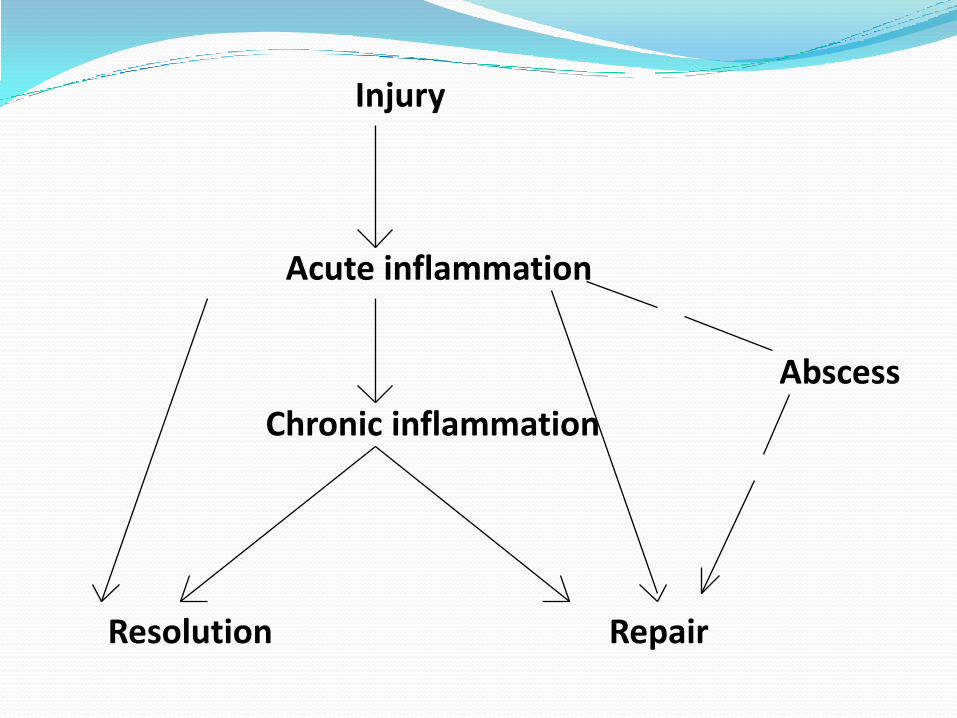
Acute inflammation
Chronic inflammation
RepairResolution
Abscess
Injury
Presentation1. Clinical presentation- cardinal signs
Redness and heat- due to increased blood flow to the inflamed site
• Swelling -caused by accumulation of fluid
• Pain - due to release of chemicals that stimulate nerve endings. Pain only happens where the appropriate sensory nerve endings exist in the inflamed area —
e.g. acute pneumonia- no pain unless the inflammation involves the parietal pleura, which has pain-sensitive nerve endings
•Loss of function has multiple causes
Presentation contd..
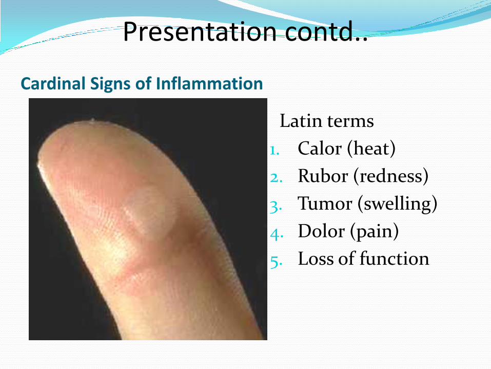
Cardinal Signs of Inflammation
Latin terms
1. Calor (heat)
2. Rubor (redness)
3. Tumor (swelling)
4. Dolor (pain)
5. Loss of function
Presentation contd..
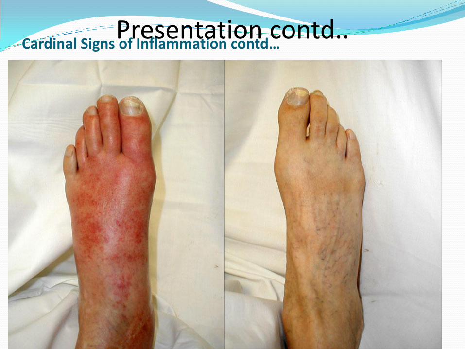
Cardinal Signs of Inflammation contd… Presentation contd..
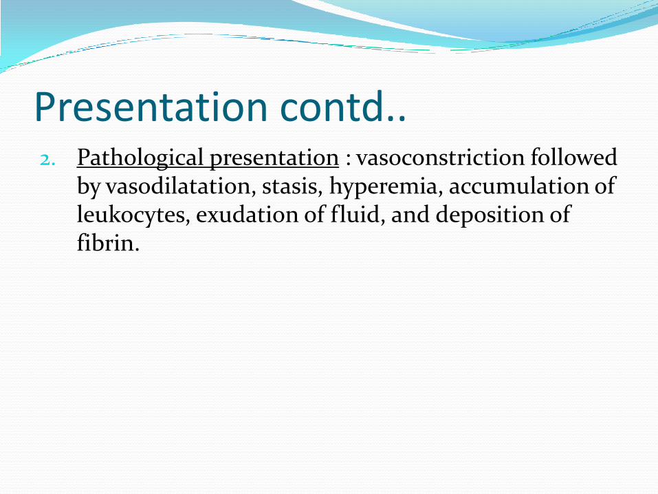
Presentation contd..2. Pathological presentation : vasoconstriction followed
by vasodilatation, stasis, hyperemia, accumulation of leukocytes, exudation of fluid, and deposition of fibrin.
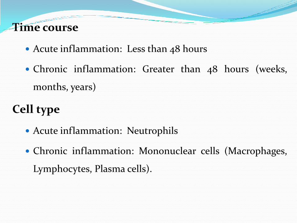
Time course
Acute inflammation: Less than 48 hours
Chronic inflammation: Greater than 48 hours (weeks,
months, years)
Cell type
Acute inflammation: Neutrophils
Chronic inflammation: Mononuclear cells (Macrophages,
Lymphocytes, Plasma cells).
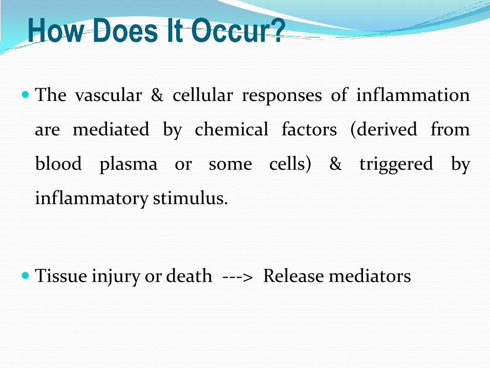
How Does It Occur? The vascular & cellular responses of inflammation
are mediated by chemical factors (derived from
blood plasma or some cells) & triggered by
inflammatory stimulus.
Tissue injury or death ---> Release mediators
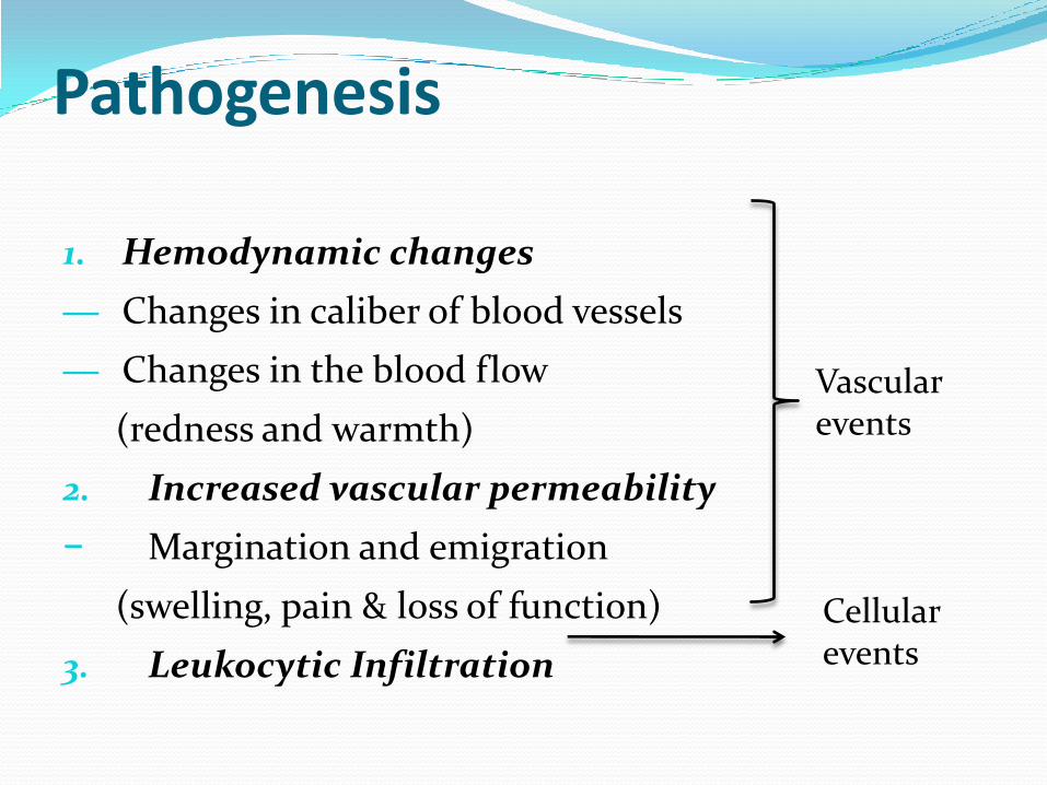
Pathogenesis 1. Hemodynamic changes
— Changes in caliber of blood vessels
— Changes in the blood flow
(redness and warmth)
2. Increased vascular permeability
− Margination and emigration
(swelling, pain & loss of function)
3. Leukocytic Infiltration
Vascular events
Cellular events
Integrated chain of events activated by chemical mediators, but perhaps transiently initiated by neurogenic mechanisms.
There is chronologic overlap among the 3 reactions and they may share common mediator mechanisms.
However, since the structural and biochemical basis of each of these responses is sufficiently different, they are best discussed separately.
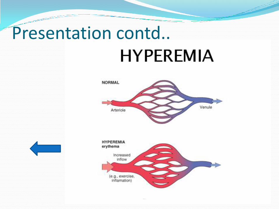
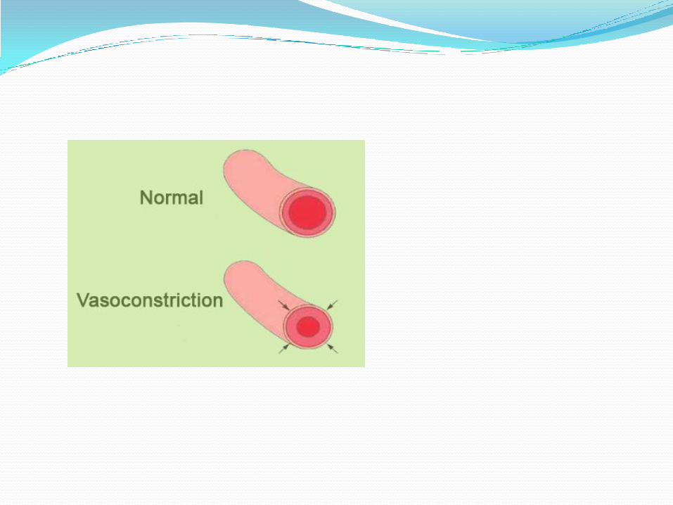
Changes in caliber of blood vessels1. Initial vasoconstriction
First response of arterioles to injury (3-5 secs or mins)
mild injury: it disappears within 3-5 seconds.
Severe injury: may remain for several minutes.
The mechanism of this vasoconstriction is unknown, but is probably neurogenic or adrenergic in origin
Due to direct mechanical stimulation of small blood vessels
Increased blood flow contd…2. Persistent vasodilatation
Follows initial vasoconstriction
Persists for the whole duration of inflammation
Initially, involves arterioles- result in opening of new capillaries and venular beds in the area.
Active process
Mainly chemical mediator mediated and is also contributed by neuronal mechanisms
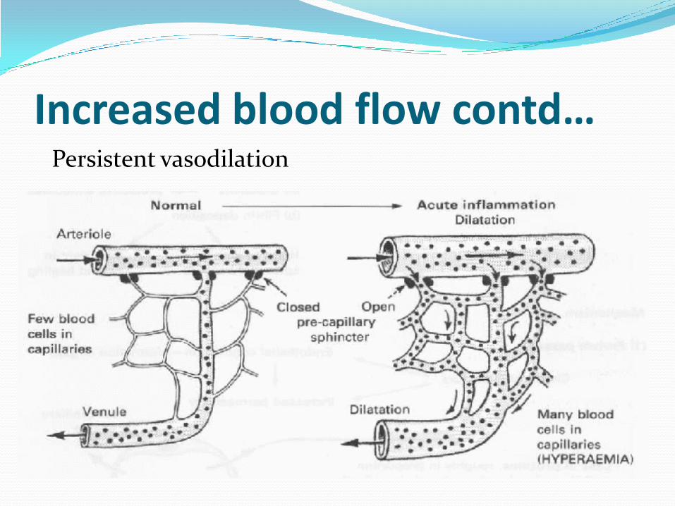
Increased blood flow contd…Persistent vasodilation
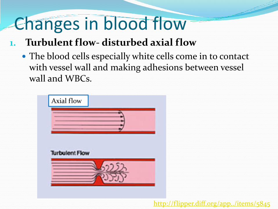
Changes in blood flow1. Turbulent flow- disturbed axial flow
The blood cells especially white cells come in to contact with vessel wall and making adhesions between vessel wall and WBCs.
http://flipper.diff.org/app../items/5845
Axial flow
Changes in blood flow contd..2. Increased velocity of blood flow –
transient (temporary)
due to the arteriolar dilatation
3. Increased amount blood flow
— Initially involves arterioles (see Persistent vasodilatation )
— Subsequent to vasodilatation, there is increased blood flow to the affected areas (this is the hallmark of the early hemodynamic changes in acute inflammation).
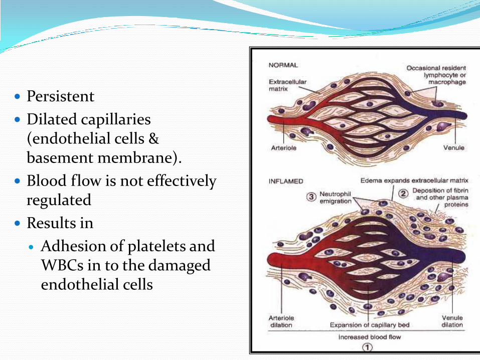
Persistent
Dilated capillaries (endothelial cells & basement membrane).
Blood flow is not effectively regulated
Results in
Adhesion of platelets and WBCs in to the damaged endothelial cells
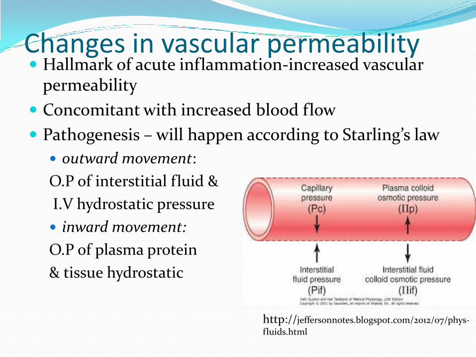
Changes in vascular permeability Hallmark of acute inflammation-increased vascular
permeability
Concomitant with increased blood flow
Pathogenesis – will happen according to Starling’s law
outward movement:
O.P of interstitial fluid &
I.V hydrostatic pressure
inward movement:
O.P of plasma protein
& tissue hydrostatic
http://jeffersonnotes.blogspot.com/2012/07/phys-
fluids.html
Outcome: retardation of blood flow- Slowing and/or stasis of the blood flow
Disrupts the laminar flow pattern and thereby displace the cellular elements to the periphery of the microvessels.
WBC fall out of the central column of flow and assume positions in contact with the endothelium.
Changes in vascular permeability contd..
Outcomes of changes in vascular permeability
1. Initial transudation - in the earliest stages of inflammation, vasodilatation, stasis increased hydrostatic pressure causes transudation of fluid
An ultrafiltrate of blood plasma
permeability of endothelium is usually normal
low protein content ( mostly albumin)
However, with the appearance of increased vascular permeability, there is exudation of large amounts of plasma proteins.
2. Exudate formation- formation of protein and cell rich extra vascular component produced mainly by inflammation ( two components- protein rich fluid and cells)
Fluid exudates
Cellular exudate
Phases of exudates formation1. Immediate transient phase
Begins: immediately after mild injury,
reaches peak : 5 - 10 minutes
phase out : within 15 to 30 minutes
Affects venules: the site of increased permeability and leakage(the capillaries are not affected).
Mediated by Histamine and other mediators
Due to contraction of endothelial cells which leads to the formation of intercellular gaps.
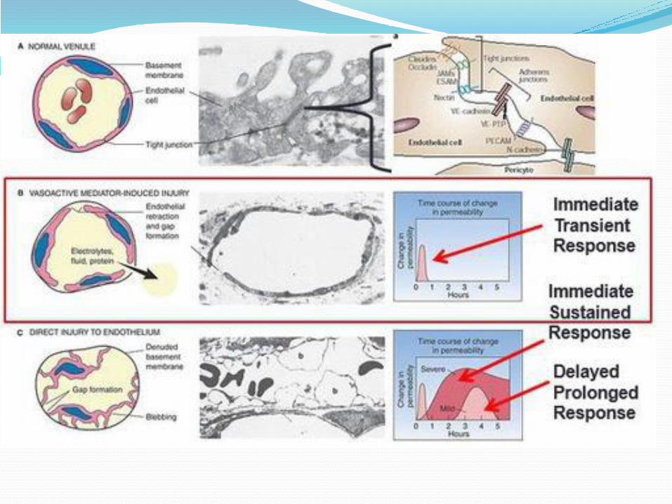
2. Immediate prolonged phase Appears immediately, high peak for several hours and
persists for days until the damaged vessels are thrombosed or repaired
Associated with severe injury causing direct endothelial cell damage
Site of increased permeability and vascular leakage- all levels of the microcirculation(venules, capillaries and arterioles.
Mechanism for increased permeability- "direct damage" to the vascular endothelium.
3. Delayed prolonged phase
Develops 4- 24 hrs(latent period) after the trauma lasts for several hours or days depending on form of injury.
Due to direct injury to the endothelium and chemical mediators
Occurs after mild to moderate thermal injury, or x-ray or ultraviolet irradiation, with certain bacterial toxins and in delayed hypersensitivity reaction.
Mechanism- unknown
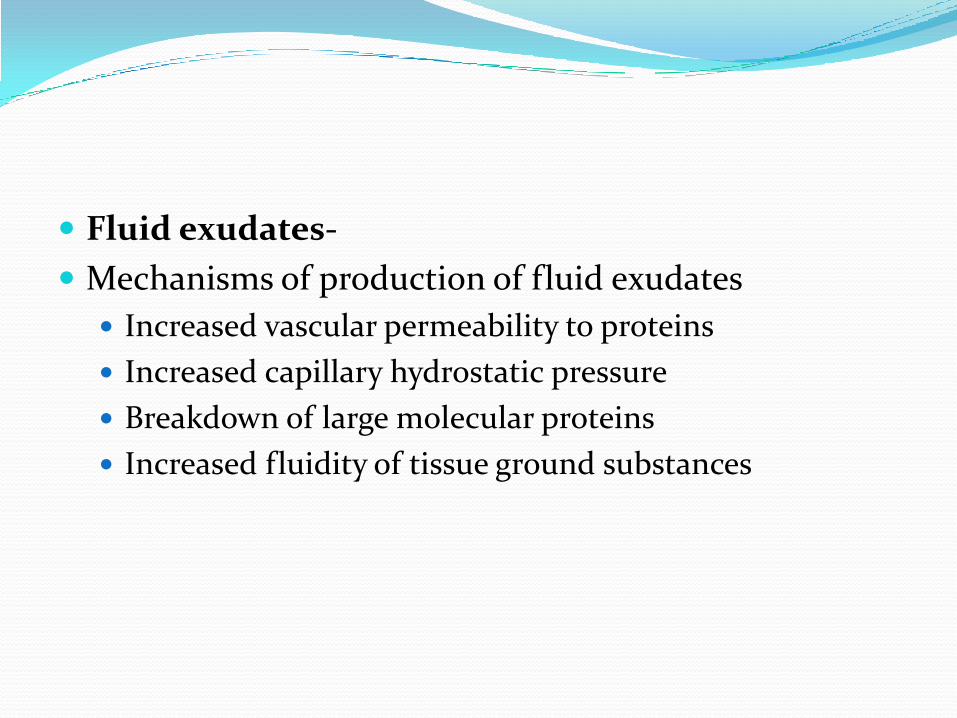
Fluid exudates-
Mechanisms of production of fluid exudates
Increased vascular permeability to proteins
Increased capillary hydrostatic pressure
Breakdown of large molecular proteins
Increased fluidity of tissue ground substances
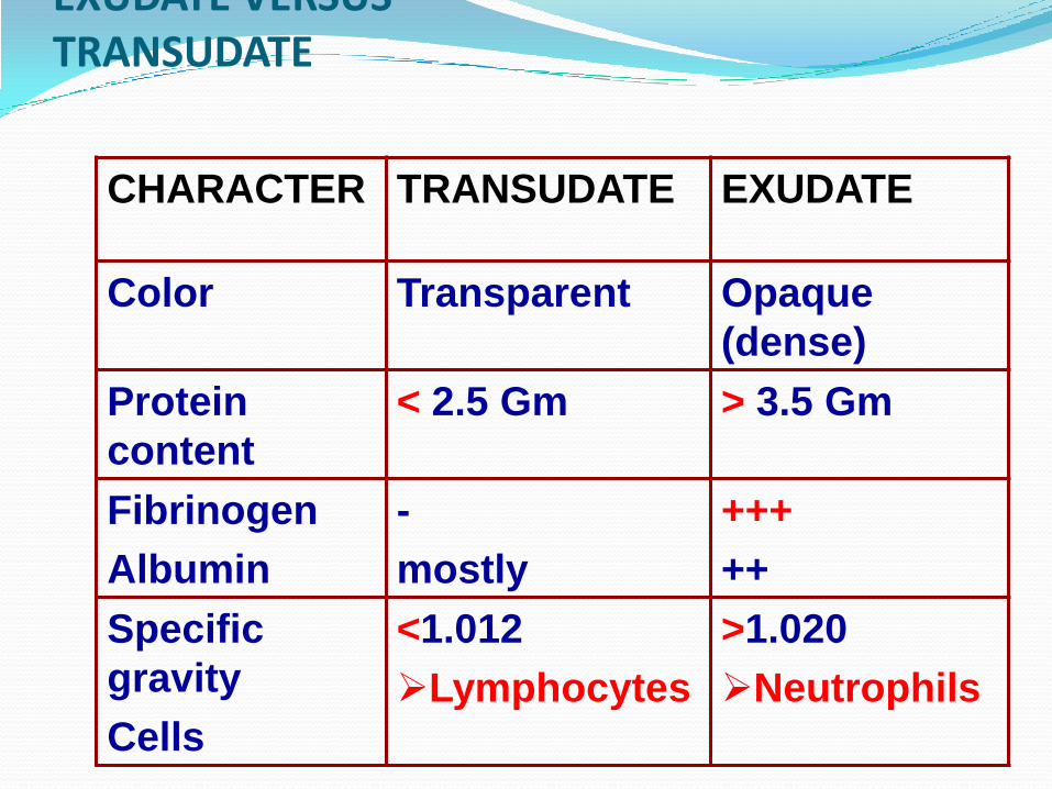
EXUDATE VERSUS TRANSUDATE
CHARACTER TRANSUDATE EXUDATE
Color Transparent Opaque
(dense)
Protein
content
< 2.5 Gm > 3.5 Gm
Fibrinogen
Albumin
-
mostly
+++
++
Specific
gravity
Cells
<1.012
Lymphocytes
>1.020
Neutrophils
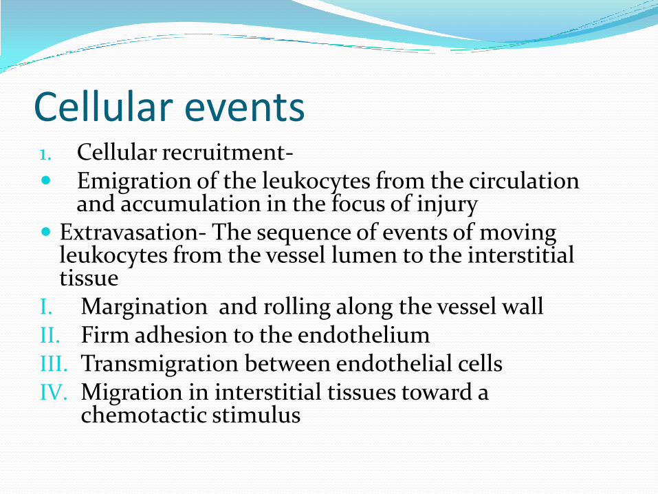
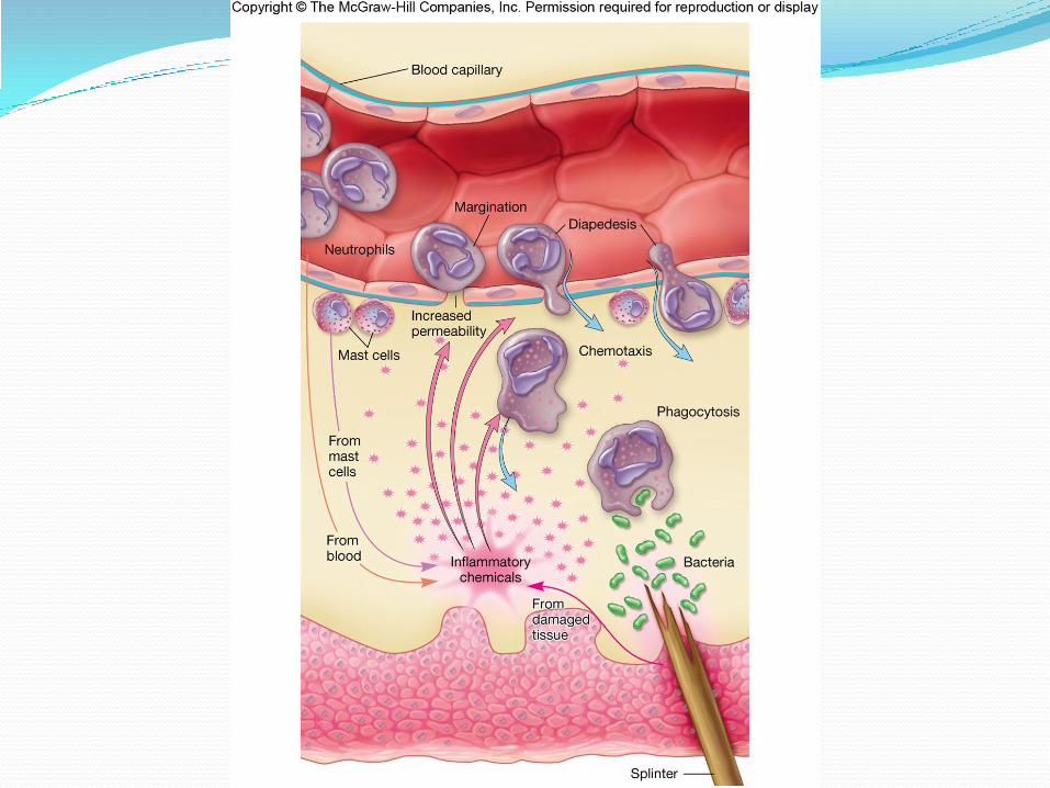
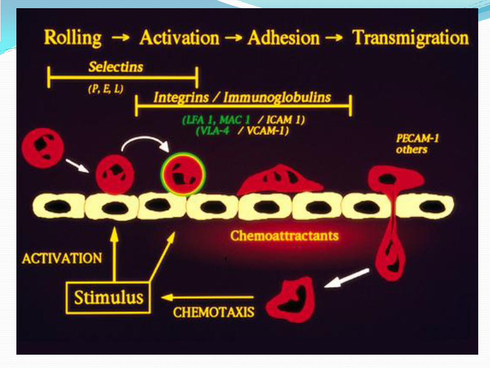
Cellular events1. Cellular recruitment- Emigration of the leukocytes from the circulation
and accumulation in the focus of injury Extravasation- The sequence of events of moving
leukocytes from the vessel lumen to the interstitial tissue
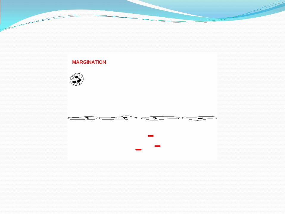
I. Margination and rolling along the vessel wall II. Firm adhesion to the endotheliumIII. Transmigration between endothelial cellsIV. Migration in interstitial tissues toward a
chemotactic stimulus
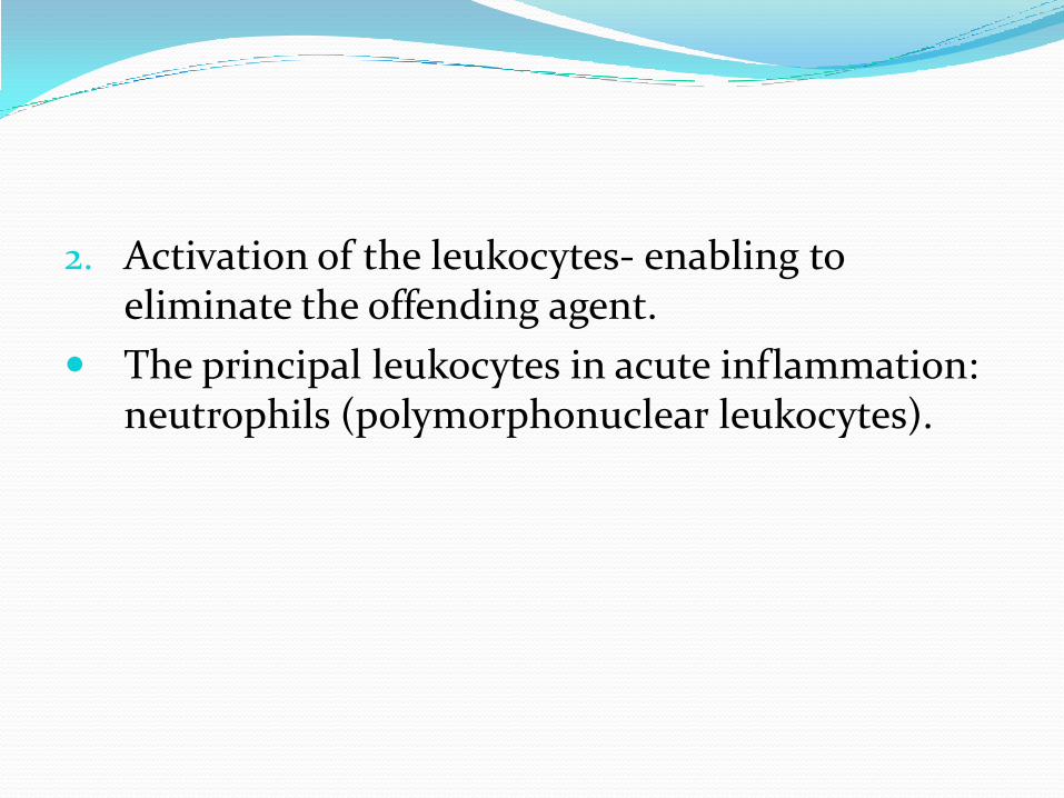
2. Activation of the leukocytes- enabling to eliminate the offending agent.
The principal leukocytes in acute inflammation: neutrophils (polymorphonuclear leukocytes).
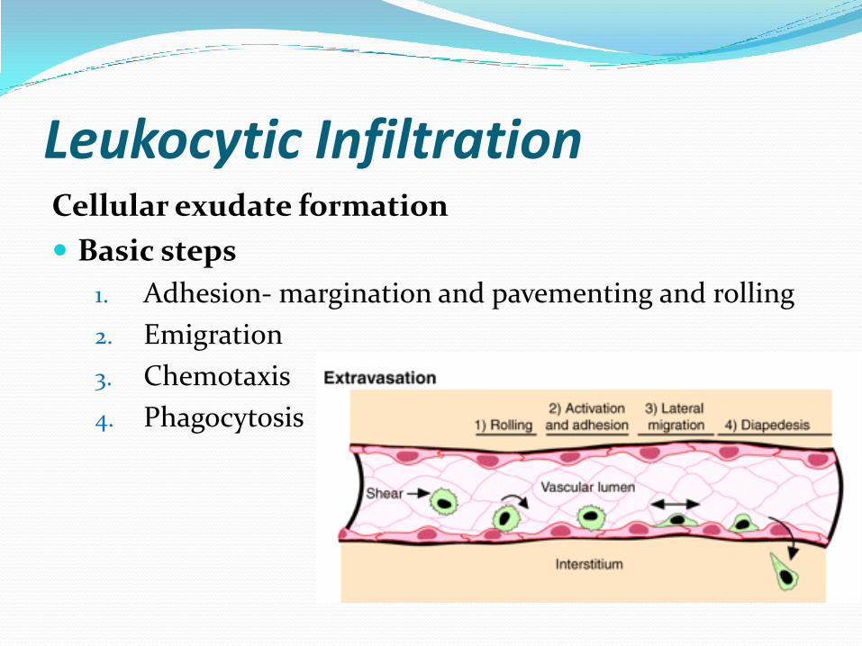
Leukocytic InfiltrationCellular exudate formation
Basic steps
1. Adhesion- margination and pavementing and rolling
2. Emigration
3. Chemotaxis
4. Phagocytosis
1. Adhesion of WBCs to the endothelium Circulating leukocytes from the central blood flow move
toward the endothelial surface. Then, initial temporary adhesions form and later more
firm adhesions occur. Margination- Leukocytes localize at outer margin of the
blood flow adjacent to the vascular endothelium. Pavementing- Leukocytes line the surface Rolling- Mediated by the action of endothelial P-and E-
selectins loosely binding to leukocytes and producing a characteristic rolling movement of the WBCs along with the endothelial surface
Rolling- Process of WBC tumbling on the endothelial surface. Process involved: Locally produced cytokines and other mediators activate the
endothelial cells.
They express adhesion molecules to which the leukocytes attach loosely.
Then, WBCs bind and detach and start tumbling which leads rolling of WBC.
The weak and transient interactions involved in rolling are mediated by the selectin family of adhesion molecules
The rolling leukocytes sense changes in the endothelium and initiate firm adhesion to endothelial surfaces.
This adhesion is mediated by integrins expressed on leukocyte cell surfaces.
2. Emigration of white cellsDefinition: the active process by which motile leukocytes escape
from the blood vessel lumen into the perivascular tissues (neutrophils, basophils, monocytes and lymphocytes all use the same pathway).
Process: Adhered white cells produce pseudopodia through the
endothelial cells Then the rest of the cytoplasm and the nucleus are pushed to the
other side
Initially neutrophils are migrated
Later monocytes are migrated
In the tissue monocytes become macrophages
Lymphocytes emigrate later and in specific types of infections (eg, viral)
Emigration of leucocytes is driven by chemokines produced in extravascular tissues, which stimulate movement of the leukocytes toward their chemical gradient.
• Diapedesis- trans-migration of WBCs across the endothelium
3. Chemotaxis
Definition-directional movement of leukocytes toward sites of infection or injury along a chemical gradient
In inflammation the neutrophils and other leucocytes move due to the chemotaxis.
Chemotaxins or chemotactic agents- The chemical agents involved in chemotaxis
Chemotaxins
activate the white cells- leukocyte activation
increase the concentration of Ca inside the cells
assembly of cytoskeletal contractile elements
including actin filaments thus aiding movements
Leukocyte activation Stimuli for activation- microbes, products of necrotic
cells, and several chemical mediators
Leukocyte activation results in the enhancement of1. Phagocytosis of particles
2. Intracellular destruction of phagocytosed microbes and dead cells by substances produced in phagosomes, including reactive oxygen and nitrogen species and lysosomal enzymes
3. Liberation of substances that destroy extracellular microbes and dead tissues, which are similar to the substances produced within phagocytic vesicles.
4. Production of mediators, including arachidonic acid metabolites and cytokines, that amplify the inflammatory reaction, by recruiting and activating more leukocytes
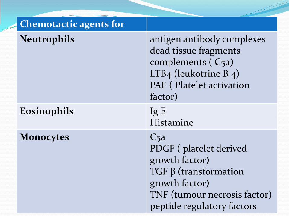
Chemotactic agents for
Neutrophils antigen antibody complexesdead tissue fragmentscomplements ( C5a)LTB4 (leukotrine B 4)PAF ( Platelet activation factor)
Eosinophils Ig EHistamine
Monocytes C5aPDGF ( platelet derived growth factor)TGF β (transformation growth factor)TNF (tumour necrosis factor)peptide regulatory factors
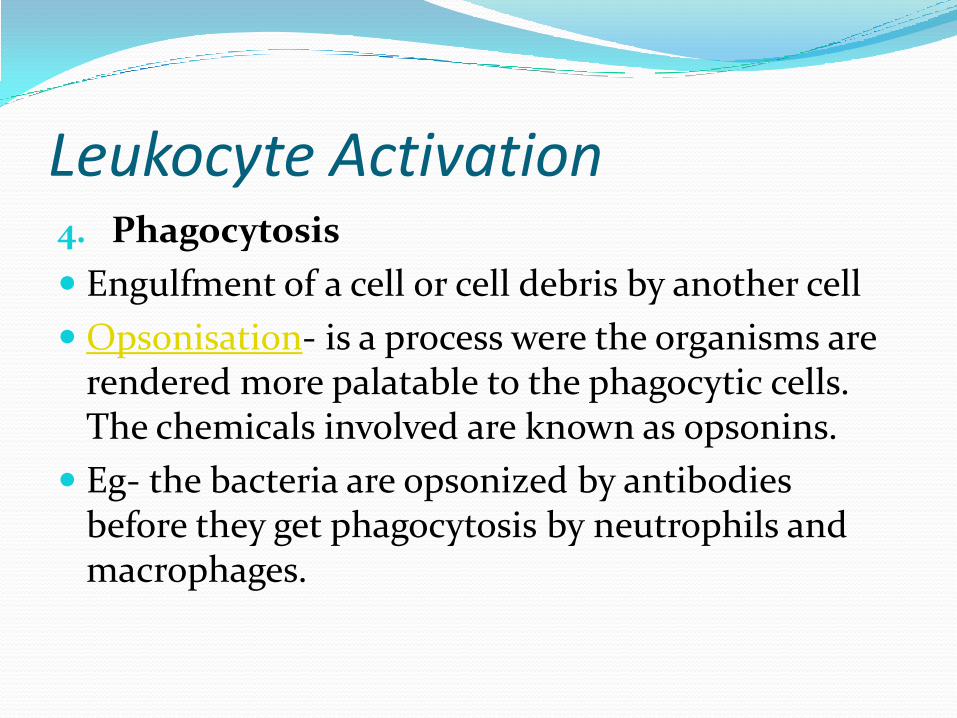
Leukocyte Activation4. Phagocytosis
Engulfment of a cell or cell debris by another cell
Opsonisation- is a process were the organisms are rendered more palatable to the phagocytic cells. The chemicals involved are known as opsonins.
Eg- the bacteria are opsonized by antibodies before they get phagocytosis by neutrophils and macrophages.
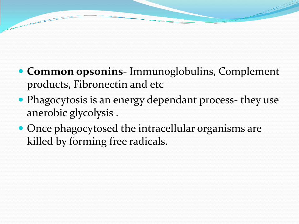
Common opsonins- Immunoglobulins, Complement products, Fibronectin and etc
Phagocytosis is an energy dependant process- they use anerobic glycolysis .
Once phagocytosed the intracellular organisms are killed by forming free radicals.
ROS Reactive oxygen species are
molecules like hydrogen peroxide
ions like the hypochlorite ion
radicals like the hydroxyl radical - most reactive among all
the superoxide anion which is both ion and radical.
A radical (also called a "free radical") is a clusters of atoms one of which contains an unpaired electron (shown in red) in its outermost shell of electrons. This is an extremely unstable configuration, and radicals quickly react with other molecules or radicals to achieve the stable configuration of 4 pairs of electrons in their outermost shell (one pair for hydrogen).
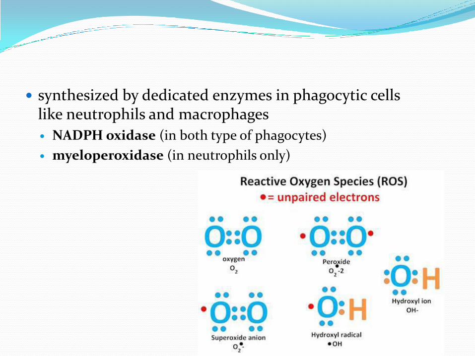
synthesized by dedicated enzymes in phagocytic cells like neutrophils and macrophages
NADPH oxidase (in both type of phagocytes)
myeloperoxidase (in neutrophils only)
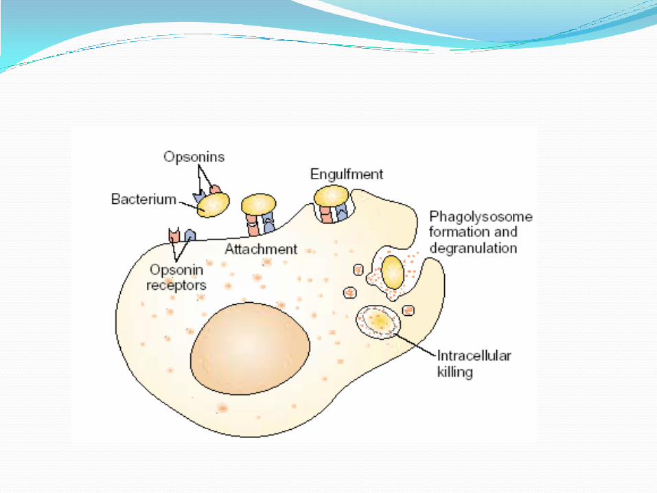
Phagocytosis(1) recognition and attachment of the particle to the
ingesting leukocyte
(2) engulfment, with subsequent formation of a phagocytic vacuole
(3) killing and degradation of the ingested material.
We will see the video
Types of acute inflammationClassification (Other Types of
Inflammation) Based on Exudate
Based on Histological Features
Based on Causative Agent
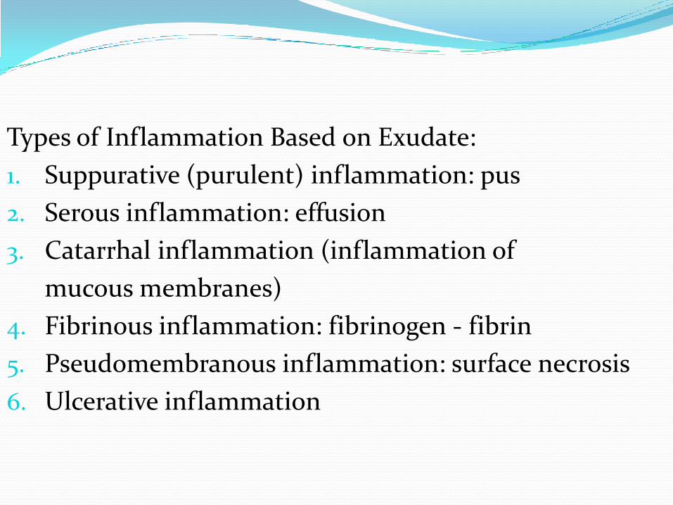
Types of Inflammation Based on Exudate:
1. Suppurative (purulent) inflammation: pus
2. Serous inflammation: effusion
3. Catarrhal inflammation (inflammation of
mucous membranes)
4. Fibrinous inflammation: fibrinogen - fibrin
5. Pseudomembranous inflammation: surface necrosis
6. Ulcerative inflammation
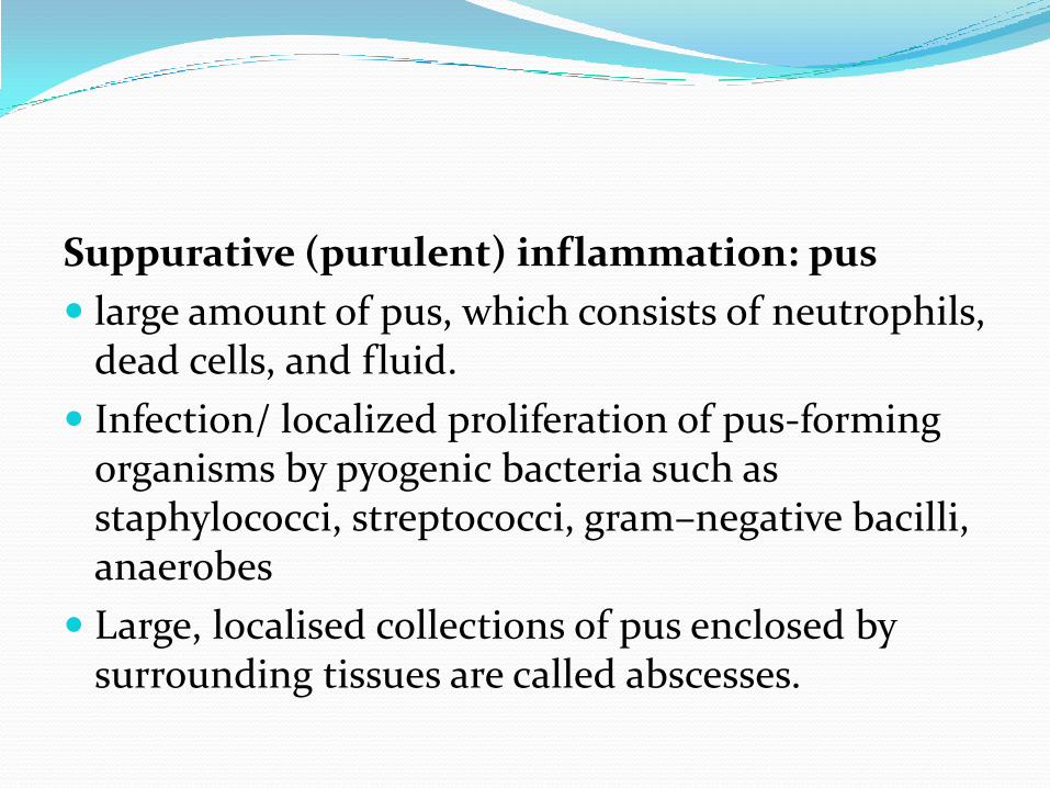
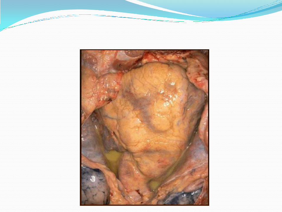
Suppurative (purulent) inflammation: pus
large amount of pus, which consists of neutrophils, dead cells, and fluid.
Infection/ localized proliferation of pus-forming organisms by pyogenic bacteria such as staphylococci, streptococci, gram–negative bacilli, anaerobes
Large, localised collections of pus enclosed by surrounding tissues are called abscesses.
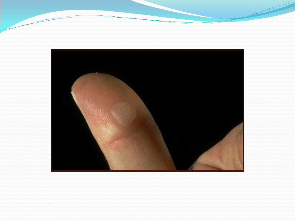
Serous inflammation: effusion
Thin, watery exudate
Example - blister in second-degree burns, viral pleuritis
Characterized by the abundant effusion of non-viscous serous fluid, commonly produced by mesothelial cells of serous membranes, but may be derived from blood plasma. Skin blisters exemplify this pattern of inflammation.
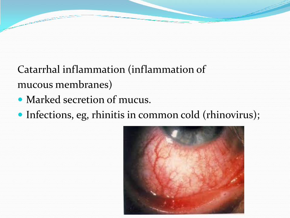
Catarrhal inflammation (inflammation of
mucous membranes)
Marked secretion of mucus.
Infections, eg, rhinitis in common cold (rhinovirus);
Types of acute inflammation
1. Fibrinous inflammation:
Resulting large increase in vascular permeability
Allows fibrin to pass through the blood vessels.
If an appropriate procoagulative stimulus is present, such as cancer cells, a fibrinous exudateis deposited.
Commonly seen in serous cavities, where the conversion of fibrinous exudate into a scar can occur between serous membranes, limiting their function.
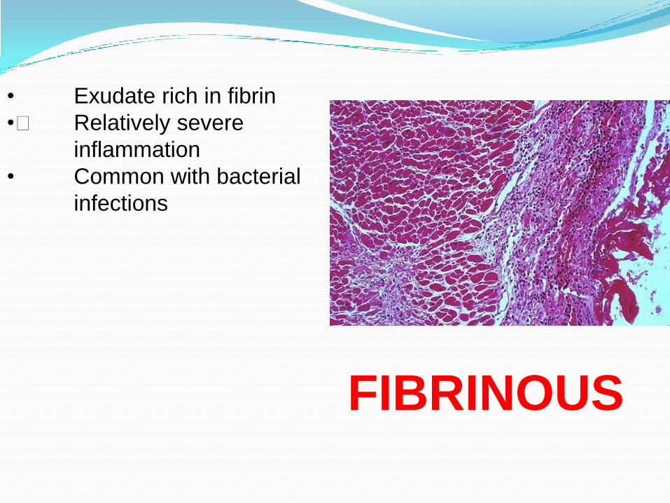
FIBRINOUS
• Exudate rich in fibrin
• Relatively severe
inflammation
• Common with bacterial
infections
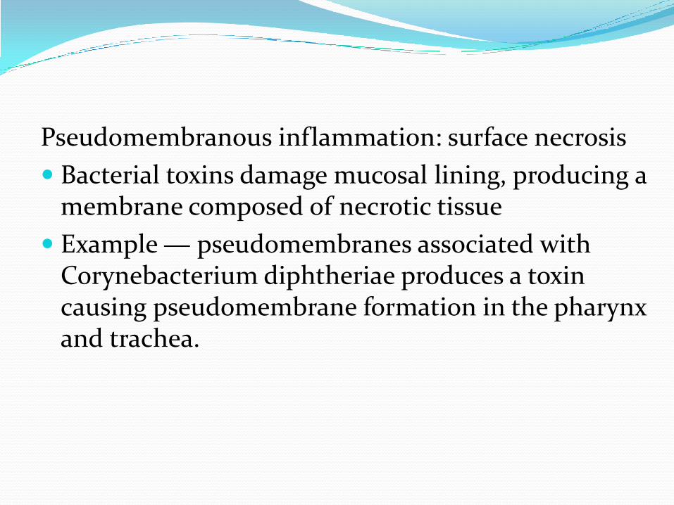
Pseudomembranous inflammation: surface necrosis
Bacterial toxins damage mucosal lining, producing a membrane composed of necrotic tissue
Example ― pseudomembranes associated with Corynebacterium diphtheriae produces a toxin causing pseudomembrane formation in the pharynx and trachea.
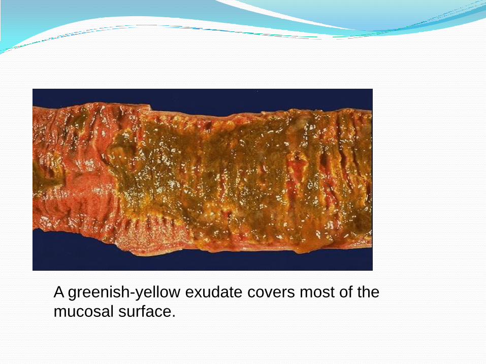
A greenish-yellow exudate covers most of the
mucosal surface.
Ulcerative inflammation:
Inflammation occurring near an epithelium can result in the necrotic loss of tissue from the surface, exposing lower layers. The subsequent excavation in the epithelium is known as an ulcer.
Example ― Ulcerative colitis
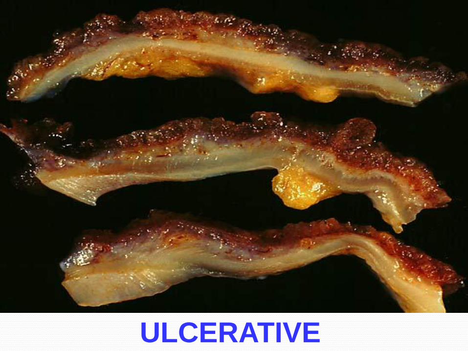
ULCERATIVE
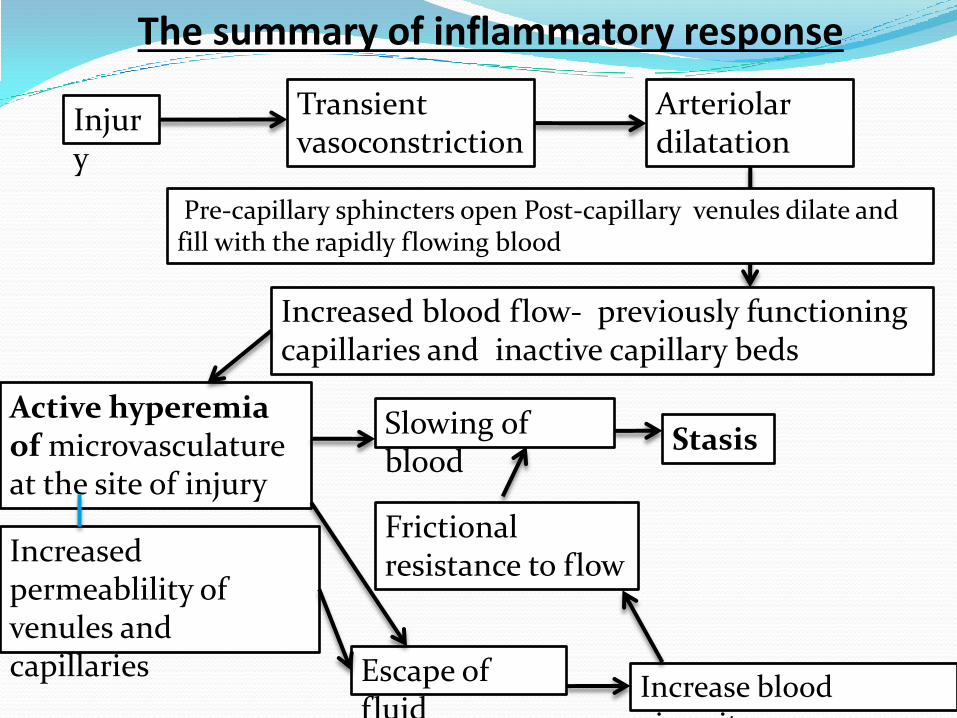
Pre-capillary sphincters open Post-capillary venules dilate and fill with the rapidly flowing blood
Injury
Arteriolar dilatation
Slowing of blood
Escape of fluid
Transient vasoconstriction
Increased blood flow- previously functioning capillaries and inactive capillary beds
Active hyperemia of microvasculature at the site of injury
Stasis
Increased permeablility of venules and capillaries
Increase blood viscosity
Frictional resistance to flow
The summary of inflammatory response
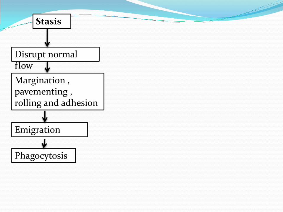
Disrupt normal flow
Stasis
Margination , pavementing , rolling and adhesion
Emigration
Phagocytosis
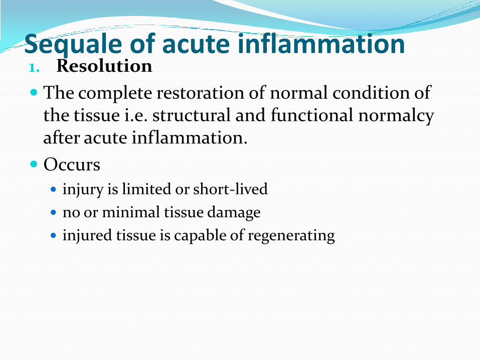
Sequale of acute inflammation1. Resolution
The complete restoration of normal condition of the tissue i.e. structural and functional normalcy after acute inflammation.
Occurs
injury is limited or short-lived
no or minimal tissue damage
injured tissue is capable of regenerating

Factors aiding the resolution
minimal cell death and tissue damage
complete elimination of causative agent
local conditions favoring the removal of debris and fluid
• Before resolution the acute inflammatory response has to be terminated.
• This involves
neutralization, decay, or enzymatic degradation of the various chemical mediators
normalization of vascular permeability
cessation of leukocyte emigration, with subsequent death (by apoptosis) of extravasated neutrophils.
Basic steps involves in complete resolution
Solution of fibrin by enzymes (polymorphs and fibrinolysins)
Removal of excess fluid by blood vessels and lymphatics
Removal of debris by phagocytotic cells
Reduction of blood flow and restoration of normal flow
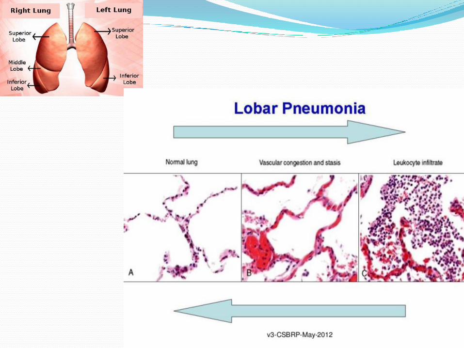
The best example for complete resolution is resolution of pneumococal lobar pneumonia.
2. Suppuration
Suppuration is the formation of pus, a mixture of living, dying and dead neutrophils and bacteria, cellular debris and sometimes globules of lipid.
The causative stimulus must be fairly persistent and is virtually always an infective agent, usually pyogenicbacteria (i.e.,Staphylococcus aureus, Streptococcus pyogenes, Neisseria species or coliform organisms).
Once pus begins to accumulate in a tissue, it become surrounded by a 'pyogenic membrane‘ consisting of emerging capillaries, neutrophils and occasional fibroblasts.
Such a collection of pus is called an abscess, and bacteria within the abscess cavity are relatively inaccessible to antibodies and to antibiotic drugs (thus, for example, acute osteomyelitis, an abscess in the bone marrow cavity, is notoriously difficult to treat).
Abscess formation:
"A localized collection of pus (suppurative inflammation)
appearing in an acute or chronic infection, and associated
with tissue destruction, and swelling.
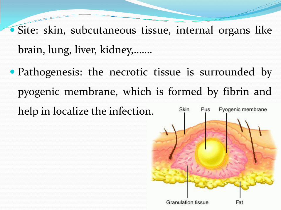
Site: skin, subcutaneous tissue, internal organs like
brain, lung, liver, kidney,…….
Pathogenesis: the necrotic tissue is surrounded by
pyogenic membrane, which is formed by fibrin and
help in localize the infection.
Evolution of an abscess
Bacteria causes tissue damage and necrosis
Bacteria multiply.
The polymorphs packed in the central zone and the periphery shows hyperemia and oedema.
Pus forms in the centre and demarcation of abscess by pyogenic membrane. Pyogenic membrane consists of newly formed capillaries, polymorphs and fibroblasats.
Pus is usually liberated through an epithelial surface and rest of the tissue is healed with a scar.
Pus is discharged in to a blood vessels multiple abscess and septicemia occurs.
Pus may solidify, calcify and later form a calcificnodule
With a partial discharge of pus chronic sinus occurs.
With the discharge in to two epithelial surfaces fistula occurs.
Organization and fibrosis
Organisation of tissues is their replacement by granulation tissue.
Organization occurs during acute inflammatory process with when there is an excessive exudation
large amounts of fibrin are formed, which cannot be removed completely by fibrinolytic enzymes from the plasma or from neutrophil polymorphs
when there is an excessive necrosis ( tissue damage)
when the local conditions are unfavorable in removing debris
in certain types of tissue ( eg- pleura)
Progression to chronic inflammation
Process
new capillaries grow into the inert material (inflammatory exudate)
macrophages migrate into the zone
fibroblasts proliferate
fibrosis
3. Chronic inflammation
If inflammatory the agent is not removed, progress to the chronic stage.
In addition to organisation of the tissue just described, the character of the cellular exudatechanges, with lymphocytes, plasma cells, and macrophages (sometimes including multinucleate giant cells) replacing the neutrophil polymorphs.

Thank you!!