in vivo stereological assessment of human cerebellar …. gunderson hjg, bendsen tf, korbo, et al....
TRANSCRIPT

P.R. Escalona1
W. M. McDonald1
P. M. Doraiswamy 1
0. B. Boyko2
M. M. Husain3
G. S. Figiel4
D. Laskowitz 1
E. H. Ellinwood, Jr. 1
K. R. R. Krishnan 1
Received October 24, 1990; revision requested February 6, 1991 ; revision received April 10, 1991 ; accepted April 17, 1991 .
This work was supported in part by NIMH grants MH-44716 and MH-4221 0.
' Department of Psychiatry, Duke University Medical Center, Durham, NC 27710. Address reprint requests to K. R. R. Krishnan, Box 3215.
2 Department of Radiology, Duke University Medical Center, Durham, NC 27710.
3 Department of Psychiatry, UT -Southwestern Medical School, Dallas , TX 75235.
' Department of Psychiatry, Washington University, St. Louis, MO 63110.
0195-6108/91/1205-0927 © American Society of Neuroradiology
927
In Vivo Stereological Assessment of Human Cerebellar Volume: Effects of Gender and Age
Intermediate T2-weighted MR images and a systematic sampling stereological method were used in 37 normal volunteers, 24 to 79 years old, to assess the effects of age and sex on cerebellar volume. Female subjects (n = 21) had significantly smaller cerebellar volumes compared with males (n = 16) of similar age (t = -3.9, p < .0008, two-tail t test). Using straight-line, univariate regression, we determined that age was not a significant predictor of cerebellar volume (R2 = 0.07, t = -1.66, p = 0.11 ), whereas gender did appear to account for a significant amount of variability in cerebellar volume (R2 = 0.33, t = -4.13, p = .0002). The mean absolute cerebellar volume in this study was 112 ml (SD ± 16) for all subjects, 104 ml (SO ± 10) for females, and 122 ml (SD ± 16) for males.
This study demonstrates the feasibility of using MR images along with a systematic stereological method to assess in vivo human cerebellar volume, thereby providing a research tool to correlate cerebellar morphology with cognitive and neuromotor function.
AJNR 12:927-929, September/October 1991
Traditionally , the cerebellum has been associated primarily with motor function [1]. However, recent studies have implicated the cerebellum in neuropsychiatric conditions [2-5]. Neuropathologic evidence, as well as CT studies, have suggested cerebellar atrophy with normal aging [6-8] .
Previous attempts to study changes in the cerebellum with aging and neuropsychiatric disorders have been hampered by the lack of in vivo methods to assess cerebellar volume. Previous efforts have used subjective scoring methods to assess morphologic changes in the cerebellum [8, 9]. With the advent of MR imaging, small changes in individual structures can be more easily analyzed . In addition, the development of a systematic sampling stereological method for volumetric determinations allows the rapid, precise, and efficient determination of the volumes of anatomic structures. We reported in a previous publication [1 0] the first application of a systematic sampling stereological method for the in vivo determination of changes in caudate volume with normal aging using MR imaging. In the present report, we apply this stereological method to evaluate the effects of age and gender on human cerebellar volume.
Materials and Methods
Subjects
All 37 subjects were normal volunteers from the community from whom written informed consent was obtained. Each subject had a complete neurologic, psychiatric, and physical examination and was determined to be free of any significant neurologic disease (stroke, transient ischemic attacks, tumor, seizure, dementia) or psychiatric illness , including drug abuse and alcoholism. The 37 subjects (16 men, 21 women) were 24 to 79 years old (mean, 54.2 [± 19 SD] years).

928 ESCALONA ET AL. AJNR:12, September/October 1991
MR Acquisition
MR imaging was performed with a 1.5-T GE Signa system. The subjects were positioned in a quadrature head coil with the canthomeatal line at 0° from the vertical axis and the laser of the imager grid centered at the nasion. Five-mill imeter-thick axial slices (interstice gap of 2.5 mm) were obtained parallel to the canthomeatalline. Spinecho acquisition parameters were 500/20/1 (TR/TEjexcitations) for the T1-weighted images and 2800/30/1 and 2800/80/1 for intermediate (first-echo) and T2-weighted (second-echo) images, respectively . The first-echo image was used to measure the cerebellar volume, which was adequate for the distinction of the cerebellar white and gray matter from surrounding CSF, bone, and vessels. Cerebellar volumes , estimated by using either the first or second echo, were highly correlated (intraclass correlation coefficient = 0.96). T1- and T2-weighted images were reviewed to exclude disease.
Volume Measurements
Cerebellar volume was measured for every slice on which it was seen by using a systematic sampling and a point-counting method (11-14). The method is based on the Cavalieri theorem of systematic sampling (3) . For this to be applicable, the structure should be sampled at fixed intervals, and the sampling should be initiated at random (14, 15). MR imaging of the brain involves consecutive sectioning of uniform thickness at fixed intervals, and the initial cut intersects the cerebellum randomly , thus satisfying the basic requirements of this method. Under these circumstances the volume of the structure is given by the formula vol = (area of cross section) x (length of structure) x (magnification factorf In this way the area of the section was determined by means of the point-counting method (11 , 12). This method is based on the random interaction of points on the structure. We used a square grid placed randomly on the cerebellum. Thus, the cerebellar volume was estimated by using the formula (slice thickness+ interstice gap) x (number of points) x (area of square on lattice grid) x (magnification factor)2 [13-15) (see Fig. 1 ).
All the measurements were made by one of the authors who was blinded to the subjects ' age and gender. The volumes represent the sum of the right and left cerebellar hemispheres and include the vermis and deep nuclei. The fourth ventricle and the CSF were excluded from the volumetric measurements.
Statistical Analysis
Data were analyzed with PC-SAS software (SAS Institute, Cary, NC). All data are reported as the mean (± SD). The two-tai led t test procedure was used to evaluate significant differences between groups, and the Pearson's correlation coefficient was used to evalu-ate correlations between variables. A p value < .05 was considered
Fig. 1.-MR image of cerebellum with square grid used to estimate cerebellar volume. Number of points that intersect the cerebellum are counted on each section (see Methods).
significant. A straight-line, univariate regression analysis was used to evaluate the relationship of both age and gender to cerebellar volume.
Results
All T2-weighted images of the cerebrum showed no abnormalities. For all subjects the mean cerebellar volume was 112 (± 16) ml with a range of 81 to 152 mi. Women had significantly smaller cerebellar volume than men (t = -3.9, df = 23.5, p < .0008, two-tailed t test) (see Table 1 ).
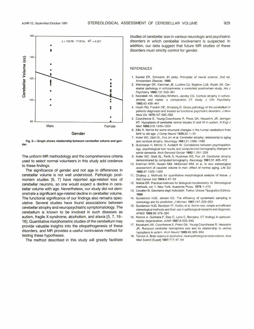
Using straight-line, univariate regression, we determined that age was not a significant predictor of cerebellar volume (R2 = 0.07, t = -1.66, p = .11) (see Fig. 2). However, gender did appear to account for a significant amount of variability in cerebellar volume (R2 = 0.33, t = -4.13, p = .0002), with women tending to have smaller cerebellar volumes than men (see Fig. 3).
Discussion
Findings in the study of cerebellar volumes in normal subjects between the ages of 24 and 79 years showed that (1) men have a significantly larger cerebellar volume than women and (2) age is not a significant predictor of cerebellar volume.
TABLE 1: Cerebellar Volumes in Males and Females
Mean age (±SD) (years) Age range (years) Cerebellar volume (ml) Cerebellar volume range (ml)
Males (n = 16)
50.8 ± 21 24-79
122 ± 16 96-152
at= -3.92 , df = 23.5, p < .0008 two-tail t test.
Females (n = 21)
55.1 ± 19 26-78
104 ± 10" 81-120
180~--------------------------------------~
y • 123.81 · 0.211aex R'2. 0.073
150 • • •
u 140
~ • Gl E 130 :I • 0 • • > 120 i'o.... _. 0 ... .!!! ... • ! 0
110 0 • ! 0
~ 0 c
100
• o• 0 • J'\
0:0 cr 0 e 0
0 90
• 0
0
80+---~n~------~-----r------r------r----~ 20 30 40 50 eo 70 80
:~es • 0 F B!~~
Age
Fig. 2.~Graph shows relationship between cerebellar volume and age.
AJNR :12, September/October 1991 STEREOLOGICAL ASSESSMENT OF CEREBELLAR VOLUME 929
160,-----------------------------------------~
y = 122.06- 17.81 2x. R2 = 0.327
• I
u 140
~ Q) • E :::::1 • 0 > 120 ... ~
~ ~ ~
100
• • •
• 80
Male Female
Gender
Fig. 3.-Graph shows relationship between cerebellar volume and gender.
The uniform MR methodology and the comprehensive criteria used to select normal volunteers in this study add credence to these findings.
The significance of gender and not age in differences in cerebellar volume is not well understood. Pathologic postmortem studies [6, 7] have reported age-related loss of cerebellar neurons, so one would expect a decline in cerebellar volume with age. Nevertheless, our study did not demonstrate a significant age-related decline in cerebellar volume. The functional significance of our findings also remains speculative. Several studies have found associations between cerebellar atrophy and neuropsychiatric symptomatology. The cerebellum is known to be involved in such diseases as autism, fragile X-syndrome, alcoholism, and ataxia [5 , 7, 16-18]. Quantitative morphometric studies of the cerebellum may provide valuable insights into the etiopathogenesis of these disorders, and MR provides a useful noninvasive method for testing these hypotheses.
The method described in this study will greatly facilitate
studies of cerebellar size in various neurologic and psychiatric disorders in which cerebellar involvement is suspected. In addition , our data suggest that future MR studies of these disorders must strictly control for gender.
REFERENCES
1. Kandel ER , Schwartz JH (eds). Principles of neural science, 2nd ed. Amsterdam: Elsevier, 1985
2. Weinberger DR, Kleinman JE, Luchins DJ , Bigelow LLB, Wyatt JW. Cerebellar pathology in schizophrenia: a controlled postmortem study. Am J Psychiatry 1980;137: 359-361
3. Nasrallah HA, McCalley-Whitters, Jacoby CG. Cortical atrophy in schizophrenia and mania: a comparative CT study. J Clin Psychiatry 1982;43 :439-441
4. Heath RG, Frankl in DE, Shraberg D. Gross pathology of the cerebellum in patients diagnosed and treated as funct ional psychiatric disorders. J Nerv Ment Dis 1979;167 :585-592
5. Courchesne E, Yeung-Courchesne R, Press GA, Hesselink JR, Jernigan HT. Hypoplasia of cerebellar vermallobules VI and VII in autism. N Eng/ J Med 1988;318: 1349-1354
6. Ellis R. Norms for some structural changes in the human cerebellum from birth to old age. J Camp Neuro/1920;32:1-33
7. Koller WC, Glatt SL, Fox JH, et al. Cerebellar atrophy: relationship to aging and cerebral atrophy. Neurology 1981 ;31 :1486-1488
8. Gutzmann H, Klimitz H, Avdaloff W. Correlations between psychopathology, psychological test results and computerized tomography changes in senile dementia. Arch Gerontal Geriatr 1982;1 :241 -259
9. Koller WC, Glatt SL, Perlik S, Huckman MS, Fox JH. Cerebellar atrophy demonstrated by computed tomography. Neurology 1981 ;31 :405-412
10. Krishnan KRR , Husain MM, McDonald WM , et al. In vivo stereological assessment of caudate volume in man: effect of normal aging. Life Sci 1990;47 : 1325-1329
11 . Chalkey J. Methods for quantitative morphological analysis of tissue. J Nat! Cancer lnst 1943;4:47-53
12. Weibel ER. Practical methods for biological morphometry. In: Stereological methods , vol. 1. New York: Academic Press, 1979:1-415
13. Cavalieri B. Geometra degli indivisibili. Torino: Unione Tipografico Editrice , 1966
14. Gunderson HJG, Jensen EG. The efficiency of systematic sampling in stereology and its prediction. J Microsc 1987;147:229-263
15. Gunderson HJG, Bendsen TF, Korbo, et al. Some new, simple and efficient stereological methods and their use in pathological research and diagnosis. APMIS 1988;96:379-394
16. Ramos A, Quintana F, Diez C, Leno C, Berciano. CT findings in spinocerebellar degeneration . AJNR 1987;8:635-640
17. Murakami JW, Courchesne E, Press GA, Yeung-Courchesne R, Hesselink JR. Reduced cerebellar hemisphere size and its relationship to vermal hypoplasia in autism. Arch Neuro/1989;46 :689-694
18. Torvick A. Brain lesions in alcoholics: neuropathological observations. Acta
Med Scand [Suppl]1987;717:47-54