in vivo biosynthesis of retinoic acid from p-carotene involves an
TRANSCRIPT

In vivo biosynthesis of retinoic acid from p-carotene involves an excentric cleavage pathway in ferret intestine
Xavier Hebuterne, Xiang-Dong Wang, Donald E. H. Smith, Guangwen Tang, and Robert M. Russell'
United States Department of Agriculture, Human Nutrition Research Center on Aging at Tufts University, 71 1 Washington Street, Boston, MA 021 11
Abstract This study was done to determine whether reti- noic acid can be produced by excentric cleavage of p-carotene in vivo. By using an inhibitor of retinaldehyde oxidation, citral, either retinaldehyde or P-carotene was incorporated in a micellar solution and perfused through the upper portion of small intestine of ferrets. After 2 h perfusion of 1 phl retinaldehyde, retinoic acid rose in portal blood (+ 3.5 k 1.3 nmol/L) and was detected in the intestinal mucosa (30 k 2 pmol/g). When citral was added at 2 mM along with retinal- dehyde, retinoic acid decreased in the portal blood and reti- noic acid was not detected in the intestinal mucosa. With or without the presence of citral (2 mM), the perfusion of kcaro- tene (10 phl) during 2 h caused a significant rise of retinoic acid in portal blood (+2.6 f 0.6 nmol/L and +4.1 & 0.6 nmol/L, respectively) and in liver; moreover, significant amounts of retinoic acid were detected in the intestinal mu- cosa (19 f 3 pmol/g and 36 * 3 pmol/g, respectively). m This study demonstrates that after intestinal perfusion of p-carotene in the ferret in vivo, a substantial amount of reti- noic acid is formed via an excentric cleavage path- way.-Hebuteme, X., X-D. Wang, D. E. H. Smith, G. Tang, and R. M. Russell. In vivo biosynthesis of retinoic acid from kcarotene involves an excentric cleavage pathway in ferret intestine. J. Lipid Res. 1996. 37: 482-492.
Supplementary key words p-carotene retinoic acid vitamin A p-carotene metabolism pcarotene absorption pcarotene cleavage
ferret
The role of p-carotene (p-C) in cancer prevention, which was first proposed by Pet0 et al. (l), has been supported by both epidemiologic (2) and biologic stud- ies (3). One possible mechanism for this protective effect is attributable to the antioxidant and free radical trapping properties of the intact p-C molecule (4). How- ever, the effects of p-C also may be attributed to one of its metabolites, retinoic acid (RA). Retinoic acid is known to induce cell differentiation (5), and this prop- erty has been used in the successful treatment of acute promyelocytic leukemia (6,7). Several RA receptor pro-
teins with DNA-binding sites have been shown to exist
In 1967, Crain, Lotspeich, and Krause (10) first de- scribed the conversion of p-C into RA by the rat intesti- nal mucosa and the transport of this acidic compound via the portal vein. It has been shown in vitro that PC is a significant precursor of RA in several mammalian tissues (11). The administration of p-C to rabbits is associated with elevated RA in serum (12), and the intestinal perfusion of p-C in the ferret raises RA level in portal blood (13). Moreover, RA has been shown to be present in human blood (14). Recently, the biosyn- thesis of 9 4 RA from 9-cis p-C has been demonstrated in vitro in human intestinal mucosa (15) and in vivo after intestinal perfusion of the ferret (16). All of these find- ings suggest that p-C may be an important precursor of RA, and the production of RA from p-C may be one explanation for the anticarcinogenic effects of p-C. How- ever, the exact mechanism of formation of RA from p-C is still unclear.
Both central and excentric cleavage pathways have been proposed for the biosynthesis of RA from p C (1 7, 18). Central cleavage converts a single p-C molecule to two molecules of retinaldehyde by cleavage at the 15,15' double bond. The retinaldehyde formed can be oxidized into RA by a retinaldehyde dehydrogenase, or the reti- naldehyde may be reduced to retinol. Excentric cleavage produces a series of P-apocarotenals of different chain lengths, all of which can be oxidized to the correspond- ing p-apocarotenoic acids including RA or be converted to retinaldehyde (Fig. 1).
(8,9).
Abbreviations: PC, kcarotene; RA, retinoic acid; HYLC, high performance liquid chromatography; HNRC, Human Nutrition Research Center on Aging.
'To whom correspondence should be addressed.
482 Journal of Lipid Research Volume 37, 1996
by guest, on Decem
ber 5, 2018w
ww
.jlr.orgD
ownloaded from

The monoterpene aldehyde, citral (3,7dimethyl-2,6- octadenial), inhibits the oxidation of retinol to RA in epidermal tissue both in vitro and in vivo (19,20). When retinaldehyde is incubated in the presence of varying concentrations of citral, RA formation is inhibited. However, after incubation of intestinal homogenates with &C and citral, it was found that citral does not inhibit the formation of papocarotenals and RA (21). These findings, therefore, suggest that, in vitro, RA can be produced from excentric cleavage of &C via a series of J3-apocarotenals. However, until now, the biosynthesis of RA via excentric cleavage of &C has not been dem- onstrated in the living animal.
Most commonly used laboratory animals do not ab- sorb &C; we recently demonstrated that the domesti- cated ferret can both absorb intact &C (22) and convert it to RA (13) which is absorbed through the portal blood. Therefore, we used the ferret model in our experiments.
The present study was undertaken to demonstrate in vivo during intestinal perfusion of the ferret that &C can be metabolized to RA despite the inhibition of retinal- dehyde dehydrogenase by citral (i.e., by an excentric cleavage mechanism).
MATERIALS AND METHODS
Chemical products All-trans-&C, all-trans-retinaldehyde, all-trans-RA, 'y-
carotene, retinyl acetate, citral, N-2-hydroxyethylpipemine N'-Zd.hanesulfonic acid (HEPES), Krebs phosphate buff-
er, a-tocopherol, oleic acid, sodium taurocholate, and other chemicals were purchased from Sigma Co. (St. Louis, MO). &Carotene was purified by chromatogra- phy on a 5% water-weakened alumina column. The purity of p-C achieved was %J8%. All solutions were prepared under red light immediately before use. All high performance liquid chromatography (HPLC) sol- vents were obtained from Baker Chemical Co. (Philipsburg, NJ) and were filtered through a 0.45 pm membrane filter.
Animals Male ferrets (Mustela putorius furo), from Marshall
Farms (North Rose, NY) were housed in an American Association of Accreditation of Laboratory Care-accred- ited animal facility at the Human Nutrition Research Center on Aging (HNRC) at Tufts University. They were fed dry ferret food (BilJacB, Win-Hy Foods, Tulsa, OK) and water ad libitum. The dry ferret food contained 0.54 pg/g &C and 17.7 pg/g retinyl esters. The diet was extracted following procedures published in the Official Method of Analysis Chemist, Inc. (1984) Chapter 43.020 and analyzed by HPLC. Surgical procedures in the experiments were approved by the Animal Care Com- mittee at Tufts University and surgery was conducted under aseptic conditions in the Division of Comparative Medicine at HNRC.
Study design We used procedures previously described (22) with
certain modifications. Briefly, after an overnight fast,
I 0-CA IIO'I'ISNR .................. 0-CAROTENE CHYLOMICRONS
LIIL'ELLBS
............... EXCENTRIC CLEAVAGE CENTRAL CLEA VAGB
v R-AI'O-CAl(O'l'ENALS +UFI'INAL -#RFlINOL
CITRAL
i.-......
TRACT ....... RETINOIC ACID *
I'OItTAL ULOOD
INTESTINAL MUCOSAL CELL
Fig. 1. Intestinal metabolism of pcarotene.
Hibuterne et al. Retinoic acid synthesis by excentric cleavage of karotene 483
by guest, on Decem
ber 5, 2018w
ww
.jlr.orgD
ownloaded from
3.0 mL corn oil was administered orally to ferrets to dilate the intestinal lymphatics. Thirty minutes later, ketamine hydrochloride (35 mg/kg) and xylazine (3 mg/kg) were administered intramuscularly to induce anesthesia. Ferrets were intubated with 3.0 mm ID endotracheal tubes, and anesthesia was maintained with 2-3% isoflurane in 100% oxygen. Anesthetized ferrets were kept on circulating hot water blankets at 38°C. Through a midline abdominal incision, the mesenteric lymph duct was cannulated using a polyethylene cathe- ter (1.27 mm OD, 0.86 mm ID; PE-90, Clay Adams, Becton Dickinson, Parsippany, NJ). The portal vein was cannulated using a heparinized polyethylene catheter (1.22 mm OD, 0.76 mm ID; PE-60, Clay Adams). Both lymph and portal tubes were secured with surgical glue. A proximal inflow catheter (0.64 cm OD, 0.32 cm ID; Tygon flexing plastic tubing, Norton, Akron, OH) was inserted into the jejunum 5 cm distally to the ligament of Treitz, the proximal outflow catheter was introduced 50 cm distally. To prevent the perfusate from washing back into the stomach or continuing into the intestine, catheters were secured by encircling ligatures. The in- testinal segment was flushed with normal saline to re- move intestinal contents.
A micellar solution containing 0.2 mM a-tocopherol, 2.5 mM oleic acid, and 10 mM sodium taurocholate in Krebs phosphate buffer was formed by sonication for 15 min at 80 W of power (Branson, Shelton, CT). All the animals received first this micellar solution for 60 min at 2.0 mL/min. The stability of p-C and retinaldehyde after incorporation into the micellar solution and after 8 h of storage at room temperature was checked by HPLC, and no oxidative products were detected. In a preliminary pilot experiment, two ferrets were studied. One of them received the micellar solution alone at the flow rate of 2.0 mL/min for 2 h. The second animal received the micellar solution containing 2 mM citral at a flow rate of 2.0 mL/min for 2 h. In experiment 1, the micellar solution containing 1 pM all-tram-retinaldehyde (group A n = 4) or 1 pM all-tram-retinaldehyde plus 2.0 mM citral (group B: n = 3), was perfused at the flow rate of 2.0 mL/min for 2 h. In experiment 2, the micellar solution containing 10 p~ all-trans p-C (group C: n = 4) or 10 pM all-truns-P-C plus 2 mM citral (group D: n = 4), was perfused at a flow rate of 2.0 mL/min for 2 h. In all experiments, the lymph drainage was collected by grav- ity in 30-min collections. The portal vein cannula was sampled every 30 min: a 1.5-mL sample was withdrawn by syringe within a 2-min period and the same volume of normal saline was simultaneously injected into the portal vein. To avoid degradation of p-C, the perfusion experiments were carried out in the dark. In all cases compounds were infused into the jejunum in a single- pass mode. After perfusion, the animals were killed by
i
i - A 3 s - 4 5 3 2 3 U .- .: Y 2 a g-carotene
B-carotene d l
+ citral 0
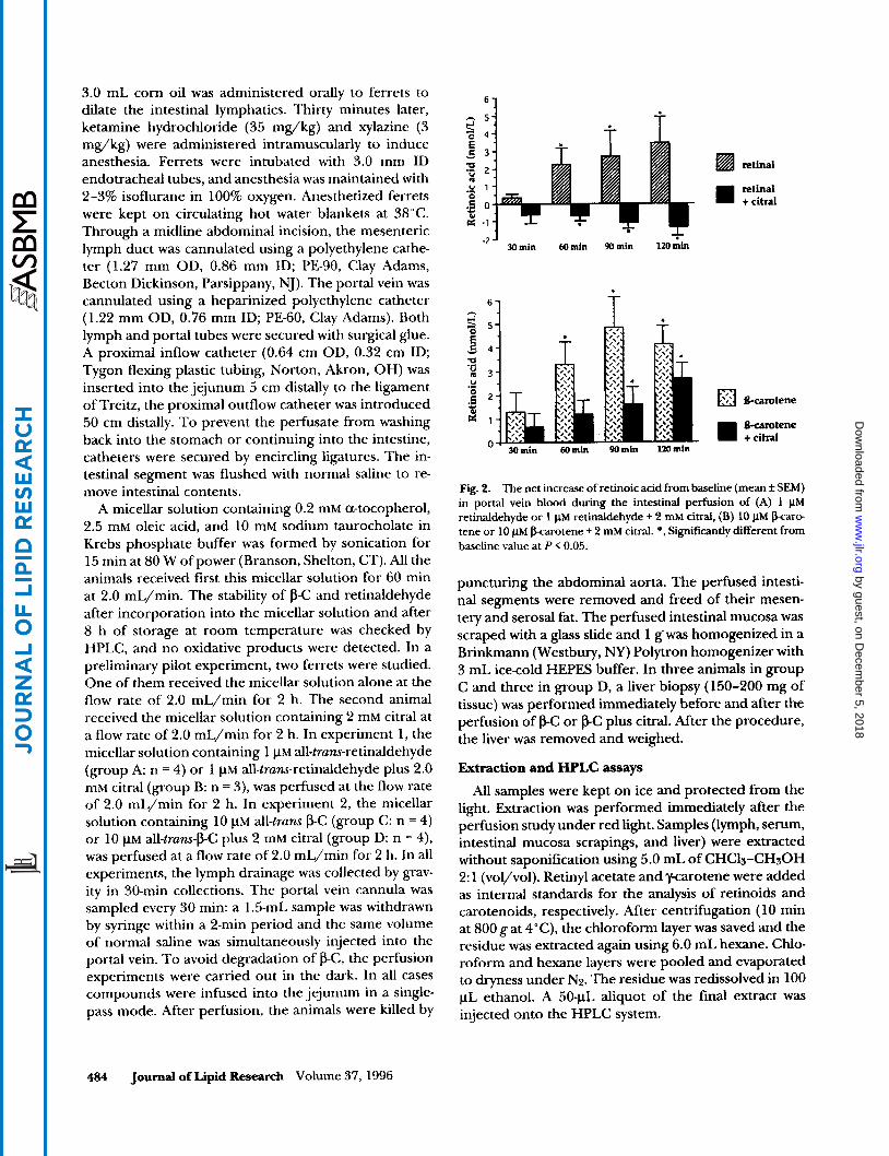
Fig. 2. The net increase of retinoic acid from baseline (mean f SEM) in portal vein blood during the intestinal perfhion of (A) 1 pM retinaldehyde or 1 pM retinaldehyde + 2 m M citral, (B) 10 pM @arc. tene or 10 pM karotene + 2 m M citral. *, Significantly different from baseline value at P < 0.05.
puncturing the abdominal aorta. The perfused intesti- nal segments were removed and freed of their mesen- tery and serosal fat. The perfused intestinal mucosa was scraped with a glass slide and 1 g' was homogenized in a Bnnkmann (Westbury, NY) Polytron homogenizer with 3 mL ice-cold HEPES buffer. In three animals in group C and three in group D, a liver biopsy (150-200 mg of tissue) was performed immediately before and after the perfusion of @C or @C plus citral. After the procedure, the liver was removed and weighed.
Extraction and HPLC assays
All samples were kept on ice and protected from the light. Extraction was performed immediately after the perfusion study under red light. Samples (lymph, serum, intestinal mucosa scrapings, and liver) were extracted without saponification using 5.0 mL of CHCls-CHsOH 2: 1 (vol/vol). Retinyl acetate and 'y-carotene were added as internal standards for the analysis of retinoids and carotenoids, respectively. After centrifugation ( 10 min at 800 gat 4"C), the chloroform layer was saved and the residue was extracted again using 6.0 mL hexane. Chlo- roform and hexane layers were pooled and evaporated to dryness under Nz. The residue was redissolved in 100 pL ethanol. A 50-pL aliquot of the final extract was injected onto the HPLC system.
484 Joumal of Lipid Research Volume 37,1996
by guest, on Decem
ber 5, 2018w
ww
.jlr.orgD
ownloaded from

E C
om
3
T i e 0 T i e oain) Fig. 3. HPLC chromatography of retinoids in ferret intestinal mucosa; (A) after the intestinal perfusion of 1 pM retinaldehyde for 2 h; (B) after the intestinal perfusion of 1 pM retinaldehyde + 2 mM citral for 2 h; (C) after the intestinal perfusion of 10 pM bcarotene for 2 h; (D) after the intestinal perfusion of 10 pM barotene + 2 mM citral for 2 h. Inserted figure shows the HPLC pattern after injection of standard retinoids. Peak identifications: (1) retinoic acid, (2) retinol, (3) retinaldehyde, (4) citral metabolite, (5) retinyl acetate added as internal standard. Mobile phase was CHsCN/THF/H20 (Solvent A 35:5:60, vol/vol/vol, 1% ammonium acetate in H20; Solvent B: 50:44:6, vol/vol/vol, 1% ammonium acetate in H20).
@Carotene, retinol, and retinyl esters (retinyl palmi- tate and retinyl stearate) were analyzed by reverse-phase HPLC on a Pecosphere-3 CIS 0.46 x 8.3 cm cartridge column (Perkin-Elmer, Norwalk, CT) using CHsCN-THF-l% ammonium acetate in water (Solvent A: 50:20:30, vol/vol/vol; Solvent B: 50:44:6, vol/vol/vol) as previously described (23, 24). A Waters 490E multi-wavelength spectrophotometer detector was set at 340 nm for retinoids and 450 nm for carotenoids, and an additional Waters 994 programmable photo diode array detector was used for measurement of maximal absorption. For the identification of RA, a modified procedure was used in which the HPLC mo- bile phase was CHsCN-THF-l% ammonium acetate in water (Solvent A 35:5:60, vol/vol/vol; Solvent B: 50:44:6, vol/vol/vol) as described earlier (22).
Individual carotenoids and retinoids were identified by co-elution with their standards, and quantified rela- tive to the recovery of internal standards, retinyl acetate, and y-carotene, by determining peak areas above ob- served baseline calibrated against known amounts of
standards. The lowest limit of detection of RA for each assay was 0.2 pmol(24).
Methylation
The positive identification of RA derived from intes- tinal perfusion with the p-C was accomplished by synthe- sizing and characterizing methyl retinoate. Three addi- tional ferrets were perfused using the procedure described above. The intestinal mucosa samples were extracted, pooled, and analyzed by HPLC. The HPLC peak that matched with the retention time of standard RA was collected and dried under N2. The residue was dissolved in 3 mL peroxide-free diethyl ether at 4°C and derivatized with diazomethane to form the methyl reti- noate. After the reaction, the excess diazomethane was removed under nitrogen and the residue was resus- pended in ethanol and subjected to HPLC as before.
Statistics
Results are expressed as means k SEM. Comparisons among the same group were performed using a one-
H 6 b u t m et al. Retinoic acid synthesis by excentric cleavage of b o t e n e 485
by guest, on Decem
ber 5, 2018w
ww
.jlr.orgD
ownloaded from
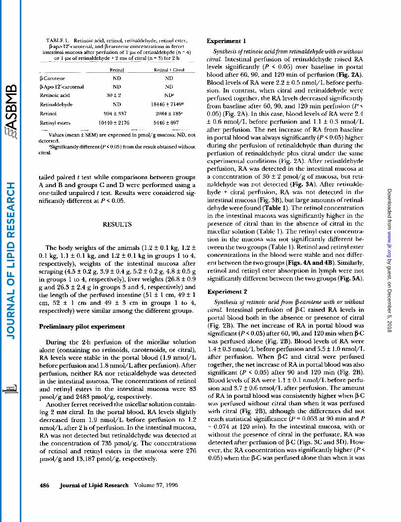
TABLE 1. Retinoic acid, retinol, retinaldehyde, retinyl ester, Bapo-l2’-carotenal, and karotene concentrations in ferret
intestinal mucosa after perfusion of 1 p~ of retinaldehyde (n = 4) or 1 FM of retinaldehyde + 2 mM of citral (n = 3) for 2 h
Retinal Retinal + Citral
FCarotene ND ND
bApol2’-carotenal ND ND
Retinoic acid 30 f 2 NtP
Retinaldehyde ND 18446f7149n
Retinol 994 f 337 2084 k 185O
Retinyl esters 10440 k 2 176 5446 f 897
Values (mean k SEM) are expressed in pmol/g mucosa; ND, not
“Significantly different (P < 0.05) from the result obtained without detected.
citral.
tailed paired t test while comparisons between groups A and B and groups C and D were performed using a one-tailed unpaired t test. Results were considered sig- nificantly different at P < 0.05.
RESULTS
The body weights of the animals (1.2 f 0.1 kg, 1.2 f 0.1 kg, 1.1 k 0.1 kg, and 1.2 f 0.1 kg in groups 1 to 4, respectively), weights of the intestinal mucosa after scraping (4.5 f 0.2 g, 3.9 f 0.4 g, 5.2 f 0.2 g, 4.8 f 0.5 g in groups 1 to 4, respectively), liver weights (26.8 f 0.9 g and 26.3 f 2.4 g in groups 3 and 4, respectively) and the length of the perfused intestine (51 f 1 cm, 49 f 1 cm, 52 f 1 cm and 49 f 3 cm in groups 1 to 4, respectively) were similar among the different groups.
Preliminary pilot experiment
During the 2-h perfusion of the micellar solution alone (containing no retinoids, carotenoids, or citral), RA levels were stable in the portal blood (1.9 nmol/L before perfusion and 1.8 nmol/L after perfusion). After perfusion, neither RA nor retinaldehyde was detected in the intestinal mucosa. The concentrations of retinol and retinyl esters in the intestinal mucosa were 83 pmol/g and 2483 pmol/g, respectively.
Another ferret received the micellar solution contain- ing 2 mM citral. In the portal blood, RA levels slightly decreased from 1.9 nmol/L before perfusion to 1.2 nmol/L after 2 h of perfusion. In the intestinal mucosa, RA was not detected but retinaldehyde was detected at the concentration of 735 pmol/g. The concentrations of retinol and retinyl esters in the mucosa were 276 pmol/g and 13,187 pmol/g, respectively.
Experiment 1
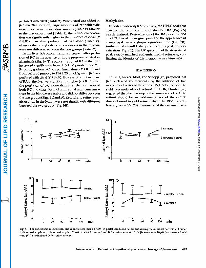
Synthesis ofretinoic acidfrom retinaldehyde with or without citral. Intestinal perfusion of retinaldehyde raised RA levels significantly (P < 0.05) over baseline in portal blood after 60, 90, and 120 min of perfusion (Fig. 2A). Blood levels of RA were 2.2 f 0.5 nmol/L before perfu- sion. In contrast, when citral and retinaldehyde were perfused together, the RA levels decreased significantly from baseline after 60, 90, and 120 min perfusion (P < 0.05) (Fig. 2A). In this case, blood levels of RA were 2.4 rf: 0.6 nmol/L before perfusion and 1.1 f 0.3 nmol/L after perfusion. The net increase of RA from baseline in portal blood was always significantly (P < 0.05) higher during the perfusion of retinaldehyde than during the perfusion of retinaldehyde plus citral under the same experimental conditions (Fig. 2A). After retinaldehyde perfusion, RA was detected in the intestinal mucosa at a concentration of 30 rf: 2 pmol/g of mucosa, but reti- naldehyde was not detected (Fig. 3A). After retinalde- hyde + citral perfusion, RA was not detected in the intestinal mucosa (Fig. 3B), but large amounts of retinal- dehyde were found (Table 1). The retinol concentration in the intestinal mucosa was significantly higher in the presence of citral than in the absence of citral in the micellar solution (Table 1). The retinyl ester concentra- tion in the mucosa was not significantly different be- tween the two groups (Table l) . Retinol and retinyl ester concentrations in the blood were stable and not differ- ent between the two groups (Figs. 4A and 4B). Similarly, retinol and retinyl ester absorption in lymph were not significantly different between the two groups (Fig. 5A).
Experiment 2 Synthesis of retinoic acid &om p-carotene with or without
citral. Intestinal perfusion of p-C raised RA levels in portal blood both in the absence or presence of citral (Fig. 2B). The net increase of RA in portal blood was significant (P < 0.05) after 60,90, and 120 min when PC was perfused alone (Fig. 2B). Blood levels of RA were 1.4 f 0.3 nmol/L before perfusion and 5.5 f 1.0 nmol/L after perfusion. When p-C and citral were perfused together, the net increase of RA in portal blood was also significant (P < 0.05) after 90 and 120 min (Fig. 2B). Blood levels of RA were 1.1 f 0.1 nmol/L before perfu- sion and 3.7 f 0.6 nmol/L after perfusion. The amount of RA in portal blood was consistently higher when p-C was perfused without citral than when it was perfused with citral (Fig. 2B), although the differences did not reach statistical significance (P = 0.053 at 90 min and P = 0.074 at 120 min). In the intestinal mucosa, with or without the presence of citral in the perfusate, RA was detected after perfusion of p-C (Figs. 3C and 3D). How- ever, the RA concentration was significantly higher (P < 0.05) when the p C was perfused alone than when it was
486 Journal of Lipid Research Volume 37, 1996
by guest, on Decem
ber 5, 2018w
ww
.jlr.orgD
ownloaded from
perfused with citral (Table 2). When citral was added to &C micellar solution, large amounts of retinaldehyde were detected in the intestinal mucosa (Table 2). Similar to the first experiment (Table l), the retinol concentra- tion was significantly higher in the presence of citral (P < 0.05) than after perfusion of gC alone (Table 2), whereas the retinyl ester concentrations in the mucosa were not different between the two groups (Table 2).
In the liver, RA concentrations increased after perfu- sion of &C in the absence or in the presence of citral in all animals (Fig. 6). The concentration of RA in the liver increased significantly from 116 k 36 pmol/g to 292 k 34 pmol/g when &C was perfused alone (P < 0.05) and from 147 f 30 pmol/g to 194 f 23 pmol/g when f3-C was perfused with citral (P< 0.05). However, the net increase of RA in the liver was significantly higher (P< 0.05) after the perfusion of f3-C alone than after the perfusion of both p-C and citral. Retinol and retinyl ester concentra- tions in the blood were stable and did not differ between the two groups (Figs. 4C and D). Retinol and retinyl ester absorption in the lymph were not significantly different between the two groups (Fig. 5B).
'.s 1 A
Methylation In order to identify R4 positively, the HPLC peak that
matched the retention time of standard RA (Fig. 7A) was derivatized. Derivatization of the RA peak resulted in a 75% loss of the original peak and the appearance of a new peak with a slower retention time (Fig. 7B). Authentic all-trans-RA also produced this peak on deri- vatization (Fig. 7C). The U V spectrum of the derivatized peak exactly matched authentic methyl retinoate, con- firming the identity of this metabolite as all-trans-RA.
DISCUSSION
In 1931, Karrer, Morf, and Schopp (25) proposed that j3-C is cleaved symmetrically by the addition of two molecules of water at the central 15,15'double bond to yield two molecules of retinol. In 1946, Hunter (26) suggested that the first step of the conversion of PC into retinol should be an oxidative attack of the central double bound to yield retinaldehyde. In 1965, two dif- ferent groups (27,28) demonstrated the enzymatic syn-
'7 c
0 1 I I I I 0 30 60 90 120 min 0 3 0 6 0 d 120 min
B
Y
- T
retinal
retinal + citral
V I I I I I I
0 30 60 90 120 min
Fig. 4. The concentrations of retinol and retinyl esters (mean It SEM) in portal vein blood before and during the intestinal perfusion of either 1 pM retinaldehyde or 1 pM retinaldehyde + 2 mM citral (A for retinol and B for retinyl esters), 10 pM karotene or 10 pM fkarotene + 2 mM citral (C for retinol and D for retinyl esters).
Hkbutenze et al. Retinoic acid synthesis by excentric cleavage of barotene 487
by guest, on Decem
ber 5, 2018w
ww
.jlr.orgD
ownloaded from

T 5
5 4
i 3
2 retinal
1
0 II yzr$
retinol retinyl esters
T
retinol retinyl esters
Fig. 5. The absorption of retinol and retinyl esters (mean + SEM) into ferret mesenteric lymph during the intestinal perfusion of (A) 1 pM retinaldehyde or 1 pM retinaldehyde + 2 mM citral or (B) 10 pM &carotene or 10 pM &carotene + 2 mM citral for 2 h.
thesis of retinol from p-C in the rat intestine. They both suggested that the process took place by means of a central cleavage mechanism forming retinaldehyde, which would be subsequently reduced to retinol. How- ever, in 1960, Glover proposed the hypothesis that p-C could undergo both central and excentric cleavage (29). This hypothesis was supported by observations in rats, that kapo-carotenals are converted into retinol or oxi- dized into their corresponding carotenoic acids (30,3 1). Such apocarotenals have been identified in the intestine of chicken after administration of g C (32).
Until recently, the existence of central or excentric pathways of p-C has been a matter of debate (17, 18,33). Some authors claimed that central cleavage does not exist (34), whereas others (35) demonstrated enzymatic conversion of PC to retinaldehyde by a cytosolic enzyme from rabbit and rat intestinal mucosa, supporting a central cleavage pathway mechanism.
Recent work by our group showed that excentric cleavage of FC does take place in vitro. The incubation of homogenates of human, monkey, ferret, and rat intestinal mucosa with p-C resulted in the formation of retinaldehyde, RA, and the p-apo-l2', lo', and 8'caro- tenals (24). The formation of p-apo-13carotenone and P-apo-14'carotenal was demonstrated in the same spe- cies (23). We also demonstrated that when intestinal homogenates are incubated with p-C, RA can be pro- duced by excentric cleavage (21) and that intestinal
perfusion of p-C in the ferret raises RA level in portal blood (13). In light of these results, the question arose whether the biosynthesis of RA via excentric cleavage of p-C can take place in vivo. This question is of importance because RA is a ligand for nuclear receptors (i.e., RAR, RXR) (8, 9) and is involved in cell regulation (5). More- over, if excentric cleavage exists in vivo, the biological activity of intermediate compounds (i.e., kapo-caro- tenals and P-apo-carotenoic acids) should be investi- gated.
In the preliminary pilot experiment we demonstrated that the perfusion of the micellar solution had no effect on the RA concentration in portal blood. Neither RA nor retinaldehyde was detected in the intestinal mucosa. When citral alone was added to the micellar solution, it resulted in a slight decrease of the RA levels in portal blood. After perfusion of citral, a significant amount of retinaldehyde was detected in the intestinal mucosa, which may result from an inhibition of oxidation of endogenous vitamin A. The high concentration of reti- nol and retinyl esters detected in the intestinal mucosa after the perfusion of citral alone may be the result of increased production of retinol from retinal-dehyde and of high retinyl ester status in the ferret that was used
In the first experiment we demonstrated that intesti- nal perfusion of retinaldehyde significantly increases RA levels in the portal blood. After retinaldehyde per- fusion, a significant amount of RA was also detected in the intestinal mucosa, but retinaldehyde was not de- tected, presumably due to its rapid conversion into either retinol or RA. When citral was added to the retinaldehyde perfusate, RA levels decreased signifi- cantly in the portal blood and were not detected in the intestinal mucosa even though a large amount of reti- naldehyde was present, reflecting blockage of the oxida- tion of retinaldehyde to RA by citral. The intestinal concentration of retinol was more than 2-fold higher
(36).
TABLE 2. Retinoic acid, retinol, retinaldehyde, retinyl ester, P-apc-12'-carotenal, and &carotene concentrations in ferret
intestinal mucosa after perfusion of 10 p~ of &carotene (n = 4) or 10 p~ of &carotene + 2 mM of citral (n = 4), for 2 h
warotene &Carotene + Citral
PCarotene 1636 k 1213 b2351+1480
PApo-1 Ycarotenal 7 9 f 10 68+ 19
Retinoic acid 36 + 3 9 f 5"
Retinaldehyde ND 10110 f 3431"
Retinol; 251 f 36 437 f 68"
Retinyl esters 7310 f 2882 6847 + 2978 Values (mean f SEM) are expressed in pmol/g mucosa; ND, not
"Significantly different (PC 0.05) from the result obtained without detected.
citral.
488 Joumd of Lipid Research Volume 37, 1996
by guest, on Decem
ber 5, 2018w
ww
.jlr.orgD
ownloaded from

400 -
300 -
200 -
100 -
O *
I %carotene + - - - A citral /
0 1 I I
Before Perfusion
After Perfusion
Fig. 6. Concentrations of retinoic acid in the ferret liver (individuals and means i SEM) before and after the intestinal perfusion of 10 HM bcarotene or 10 pM barotene + 2 mM citral for 2 h. *Significantly different from baseline value at P < 0.05.
after perfusion of retinaldehyde and citral than after perfusion of retinaldehyde alone (Table l), suggesting an increase in the reduction of the accumulated retinal- dehyde to retinol.
In this model, where the lymph duct is cannulated, it is not surprising to see that portal concentrations of retinyl esters did not change during perfusion as most are absorbed in the lymph. There was no effect of citral on retinyl ester absorption in lymph due to slow absorp- tion and presence of endogenous retinol and retinyl esters at the beginning of the study. Thus, our first experiment clearly demonstrates that after intestinal perfusion in the ferret, citral blocks the oxidation of retinaldehyde to RA, which results in an accumulation of retinaldehyde and retinol in the intestinal mucosa.
In the second experiment, we proceeded with the perfusion of p C under the same conditions to confirm that RA can arise from excentric cleavage of PC in vivo. From the work of Napoli and Race (11) we chose a concentration of 10 pM of p C to study the effect of citral on RA formation from PC. Retinoic acid levels obtained in the portal blood and in the intestinal mucosa after perfusion of 1 pM retinaldehyde or 10 pM p C without citral were quite similar (Tables 1 and 2). As observed in the perfusion of retinaldehyde plus citral, the perfu- sion of a mixture of p-C plus citral resulted in the accumulation of retinaldehyde and an increase of the concentration of retinol in the intestinal mucosa of the ferret (Fig. 3, Tables 1 and 2). This result demonstrates once again that at this concentration, citral blocks the
oxidation of retinaldehyde to RA and increases the reduction of retinaldehyde to retinol.
After pC perfusion, if all the FtA were being produced via oxidation of retinaldehyde resulting from the central cleavage mechanism, we would not have expected to find any RA in the mucosa or in the portal blood after the perfusion of a mixture of pC plus citral. However, contrary to the first experiment, RA was still detected in the mucosa (Fig. 3D), and RA levels increased signifi- cantly in portal blood (Fig. 2B) after the perfusion of p C plus citral. Moreover, the RA concentration in the liver increased in all the animals perfused with f3-C (Fig. 6), regardless of the presence of citral, and p-apo-12’caro- tenal was detected in the intestinal mucosa. These find- ings clearly demonstrate for the first time in the living animal that RA can be formed by a pathway other than oxidation of retinaldehyde (i.e., via excentric cleavage).
The results of this study are consistent with the ‘‘goxi- dation mechanism” proposed by Glover (29), which involves cleavage at several double bonds of the conju- gated system. This cleavage continues with successive removal of two carbon units until the 15,15‘ double bond is reached. At this point further poxidation is blocked by the methyl group located on C13. The recent isolation of apocarotenals and particularly papo-13- carotenone and Fapo-l4’-carotenal from the intestine of different species (23, 24) strongly supports this hy- pothesis. In order to provide some information about the presence of intermediates, e.g., fLapocarotenoic acids, we perfused additional ferrets (n = 3) with j3-C
Hibuterne et al. Retinoic acid synthesis by excentric cleavage of barotene 489
by guest, on Decem
ber 5, 2018w
ww
.jlr.orgD
ownloaded from

4
Fig. 7. HPLC chromatography of ferret intestinal mucosa (A) after the perfusion of 10 pM of barotene for 2 h (B) after derivatization with ethereal diazomethane and an additional HPLC separation. (C) HPLC chromatography of standard methyl retinoate. Peak identifications: ( 1 ) retinoic acid, (2) retinol, (3) retinyl acetate added as internal standard, (4) methyl retinoate. Mobile phase was CHsCN/THF/H20 (Solvent A: 35:5:60, vol/vol/vol, 1% ammonium acetate in H20; Solvent B: 50:44:6, vol/vol/vol, 1% ammonium acetate in H20).
using the same protocol. We still could not detect any additional intermediate besides papo-l2’-carotenal. We then perfusated 5 pM papo-l4’-carotenal (at a concen- tration which is much higher than a physiologic level) through the ferret intestine (n = 5; X-D. Wang, C. S. Jean, and R. M. Russell, unpublished data). The results showed that P-apo-l4’-carotenal disappeared rapidly and only retinol and small amounts of retinoic acid and P-apo-l4’-carotenoic acid were detected in the intestinal mucosa, which suggests that in vivo these intermediates may be converted rapidly to retinoids and other polar metabolites.
An interesting aspect of this study is the demonstra- tion that the addition of citral to the p-C micellar solu- tion resulted in a 47% decrease in the RA concentration in the intestine and a 36% decrease in the RA concen- tration in the portal blood after 2 h perfusion, as com- pared to the perfusion of p-C alone. This finding sug- gests that after perfusion of p-C, less than 50% of the RA is formed via oxidation of retinaldehyde (i.e., via central cleavage). However, after perfusion, the net increase from baseline in RA in the liver was 73% lower when citral was added to the p-C micellar solution versus when citral was not added to the pC micellar solution; in the portal blood, after 60 and 90 min perfusion, the net increase of RA was about 60% lower with the perfu- sion of citral versus without the perfusion of citral, suggesting that more than 60% of the RA is formed directly via oxidation of retinaldehyde (i.e., via central cleavage). Because the data in the pilot study indicate that less than 10% of the decrease in RA concentration in the portal blood was due to citral alone, the decrease in RA concentration in the portal blood may overesti- mate the inhibition of RA formation from p-C when citral is added. It is not surprising that the results
obtained in the last blood sample and intestine taken just before the end of the experiment (after 120 min perfusion) are in close agreement, as the newly formed R4 leaves the intestine via the portal route. On the other hand, the net increase of RA in the liver is a reflection of the flow of RA coming via the portal vein during the entire 2-h perfusion. Discrepancies between intestine and liver results can also be explained by limits on the capacity of absorption of RA from the intestine into blood and/or redistribution of the RA produced to other organs than the liver. Retinoic acid can also be further oxidized to degradation products or undergo glucuronidation.
Although the results of the present study do not allow us to determine with accuracy the exact importance of central versus excentric pathways in the metabolism of p-C, this study demonstrates for the first time in vivo that a substantial amount of RA is formed via a pathway that bypasses retinaldehyde oxidation, i.e., by excentric cleavage. It is possible that some retinaldehyde results from the degradation of the 0-apocarotenals formed from an excentric pathway (Fig. 1) resulting in an un- derestimate of excentric cleavage. It has been shown that the incubation of papo8-carotenals or papo-12’- carotenals with human intestinal homogenates results in the production of retinaldehyde, but this pathway seems to be less important than the formation of RA in vitro, as the P-apo-carotenals produced 2.7- to 6.6-times more RA than retinaldehyde (21). Moreover, the addi- tion of citral to papo-carotenal incubations decreased the formation of RA by only 14% (21). It is interesting to note the results of the work of Sharma, Mathur, and Ganguly (3 l), who evaluated the relative biopotencies of P-apo-carotenals compared with p-C. In a curative growth assay in the rat, the biopotencies were 72, 78,
490 Journal of Lipid Research Volume 37, 1996
by guest, on Decem
ber 5, 2018w
ww
.jlr.orgD
ownloaded from

and 72% of that for &C on a molar basis for bapo-S’, lo’, and 12’-carotenal, respectively. However, the rela- tive amount of vitamin A in the liver for the bap0-12’- carotenal was only 20% of that for PC, suggesting that a large fraction of this &apo-carotenal may be trans- formed into RA, while only a small fraction is ultimately reduced to the corresponding alcohol.
In summary, this study demonstrates unambiguously for the first time in vivo that an excentric cleavage mechanism of p-C metabolism is involved in the biosyn- thesis of RA in the ferret intestine. This excentric cleav- age mechanism represents a quantitatively important pathway in the metabolism of p-C into RA.m
The authors thanks Dr. Norman I. Krinsky of Tufts University School of Medicine for his suggestions and Dr. Elisabeth J. Johnson of Human Nutrition Research Center of Aging at Tufts University for her help during the preparation of the manuscript. This study was funded in part by funds of the U.S. Department of Agriculture, Agricultural Research Service un- der contract number 53-3KO6-01, and by grant ROl-CA-49195- 01 from the National Institutes of Health. The stay in USA of Dr. Xavier HCbuterne was possible with the support of the University of Nice (France) School of Medicine and with a grant from Beaufour Laboratories (Paris, France). The con- tents of this publication do not necessarily reflect the views or policies of the U.S. Department of Agriculture, nor does mention of trades names, commercial products, or organiza- tions imply endorsement by the U.S. Government. Manuscript received 21 lune 1995 and in revbed form 4 December 1995.
REFERENCES
1.
2.
3.
4.
5.
6.
7.
Peto, R., R. Doll, J. D. Buckley, and M. B. Sporn. 1981. Can dietary ba ro tene materially reduce human cancer rates? Nature. 290 201-208. Ziegler, R. G. 1991. Vegetables, fruits, and carotenoids and the risk of cancer. Am.J. Clin. Nutr. 53: 251s-259s. Krinsky, N. I. 1991. Effects of carotenoids in cellular and animal systems. Am.J Clin. Nutr. 53: 238s-2463. Burton, G. W., and K. U. Ingold. 1984. pcarotene: an unusual type of lipid antioxidant. Science. 244: 569-573. Strickland, S., and V. Mahdavi. 1978. The induction of differentiation in teratocarcinoma stem cells by retinoic acid. Cell. 15: 393-403. Chen, Z. X., Y. Q. Xue, R. F. Tao, X. M. Xia, C. Li, W. Y. Zu, X. Z. Yao, and B. J. Ling. 1991. A clinical and experi- mental study of all-trans retinoic acid treated acute promyelocytic leukemia patients. Blood. 78: 1413-1419. Warrell, R. P., S. R. Frankel, W. H. Miller, D. A. Schein- berg, L. M. Itri, W. N. Hittelman, R. Vyas, M. Andreeff, A. Tafuri, A. Jakubowski, J. Gabrilove, M. S. Gordon, and E. Dmitrovsky. 1991. Differentiation therapy of acute
promyelocytic leukemia with tretinoin (all-trans-retinoic acid). N. Eng1.J. Med 324 1385-1393.
8. Heyman, R. A., D. J. Mangelsdorf, J. A. Dyck, R. B. Stein, G. Eichele, R. M. Evans, and C. Thaller. 1992. 9-Cis-reti- noic acid is a high affinity ligand for the retinoid X receptor. Cell. 68: 397-406.
9. Petkovich, M., N. J. Brand, A. Krust, and P. Chambon. 1987. A human retinoic acid receptor which belongs to the family of nuclear receptors. Nature. 330: 444-450.
10. Crain, F. D., F. J. Lotspeich, and R. F. Krause. 1967. Biosynthesis of retinoic acid by intestinal enzymes of the rat. J. Lipid Res. 8: 249-254.
11. Napoli, J. L., and K. R. Race. 1988. Biogenesis of retinoic acid from barotene. Differences between the metabo- lism of ba ro tene and retinal. J. Biol. Chem. 263:
12. Folman, Y., R. M. Russell, G. Tang, and G. Wolf. 1989. Rabbits fed on pcarotene have higher serum levels of all-trans retinoic acid than those receiving no barotene. Br.J. Nutr. 62: 195-201.
13. Wang, X. D., R. M. Russell, R. P. Marini, G. Tang, G. G. Dolnikowski, J. G. Fox, and N. I. Krinsky. 1993. Intestinal perfusion of @carotene in the ferret raises retinoic acid level in portal blood. Biochim. Biophys. Acta. 1167:
14. De Leenheer, A. P., W. E. Lambert, and I. Claeys. 1982. All-trans retinoic acid: measurement of reference value in human serum by high performance liquid chromatogra- phy. J. Lipid Res. 23: 1362-1367.
15. Wang, X. D., N. I. Krinsky, P. N. Benotti, and R. M. Russell. 1994. Biosynthesis of 9-cis-retinoic acid from 9 4 - pcarotene in human intestinal mucosa in vitro. Arch. Biochem. Biophys. 313: 152-155.
16. Hkbuterne, X., X. D. Wang, E. J. Johnson, N. I. Krinsky, and R. M. Russell. 1995. Intestinal absorption and meta- bolism of 9-cis-barotene in vivo: biosynthesis of 9-ckret- inoic acid. J. Lipid Res. 36: 1264-1273.
17. Ganguly, J., and P. S. Sastry. 1985. Mechanism of conver- sion of pcarotene into vitamin A central cleavage versus random cleavage. World Rev. Nutr. Diet. 45: 198-220.
18. Krinsky, N. I., X. D. Wang, G. Tang, and R. M. Russell. 1993. Conversion of carotenoids to retinoids. In Reti- noids: Progress in Research and Clinical Applications. M. Dekker, M. A. Livrea and L. Packer, editors. New York,
17372-17377.
159-164.
19.
20.
21.
22.
23.
Basel, Hong-Kong. 1-16. Connor, M. J., and M. H. Smit. 1987. Terminal group oxidation of retinol by mouse epidermis. Inhibition in vitro and in vivo. Biochem. J. 244: 489-492. Connor, M. J. 1988. Oxidation of retinol to retinoic acid as a requirement for biological activity in mouse epider- mis. Cancer Res. 48: 7038-7040. Wang, X. D., N. I. Krinsky, G. Tang, and R. M. Russell. 1992. Retinoic acid can be produced from excentric cleavage of ba ro tene in human intestinal mucosa. Arch. Biochem. Biophys. 293: 298-304. Wang, X. D., N. I. Krinsky, R. P. Marini, G. Tang, J. Yu, R. Hurley, J. G. Fox, and R. M. Russell. 1992. Intestinal uptake and lymphatic absorption of ba ro tene in ferrets: a model for human pcarotene metabolism. Am. J. Physiol.
Tang, G., X. D. Wang, R. M. Russell, and N. I. Krinsky. 1991. Characterization of P-apo-13carotenone and papo- 14’carotenal as enzymatic products of the excentric cleav- age of barotene. Biochemistry. 30: 9829-9834.
263: G480-G486.
Hkbuterne et al. Retinoic acid synthesis by excentric cleavage of harotene 491
by guest, on Decem
ber 5, 2018w
ww
.jlr.orgD
ownloaded from

24. Wang, X. D., G. Tang, J. G. Fox, N. I. Krinsky, and R. M. Russell. 1991. Enzymatic conversion of p-carotene into kapocarotenals and retinoids by human, monkey, ferret, and rat tissues. Arch. Biochem. Biophys. 285: 8-16.
25. Karrer, P., R. Morf, and K. Schopp. 1931. Zur kenntnis des vitamins-A aus fishtranen 11. Helv. Chim. Acta. 14:
26. Hunter, R. F. 1946. The conversion of carotene into vitamin A. Nature. 158: 257-260.
27. Goodman, D. S., and H. S. Huang. 1965. Biosynthesis of vitamin A with rat intestinal enzymes. Science. 149:
28. Olson, J. A., and 0. Hayaishi. 1965. The enzymatic cleav- age of pcarotene into vitamin A by soluble enzymes of rat liver and intestine. Proc. Natl. Acud. Sci. USA. 54:
29. Glover, J. 1960. The conversion of pcarotene to vitamin A. In Vitamins and Hormones. R. S. Harris and D. J. Ingle, editors. Academic Press, New York and London. 18:
30. Glover, J., and E. R. Redfearn. 1954. The mechanism of the transformation of p-carotene into vitamin A in vivo. Biochem. J. 158: XV-XV.
1431- 1436.
879-880.
1364- 1370.
371-386.
31. Sharma, R. V., S. N. Mathur, andJ. Ganguly. 1976. Studies of the relative biopotencies and intestinal absorption of different apo-p-carotenoids in rats and chickens. Biochem
32. Sharma, R. V., S. N. Mathur, A. A. Dimitrovskii, R. Dah, and J. Ganguly. 1977. Studies on the metabolism o f kcarotene and apo-&carotenoids in rats and chickem. Biochem. Biophys. Acta. 486: 183- 194.
33. Olson, J. A. 1993. The irresistible fascination of carotc- noids and vitamin A. Am. J. Clin. Nutr. 57: 833-839.
34. Hansen, S., and W. Maret. 1988. Retinal is not formed in vitro by enzymatic central cleavage of barotene. Biochem-
35. Lakshman, M. R., I. Mychkovsky, and M. Attlesey. 1989. Enzymatic conversion of all-trans-fkarotene to retinal by a cytosolic enzyme from rabbit and rat intestinal mucosa. Proc. Natl. Acad. Sci. USA. 86: 9124-9128.
36. Schweigert, F. J., 0. A. Ryder, W. A. Rambeck, and H. Zucker. 1990. The majority of vitamin A is transported as retinyl esters in the blood of most carnivores. Comp. Biochem. Physiol. 95A: 573-578.
J. 158: 377-383.
&tv. 27: 200-206.
492 Journal of Lipid Research Volume 37, 1996
by guest, on Decem
ber 5, 2018w
ww
.jlr.orgD
ownloaded from