in vitro antidiabetic activity of three fractions of methanol … · · 2016-08-10fraction 17...
TRANSCRIPT

Received on: 20/06/2014 Accepted on: 30/07/2014 Published on: 15/08/2014
Govindappa M Department of Biotechnology, Shridevi Institute of Engineering & Technology, Sira Road, Tumkur-572 106, Karnataka, INDIA Email:[email protected]
QR Code for Mobile users
DOI: 10.15272/ajbps.v4i34.520
In Vitro Antidiabetic Activity of Three Fractions of Methanol Extracts of Loranthus Micranthus,
Identification of Phytoconstituents by GC-MS and Possible Mechanism Identified by GEMDOCK Method
Channabasava, Govindappa M*, Chandrappa CP and Sadananda TS
Natural Products Laboratory, Department of Biotechnology, Shridevi Institute of Engineering & Technology, Sira Road, Tumkur-572 106, Karnataka, INDIA
Abstract Three (17, 20, 21) fractions of methanol extracts of Loranthus micranthus yielded different phytochemicals was confirmed by GC-MS analysis. The fraction 17 yielded 13 different phytochemicals followed by 20 (11 compounds) and 21 (12 compounds). More antidiabetic compounds were observed in fraction 17. In vitro antidiabetic activity was performed using these three column fractions for inhibition of α-amylase, α-glucosidase, sucrase and glucose. Strong antidiabetic activity was observed in 17 fraction and they inhibited all the above enzymes at rate followed by 21 and 20. The common phytochemical in all the extract is octadecenoic acid. It was used for GEMDOCK to study the interaction with enzymes. octadecenoic acid can be seperated and used as antidiabetic agent. Keywords: Loranthus micranthus, antidiabetic, GC-MS, enzymatic inhibitions, GEMDOCKing
Cite this article as:
Channabasava, Govindappa M*, Chandrappa CP and Sadananda TS. In Vitro Antidiabetic Activity of Three Fractions of Methanol
Extracts of Loranthus Micranthus, Identification of Phytoconstituents by GC-MS and Possible Mechanism Identified by GEMDOCK
Method. Asian Journal of Biomedical and Pharmaceutical Sciences; 04 (34); 2014; 34-41.

Govindappa M. et al: Asian Journal of Biomedical and Pharmaceutical Sciences; 4(34) 2014, 34-41.
© Asian Journal of Biomedical and Pharmaceutical Sciences, all rights reserved. Volume 4, Issue 34, 2014. 35
INTRODUCTION Diabetes mellitus is a multiple metabolic disorder characterized by high blood glucose level and its improper management leading to multiple chronic complications. Increased level of blood glucose may be due to inefficiency of pancreatic cells in insulin secretion or cellular resistance towards insulin uptake. International Diabetes Federation`s (IDF) recent estimates indicate that 8.3% of adults – 382 million people worldwide and 65.1 million people in India have diabetes, and the number of people with the disease is set to rise beyond 592 million in less than 25 years [1]. Herbal drugs are being the choice over synthetic drugs for having multiple therapeutic properties and fewer side effects. Even there are more than 1000 plant species being used for the treatment of Type 2 diabetes mellitus (advanced stage) worldwide [2], the diabetic cases are increasing at an alarming rate. This shows the necessities and the importance of more alternate and effective antidiabetic drugs and their systematic studies. Loranthus micranthus (Loranthaceae) is a hemiparasitic shrub commonly known as African mistletoe. It has been reported to have antidiabetic, antimicrobial and studies reveal that composition and biological activities of mistletoe are dependent on harvesting period, locality and host tree species [3], immunomodulatory and antimotility activities [4]. It decreases the blood glucose level and controls the loss of body weight in diabetes mellitus [5]. The review of literature reveals no reports on mistletoe, which are present in India. Our present work was aimed to carry out in vitro antidiabetic activities by enzymatic inhibition studies of Loranthus micranthus. Solvent, methanol was selected for extraction. α-amylase, α-glucosidase, sucrase and glucose diffusion models were used for antidiabetic studies. Further, it has been confirmed by GC-MS studies in the identification of responsible antidiabetic phytoconstituents present in Loranthus micranthus extract. The protein-ligand docking is the prediction of ligand conformation and orientation relative to the active site of a target protein. A computer aided docking process, identifying the lead compounds by minimizing the energy of intermolecular interactions, is an important approach for structure based drug design [6] and it is a very useful tool to give strong evidence for in vitro experiments. Ligand-molecule interaction study was conducted by using GEMDOCK method [7] to know systemic evaluation in identifying scoring functions. This method will know how our molecules interact with ligands and how they inhibit their function.
MATERIALS AND METHODS: Chemicals and reagents p-nitrophenyl-α-d-glucopyranoside, p-nitrophenyl-β-d-glucopyranoside, β-glucosidase from almonds and 3-5-dinitrosalisylic acid were purchased from Sisco Research Laboratory, India. A glucose oxidase/peroxidase assay kit was purchased from Agappe Diagnostics, India. α-amylase (23 u/mg solid) was purchased from Sigma-Aldrich, India. All the chemicals and reagents used in the study were of extra pure analytical grade. Collection and processing of plant The fresh leaves of Loranthus micranthus growing on the host plant Azadirachta indica collected in the month of April, 2009 during the flowering period at DC Bunglow, Tumkur, Karnataka, India and identified using authenticated herbarium from the Department of Studies in Botany, University of Mysore, Mysore. The plant material was washed with distilled water three times, shade-air dried (26+20C) and pulverized to a coarse powder in a mechanical grinder, passed through a 40 mesh sieve and stored in airtight container for further work. Preparation of crude extracts 25 g/100ml of powdered Loranthus micranthus was kept for solvent extraction in a rotary shaker at 370C, 72 rpm for 48 h. The methanol was used with increasing order of their polarity. The solvent extract was centrifuged at 6000 rpm for 10 min and then filtered with Whatman No. 1 filter paper and evaporated at a constant temperature of 620C in hot air oven until a very concentrated extract was obtained. Column chromatography Column chromatography is a type of adsorption chromatography technique. Here stationary phase is a silica gel packed in a vertical column. Cotton wool was plugged at the bottom of knob to hold stationary phase to allow only the solvent and sample. Silica was activated at 1100 C for 20 min to remove moisture content. Silica slurry was prepared by starting eluent and packed sufficiently in the column. The column was eluted with an initial eluent to remove further impurities. The extract sample to be separated was placed on the top of packed stationary phase without disturbing the silica bed. The gradient elution was carried out using solvents as an increase in their polarity. The solvents used are hexane, hexane and toluene (2:1, 1:1, 1:2), toluene, toluene and ethyl acetate (2:1, 1:1, 1:2), ethyl acetate, ethyl acetate and methanol (2:1, 1:1, 1:2) and methanol in different ratios. 25 eluted fractions were periodically collected at regular volumes of 5 ml each. Further TLC was carried out for each fraction to confirm the separation of single compounds.
Govindappa M. et al: Asian Journal of Biomedical and Pharmaceutical Sciences; 4(34) 2014, 34-41.
© Asian Journal of Biomedical and Pharmaceutical Sciences, all rights reserved. Volume 4, Issue 34, 2014. 36
GC – MS analysis GC-MS analysis of methanolic fractions of Loranthus micranthus was carried out with Agilent 7890-A having an MS detector 5975-C, ionization for MS is electron impact ionization. Mass analyzer was Quadrupole. The peaks were analyzed using data analysis software NIST-2008. An experiment was carried in column HP- 5 ms, dimensions- 30m L x 0.25mm ID x 0.25um film thickness. The initial temperature ramp was maintained at 40ºC, hold time -2 min. At the end the temperature ramp was 310ºC and hold time was 10 min. The rate of temperature ramp was 10ºC/min. The experiment was programmed with total run time 34 min, helium was used as a carrier gas at a constant flow rate of 1.0 ml/min, split less flow 1ml/min. Injection volume was 1µl with scan mass range 30m/z – 600m/z having positive polarity (+ve). Identification of phytoconstituents Interpretation of GC-MS mass-spectra were carried out using the database of National Institute Standard and Technology- 2008 (NIST-2008) having more than 62,000 patterns. The spectrums of the unknown components were compared with the spectrum of known components of NIST library and the parameters viz., molecular weight, structure of the components, total ionic chromatograms and ionization chromatograms were ascertained in naming the particular compound. In vitro antidiabetic activity Assay of α-amylase inhibitory activity The effect of three fractions of methanol extract of Loranthus micranthus on α-amylase activity was studied using an enzyme–starch system [8]. Three fractions (1–5%) were mixed by stirring with 25 mL of 4% potato starch in a beaker; 100 mg of α-amylase was added to the starch solution, stirred vigorously and incubated at 37°C for 60 min. After the incubation period, 0.1M NaOH was added to terminate enzyme activity. The mixture was centrifuged (3000 g; 15 min) and the maltose content in the supernatant was determined. Assay of α-glucosidase inhibitory activity α-glucosidase inhibitory activity was assayed according to the method of Honda and Hara [9]. Enzyme solution (10 µL) and three fractions of methanol extract (10–50 µL) were incubated together for 10 min at 37°C and the volume was made up to 210 µL with maleate buffer, pH 6.0. The enzyme reaction was started by adding 200 µL of 2 mM p-nitrophenyl-α-d-glucopyranoside solution and further incubated at 37°C for 30 min. The reaction was terminated by treating the mixture in a boiling water bath for 5 min. After the addition of 1.0 mL of 0.1 M disodium hydrogen phosphate solution, absorption of the liberated p-nitrophenol was read at 400 nm.
Assay of sucrase inhibitory activity The effect of three fractions of methanol extract of Loranthus micranthus on sucrase activity was assayed according to the method of Honda and Hara [9]. The enzyme solution (10 µL) and three fractions of methanol extract (10–50 µL) were incubated together for 10 min at 37°C and the volume was made up to 200 µL with maleate buffer (pH 6.0). The enzyme reaction was started by adding 100 µL sucrose solution (60 mM). After 30 min, the reaction was terminated by adding 200 µL of 3, 5-dinitrosalicylic acid reagent and treating the mixture in a boiling water bath for 5 min. The absorbance of the solution was read at 540 nm. The percent inhibitory activities were calculated using the following formula: %Inhibition= (AbsControl-AbsSample) x 100/AbsControl Where, AbsControl is the absorbance of the control reaction (containing all reagents except the test sample) and AbsSample is the absorbance of the test sample. An untreated enzyme solution was used as the control. All experiments were carried out in triplicate. Assay of Loranthus micranthus on glucose diffusion A method described by Gallagher et al. [10] was used to evaluate the effects of three fractions of methanol extract of Loranthus micranthus on glucose movement in vitro. The in vitro model consisted of a dialysis tube (6cm X 29.31 mm) (Himedia LA393-5MT-2010) in which 6 ml of plant extract and 2 ml of 0.15 M NaCl containing 1.65 mM D-glucose was added. The dialysis tube was sealed at each end and placed in a centrifuge tube containing 45 ml 0.15 M NaCl. The tubes were placed on an orbital shaker water bath and incubated at 370 C for 3 h. The movement of glucose into the external solution was provided. The concentration of glucose within the dialysis tube was measured and control tests were conducted in the absence of plant extracts. Glucose concentrations were analyzed by enzymatic method using the glucose oxidase kit. All tests were carried out in triplicate and the results were presented as means + SD. GEMDOCK Parameters We developed a molecular docking approach termed GEMDOCK (Generic Evolutionary Method for molecular DOCKing). The GEMDOCK software is available on the web at http://gemdock.life.netu.edu.tw. Setting of GEMDOCK parameters, such as an initial step size, family competition length (L-2), population size (N=300) and combination probability (pc=0.3) in this work. The GEMDOCK optimization stops when either the convergence is a certain threshold value or the interactions exceeds a maximal present value, which was set to 70. Therefore, GEMDOCK generated 1200 solutions in one generation and terminated after it
Govindappa M. et al: Asian Journal of Biomedical and Pharmaceutical Sciences; 4(34) 2014, 34-41.
© Asian Journal of Biomedical and Pharmaceutical Sciences, all rights reserved. Volume 4, Issue 34, 2014. 37
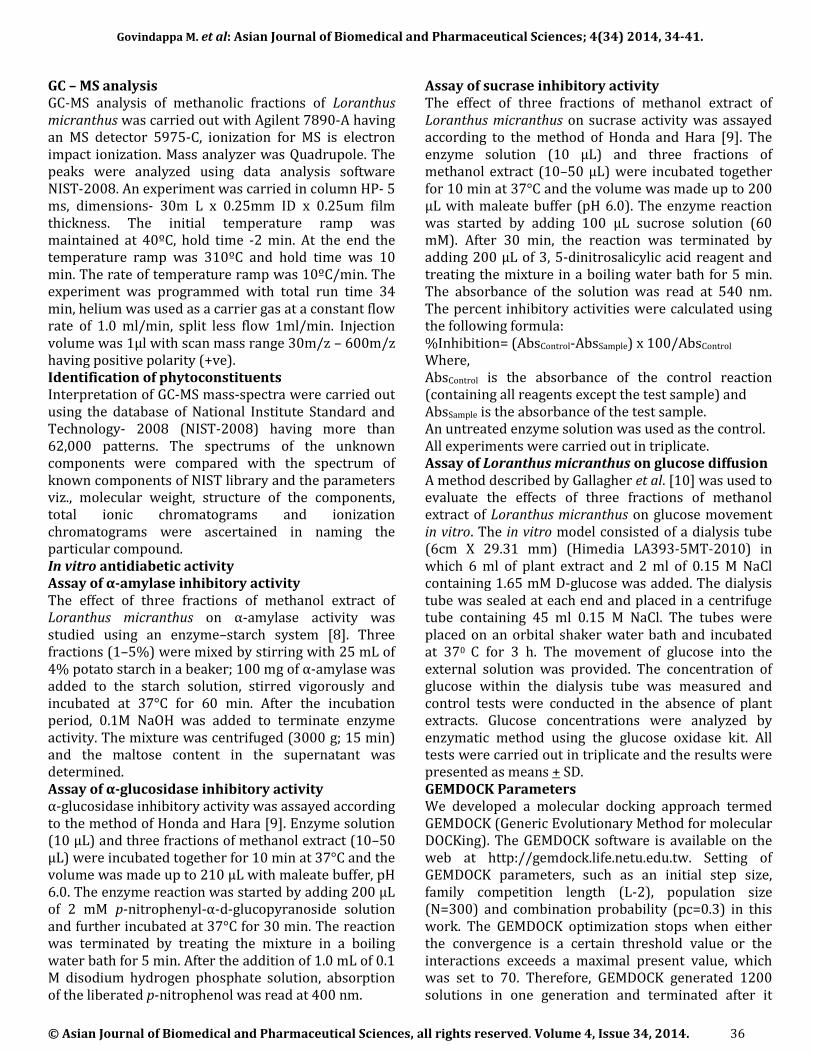
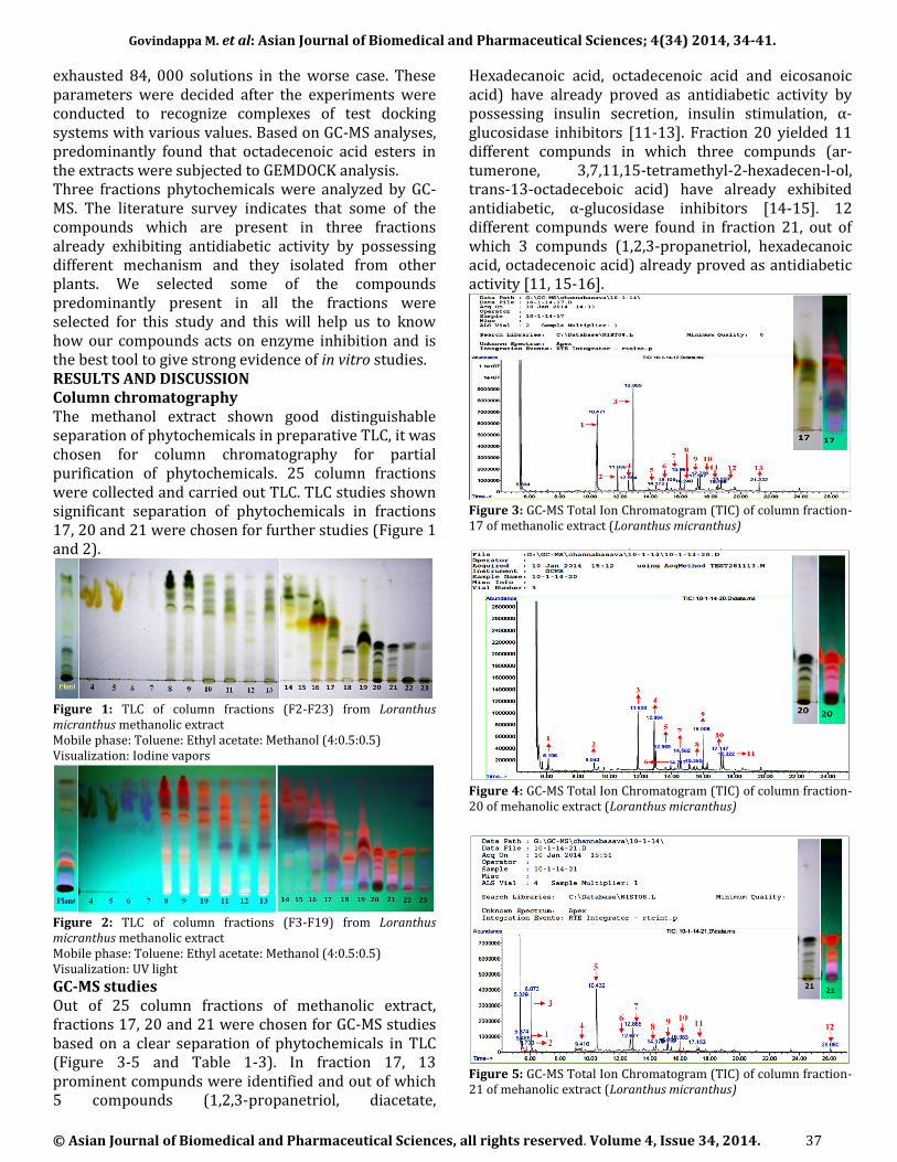
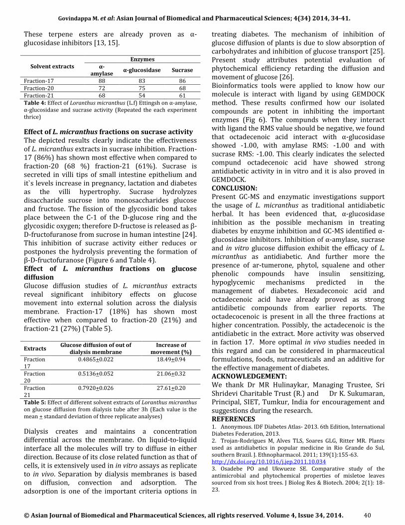
exhausted 84, 000 solutions in the worse case. These parameters were decided after the experiments were conducted to recognize complexes of test docking systems with various values. Based on GC-MS analyses, predominantly found that octadecenoic acid esters in the extracts were subjected to GEMDOCK analysis. Three fractions phytochemicals were analyzed by GC-MS. The literature survey indicates that some of the compounds which are present in three fractions already exhibiting antidiabetic activity by possessing different mechanism and they isolated from other plants. We selected some of the compounds predominantly present in all the fractions were selected for this study and this will help us to know how our compounds acts on enzyme inhibition and is the best tool to give strong evidence of in vitro studies. RESULTS AND DISCUSSION Column chromatography The methanol extract shown good distinguishable separation of phytochemicals in preparative TLC, it was chosen for column chromatography for partial purification of phytochemicals. 25 column fractions were collected and carried out TLC. TLC studies shown significant separation of phytochemicals in fractions 17, 20 and 21 were chosen for further studies (Figure 1 and 2).
Figure 1: TLC of column fractions (F2-F23) from Loranthus micranthus methanolic extract Mobile phase: Toluene: Ethyl acetate: Methanol (4:0.5:0.5) Visualization: Iodine vapors
Figure 2: TLC of column fractions (F3-F19) from Loranthus micranthus methanolic extract Mobile phase: Toluene: Ethyl acetate: Methanol (4:0.5:0.5) Visualization: UV light GC-MS studies Out of 25 column fractions of methanolic extract, fractions 17, 20 and 21 were chosen for GC-MS studies based on a clear separation of phytochemicals in TLC (Figure 3-5 and Table 1-3). In fraction 17, 13 prominent compunds were identified and out of which 5 compounds (1,2,3-propanetriol, diacetate,
Hexadecanoic acid, octadecenoic acid and eicosanoic acid) have already proved as antidiabetic activity by possessing insulin secretion, insulin stimulation, α-glucosidase inhibitors [11-13]. Fraction 20 yielded 11 different compunds in which three compunds (ar-tumerone, 3,7,11,15-tetramethyl-2-hexadecen-l-ol, trans-13-octadeceboic acid) have already exhibited antidiabetic, α-glucosidase inhibitors [14-15]. 12 different compunds were found in fraction 21, out of which 3 compunds (1,2,3-propanetriol, hexadecanoic acid, octadecenoic acid) already proved as antidiabetic activity [11, 15-16].
Figure 3: GC-MS Total Ion Chromatogram (TIC) of column fraction-17 of methanolic extract (Loranthus micranthus)
Figure 4: GC-MS Total Ion Chromatogram (TIC) of column fraction-20 of mehanolic extract (Loranthus micranthus)
Figure 5: GC-MS Total Ion Chromatogram (TIC) of column fraction-21 of mehanolic extract (Loranthus micranthus)

Govindappa M. et al: Asian Journal of Biomedical and Pharmaceutical Sciences; 4(34) 2014, 34-41.
© Asian Journal of Biomedical and Pharmaceutical Sciences, all rights reserved. Volume 4, Issue 34, 2014. 38
Fraction-17
Peak No
Retention time (min)
Identified compound name
Biological activity Source Reference
1 10.471 1,2,3-Propanetriol, diacetate Insulin secretion - (Wuttke et al.,
2013) 2 11.836 1-Tetradecene - - -
3 12.554 2,5-Cyclohexadiene-1,4-dione, 2,6-bis(1,1-
dimethylethyl)- - - -
4 12.865 Phenol, 2,4-bis(1,1-dimethylethyl)- - - -
5 14.372 1-{2-[3-(2-Acetyloxiran-2-yl)-1,1-
dimethylpropyl]cycloprop-2-enyl}ethanone - - -
6 15.109 Unknown compound Unknown - -
7
15.983 Hexadecanoic acid, methyl ester
Anti-inflammatory activity - (Saeed et al.,
2012) Vasodilator - (Lee et al., 2010)
Release of insulin stimulation - (Parker et al.,
2003)
Anti-diabetic activities - (Zuraini et al.,2012)
8 16.248 1,2-Benzenedicarboxylic acid, butyl octyl ester - - -
9
17.147 9-Octadecenoic acid (Z)-, methyl ester alpha-glucosidase inhibitors -
(Artanti et al., 2012)
alpha-glucosidase inhibitors Oncoba spinosa
(Balogun et al., 2013)
10
17.296 Octadecanoic acid, methyl ester alpha-glucosidase inhibitors -
(Artanti et al., 2012)
alpha-glucosidase inhibitors Oncoba spinosa
(Balogun et al., 2013)
11
18.499 Eicosanoic acid, methyl ester alpha-glucosidase inhibitors -
(Artanti et al., 2012)
alpha-glucosidase inhibitors Oncoba spinosa
(Balogun et al., 2013)
12 18.693 4,8,12,16-Tetramethylheptadecan-4-olide
- - -
13 21.222
2,6,10,14,18,22-Tetracosahexaene,
2,6,10,15,19,23-hexamethyl-, (all-E)- (Squalene) Antioxidant effect ,
antimicrobial - (Ryszard, 2009)
Table 1: GC-MS detection of phytoconstituents of column fraction-17
Fraction-20
Peak No
Retention time (min)
Identified compound name
Biological activity Source Reference
1 6.106 - - - - 2 9.043 Nitrobenzene Analgesic additive - - 3 11.830 1-Tetradecene - - -
4 12.884 Phenol, 2,4-bis(1,1-dimethylethyl)- Antifungal activity Avocado roots (Rangel et al., 2013)
Antioxidant (Yoon et al., 2006) 5 12.969 Dodecanoic acid, methyl ester Osteoporosis - Wikipedia
6 14.217 Ar-tumerone (Terpene) Antioxidant, antidiabetic and
antiinflammatory - (Elmazar et al., 2013)
Antioxidant, antidiabetic Zingiber officinale (Lucia et al., 2013) 7 14.562 Methyl tetradecanoate - - -
8 15.395 3,7,11,15-Tetramethyl-2-hexadecen-1-
ol (Phytol)
Antibacterial antifungal
Ginkgo biloba (Tao et al., 2013)
Anticonvulsant - (Costa et al., 2012) Antiarthritis - (Hultqvist et al., 2006)
Insulin sensitizing/anti-diabetic effect
- (Elmazar et al., 2013)
9 16.008
Benzenepropanoic acid, 3,5-bis(1,1-dimethylethyl)-4-hydroxy-, methyl
ester
- - -
10 17.147 trans-13-Octadecenoic acid, methyl
ester alpha-glucosidase inhibitors - (Artanti et al., 2012)
11 17.322
Methyl 16-methyl-heptadecanoate - - -
Table 2: GC-MS detection of phytoconstituents of column fraction-20
Effect of L. micranthus fractions on α-amylase activity Inhibitory activities of three fractions of L. micranthus on α-amylase were studied using α-amylase starch model system. Inhibition of α-amylase activity of
column fraction-17 (88%) was found more when compared to fraction-20 (72%) and fraction-21 (68%) (Figure 6 and Table 4). α-amylase is an enzyme that hydrolyzes alpha bonds of large, alpha-linked

Govindappa M. et al: Asian Journal of Biomedical and Pharmaceutical Sciences; 4(34) 2014, 34-41.
© Asian Journal of Biomedical and Pharmaceutical Sciences, all rights reserved. Volume 4, Issue 34, 2014. 39
polysaccharides, such as starch and glycogen. It is the major form of amylase found in humans and other mammals. Since α-amylase plays an important role in digestion of starch and glycogen, it is considered a strategy for the treatment of disorders in carbohydrate uptake, such as diabetes and obesity [17] to reduce postprandial glucose level. Hence α-Amylase inhibitors
may be of value as novel therapeutic agents [18]. However, inhibition of α-amylase by the phytochemicals of plants could be conclusively attributed to the presence of flavonoids [19], phenols [20].
Fraction-21 Peak
No Retention time
(min) Identified compound
name Biological activity Source Reference
1 5.465 Glycolaldehyde dimethyl acetal - - 2 5.730 Acetamide, 2,2,2-trifluoro-N-methyl- - - - 3 9.410 1,2,3-Propanetriol, monoacetate Antidote - (Chenoweth et al., 1951) 4 10.432 1,2,3-Propanetriol, diacetate Insulin secretion - (Wuttke et al., 2013) 5 12.677 1,2-Propanediol, 2-acetate - - -
6 12.865 Phenol, 2,4-bis(1,1-dimethylethyl)- Antifungal activity Avocado roots (Rangel et al., 2013)
Antioxidant (Yoon et al., 2006) 7 14.378 5-t-Butyl-4-methylimidazole - - -
8 15.116 Spiro[2,4,5,6,7,7a-hexahydro-2-oxo-
4,4,7a-trimethylbenzofuran]-7,2'-(oxirane)
- - -
9 15.2 p-Bromoatropine - - -
10 15.983 Hexadecanoic acid, methyl ester
Anti-inflammatory activity - (Saeed et al., 2012) Vasodilator - (Lee et al., 2010)
Release of insulin stimulation (Parker et al., 2003) Anti-diabetic activities - (Zuraini et al.,2012)
11 17.153 trans-13-Octadecenoic acid, methyl
ester alpha-glucosidase inhibitors - (Artanti et al., 2012)
12 26.08 Unknown semicarbazone - -
Table 3: GC-MS detection of phytoconstituents of column fraction-21
Effect of L. micranthus fractions on α-glucosidase activity Three fractions of L. micranthus showed significant inhibition of α-glucosidase enzyme activity. Fraction-17 (83%) and fraction-20 (75%) shown comparatively of similar effectiveness and fraction-21 (54%) was shown less inhibition of enzyme activity (Figure 6 and Table 4).
Figure 6: Effect of Loranthus Micranthus on α-amylase, α-glucosidase and sucrose activity
α-glucosidase is an enzyme located on the brush border of enterocytes of jejunum [21]. It binds to disaccharides and oligosaccharides, and cleaves terminal, non-reducing 1,4-alpha bonds and breaks down to single α- glucose molecule depending upon the substrate. It is proposed that alpha-glucosidase in the glucosidic path plays an important part in complementing phosphorolytic pathway in the liver’s metabolic response to energy demands [22].
Figure 6: Effect of hexadecenoic acid on three different enzymes A. on α-glucosidase, B. amylase, C. sucrase
α-glucosidase inhibitors block the action of enzyme in the small intestine, which is rate-limiting in the conversion of oligosaccharides to monosaccharides necessary for gastrointestinal absorption. The main benefits attributed to α-glucosidase inhibitors are, reduction in both postprandial glycemic levels and the total range of postprandial glucose levels [23]. The GC-MS study of L. micranthus proves the presence of known α-glucosidase inhibitors, 9-octadecenoic acid (Z) - methyl ester, octadecanoic acid methyl ester, eicosanoic acid methyl ester in methanolic extracts.
Govindappa M. et al: Asian Journal of Biomedical and Pharmaceutical Sciences; 4(34) 2014, 34-41.
© Asian Journal of Biomedical and Pharmaceutical Sciences, all rights reserved. Volume 4, Issue 34, 2014. 40
These terpene esters are already proven as α-glucosidase inhibitors [13, 15].
Solvent extracts Enzymes
α-amylase
α-glucosidase Sucrase
Fraction-17 88 83 86 Fraction-20 72 75 68 Fraction-21 68 54 61 Table 4: Effect of Loranthus micranthus (L.f) Ettingsh on α-amylase, α-glucosidase and sucrase activity (Repeated the each experiment thrice)
Effect of L. micranthus fractions on sucrase activity The depicted results clearly indicate the effectiveness of L. micranthus extracts in sucrase inhibition. Fraction-17 (86%) has shown most effective when compared to fraction-20 (68 %) fraction-21 (61%). Sucrase is secreted in villi tips of small intestine epithelium and it`s levels increase in pregnancy, lactation and diabetes as the villi hypertrophy. Sucrase hydrolyzes disaccharide sucrose into monosaccharides glucose and fructose. The fission of the glycosidic bond takes place between the C-1 of the D-glucose ring and the glycosidic oxygen; therefore D-fructose is released as β-D-fructofuranose from sucrose in human intestine [24]. This inhibition of sucrase activity either reduces or postpones the hydrolysis preventing the formation of β-D-fructofuranose (Figure 6 and Table 4). Effect of L. micranthus fractions on glucose diffusion Glucose diffusion studies of L. micranthus extracts reveal significant inhibitory effects on glucose movement into external solution across the dialysis membrane. Fraction-17 (18%) has shown most effective when compared to fraction-20 (21%) and fraction-21 (27%) (Table 5).
Extracts Glucose diffusion of out of
dialysis membrane Increase of
movement (%) Fraction 17
0.4865+0.022 18.49+0.94
Fraction 20
0.5136+0.052 21.06+0.32
Fraction 21
0.7920+0.026 27.61+0.20
Table 5: Effect of different solvent extracts of Loranthus micranthus on glucose diffusion from dialysis tube after 3h (Each value is the mean + standard deviation of three replicate analyses)
Dialysis creates and maintains a concentration differential across the membrane. On liquid-to-liquid interface all the molecules will try to diffuse in either direction. Because of its close related function as that of cells, it is extensively used in in vitro assays as replicate to in vivo. Separation by dialysis membranes is based on diffusion, convection and adsorption. The adsorption is one of the important criteria options in
treating diabetes. The mechanism of inhibition of glucose diffusion of plants is due to slow absorption of carbohydrates and inhibition of glucose transport [25]. Present study attributes potential evaluation of phytochemical efficiency retarding the diffusion and movement of glucose [26]. Bioinformatics tools were applied to know how our molecule is interact with ligand by using GEMDOCK method. These results confirmed how our isolated compounds are potent in inhibiting the important enzymes (Fig 6). The compunds when they interact with ligand the RMS value should be negative, we found that octadecenoic acid interact with α-glucosidase showed -1.00, with amylase RMS: -1.00 and with sucrase RMS: -1.00. This clearly indicates the selected compund octadecenoic acid have showed strong antidiabetic activity in in vitro and it is also proved in GEMDOCK. CONCLUSION: Present GC-MS and enzymatic investigations support the usage of L. micranthus as traditional antidiabetic herbal. It has been evidenced that, α-glucosidase inhibition as the possible mechanism in treating diabetes by enzyme inhibition and GC-MS identified α-glucosidase inhibitors. Inhibition of α-amylase, sucrase and in vitro glucose diffusion exhibit the efficacy of L. micranthus as antidiabetic. And further more the presence of ar-tumerone, phytol, squalene and other phenolic compounds have insulin sensitizing, hypoglycemic mechanisms predicted in the management of diabetes. Hexadeconoic acid and octadecenoic acid have already proved as strong antidibetic compounds from earlier reports. The octadecocenoic is present in all the three fractions at higher concentration. Possibly, the actadecenoic is the antidiabetic in the extract. More activity was observed in faction 17. More optimal in vivo studies needed in this regard and can be considered in pharmaceutical formulations, foods, nutraceuticals and an additive for the effective management of diabetes. ACKNOWLEDGEMENT: We thank Dr MR Hulinaykar, Managing Trustee, Sri Shridevi Charitable Trust (R.) and Dr K. Sukumaran, Principal, SIET, Tumkur, India for encouragement and suggestions during the research. REFERENCES 1. Anonymous. IDF Diabetes Atlas- 2013. 6th Edition, International Diabetes Federation, 2013. 2. Trojan-Rodrigues M, Alves TLS, Soares GLG, Ritter MR. Plants used as antidiabetics in popular medicine in Rio Grande do Sul, southern Brazil. J. Ethnopharmacol. 2011; 139(1):155-63. http://dx.doi.org/10.1016/j.jep.2011.10.034 3. Osadebe PO and Ukwueze SE. Comparative study of the antimicrobial and phytochemical properties of misletoe leaves sourced from six host trees. J Biolog Res & Biotech. 2004; 2(1): 18-23.

Govindappa M. et al: Asian Journal of Biomedical and Pharmaceutical Sciences; 4(34) 2014, 34-41.
© Asian Journal of Biomedical and Pharmaceutical Sciences, all rights reserved. Volume 4, Issue 34, 2014. 41
4. Osadebe PO and Uzochukwu IC. Chromatographic and anti-motility studies on extract of Loranthus micranthus Linn. J. Pharm. Allied Sci. 2006; 3(1): 263-268. 5. Obatomi DK, Bikomo EO and Temple VC. Antidiabetic properties of African Mistletoe in streptozotocin induced diabetic rats. J. Ethnopharmacol. 1994; 43:13-17. http://dx.doi.org/10.1016/0378-8741(94)90111-2 6. Kuntz ID. Structure-based strategies for drug design and discovery. Science. 1992; 257:1078–1082. http://dx.doi.org/10.1126/science.257.5073.1078 7. Yang JM and Chen CC. GEMDOCK: A Generic Evolutionary Method for Molecular Docking. PROTEINS: Structure, Function, and Bioinformatics. 2004; 55:288–304. http://dx.doi.org/10.1002/prot.20035 8. Ou SK, Kwok Y and Li LF. In vitro study of the possible role of dietary fiber in lowering postprandial serum glucose. J. Agri. Food Chem. 2001; 49:1026–1029. http://dx.doi.org/10.1021/jf000574n 9. Honda M and Hara Y. Inhibition of rat small intestinal sucrase and á-glucosidase activities by tea polyphenols. Bioscience, Biotechnology, Biochemistry. 1993; 57: 123-124. http://dx.doi.org/10.1271/bbb.57.123 10. Gallagher AM, Flatt PR, Duffy D and Adbel-wahad YHA. The effects of traditional antidiabetic plants on in vitro glucose diffusion. Nutraceutical Research. 2003; 23: 413-424. 11. Wuttke A, Idevall-Hagren O and Tengholm A. P2Y₁ receptor-dependent diacylglycerol signaling microdomains in β cells promote insulin secretion. FASEB J. 2013; 27(4):1610-20. http://dx.doi.org/10.1096/fj.12-221499 12. Parker SM, Moore PC, Johnson LM and Poitout V. Palmitate potentiation of glucose-induced insulin release: a study using 2-bromopalmitate. Metabolism. 2003; 52(10):1367-71. http://dx.doi.org/10.1016/S0026-0495(03)00279-8 13. Balogun OS, Oladosu IA, Akinnusi A and Zhiqiang L. Fatty acids composition, α-glucosidase inhibitory potential and cytotoxicity activity of Oncoba spinosa Forssk. Elixir Appl. Chem. 2013; 59: 15637-15641. 14. Elmazar MM, El-Abhar HS, Schaalan MF, Farag NA. Phytol/Phytanic acid and insulin resistance: potential role of phytanic acid proven by docking simulation and modulation of biochemical alterations. PLoS One. 2013; 8(1):e45638. http://dx.doi.org/10.1371/journal.pone.0045638 15. Artanti N, Tachibana S, Kardono LB, Sukiman H. Isolation of alpha-glucosidase inhibitors produced by an endophytic fungus, Colletotrichum sp. TSC13 from Taxus sumatrana. Pak J Biol Sci. 2012; 15(14):673-9. http://dx.doi.org/10.3923/pjbs.2012.673.679 16. Zuraini A, Zamhuri KF, Yaacob A, Siong CH, Selvarajah M, Ismail A and Hakim MN. In vitro anti-diabetic activities and chemical analysis of polypeptide-k and oil isolated from seeds of Momordica charantia (Bitter Gourd). Molecules. 2012; 17: 9631 – 9640. http://dx.doi.org/10.3390/molecules17089631 17. de Sales PM, de Souza PM, Simeoni LA, Pérola de OM and Silveira D. α-amylase inhibitors: A Review of raw material and isolated compounds from plant source. J. Pharm Pharmaceut Sci. 2012; 15(1):141–183. 18. Plus W and Keup U. Influence of an alpha-amylase inhibitor (Bay d 7791), Diabetolgia, 1973; 9: 97-101. 19. Adisakwattana S, Jiphimai P, Prutanopajai B, Chanathong S. Sapwarobol. Evaluation of á- glucosidase, á-amylase and protein glycation inhibitory activities of edible plants. Int. J. Food Sciences and Nutrition. 2010; 61(3):295-305. http://dx.doi.org/10.3109/09637480903455963 20. Rohn S, Rawel HM and Kroll J. Inhibitory effects of plant phenols on the activity of selected enzymes. Journal of Agriculture Food Chemistry. 2002;50(12):3566-3571. http://dx.doi.org/10.1021/jf011714b
21. Lebovitz EH. Alpha-glucosidase inhibitors. Endocrinology and Metabolism Clinics. 1997; 26(3):540-551. 22. Mehrani H and Storey KB. Characterization of alpha-glucosidases from rainbow trout liver. Arch. Biochem. Biophys. 1993; 306(1):188–194. http://dx.doi.org/10.1006/abbi.1993.1499 23. Truscheit E, Frommer W, Junge B, Muller L, Schmidt DD and Wingender W. Chemistry and biochemistry of microbial á-glucosidase inhibitors; Angew. Chem Int Ed Engl. 1981; 20:744-761. http://dx.doi.org/10.1002/anie.198107441 24. Zagalak B and Curtius HC. The mechanism of the human intestinal sucrase action. Biochemical and Biophysical Research Communications. 1975; 62(3): 503–509. http://dx.doi.org/10.1016/0006-291X(75)90427-1 25. Chhetri R, Basnet D, Chiu PF, Kalikotay S, Chhetri G and Parajuli S. Current status of ethnomedicinal plants in the Darjeeling, Himalaya. Current Science. 2005;89(2):268-268. 26. Subramanian VS, Marchant JS, Reidling JC and Said HM. N-Glycosylation is required for Na+- dependent vitamin C transporter functionality. Biochemistry, Biophysics, Research Communication. 2008; 374:123-127. http://dx.doi.org/10.1016/j.bbrc.2008.06.120