in this issue - cancer discoverytion of mae-proximal genes was linked to poor patient outcome in...
TRANSCRIPT

IN THIS ISSUE
JULY 2018�CANCER DISCOVERY | 781
IN THIS ISSUE
Genetic alterations in the fi bro-blast growth factor receptor 3 (FGFR3) frequently drive tumori-genesis in patients with urothelial carcinoma. In preclinical and early clinical studies, the orally bio-available, selective, ATP-compet-itive FGFR1–3 inhibitor BGJ398 had activity against FGFR-altered tumors. In a phase I trial, BGJ398
induced tumor regression in 4 of 5 patients with previously treated FGFR3-mutant advanced urothelial carcinoma, and Pal, Rosenberg, and colleagues evaluated the safety and effi cacy of BGJ398 in an expansion cohort of 67 patients with FGFR3-altered urothelial carcinoma. The primary endpoint was the response rate. Overall, objective responses were achieved in
17 of 67 patients (25.4%), and an additional 26 of 67 patients (38.8%) achieved stable disease. Among patients who achieved complete or partial responses, the median duration of response was 5 months, and for patients with a best response of stable disease the median duration of stable disease was 2 months. BGJ398 had an acceptable safety profi le, with manageable and reversible grade 3–4 adverse events occurring in 68.7% of patients. FGFR3 resistance mutations identifi ed in preclinical studies were detected in cell-free DNA (cfDNA) from 4 patients during treatment, suggesting potential resistance mechanisms. Together, these fi ndings indicate that BGJ398 has single-agent antitumor activity in FGFR3-altered urothelial carcinoma and support further investigation of BGJ398 for the treatment of patients with FGFR3-altered tumors. ■
See article, p. 812.
• The FGFR inhibitor BGJ398 achieved responses in 25.4% of patients with FGFR3-altered urothelial carcinoma.
• cfDNA analysis detected mutations that may induce resistance to BGJ398 in patients.
• BGJ398 warrants further investiga-tion for the treatment of patients with FGFR3-altered tumors.
An FGFR Inhibitor Has Activity in FGFR3-Altered Urothelial Carcinoma
The majority of patients with non–small cell lung cancer (NSCLC) do not respond to PD-1/PD-L1 blockade. PD-L1 expres-sion and tumor mutation bur-den (TMB) are associated with a higher likelihood of antitumor response, but the factors driving primary resistance to PD-1 block-ade remain largely unknown.
Skoulidis, Goldberg, Greenawalt, and colleagues found that mutations in STK11 are associated with resistance to PD-1 inhibition in KRAS-mutant lung cancer. In the Stand Up To Cancer cohort of 174 patients with lung adenocarci-noma, objective responses were achieved in 7.4% of patients with cooccurring STK11 and KRAS alterations (KL), 35.7% of patients with co-occurring KRAS and TP53 alterations (KP), and 28.6% of patients with only KRAS mutations (K).
Furthermore, KL patients exhibited shorter progression-free and overall survival compared with K patients. Similarly, in 44 patients with NSCLC treated with anti–PD-1 in the phase III CheckMate-57 trial, KL patients had a 0% response rate, KP patients had a 57.1% response rate, and K patients had a 18.2% response rate. STK11 alterations were enriched in PD-L1–negative tumors with an intermediate to high TMB. However, genomic alterations in STK11 were also associ-ated with primary resistance to PD-1 blockade in patients with PD-L1–positive tumors. Moreover, in a mouse model of KRAS-mutant lung adenocarcinoma, Stk11 deletion induced de novo resistance to PD-1 blockade. Altogether, these fi ndings demonstrate that STK11 alterations confer primary resistance to PD-1/PD-L1 blockade and suggest that genomic profi ling may be benefi cial to identify patients likely to derive clinical benefi t from PD-1 inhibition. ■
See article, p. 822.
• In patients with KRAS-mutant lung cancer, STK11 alterations are linked to resistance to anti–PD-1 therapy.
• STK11 deletion induced de novo resistance to PD-1 blockade in mice with KRAS-mutant lung cancer.
• Genomic profiling may identify patients with lung cancer likely to benefit from anti–PD-1 therapy.
STK11 Alterations Confer Primary Resistance to PD-1 Inhibition
Research. on March 4, 2020. © 2018 American Association for Cancercancerdiscovery.aacrjournals.org Downloaded from

IN THIS ISSUE
782 | CANCER DISCOVERY�JULY 2018 www.aacrjournals.org
Oncogenic mutations or rear-rangements affecting the recep-tor tyrosine kinase (RTK) RET occur in multiple tumor types including non–small cell lung cancer (NSCLC), medullary thy-roid cancer (MTC), and papil-lary thyroid cancer (PTC). RET activation drives tumorigenesis in these tumors, but no specifi c
RET-targeted therapeutics have been developed. Multiki-nase inhibitors (MKI) have exhibited limited effi cacy against RET, but also have signifi cant off-target toxicities. Subbiah, Gainor, and colleagues sought to develop a more potent and selective RET inhibitor. A kinase inhibitor library screen iden-tifi ed putative RET inhibitors with activity against wild-type RET and oncogenic variants (M918T, V804L, V804M), and iterative medicinal chemistry optimization yielded BLU-667.
In vitro, BLU-667 potently and selectively inhibited RET, with a greater than 10-fold increase in potency over previously evaluated MKIs. BLU-667 treatment inhibited RET signaling, including signaling driven by RET fusions, to suppress the proliferation of RET-driven tumor cells, with the potential to overcome resistance conferred by the gatekeeper mutations V804L and V804M. In vivo, BLU-667 had potent antitumor activity against RET-driven NSCLC, MTC, and colorectal cancer. BLU-667 is under investigation in a fi rst-in-human phase I dose-escalation study, and four early patients, two patients with RET-mutant MTC and two patients with RET-rearranged NSCLC, experienced durable partial responses without notable off-target toxicity. The development of BLU-667 may allow for therapeutic targeting of RET in multiple RET-driven tumor types, and early clinical responses support further clinical investigation of BLU-667. ■
See article, p. 836.
• BLU-667 inhibits RET-driven signal-ing and tumor growth with less toxi city than multikinase inhibitors.
• BLU-667 has activity against wild-type RET, multiple RET fusions, and RET with gatekeeper mutations.
• BLU-667 produced durable clinical responses in four patients with RET-driven tumors.
BLU-667 Is a Potent Selective RET Inhibitor with Clinical Activity
Metastasis is associated with aberrant gene expression pro-grams. However, few metastasis-specifi c driver mutations have been identifi ed, and the mecha-nisms by which metastatic traits are acquired remain poorly understood. To determine the role of enhancer perturbation on metastasis gene expression, Rod-
rigues and colleagues performed high-throughput enhancer profi ling using ChIP-seq and chromatin conformation cap-ture by Hi-C in metastatic models of VHL-mutant clear cell renal cell carcinoma (ccRCC). Identifi ed metastasis-associ-ated enhancers (MAE) were distributed in clusters, promoted expression of metastasis-associated genes, and were strongly correlated with those observed in human ccRCC samples. Data from The Cancer Genome Atlas revealed that activa-tion of MAE-proximal genes was linked to poor patient
outcome in ccRCC, and inhibition of two MAEs (MAE-1 and MAE-126) enriched for binding of the transcriptional cofactor p300 resulted in reduced metastatic colonization in vivo. Mechanistically, the MAE-1/MAE-126 cluster promoted expression of the chemokine receptor CXCR4, which has been previously linked to metastatic progression, by evicting PRC2 from the CXCR4 promoter. Consistent with these fi ndings, CXCR4 expression was suffi cient to rescue metastatic colo-nization in cells where MAE-1 was inactivated. MAE-126 was activated by the canonical VHL–HIF2A pathway, and MAE-1 was activated by NF-κB signaling. MAE-1 was active in lymph-oblastoid cell lines, suggesting that it may be a functionally conserved tissue-restricted lymphoid enhancer co-opted by ccRCC cells to promote metastasis. This characterization of the metastatic enhancer landscape provides insight into epigenetic mechanisms that drive metastatic gene expression programs. ■
See article, p. 850.
• Metastasis-associated enhancers (MAE) promote metastatic coloniza-tion and poor outcomes in renal cancer.
• MAE-1 and MAE-126 enhance expression of the chemokine receptor CXCR4 to drive metastasis.
• Co-option of cross-lineage MAEs may drive the acquisition of a meta-static phenotype in renal cancer.
Activation of Enhancer Modules Drives Renal Cell Carcinoma Metastasis
Research. on March 4, 2020. © 2018 American Association for Cancercancerdiscovery.aacrjournals.org Downloaded from
IN THIS ISSUE
JULY 2018�CANCER DISCOVERY | 783
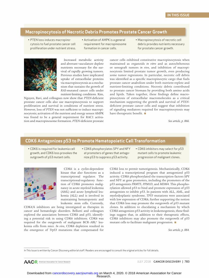
Increased metabolic activity and aberrant vasculature deplete nutrients necessary for the sur-vival of rapidly growing tumors. Previous studies have implicated uptake of extracellular proteins via macropinocytosis as a mecha-nism that sustains the growth of RAS-mutated cancer cells under nutrient-limiting conditions. Kim,
Nguyen, Ravi, and colleagues now show that PTEN-defi cient prostate cancer cells also use macropinocytosis to support proliferation and survival in conditions of nutrient stress. However, loss of PTEN was not suffi cient to induce macropi-nocytosis; activation of the nutrient and energy sensor AMPK was found to be a general requirement for RAC1 activa-tion and macropinosome formation. PTEN-defi cient prostate
cancer cells exhibited constitutive macropinocytosis when maintained as organoids in vitro and as autochthonous or xenograft tumors in vivo, and inhibition of macropi-nocytosis limited prostate tumor growth, even producing some tumor regressions. In particular, necrotic cell debris was identifi ed as a specifi c macropinocytic cargo that fuels prostate cancer anabolism under both nutrient-replete and nutrient-limiting conditions. Necrotic debris contributed to prostate cancer biomass by providing both amino acids and lipids. Taken together, these fi ndings defi ne macro-pinocytosis of extracellular macromolecules as a critical mechanism supporting the growth and survival of PTEN-defi cient prostate cancer cells and suggest that inhibition of signaling mediators required for macropinocytosis may have therapeutic benefi t. ■
See article, p. 866.
• PTEN loss induces macropino-cytosis to fuel prostate cancer cell proliferation under nutrient stress.
• Activation of AMPK is a general requirement for macropinosome formation in cancer cells.
• Macropinocytosis of necrotic cell debris provides nutrients necessary for prostate cancer growth.
Macropinocytosis of Necrotic Debris Promotes Prostate Cancer Growth
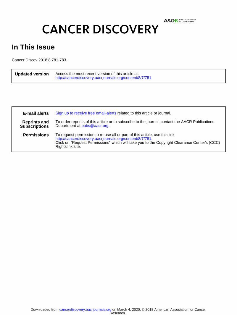
CDK6 is a cyclin-dependent kinase that also functions as a transcriptional regulator. The transcriptional-regulatory func-tion of CDK6 promotes malig-nancy in acute myeloid leukemia (AML) and acute lymphoid leu-kemia (ALL) and is involved in maintaining hematopoietic and leukemic stem cells. Currently,
CDK4/6 inhibitors are being investigated as therapies in cancer and hematologic disorders. Bellutti and colleagues explored the association between CDK6 and p53, identify-ing a potential risk in using CDK6 inhibitors. CDK6 was required for the outgrowth of malignant BCR–ABL+ leu-kemia cells from mice. In vivo, CDK6 depletion resulted in the emergence of Trp53 mutations that compensated for
CDK6 loss to permit tumorigenesis. Mechanistically, CDK6 induced a transcriptional program that antagonized p53 activity. CDK6 phosphorylated the transcription factors SPY and NFY at gene promoters, including the promoters of the p53 antagonists PRMT5, PPM1D, and MDM4. This phospho-rylation allowed p53 to bind and promote expression of p53 antagonists to inhibit p53. In patients with ALL, AML, and myelodysplastic syndrome, TP53 mutations were associated with low expression of CDK6, further supporting the notion that CDK6 loss may promote the outgrowth of p53 mutant clones. In addition to elucidating a mechanism by which CDK6 antagonizes p53 activity in leukemogenesis, these fi nd-ings suggest that, in addition to their therapeutic effects, CDK6 inhibitors may also promote the outgrowth of p53 mutant cells to facilitate malignant progression. ■
See article, p. 884.
• CDK6 is required for leukemia cell growth, and CDK6 loss provokes the outgrowth of p53 mutant cells.
• CDK6 phosphorylates SPY and NFY at promoters of genes that antago-nize p53 to suppress p53 activity.
• CDK6 inhibitors may select for p53 mutant cells to promote leukemic progression of malignant clones.
CDK6 Antagonizes p53 to Promote Hematopoietic Cell Transformation
In This Issue is written by Cancer Discovery editorial staff. Readers are encouraged to consult the original articles for full details.
Research. on March 4, 2020. © 2018 American Association for Cancercancerdiscovery.aacrjournals.org Downloaded from
2018;8:781-783. Cancer Discov In This Issue
Updated version
http://cancerdiscovery.aacrjournals.org/content/8/7/781
Access the most recent version of this article at:
E-mail alerts related to this article or journal.Sign up to receive free email-alerts
SubscriptionsReprints and
To order reprints of this article or to subscribe to the journal, contact the AACR Publications
Permissions
Rightslink site. Click on "Request Permissions" which will take you to the Copyright Clearance Center's (CCC)
.http://cancerdiscovery.aacrjournals.org/content/8/7/781To request permission to re-use all or part of this article, use this link
Research. on March 4, 2020. © 2018 American Association for Cancercancerdiscovery.aacrjournals.org Downloaded from