imaging ischemic heart disease: role of spect and pet. · imaging ischemic heart disease: role of...
TRANSCRIPT

Hein J. VerberneAcademic Medical Center, University of Amsterdam,
Amsterdam, Netherlands
Imaging ischemic heart disease: role of SPECT and PET.
Focus on Patients with Known CAD
International Conference on Integrated Medical Imaging in Cardiovascular Diseases (IMIC 2016)

Disclosure
• Nothing to disclose
Known CAD: Nuclear CardiologyIMIC 2016

Learning objectives
• Discuss the application of appropriate use criteria for SPECT and PET in ischemic heart disease
• Examine new insights in myocardial perfusion imaging: future
Known CAD: Nuclear CardiologyIMIC 2016

Goals
• What is “known” coronary artery disease (CAD):– Definition
• What is the evidence for what indication:– Ischemia detection vs. prognosis
• Clinical implications: – How to use the (imaging) findings
Known CAD: Nuclear CardiologyIMIC 2016

Continuum
Known CA: Nuclear CardiologyEANM 2014
ACCF Circulation 2012;126:e354‐e471
Known CAD: Nuclear CardiologyIMIC 2016

Definition
• Known CAD implies:– Already established:
• Guidelines:– Stable Coronary Artery Disease (ESC 2013) – Stable Ischemic Heart Disease (ACCF 2012)
• Adult patients:– with stable known IHD (including angiographically proven, post MI), including recurrent (renewed‐onset) chest pain and patients with stable pain syndromes.
ESC Eur Heart J 2013;34:2949–3003ACCF Circulation 2012;126:e354‐e471
Known CAD: Nuclear CardiologyIMIC 2016

Definition
• Stable angina pectoris or other symptoms felt to be related to coronary artery disease (CAD) such as dyspnea;
• Previously symptomatic with known obstructive or non‐obstructive CAD, who have become asymptomatic with treatment and need (regular) follow‐up
ESC Eur Heart J 2013;34:2949–3003ACCF Circulation 2012;126:e354‐e471
Known CAD: Nuclear CardiologyIMIC 2016

Definition
• Stable angina pectoris or other symptoms felt to be related to coronary artery disease (CAD) such as dyspnea;
• Previously symptomatic with known obstructive or non‐obstructive CAD, who have become asymptomatic with treatment and need (regular) follow‐up.
ESC Eur Heart J 2013;34:2949–3003ACCF Circulation 2012;126:e354‐e471
Known CAD: Nuclear CardiologyIMIC 2016

Main features of SCAD
Pathogenesis• Stable anatomical atherosclerotic and/or
functional alterations of epicardial vessels and/or microcirculation
Natural history• Stable symptomatic or asymptomatic phases
which may be interrupted by ACSMechanisms of myocardial ischaemia• Fixed or dynamic stenoses of epicardial coronary
arteries;• Microvascular dysfunction;• Focal or diffuse epicardial coronary spasm;• Overlap of above mechanisms in the same
patient and may change over time.Known CAD: Nuclear Cardiology
IMIC 2016

Prognosis
• stable CAD:– individual’s prognosis can vary considerably:
• Reduction of Atherothrombosis for Continued Health (REACH) registry: – included very high‐risk patients, many with peripheral arterial disease or previous MI and almost 50% with diabetes.
• Annual mortality rate of 2.9%.
• Annual mortality rate in patients with non‐obstructive plaques is 0.63%.
JAMA 2007;297:1197–1206
Known CAD: Nuclear CardiologyIMIC 2016

Prognosis
• Therefore important to identify:– those patients with a less severe form of disease and a good prognosis (i.e. no unnecessary tests and revascularization procedures).
– those patients with more severe forms of disease, who may benefit from more aggressive investigation and—potentially—intervention, including revascularization.
Known CAD: Nuclear CardiologyIMIC 2016

Pre test probability
Genders et al. Eur Heart J 2011;32:1316–1330
• White: pre‐test probability: <15% done• Blue: pre‐test probability: 15‐65% consider non‐invasive testing• Light red: pre‐test probability: 66‐85% non‐invasive testing• Red: pre‐test probability: >85% non‐invasive testing prognosis
diagnosis
Known CAD: Nuclear CardiologyIMIC 2016

Data
ACCF etc. JACC 2009;53:2201‐29ESC Eur Heart J 2013;34:2949‐3003
ACCF Circulation 2012;126:e354‐e471
Known CAD: Nuclear CardiologyIMIC 2016

Understanding the evidence
Known CAD: Nuclear CardiologyIMIC 2016
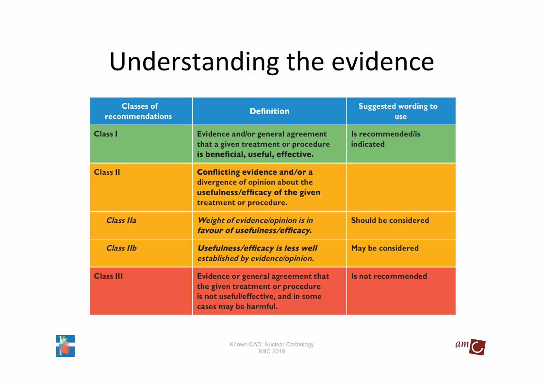
Understanding the evidence
Known CAD: Nuclear CardiologyIMIC 2016

Understanding the evidence
• An appropriate imaging study:– expected incremental information in combination with clinical judgment:
– exceeds the expected negative consequences by a sufficiently wide margin for a specific indication that the procedure is generally considered acceptable care and a reasonable approach for the indication.
ACCF etc. JACC 2009;53:2201‐29
Known CAD: Nuclear CardiologyIMIC 2016

Understanding the evidence
– Score 7–9:• Appropriate (test is generally acceptable and is a reasonable approach for the indication).
– Score 4–6:• Uncertain (test may be generally acceptable and maybe a reasonable approach for the indication).
– Score 1–3:• Inappropriate (test is not generally acceptable and is not a reasonable approach for the indication).
ACCF etc. JACC 2009;53:2201‐29
Known CAD: Nuclear CardiologyIMIC 2016

Appropriate use criteria
ACCF etc. JACC 2009;53:2201‐29
Indication Description ScoreRisk Assessment With Prior Test Results and/or Known Chronic Stable CAD Prior Noninvasive
Evaluation
•Equivocal, borderline, or discordant stress testing where obstructive CAD remains a concern
A (8)
Risk Assessment With Prior Test Results and/or Known Chronic Stable CAD New or Worsening
Symptoms
•Abnormal coronary angiography OR abnormal prior stress imaging study A (9)
Risk Assessment With Prior Test Results and/or Known Chronic
Stable CAD Coronary Angiography (Invasive or
Noninvasive)
•Coronary stenosis or anatomic abnormality of uncertain significance
•Evaluation of ischaemic equivalent
A (9)
A (8)
Known CAD: Nuclear CardiologyIMIC 2016

Appropriate use criteria
ACCF etc. JACC 2009;53:2201‐29
Indication Description ScoreRisk Assessment:
Postrevascularization (PCI or CABG)†Symptoma c
•Evaluation of ischaemic equivalent A (8)
Risk Assessment: Postrevascularization
(PCI or CABG)†Asymptoma c
•Incomplete revascularization•Additional revascularization feasible
•Greater than or equal to 5 years after CABG
A (7)
A (7)
Known CAD: Nuclear CardiologyIMIC 2016

Appropriate use criteria
ACCF etc. JACC 2009;53:2201‐29
Indication Description Score
Risk Assessment: Within 3 Months of an ACS STEMI
•Hemodynamically stable, no recurrent chest pain symptoms or no signs of HF•To evaluate for inducible ischaemia•No prior coronary angiography
A (8)
Risk Assessment: Within 3 Months of an ACS UA/NSTEMI
•Hemodynamically stable, no recurrent chest pain symptoms or no signs of HF•To evaluate for inducible ischaemia•No prior coronary angiography
A (9)
Known CAD: Nuclear CardiologyIMIC 2016

Appropriate use criteria
ACCF etc. JACC 2009;53:2201‐29
Indication Description ScoreAssessment of
Viability/Ischemia Ischemic Cardiomyopathy / Assessment of Viability
•Known severe LV dysfunction•Patient eligible for revascularization A (9)
Known CAD: Nuclear CardiologyIMIC 2016

Use of AUCAppropriateUncertainInappropriate
Hendel et al. JACC 2010;55:156‐62
N=5928
71%
14%
15%
23.4
0.46.8
63
1.16.6
0
10
20
30
40
50
60
70
Low Moderate High
TotalAsymptomatic
• Multivariate analysis for inappropriateness:• Asymptomatic OR: 22.5 (95%CI:15.2–33.2)
“known” CAD:• The second most frequent inappropriate
indication was the performance of SPECT imaging ≤2 years after PCI in an asymptomatic patient (23.8%).
Inapprop
riate ra
te (%
)
CAD risk
Known CAD: Nuclear CardiologyIMIC 2016

Use of AUC
Hendel et al. JACC 2010;55:156‐62
Period 1: baseline. Period 2: after availability of “on‐demand” reports. Period 3: after delivery of specific site and aggregate reports.
An objective automated computer algorithm calculated appropriateness
Known CAD: Nuclear CardiologyIMIC 2016

Use of AUC
Hendel et al. JACC 2010;55:156‐62
Period 1: baseline. Period 2: after availability of “on‐demand” reports. Period 3: after delivery of specific site and aggregate reports.
An objective automated computer algorithm calculated appropriateness
The single site with substantial change in the rate of inappropriate test use:
Initiated discussions to educate physicians on compliance with the AUC.
Known CAD: Nuclear CardiologyIMIC 2016

Testing asymptomatic patients at risk for SCAD
Recommendation Class LevelIn low‐ or intermediate‐risk (based on SCORE) asymptomatic
adults stress imaging tests are not indicated for furtherCV risk assessment.
III C
ESC Eur Heart J 2013;34:2949‐3003
Known CAD: Nuclear CardiologyIMIC 2016
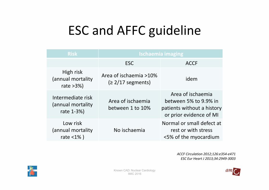
ESC and AFFC guideline
ACCF Circulation 2012;126:e354‐e471ESC Eur Heart J 2013;34:2949‐3003
Risk Ischaemia imaging
ESC ACCFHigh risk
(annual mortality rate >3%)
Area of ischaemia >10% (≥ 2/17 segments) idem
Intermediate risk(annual mortality
rate 1‐3%)
Area of ischaemia between 1 to 10%
Area of ischaemia between 5% to 9.9% in
patients without a history or prior evidence of MI
Low risk(annual mortality
rate <1% )No ischaemia
Normal or small defect at rest or with stress
<5% of the myocardium
Known CAD: Nuclear CardiologyIMIC 2016

Risk stratification using ischaemia testing ESC
Recommendation Class LevelRisk stratification using preferably stress imaging is
recommended in patients with SCAD after significant change in symptom level.
I B
Stress imaging is recommended for risk stratification in patients with known SCAD and a deterioration in symptoms if the site
and extent of ischaemia would influence clinical decision making I B
ESC Eur Heart J 2013;34:2949‐3003
Known CAD: Nuclear CardiologyIMIC 2016

Risk stratification using ischaemia testing ACCF
Recommendation Class LevelEither exercise or pharmacological stress with imaging is
recommended for risk assessment in patients with SCAD who are being considered for revascularization of known coronary
stenosis of unclear physiological significance.
I B
ACCF Circulation 2012;126:e354‐e471
Known CAD: Nuclear CardiologyIMIC 2016

Re‐assessment in patients with SCAD ESC
Recommendation Class LevelAn exercise ECG or stress imaging if appropriate is
recommended in the presence of recurrent or new symptoms once instability has been ruled out.
I C
ESC Eur Heart J 2013;34:2949‐3003
Known CAD: Nuclear CardiologyIMIC 2016

Re‐assessment in patients with SCAD ACCF
Recommendation Class LevelStress imaging with nuclear MPI or echocardiography is
recommended in patients with known SCAD who have new or worsening symptoms not consistent with unstable angina.
I B
ACCF Circulation 2012;126:e354‐e471
Known CAD: Nuclear CardiologyIMIC 2016

Management based risk assessment
ESC Eur Heart J 2013;34:2949‐3003
Confirmed diagnosis SCAD
Low risk High event risk
Continue OMT
Trial ofOMT
Symptoms improved
Intermediate event risk
CAG (+ PCI option) + OMT OMT and consider CAG
Intensify medical treatment
Symptoms improved
Yes
Yes
No
No
Known CAD: Nuclear CardiologyIMIC 2016

Revascularization of SCAD in patients OMT ESC
Recommendation To improveprognosis
To improvesymptoms
persistent onOMT
Class Level Class Level
Proven large area of ischaemia (>10% LV) I B I BDyspnoea/cardiac heart failure with >10%
ischaemia/viability supplied by stenosis >50% IIb B IIa B
No limiting symptoms with OMT in vessel other than left main or proximal LAD or single remainingvessel or vessel subtending area of ischaemia <10%
of myocardium or with FFR ≥0.80.
III A III C
ESC Eur Heart J 2013;34:2949‐3003
Known CAD: Nuclear CardiologyIMIC 2016

Risk stratification using ischaemia testing ACCF
Recommendation Class LevelEither exercise or pharmacological stress with imaging is
recommended for risk assessment in patients with SCAD who are being considered for revascularization of known coronary
stenosis of unclear physiological significance.
I B
Of interestA request to perform either a) more than 1 stress imaging study or b) a stress imaging study and a CCTA at the same time is not
recommended for risk assessment in patients with SCAD.III C
ACCF Circulation 2012;126:e354‐e471
Known CAD: Nuclear CardiologyIMIC 2016

Revascularization of SCAD in patients OMT
ESC Eur Heart J 2013;34:2949‐3003
Significant CAD + ischaemia (>10% myocardium) + OMT
Revascularization possible Revascularization not possible
Anatomical factorsClinical factorsTechnical factorsLocal factors
Failure
Refractory angina
Stem cell therapy?Spinal cord stimulation?
Etc.CABG hybrid PCI
Known CAD: Nuclear CardiologyIMIC 2016

• ♂ 79 year• history:
– 1988 inferior MI– 2000 atypical AP, medical treatment– 2013 change of cardiologist
• RF:– Hypertension, DM, smoking
• Co‐morbidity: arthritis• EGG:
Myocardial perfusion
Known CAD: Nuclear CardiologyIMIC 2016

ECG at rest
Myocardial perfusion
Known CAD: Nuclear CardiologyIMIC 2016

ECG after Adenosine
Myocardial perfusion
Known CAD: Nuclear CardiologyIMIC 2016

Nuclear Cardiologymyocardial perfusion
Known CAD: Nuclear CardiologyIMIC 2016

SA SA
HLA HLA
VLA VLA
EF = 38%EF = 36%
GE Healthcare training session: CARDIOVASCULAR IMAGINGJanuary 2014 . Amsterdam . The Netherlands3rd BALKAN CONGRESS OF NUCLEAR MEDICINE
24 – 26 April 2014 . Bucharest . Romania3rd BALKAN CONGRESS OF NUCLEAR MEDICINE24 – 26 April 2014 . Bucharest . Romania
Stress Rest
Myocardial perfusion
Known CAD: Nuclear CardiologyIMIC 2016

LADRCA
Myocardial perfusion
Known CAD: Nuclear CardiologyIMIC 2016

• ♂ 79 year• history:
– 1988 inferior MI– 2000 atypical AP, medical treatment– 2013 change of cardiologist
• MPS:– Ischemia
• CAG:– 2 vessel disease and successful PCI of RCA and LAD
• 3 months later recurrence of angina!
Myocardial perfusion
Known CAD: Nuclear CardiologyIMIC 2016

MPI post PCI RCA
Myocardial perfusion
Known CAD: Nuclear CardiologyIMIC 2016

Stress RestSA SA
HLA HLA
VLA VLA
EF = 53%EF = 53%
Myocardial perfusion
Known CAD: Nuclear CardiologyIMIC 2016

RCA Post‐PCI LAD Post‐PCI
Myocardial perfusion
Known CAD: Nuclear CardiologyIMIC 2016

Known SCAD and normal MPI
The annualized cardiac mortality rate was 0.9%
Ottenhof et al. J Nucl Cardiol 2013;20:748–54
N= 266, CAD: previous myocardial infarction and/or previous coronary
revascularizationNormal MPI during stress and at rest (<2 segments)Median follow‐up of 12 years (range 8 to 21 years)
Known CAD: Nuclear CardiologyIMIC 2016

Appropriate use criteria
ACCF etc. JACC 2009;53:2201‐29
Indication Description ScoreRisk Assessment with Prior Test Results and/or Known Chronic Stable CAD Asymptomatic Prior Coronary Calcium Agatston
Score
•High CHD risk•Agatston score between 100 and 400
•Agatston score greater than 400
A (7)
A (7)
Known CAD: Nuclear CardiologyIMIC 2016

Conclusions
• Role of MPI in patients with know SCAD is well validated:– Adequate representation in guidelines
• Adherence to guidelines or appropriate use criteria is essential:– Adequate patient care – Trainable!
Known CAD: Nuclear CardiologyIMIC 2016

Therefore, important to realize that:
Garbage in = Garbage out
As a consequence check the quality of the acquired and processed data!!
Myocardial perfusionQuality is essential!
Known CAD: Nuclear CardiologyIMIC 2016

Call for abstracts & clinical casesDeadline 21 Nov 2016
JOIN US IN VIENNA, AUSTRIA!

