identification and characterization of binding sites on
TRANSCRIPT

pubs.acs.org/BiochemistryPublished on Web 10/07/2009r 2009 American Chemical Society
Biochemistry 2009, 48, 10591–10600 10591
DOI: 10.1021/bi901330g
Identification and Characterization of Binding Sites on S100A7, a Participant in Cancerand Inflammation Pathways†
Rafael Le�on,‡ Jill I. Murray,‡ Gina Cragg,§ Benjamin Farnell,§ Nathan R. West, ) Tamara C. S. Pace,‡ Peter H. Watson, )
Cornelia Bohne,*,‡ Martin J. Boulanger,*,§ and Fraser Hof*,‡
‡Department of Chemistry, University of Victoria, P.O. Box 3065, Victoria, British Columbia V8W 3V6, Canada, §Department ofBiochemistry and Microbiology, University of Victoria, P.O. Box 3065, Victoria, British Columbia V8W 3V6, Canada, and )Deeley
Research Centre, BC Cancer Agency, 2410 Lee Avenue, Victoria, British Columbia V8R 6V5, Canada
Received July 31, 2009; Revised Manuscript Received October 7, 2009
ABSTRACT: S100A7 (psoriasin) is a member of the S100 family of signaling proteins. It is implicated in andconsidered a therapeutic target for inflammation and cancer, yet no small molecule ligands for S100A7 havebeen identified. To begin the development of specific small molecule inhibitors of S100A7 function, we haveused a series of surface binding fluorescent dyes to probe the surface hydrophobic sites. Two naphthalene-based dyes (2,6-ANS and 1,8-ANS) were found to bind S100A7 in a distinct cleft. We characterized thebinding interaction by determining both the structure of S100A7 bound to 2,6-ANS and the structure ofS100A7 bound to 1,8-ANS to 1.6 A. In both cases, two molecules of dye were docked such that thenaphthalene groups were positioned in two symmetry-related grooves that are formed by the N-terminalhelices of each monomer. We observed that Met12 acts as a gatekeeper to the binding cleft, adopting an“open” conformation for the more elongated 2,6-ANS while remaining in a “closed” conformation for themore compact 1,8-ANS. Steady-state fluorescence experiments revealed that S100A7 binds two copies of 2,6-ANS, each with a Kd of 125 μM. Time-resolved fluorescence lifetime measurements indicated that the twomolecules of 2,6-ANS bind in two independent binding sites with different fluorescence lifetimes, suggestingthat the S100A7 homodimer is not perfectly symmetric in solution. Isothermal titration calorimetry studiesdemonstrate that S100A7 has a higher affinity for 2,6-ANS than 1,8-ANS. Yeast two-hybrid studies were alsoused to probe contributions of individual residues of an S100A7 triple mutant with respect to Jab1 binding.Mutation of Leu78, which forms part of the Met12 cleft occupied by 2,6-ANS, reduced the level of Jab1binding, suggesting a potentially important role for the Met12 hydrophobic pocket in defining a Jab1interface. Additional Y2H studies also delineate contributions of Gln88 and in particular Asp56 that showsthemost significant abrogated binding to Jab1. Collectively, these data suggest a complex interaction betweenS100A7 and the much larger Jab1. These studies form the basis for the development of small moleculereporters and modifiers of S100A7 form and function.
S100A7, also known as psoriasin, is a member of the S100family of EF-hand calcium-binding signaling proteins. The S100proteins are expressed exclusively in vertebrates and represent thelargest subgroup within the superfamily of EF-hand Ca2þ-binding proteins (1). To date, more than 20 human S100 genesare known, of which 17 are tightly clustered in a region of thehuman 1q21 chromosome that is frequently rearranged incancers (2-4).
S100A7 was first observed as a protein highly upregulated inlesions of psoriatic skin (5) and is considered to play a role ininflammation processes. It is upregulated and excreted from cellsin the epidermis during inflammation and is a chemotactic factorfor keratinocytes (6, 7) and leukocytes (8). Its cell migrationinducing properties are mediated by the receptor for advanced
glycation end products (RAGE)1 in a Zn2þ-dependent man-ner (8). In this extracellular context, S100A7 has also beenimplicated as a host-defense protein that selectively kills Escher-ichia coli on the surface of skin (9, 10).
Within cells, S100A7 apparently lives a different life. Intracel-lular S100A7 expression is predominantly associated with squa-mous cell tumor subtypes in cancers of the lung, head and neck,cervix, and bladder (11-14). S100A7 has also been found to beexpressed in nonsquamous tumors, including gastric cancer,melanoma, and breast cancer (15-17). S100A7 has been mostheavily studied in breast tissue. Expression is absent or almostundetectable in normal breast epithelial cells. The frequency and
†F.H. and M.J.B. are Career Scholars of the Michael Smith Founda-tion for Health Research, and M.J.B. is supported by a CIHR NewInvestigator Award. This work was supported by the Canadian BreastCancer Foundation and NSERC.*To whom correspondence should be addressed. C.B. and F.H.:
telephone, (250) 721-7193; fax, (250) 721-7147; e-mail, [email protected] [email protected].: telephone, (250) 721-7072; fax, (250) 721-8855;e-mail, [email protected].
1Abbreviations: 1,8-ANS, 1-anilinonaphthalene-8-sulfonic acid; 2,6-ANS, 2-anilinonaphthalene-6-sulfonic acid; 3-AT, 3-aminotriazole; Bis-ANS, 4,40-bis(1-anilinonaphthalene-8-sulfonate); DCIS, ductal carci-noma in situ; E-FABP, epidermal fatty acid binding protein; EGF,epidermal growth factor; EGFR, epidermal growth factor receptor;IRF, instrument response function; ITC, isothermal titration calorim-etry; Jab1, c-Jun activation domain binding protein-1; MHC, majorhistocompatibility complex; PDB, Protein Data Bank; RAGE, receptorfor advanced glycation end products; RanBPM, Ran-binding protein inthe microtubule-organizing center; S100A73, S100A7Asp56Gly/Leu78-Met/Gln88Lys triple mutant.
10592 Biochemistry, Vol. 48, No. 44, 2009 Le�on et al.
level of expression increase in ductal carcinoma in situ (DCIS)lesions associated with an increasing risk of progression toinvasive cancer; its highest level of expression is seen in high-grade DCIS, where it is among the most highly expressedproteins (18, 19). Also, S100A7 expression correlates withincreases in genes associated withMHC class II receptor activity,antigen processing and antigen presentation, and immune cellactivation. These results are consistent with a role for S100A7 inmodulating the immune response which may be a factor in earlybreast tumor progression (20).
S100 proteins are known in general to each have manyinteraction partners (21). The proteins for which direct interac-tions with S100A7 are described in the literature includeRAGE (8), RanBPM (22), transglutaminase (23), andE-FABP (24, 25). S100A7 also interacts with the signalingmasterregulator Jab1 (c-jun activation domain binding protein-1) (26).The formation of the S100A7-Jab1 complex is implicated in thedevelopment of early stage breast cancer (27). In breast cancercells, the binding of S100A7 causes the regulatory protein Jab1 totranslocate from the cytoplasm to the nucleus, where it stimulatesa variety of downstream tumorigenic effects (28). Further, bothS100A7 and Jab1 have been linked to the epidermal growthfactor (EGF) and its tyrosine receptor kinase (EGFR), a pathwaythat contributes to a variety of cancers (29, 30). Treatment ofbreast cells with EGF induces S100A7 expression, while down-regulation of S100A7 produces cells with reduced sensitivity toEGF-triggered angiogenesis and osteoclast formation. Further,treatment of breast cells with EGF causes translocation of Jab1to the nucleus (the same effect that was previously observed upontreatment of cells with S100A7). Together, these results suggestthat the S100A7-Jab1 complex is intimately connected to thiswell-known cancer/angiogenesis pathway. With a growing un-derstanding of the dual roles of S100A7 as a mediator of disease-related cell migration (6-8, 31) and cell growth (29, 30) path-ways, it has been proposed as a therapeutic target for thetreatment of inflammation (8) and cancer (27, 28, 32).
The X-ray crystal structure of S100A7 has been determined ata resolution of 2.05 A (33, 34). S100A7 exists as a homodimer thatbinds two Ca2þ ions in EF-hand motifs and two Zn2þ ions atbinding sites that span the dimer interface. In spite of thisstructural data and the wealth of data on biological functionsand interaction partners, structural information on the participa-tion of S100A7 in any of its complexes is scarce, and in some casesvery crude: the triple mutation of putative Jab1-binding residueson S100A7 (Asp56Gly/Leu78Met/Gln88Lys) abrogates bindingto Jab1 and eliminates all of the known downstream tumorigeniceffects associated with S100A7 expression in breast cancercells (27); the S100A7 homologue S100A15 (93% sequenceidentity, differing mainly in the C-terminal region) does not bindto RAGE in the same assays that confirmed S100A7’s directinteraction with this receptor (8); an S100A7 mutant lackingN-terminal residues 1-34 and the C-terminal Zn2þ-bindingmotif (residues 81-101) is still active at killing E. coli (10).
No small molecule ligands for S100A7 are known, and veryfew ligand-binding sites on any S100 protein have beenreported (35-39). We sought to probe the surface features ofS100A7 that might give rise to binding of biological partners andpotential therapeutic agents. To this end, we have studied theinteraction of S100A7 with a collection of surface-bindingfluorescent dyes. The fluorescence intensities of certain dyes aregreatly affected by the mobility and polarity of their microenvir-onments (40), and several studies have used such dyes to identify
surface hydrophobic sites on proteins (41-44). Time-resolved(lifetime) fluorescence studies are even more sensitive to fluor-ophore binding environments; they can discriminate betweensimilar emissive species where the lifetime and intensity arealtered by extremely subtle changes in the vicinity of thefluorophore (45, 46).
We have identified a distinct binding site on the surface ofS100A7 and characterized the molecular complexes of S100A7with two isomeric dyes (2,6-ANS and 1,8-ANS) using X-raycrystallography, steady-state fluorescence, nanosecond time-re-solved fluorescence, and isothermal titration calorimetry. Thesedata instruct the studies of S100A7 in complex with its molecularpartners and will guide the development of specific S100A7-binding small molecules for the study and modification ofS100A7’s numerous important biological functions.
EXPERIMENTAL PROCEDURES
Materials. Buffers and solutions were freshly prepared everyday. 8-Anilinonaphthalene-1-sulfonic acid (1,8-ANS) and 6-anilinonaphthalene-2-sulfonic acid (2,6-ANS) were purchasedfrom Invitrogen (Molecular Probes Inc.). These probes were usedwithout further purification. All other reagents were of analyticalgrade.Production and Purification of Soluble S100A7. Human
S100A7 was amplified by PCR and cloned into the pET32aexpression vector (Novagen) in framewith thioredoxin and anN-terminal hexahistidine tag. Recombinant expression of S100A7was conducted in E. coli Rosetta-Gammi B (Invitrogen,Carlsbad, CA) grown in 2�YT media (DIFCO, Sparks, MD)supplemented with 50 μg/mL kanamycin (Sigma). The cells weregrown at 37 �C to an OD600 of ∼0.8. The temperature waslowered to 30 �C. At an OD600 of 1, expression was induced with0.75 mM isopropyl D-thiogalactoside (IPTG) for 8 h. Theharvested cells were resuspended in 20 mM HEPES buffer (pH8) with 20 mM imidazole and 500 mM NaCl (resuspensionbuffer) and lysed using a French press (SLM Instruments). Thecrude cell extract was centrifuged at 16000 rpm for 45 min toremove insolublematerial and the supernatant applied directly toa Ni-NTA column (Qiagen) equilibrated with resuspensionbuffer. S100A7 fractions eluted with an increasing concentrationof imidazole were analyzed via SDS-PAGE, pooled on the basisof purity, and concentrated. The S100A7 hexahistidine tag wasproteolytically removed with thrombin (Novagen) and theprotein purified on a SuperdexTM S-75 gel filtration column(Amersham Biosciences) equilibrated with 20 mM HEPES and150 mMNaCl. The protein concentration was defined by aminoacid analysis, by which an extinction coefficient ε280 of 57020M-1 cm-1 was determined. A stock solution of 800 μM S100A7wasmaintained at 4 �C. For each experiment, protein was dilutedto a mother solution of 10 μM in buffer [40 mM Tris-HCl(pH 7.2), 100 mMKCl, and 2 mMCaCl2] before being diluted tothe final experimental conditions.Crystallization, Data Collection, and Processing. Puri-
fied S100A7 was concentrated to 17 mg/mL and crystallizedusing the sitting drop vapor diffusion method at 18 �C in 0.2 Mtrisodium citrate and 0.1M sodium cacodylate (pH 6.5). Crystalswere soaked with either 2,6- or 1,8-anilinonaphthalenesulfonate(ANS) at a final concentration of 10 mM for 4 h and frozen at100 K directly in the cryo stream. Diffraction data were collectedon a Rigaku R-axis IVþþ area detector coupled to an MM-002X-ray generator with Osmic “blue” optics and an Oxford

Article Biochemistry, Vol. 48, No. 44, 2009 10593
Cryostream 700 instrument. Diffraction data to 1.6 A wereprocessed using Crystal Clear with d*trek (47). Data collectionand refinement statistics are listed in Table 1.Structure Solution and Refinement. All refinement steps
were conducted using the CCP4 suite of programs (48). Initialphases were obtained by molecular replacement (MR) usingMOLREP (49) with themonomeric formof native S100A7 (PDBentry 2PSR) used as the search model. Solvent atoms wereselected using COOT (50), and the overall structure was refinedwith REFMAC (51). Stereochemical analysis of the refinedS100A7 structure was performed with PROCHECK andSFCHECK in CCP4 (48) with the Ramachandran plot showingexcellent stereochemistry with more than 98% of the residues inthe favored conformations for both structures and no residuesmodeled in disallowed orientations.Overall, 5%of the reflectionswere set aside for calculation ofRfree. The coordinates for S100A7in complex with 2,6- and 1,8-ANS have been deposited in theRCSB PDB as entries 2wos (structure factors, r2wosssf) and2wor (structure factors, r2worsf), respectively.Steady-State Fluorescence. Steady-state fluorescence spec-
tra were recorded on a PTI QM-2 fluorimeter at 25 �C. Theexcitation wavelength was 319 nm, and spectra were collected
between 350 and 600 nm. The excitation and emission slits wereset with bandpasses of 3 and 6 nm, respectively. Fluorescencespectra were corrected for the baseline spectrum using a solutioncontaining all compounds except ANS, ensuring that artifacts,such as Raman emission of the solvent and emission from theprotein, were subtracted from the fluorescence spectra. Theemission from the protein was<1%of the fluorescence intensityfor all solutions. Fluorescence intensities were determined byintegrating the corrected spectra between 415 and 430 nm.Fluorescence Binding Assays. Samples of 2,6-ANS (0.5
μM) and varying concentrations of S100A7 were prepared in 40mM Tris (pH 7.2), 100 mM KCl, 2 mM CaCl2 buffer, mixedgently by pipet, and incubated at room temperature for at least 30min. Each sample was transferred to the cuvette by pipet andincubated in the fluorimeter, in the dark, for 10 min before thefluorescence scan was initiated. After the fluorescence scan wascompleted, the sample was incubated in the dark for 5 min andthen a second fluorescence scan was initiated. Fluorescencespectra were recorded for each S100A7 concentration in thepresence and absence of 2,6-ANS, as well as for controls of 2,6-ANS only and buffer only. The emission from ANS in buffer isless than 2% of the intensity from ANS at the lowest proteinconcentration examined. As this is smaller than the errors in thefluorescencemeasurements, the emission fromANS inbufferwasnot taken into account. The concentration of protein wascalculated as the homodimer form, since this is the only speciesthat has been observed in solution. The fluorescence spectra ateach protein concentration were integrated, and the binding wasanalyzed assuming the equilibrium in eq 1, where the protein hasn identical, independent binding sites for 2,6-ANS.
ANSþnPaANS-P ð1ÞThis is equivalent to 1:1 binding between ANS and the protein,with the effective concentration of available protein binding sitesbeing n[P]. S100A7 has two binding sites for 2,6-ANS (asdetermined by crystallography), and n was fixed to 2 for all fits.The binding isotherm (eq 2) can be derived from the definition ofthe equilibrium constant, and the mass balance equations (52):
ΔI ¼ F
2KdþnPTþLT -
ffiffiffiffiffiffiffiffiffiffiffiffiffiffiffiffiffiffiffiffiffiffiffiffiffiffiffiffiffiffiffiffiffiffiffiffiffiffiffiffiffiffiffiffiffiffiffiffiffiffiffiffiffiffiðKdþnPTþLTÞ2 -4nPTLT
q� �ð2Þ
where Kd is the dissociation constant, PT is the total proteinconcentration, LT is the total ligand concentration, and F is afluorescence scaling factor.
Assuming that the protein concentration is in excess of theligand concentration, [P] ∼ PT, and the following bindingisotherm can be derived (eq 3).
ΔI ¼ φnPT
KdþnPTð3Þ
where φ is a fluorescence scaling factor. This equation can belinearized to give eq 4.
1
ΔI¼ Kd
φnPTþ 1
φð4Þ
The equilibrium constant is determined from the curved plot (eq2) as this results in the proper weighting of uncertainties. Thelinearity of the double-reciprocal plot (eq 4) supports the validityof the assumption that the binding sites are independent.
Experiments were also conducted with 2,6-ANS (from 0 to 100μM) added to a constant concentration of S100A7 (1 μM) in
Table 1: Data Collection and Refinement Statisticsa
2,6-ANS 1,8-ANS
Data Collection
space group P43212 P212121a, b, c (A) 51.66, 51.66,
117.24
51.60, 51.60,
116.91
R, β, γ (deg) 90, 90, 90 90, 90, 90
wavelength (A) 0.9760 0.9760
resolution (A) 47.30-1.70 47.21-1.70
no. of measured
reflections
335832 479650
no. of unique
reflections
18275 18199
redundancy 18.4 (15.7) 26.4 (22.5)
completeness (%) 99.9 (99.9) 100 (100)
I/σ(I) 33.2 (10.9) 34.1 (9.3)
Rmergeb 0.068 (0.242) 0.072 (0.349)
Refinement
Rcrystc/Rfree
d 0.182/0.210 0.188/0.212
no. of atoms
protein 769 769
solvent 99 96
ligand 21 21
Ca2þ/Zn2þ 1/1 1/1
B values (A2)
protein 16.08 14.47
solvent 27.47 27.14
ligand 28.32 13.48
Ca2þ/Zn2þ 12.38/12.98 10.45/12.63
root-mean-square
deviation from
ideality
bond lengths (A) 0.036 0.033
bond angles (deg) 2.753 2.563
aValues in parentheses are for the highest-resolution shell. bRmerge =Phkl|I- ÆIæ|/
PhklI, where I is the intensity of unique reflection hkl and ÆIæ is
the average over symmetry-related observations of unique reflection hkl.cRcryst =
P|Fobs - Fcalc/
PFfobs, where Fobs and Fcalc are the observed and
the calculated structure factors, respectively. dRfree is the R value using 5%of the reflections randomly chosen and omitted from refinement.

10594 Biochemistry, Vol. 48, No. 44, 2009 Le�on et al.
40 mM Tris-HCl, 100 mM KCl, and 2 mM CaCl2 (pH 7.2).Following each addition of ANS, the solution was mixed andallowed to equilibrate for 30 min. For each solution of proteinwith ANS, a control solution of ANS alone in buffer wasprepared.Time-Resolved Fluorescence Measurements. Fluores-
cence decays were measured with an OB920 Edinburgh single-photon counter with a hydrogen flash lamp as the excitationsource. The excitation and emission wavelengths were set to 319and 423 nm, respectively, and the bandwidths for the excitationand emission monochromators were 16 nm. An iris was used todecrease, when necessary, the collection rate to less than 2.5% ofthe excitation rate. The number of counts in the channel ofmaximum intensity was 2000, and control experiments thatincluded 10000 counts did not show any differences in therecovered values for the lifetimes and pre-exponential factors.All measurements were performed at 25 �C. A Ludox suspensionwas used to collect the instrument response function (IRF) at theexcitation wavelength. The IRF was deconvoluted from the datain the fitting procedure using the Edinburgh software, and thedecays were fit to a sum of exponentials (eq 5).
It ¼ I0Xi
1
ðAie-t=τi Þ ð5Þ
where τi is the lifetime of each species and Ai the correspondingpre-exponential factor, where the sum of all pre-exponentialfactors is unity (eq 6).
Xi
1
Ai ¼ 1 ð6Þ
The values of χ2 (0.9-1.1) and visual inspection of the residualsand autocorrelation were used to determine the quality of thefit (53).Isothermal TitrationCalorimetry. ITCmeasurementswere
taken using a VP-ITC calorimeter fromMicroCal, LLC. S100A7and ANS isomers were dissolved in 40 mM Tris-HCl, 100 mMKCl, and 2 mM CaCl2 at final concentrations of 10 μM and 2mM, respectively. The samples were degassed under vacuumprior to titrations. The reference cell was filled with Milli-Qwater. The titration of S100A7 with 1,8-ANS and 2,6-ANSinvolved 28 injections of ligand solution (the first injection was2 μL, and all remaining injections were 10 μL). In all cases, theligand solution was injected at 4 min intervals. The syringestirring speed as set at 307 rpm, and the temperature of thetitration cell was set at 30 �C.The heat of ligand dilution in buffer,determined in separate experiments, was subtracted from thetitration data. Raw data were integrated and processed withOrigin 7.0 provided by the manufacturer. The first injection ineach experiment was not taken into account for the analysis.Three independent experiments with 2,6-ANS and two indepen-dent experiments with 1,8-ANS were analyzed separately.Yeast Two-Hybrid Assays. For yeast two-hybrid studies,
amino acids 42-335 of human Jab1 were expressed as a fusionwith the GAL4 DNA binding domain in bait plasmid pGBT9(Clontech). Wild-type or mutant S100A7 (generated throughPCR mutagenesis) was expressed as a fusion with the GAL4activation domain in prey plasmid pACT2.2 (Addgene plasmid11343, deposited by G. Caldwell). Saccharomyces cerevisiaestrain Y190 (ATCC) was cotransformed with each constructusing the lithium acetate method (54). Relative S100A7-Jab1
interaction strength was assessed by plating transformed cellsonto selective medium lacking leucine, tryptophan, and histidine(Sigma) with various concentrations (10, 25, or 50 mM) of thehistidine analogue 3-aminotriazole (3-AT), or onto leucine/tryptophan deficient control plates. Colony formation on selec-tive relative to control medium was assessed in triplicate afterapproximately 1 week. Differences were assessed using a Stu-dent’s t test.
RESULTS
S100A7 was produced in E. coli as a soluble protein andpurified to homogeneity. Analysis using gel filtration chroma-tography showed that S100A7 existed as a stable dimer consistentwith previous reports (33). S100A7 crystallized with one mono-mer in the asymmetric unit of the tetragonal unit cell with thesecond molecule of the physiological dimer generated by thecrystallographic 2-fold axis. Crystals of S100A7were soakedwitha variety of probe molecules, including Nile Red, Bis-ANS, 2,6-ANS, and 1,8-ANS, and were re-examined via X-ray diffraction.Only crystals treated with 2,6-ANS and 1,8-ANS gave newdensity indicative of binding under these conditions. S100A7costructures with each ANS isomer were refined to a finalresolution of 1.6 A with the final models starting at Ser1 andextending through Ser96 (Figure 1). Each S100A7 monomercoordinates one Ca2þ ion and contributes two of the four ligandsto the coordination of each of the two Zn2þ ions bound at thehomodimer interface. Both S100A7-ANS costructures exhibitunambiguous electron density for the protein backbone andbound dye molecules with low temperature factors and excellentstereochemistry, with more than 98% of the residues adoptingthe most favorable conformation and no residues in the dis-allowed conformation. Final data collection and refinementstatistics are presented in Table 1.
The S100A7 Ca2þ ions in the 2,6- and 1,8-ANS costructuresdeviate less than 0.25 A2 [root-mean-square deviation (rmsd)]from the previously reported native S100A7 structure (34),indicating the binding of the dyes does not significantly perturbthe monomer fold or homodimer structure. Both 2,6- and 1,8-ANS dock into the same groove on S100A7 formed by the N-terminal helices of each monomer. The binding of the moreelongated 2,6-ANS isomer results in a side chain conformationalchange in which the aniline ring displaces the side chain ofMet12by more than 5 A such that it adopts an “open” conformation.The gatekeeper Met12 side chain swings back into the spaciousneighboring Leu78 pocket and opens its own pocket to accom-modate the incoming small molecule, all without significantmovement by any other neighboring residues. The naphthalenegroup of 2,6-ANS is positioned directly between the N-terminalhelices and packs against Gly11 from one monomer and Ala5and Ser8 from the second monomer. The sulfonate group isdirected away from S100A7 toward solvent and is stabilizedthrough interactions with the side chain of Gln4. A comparisonof the 2,6- and 1,8-ANS costructures reveals that the naphthalenegroups of the two dye molecules adopt similar orientationsbetween the N-terminal helices. The more compact structure ofthe 1,8-ANS isomer, however, results in the aniline group beingdirected away from the core of the dimer interface. As a result, theside chain of Met12 is not displaced and instead adopts the“closed” position observed in the native S100A7 structure. Theside chain Lys18 on the surface of S100A7 is, however, displacedby ∼3.4 A to accommodate the aniline group of 1,8-ANS.

Article Biochemistry, Vol. 48, No. 44, 2009 10595
The solution binding of 1,8-ANS and 2,6-ANS to S100A7 wasstudied using steady-state and time-resolved fluorescence experi-ments. Both isomers of ANS are weakly fluorescent in aqueoussolution, while fluorescence is enhanced when they are located ina constrained environment. Titration of S100A7 (1 μM)with 1,8-ANS did not result in any observable increase in the fluorescenceemission of the dye, up to a concentration of 100 μM, and nofurther fluorescence studies were conducted with 1,8-ANS. Asimilar lack of response was also observed for Bis-ANS and NileRed. Titration of S100A7 (1 μM) with the 2,6-ANS isomerresulted in fluorescence enhancement relative to free 2,6-ANS,which is indicative of binding (Figure 2). The increase influorescence intensity was accompanied by a blue shift of
40 nm, a result typical of an ANS dye bound in a nonpolarenvironment (45). S100A7 contains noTrp residues, and thus, theinherent protein fluorescence is insignificant under these experi-mental conditions.
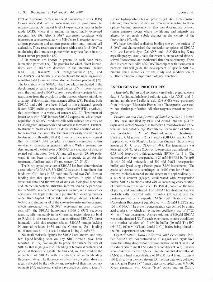
At a constant concentration of S100A7 (1 μM), the 2,6-ANSfluorescence emission intensity (I - I0) increased throughout thetitration with 2,6-ANS (from 0 to 100 μM). The plot of I - I0versus ANS concentration is sigmoidal and does not level off athigher ANS concentrations (Figure 3A). This is indicative ofbinding to additional weaker sites at the higher ANS:proteinratios, and this protocol is not suitable for recovery of Kd for thehigher-affinity ANS binding sites. In addition, at high concentra-tions of ANS, the absorbance at the excitation wavelength ishigh, and there is no longer a linear relationship betweenfluorescence intensity and concentration.
Under conditions where there is an excess of S100A7, 2,6-ANSshould selectively bind to the highest-affinity sites available, andthe changes observed in the fluorescence intensities can beattributed to binding at these sites. Addition of S100A7 (from0 to 250 μM) to a constant 2,6-ANS concentration (0.5 μM) wasconducted. As the amount of protein was increased, the fluores-cence intensity increased and a blue shift in the emission max-imum was again observed. A Kd of 125 ( 11 μM was recoveredfrom the nonlinear fit of the fluorescence intensity to eq 2 whenthe number of independent binding sites is fixed to 2 (Figure 3B).The double-reciprocal plot for the data (Figure 3C) was linear,indicating that the assumption of independent sites was reason-able and that the Kd values for these two sites are similar.
Steady-state fluorescence spectra are a sum of the contribu-tions from each emissive species in solution. Time-resolved
FIGURE 1: Probing surface pockets of S100A7 with isomers of ANS. Cartoon representation of the symmetry-calculated dimer of S100A7 incomplex with (A) 2,6-ANS and (B) 1,8-ANS. Binding of 2,6-ANS to S100A7 displaces the side chain of Met12 (gray) relative to the positionadopted in the native structure (blue), exposing a deep tunnel that spans the S100A7 dimer. The 1,8-ANS, however, does not penetrate as deeplyinto S100A7 and as a result does not displace the side chain ofMet12. The 2Fo- Fc electron density maps of 2,6- and 1,8-ANS calculated at 1.2σare shown in the bottom left and right panels, respectively.
FIGURE 2: Fluorescence spectra (λex = 319 nm) of 0.5 μM 2,6-ANSin the presence of increasing concentrations of S100A7: (a) 0, (b) 30,(c) 50, and (d) 250 μM. The inset shows normalized fluorescencespectra of 2,6-ANS (0.5 μM) bound to 250 μM S100A7 (1) and inbuffer (2). Buffer consisted of 40 mMTris-HCl, 100 mMKCl, and 2mM CaCl2 (pH 7.2) at 25 �C.
10596 Biochemistry, Vol. 48, No. 44, 2009 Le�on et al.
studies, however, can differentiate fluorophores that have thesame emission spectra but different emission efficiencies, i.e.,lifetimes. Time-resolved fluorescence experiments were con-ducted to determine the fluorescence lifetimes of 2,6-ANS underconditions similar to those used in steady-state fluorescenceexperiments. For 2,6-ANS in the presence of S100A7, all lifetimedecays were fitted to a sum of exponentials (eq 5).
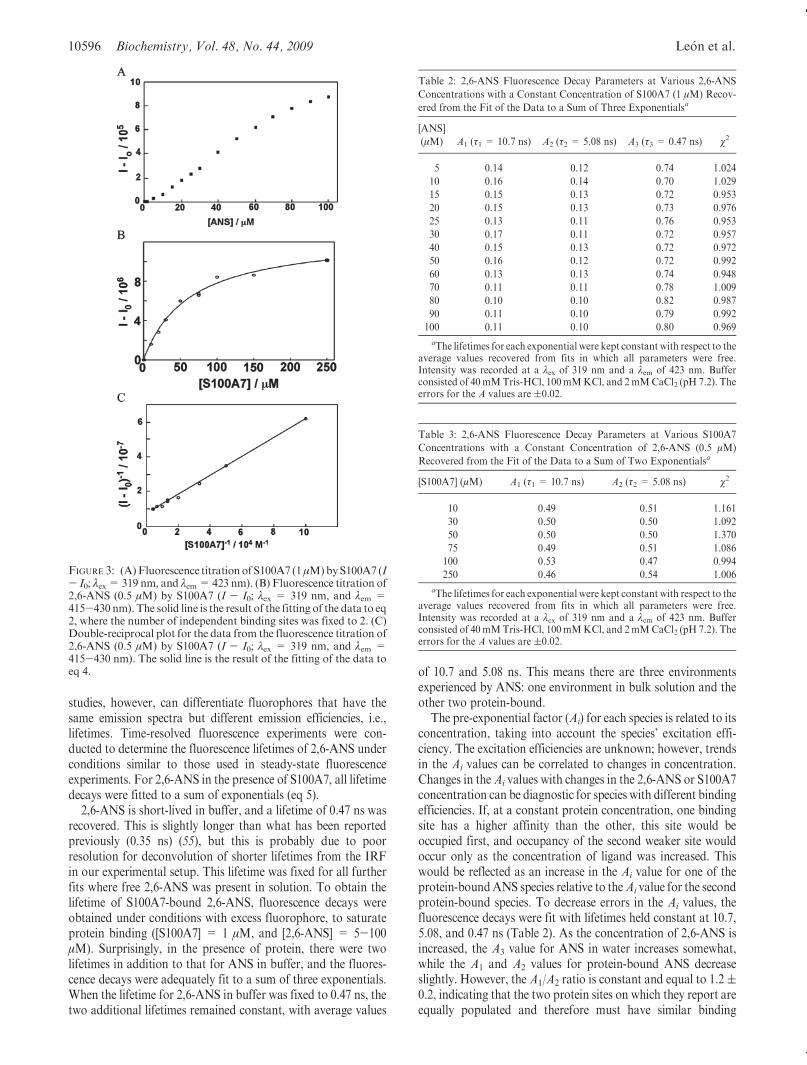
2,6-ANS is short-lived in buffer, and a lifetime of 0.47 ns wasrecovered. This is slightly longer than what has been reportedpreviously (0.35 ns) (55), but this is probably due to poorresolution for deconvolution of shorter lifetimes from the IRFin our experimental setup. This lifetime was fixed for all furtherfits where free 2,6-ANS was present in solution. To obtain thelifetime of S100A7-bound 2,6-ANS, fluorescence decays wereobtained under conditions with excess fluorophore, to saturateprotein binding ([S100A7] = 1 μM, and [2,6-ANS] = 5-100μM). Surprisingly, in the presence of protein, there were twolifetimes in addition to that for ANS in buffer, and the fluores-cence decays were adequately fit to a sum of three exponentials.When the lifetime for 2,6-ANS in buffer was fixed to 0.47 ns, thetwo additional lifetimes remained constant, with average values
of 10.7 and 5.08 ns. This means there are three environmentsexperienced by ANS: one environment in bulk solution and theother two protein-bound.
The pre-exponential factor (Ai) for each species is related to itsconcentration, taking into account the species’ excitation effi-ciency. The excitation efficiencies are unknown; however, trendsin the Ai values can be correlated to changes in concentration.Changes in theAi values with changes in the 2,6-ANS or S100A7concentration can be diagnostic for species with different bindingefficiencies. If, at a constant protein concentration, one bindingsite has a higher affinity than the other, this site would beoccupied first, and occupancy of the second weaker site wouldoccur only as the concentration of ligand was increased. Thiswould be reflected as an increase in the Ai value for one of theprotein-boundANS species relative to theAi value for the secondprotein-bound species. To decrease errors in the Ai values, thefluorescence decays were fit with lifetimes held constant at 10.7,5.08, and 0.47 ns (Table 2). As the concentration of 2,6-ANS isincreased, the A3 value for ANS in water increases somewhat,while the A1 and A2 values for protein-bound ANS decreaseslightly. However, the A1/A2 ratio is constant and equal to 1.2(0.2, indicating that the two protein sites on which they report areequally populated and therefore must have similar binding
FIGURE 3: (A)Fluorescence titration of S100A7 (1μM)byS100A7 (I- I0; λex= 319 nm, and λem=423 nm). (B) Fluorescence titration of2,6-ANS (0.5 μM) by S100A7 (I - I0; λex = 319 nm, and λem =415-430 nm). The solid line is the result of the fitting of the data to eq2, where the number of independent binding sites was fixed to 2. (C)Double-reciprocal plot for the data from the fluorescence titration of2,6-ANS (0.5 μM) by S100A7 (I - I0; λex = 319 nm, and λem =415-430 nm). The solid line is the result of the fitting of the data toeq 4.
Table 2: 2,6-ANS Fluorescence Decay Parameters at Various 2,6-ANS
Concentrations with a Constant Concentration of S100A7 (1 μM) Recov-
ered from the Fit of the Data to a Sum of Three Exponentialsa
[ANS]
(μM) A1 (τ1 = 10.7 ns) A2 (τ2 = 5.08 ns) A3 (τ3 = 0.47 ns) χ2
5 0.14 0.12 0.74 1.024
10 0.16 0.14 0.70 1.029
15 0.15 0.13 0.72 0.953
20 0.15 0.13 0.73 0.976
25 0.13 0.11 0.76 0.953
30 0.17 0.11 0.72 0.957
40 0.15 0.13 0.72 0.972
50 0.16 0.12 0.72 0.992
60 0.13 0.13 0.74 0.948
70 0.11 0.11 0.78 1.009
80 0.10 0.10 0.82 0.987
90 0.11 0.10 0.79 0.992
100 0.11 0.10 0.80 0.969
aThe lifetimes for each exponential were kept constant with respect to theaverage values recovered from fits in which all parameters were free.Intensity was recorded at a λex of 319 nm and a λem of 423 nm. Bufferconsisted of 40mMTris-HCl, 100mMKCl, and 2mMCaCl2 (pH 7.2). Theerrors for the A values are (0.02.
Table 3: 2,6-ANS Fluorescence Decay Parameters at Various S100A7
Concentrations with a Constant Concentration of 2,6-ANS (0.5 μM)
Recovered from the Fit of the Data to a Sum of Two Exponentialsa
[S100A7] (μM) A1 (τ1 = 10.7 ns) A2 (τ2 = 5.08 ns) χ2
10 0.49 0.51 1.161
30 0.50 0.50 1.092
50 0.50 0.50 1.370
75 0.49 0.51 1.086
100 0.53 0.47 0.994
250 0.46 0.54 1.006
aThe lifetimes for each exponential were kept constant with respect to theaverage values recovered from fits in which all parameters were free.Intensity was recorded at a λex of 319 nm and a λem of 423 nm. Bufferconsisted of 40mMTris-HCl, 100mMKCl, and 2mMCaCl2 (pH 7.2). Theerrors for the A values are (0.02.

Article Biochemistry, Vol. 48, No. 44, 2009 10597
constants. The additional weak binding of 2,6-ANS to S100A7observed in the steady-state studies under these conditions is notapparent in the time-resolved studies. This is most likely becausethe increase in the lifetime of 2,6-ANS in these weak binding sitesis minimal and the population of these sites is small.
Experiments conducted with excess protein ([2,6-ANS] = 0.5μM, and [S100A7]= 10-250 μM) yielded results similar to thosewith excess fluorophore. Under these conditions, there is nocontribution to the decay from ANS in buffer, indicating that allANS is bound, and all the fluorescence decays were adequately fitto the sum of two exponentials. The two lifetimes recovered weresimilar to the lifetimes of 10.7 and 5.08 ns observed above,demonstrating that the same binding sites are occupied underboth sets of conditions. To decrease errors in the recovered Ai
values, fits were performed where these two lifetimes were heldconstant (Table 3). There were no changes in the A values as theprotein concentration was increased, and the A1/A2 ratio wasconstant at 0.9 ( 0.1, indicating again that the populations ofthese two protein-bound species are equal and that they musthave similar association constants for the protein.
Because of the lack of fluorescence enhancement from bound1,8-ANS, we used isothermal titration calorimetry (ITC) tocompare the binding of the two ANS isomers to S100A7.Isolation of higher-affinity sites by inverse titration of S100A7into 2,6-ANS was impossible because of the prohibitive amountof protein required for such ITCmeasurements. Instead, titrationof solutions of each ANS isomer (2 mM) into S100A7 (10 μM)was conducted at 30 �C. Each injection of 1,8-ANS into S100A7gave slightly exothermic peaks and little curvature, suggestive ofvery weak binding (Figure 4B). The titration of 2,6-ANS intoS100A7 under identical conditions gives larger exotherms andgreater curvature (Figure 4A). Fitting the multiple parameters(Kassoc, ΔH, ΔS, and stoichiometry n) to unique solutions was
impossible in both cases, as expected for the multiple higher- andlower-affinity binding sites that are engaged during these ANS-into-protein titrations. Nevertheless, we can conclude from theseITC data that 2,6-ANS binds to S100A7 with a more negativeΔH and a higher affinity versus those of 1,8-ANS.
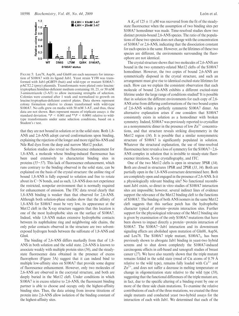
S100A7 exerts tumorigenic effects in breast cells through aninteraction with Jab1 that can be abrogated upon mutation ofthree residues (Asp56Gly, Leu78Met, and Gln88Lys), known asthe S100A7 triple mutant (S100A73) (27). The triple mutant hasbeen extensively studied in engineered cancer cells lines and usingthe yeast two-hybrid assay (27). However, the impact of eachindividual mutation has not been assessed. Of particular interestis Leu78, which lies directly adjacent to the ANS binding site. Toassess the effect of each mutation individually, especially Leu78-Met, we produced three singlymutated S100A7 clones for use in ayeast two-hybrid assay with Jab1, in addition to wild-type andtriple mutant S100A7. We observed that each single mutationwas sufficient to abrogate yeast growth on selective medium(Figure 5), revealing each of the three mutations (Asp56Gly,Leu78Met, andGln88Lys) is detrimental to Jab1 interaction. Theuse of progressively more stringent selection conditions(increasing concentrations of 3-AT) allows the ranking of theeffect at each residue, in the following order: Asp56 > Leu78 >Gln88 (Figure 5).
DISCUSSION
X-ray cocrystal structures reveal that both 2,6-ANS and 1,8-ANS bind at the same Met12 cleft in S100A7 (Figure 1), whiletreatments with bis-ANS or Nile Red did not result in theobservation of any bound dye at the Met12 pocket or at anyother surface site on the protein. Solution studies with the twolarger dyes also show no fluorescence enhancement, suggesting
FIGURE 4: Isothermal titration calorimetry (ITC) data for titration of (A) 2,6-ANS and (B) 1,8-ANS into a solution of S100A7. The S100A7concentration in the cell was 10 μM; the syringe concentrations of 2,6-ANS and 1,8-ANS were 2 mM. The top panel of each figure shows rawtitration data arising from10μL injections,while the bottompanel shows integrated heats of binding for each injection point corrected for heats ofdissolution determined in separate experiments (not shown). Unique fitting solutions could not be determined.
10598 Biochemistry, Vol. 48, No. 44, 2009 Le�on et al.
that they are not bound in solution or in the solid state. Both 1,8-ANS and 2,6-ANS adopt curved conformations upon binding,explaining the rejection of the longer andmore rigid bis-ANS andNile Red dyes from the deep and narrow Met12 pocket.
Solution studies also reveal no fluorescence enhancement for1,8-ANS, a molecule whose binding-induced fluorescence hasbeen used extensively to characterize binding sites inproteins (55-57). This lack of fluorescence enhancement, whichruns contrary to the binding observed in the solid state, can beexplained on the basis of the crystal structure: the aniline ring ofbound 1,8-ANS is fully exposed to solution and free to rotateabout its C-N bonds, and as such, 1,8-ANS does not experiencethe restricted, nonpolar environment that is normally requiredfor enhancement of emission. The ITC data reveal clearly that1,8-ANS binding is weaker than that observed for 2,6-ANS.Although both solution-phase studies show that the affinity of1,8-ANS for S100A7 must be very low, its appearance at theMet12 cleft in the X-ray structure suggests that this location isone of the most hydrophobic sites on the surface of S100A7.Indeed, while 1,8-ANS makes extensive hydrophobic contactsbetween its naphthalene ring and neighboring side chains, theonly polar contacts observed in the structure are two solvent-exposed hydrogen bonds between the sulfonate of 1,8-ANS andGln4.
The binding of 2,6-ANS differs markedly from that of 1,8-ANS in both solution and the solid state. 2,6-ANS is known toassociate weakly with multiple protein sites (56), and the steady-state fluorescence data obtained in the presence of excessfluorophore (Figure 3A) suggest that it can indeed bind tomultiple low-affinity sites on S100A7 that provide some degreeof fluorescence enhancement. However, only two molecules of2,6-ANS are observed in the cocrystal structure, and both aredeeply buried in the Met12 cleft. Under conditions in whichS100A7 is in excess relative to 2,6-ANS, the fluorescent bindingpartner is able to choose and occupy only the highest-affinitybinding sites. Thus, the data arising from inverse titrations ofprotein into 2,6-ANS allow isolation of the binding constant ofthe highest-affinity sites.
A Kd of 125( 11 μMwas recovered from the fit of the steady-state fluorescence when the assumption of two binding sites perS100A7 homodimer was made. Time-resolved studies show twodistinct protein-bound 2,6-ANS species. The ratio of the popula-tions of these two species does not change with the concentrationof S100A7 or 2,6-ANS, indicating that the dissociation constantfor each species is the same.However, as the lifetimes of these twospecies are different, the environments surrounding the fluor-ophore are not identical.
The crystal structure shows that twomolecules of 2,6-ANS arebound in the two symmetry-related Met12 clefts of the S100A7homodimer. However, the two copies of bound 2,6-ANS aresymmetrically disposed in the crystal structure, and such anarrangement must give rise to identical excited-state lifetimes foreach. How can we explain the consistent observation that eachmolecule of bound 2,6-ANS exhibits a different excited-statelifetime under the large range of conditions studied? It is possiblethat in solution the different environments for each copy of 2,6-ANS arise from differing conformations of the two bound copiesof 2,6-ANS within a perfectly symmetric S100A7 dimer. Analternative explanation arises if one considers that S100A7consistently exists in solution as a homodimer with brokensymmetry. Indeed, S100A7 was previously reported to crystallizeas a nonsymmetric dimer in the presence of low Zn2þ concentra-tions, and that structure reveals striking dissymmetry in theMet12 region (34). It is possible that a similar nonsymmetricstructure of S100A7 is significantly populated in solution.Whatever the structural explanation, the use of time-resolvedfluorescence here reveals a loss of symmetry for the S100A7-2,6-ANS complex in solution that is invisible to steady-state fluor-escence titrations, X-ray crystallography, and ITC.
One of the two Met12 clefts is open in structure 3PSR (34).Both are closed in structures 1PSR and 2PSR (33, 34). Both arepartially open in the 1,8-ANS costructure determined here. Bothare completely open and engaged in the presence of 2,6-ANS. Is ita physiologically relevant binding site? No functional recombi-nant Jab1 exists, so direct in vitro studies of S100A7 interactionsites are impossible; however, several indirect lines of evidencesupport the relevance of the Met12 binding cleft to the functionsof S100A7. The binding of both ANS isomers in the sameMet12cleft suggests that this surface patch has the hydrophobiccharacter typical of protein-protein interaction sites. Furthersupport for the physiological relevance of the Met12 binding siteis given by examination of the only S100A7 mutations that havebeen shown to impact directly a protein-protein interaction ofS100A7. The S100A7-Jab1 interaction and its downstreamsignaling effects are abolished upon mutation of Gln88, Asp56,and Leu78. The S100A7 triple mutant, S100A73, has beenpreviously shown to abrogate Jab1 binding in yeast-two hybridscreens and to shut down completely the S100A7-inducedcarcinogenic effects in cell-based and xenograft studies of breastcancer (27). We have also recently shown that the triple mutantremains folded in the solid state (rmsd of CR atoms of 0.79 Arelative to the wild type), remains fully loaded with Ca2þ andZn2þ, and does not suffer a decrease in melting temperature orchange in oligomerization state relative to the wild type (58),suggesting that the functional differences of the triplemutant are,in fact, due to the specific altering of a binding event by one ormore of the three side chain mutations. To examine the relativecontributions of each of the three mutations, we created the threesingle mutants and conducted yeast two-hybrid assays for theinteraction of each with Jab1. We determined that each of the
FIGURE 5: Leu78, Asp56, and Gln88 are each necessary for interac-tion of S100A7 with its ligand Jab1. Yeast strain Y190 was trans-formed with Jab1-pGBT9 (bait) and wild-type or mutant S100A7-pACT2.2 (prey) plasmids. Transformants were plated onto leucine/tryptophan/histidine-deficient medium containing 10, 25, or 50 mM3-aminotriazole (3-AT) to allow increasing strengths of selection.Colonies were counted after 1 week and normalized to growth onleucine/tryptophan-deficient control plates. Data shown representcolony formation relative to clones transformed with wild-typeS100A7. No cells grew on media with 50 mM 3-AT, and thus, thesedata are not shown. Bars represent means of triplicate assays ( thestandard deviation. *P < 0.001 and **P < 0.0001 relative to wild-type transformants under same selection conditions, based on aStudent’s t test.

Article Biochemistry, Vol. 48, No. 44, 2009 10599
three single mutations significantly weakens the S100A7-Jab1interaction relative to that of wild-type S100A7, and to a degreethat is similar to the overall effect of the triple mutant S100A73(Figure 5). While Asp56Gly shows the strongest effect, theLeu78Met mutation that is directly adjacent to the ANS bindingsite is also strong despite the conservative nature of this hydro-phobic-for-hydrophobic mutation. Two copies of Leu78 line thehydrophobic gorge along the C2 axis of the S100A7 homodimer,and the binding of 2,6-ANS to S100A7 causes each monomer’sneighboring Met12 residue to undergo a well-ordered reorienta-tion that significantly alters the shape of the continuous hydro-phobic Met12-Leu78 recognition surface. In the absence of anS100A7 costructure with any of its protein binding partners, wecannot speculate in any detail about the orientations of Met12and Leu78 that might be adopted in the complexes, but all of thisevidence, taken together, leads us to propose that theMet12 cleftdiscovered in this study due to its occupation by ANS is relevantto one or more of the physiologically relevant complexes formedby S100A7.
The importance of S100 proteins as signaling proteins withtissue- and pathology-specific distributions is increasing, but fewexamples of small molecule binding have been reported for anymembers of this family (35-39). Among S100 proteins, S100A7is typical in its lack of mutation and structural data relating tofunctions and formation of complexes. The demonstration of theMet12 cleft as a small molecule binding site with the potentialalso to be a protein-protein binding site is an important step inunderstanding and inhibiting protein-protein complexes ofS100A7. We are currently using these data to pursue smallmolecules that report on and modify the interaction of S100A7with Jab1.
ACKNOWLEDGMENT
We thank Melanie Olsen for the contribution of the S100A7plasmid.
REFERENCES
1. Marenholz, I., Heizmann, C.W., and Fritz, G. (2004) S100 proteins inmouse andman: From evolution to function and pathology (includingan update of the nomenclature). Biochem. Biophys. Res. Commun.322, 1111–1122.
2. Engelkamp, D., Schafer, B. W., Mattei, M. G., Erne, P., andHeizmann, C. W. (1993) Six S100 genes are clustered on humanchromosome 1q21: Identification of two genes coding for the twopreviously unreported calcium-binding proteins S100D and S100E.Proc. Natl. Acad. Sci. U.S.A. 90, 6547–6551.
3. Schafer, B. W., Wicki, R., Engelkamp, D., Mattei, M. G., andHeizmann, C. W. (1995) Isolation of a YAC clone covering a clusterof nine S100 genes on human chromosome 1q21: Rationale for a newnomenclature of the S100 calcium-binding protein family. Genomics25, 638–643.
4. Sturchler, E., Cox, J. A., Durussel, I., Weibel, M., and Heizmann,C. W. (2006) S100A16, a novel calcium-binding protein of the EF-hand superfamily. J. Biol. Chem. 281, 38905–38917.
5. Madsen, P., Rasmussen,H.H., Leffers, H., Honore, B., Dejgaard,K.,Olsen, E., Kiil, J., Walbum, E., Andersen, A. H., and Basse, B.; et al.(1991) Molecular cloning, occurrence, and expression of a novelpartially secreted protein “psoriasin” that is highly up-regulated inpsoriatic skin. J. Invest. Dermatol. 97, 701–712.
6. Boniface, K., Bernard, F.-X., Garcia, M., Gurney, A. L., Lecron,J.-C., and Morel, F. (2005) IL-22 Inhibits Epidermal Differentiationand Induces Proinflammatory Gene Expression and Migration ofHuman Keratinocytes. J. Immunol. 174, 3695–3702.
7. Boniface, K., Diveu, C.,Morel, F., Pedretti, N., Froger, J., Ravon, E.,Garcia, M., Venereau, E., Preisser, L., Guignouard, E., Guillet, G.,Dagregorio, G., Pene, J., Moles, J.-P., Yssel, H., Chevalier, S.,Bernard, F.-X., Gascan, H., and Lecron, J.-C. (2007) Oncostatin MSecreted by Skin Infiltrating T Lymphocytes Is a Potent Keratinocyte
Activator Involved in Skin Inflammation. J. Immunol. 178, 4615–4622.
8. Wolf, R., Howard, O. M. Z., Dong, H.-F., Voscopoulos, C.,Boeshans, K., Winston, J., Divi, R., Gunsior, M., Goldsmith, P.,Ahvazi, B., Chavakis, T., Oppenheim, J. J., and Yuspa, S. H. (2008)Chemotactic Activity of S100A7 (Psoriasin) Is Mediated by theReceptor for Advanced Glycation End Products and PotentiatesInflammation with Highly Homologous but Functionally DistinctS100A15. J. Immunol. 181, 1499–1506.
9. Glaser, R., Harder, J., Lange, H., Bartels, J., Christophers, E., andSchroder, J.-M. (2005) Antimicrobial psoriasin (S100A7) protectshuman skin from Escherichia coli infection. Nat. Immunol. 6, 57.
10. Lee, K. C., and Eckert, R. L. (2006) S100A7 (Psoriasin): Mechanismof Antibacterial Action in Wounds. J. Invest. Dermatol. 127, 945.
11. Zhang, H., Zhao, Q., Chen, Y., Wang, Y., Gao, S., Mao, Y., Li, M.,Peng, A., He, D., and Xiao, X. (2008) Selective expression of S100A7in lung squamous cell carcinomas and large cell carcinomas but not inadenocarcinomas and small cell carcinomas. Thorax 63, 352–359.
12. Celis, J. E., Rasmussen, H. H., Vorum, H., Madsen, P., Honore, B.,Wolf, H., andOrntoft, T. F. (1996) Bladder squamous cell carcinomasexpress psoriasin and externalize it to the urine. J. Urol. 155, 2105–2112.
13. Ralhan, R., DeSouza, L. V., Matta, A., Chandra Tripathi, S.,Ghanny, S., Datta Gupta, S., Bahadur, S., and Siu, K. W. M.(2008) Discovery and Verification of Head-and-neck Cancer Biomar-kers by Differential Protein Expression Analysis Using iTRAQLabeling, Multidimensional Liquid Chromatography, and TandemMass Spectrometry. Mol. Cell. Proteomics 7, 1162–1173.
14. Webb, M., Emberley, E. D., Lizardo, M., Alowami, S., Qing, G.,Alfia’ar, A., Snell-Curtis, L. J., Niu, Y., Civetta, A., Myal, Y., Shiu,R., Murphy, L. C., and Watson, P. H. (2005) Expression analysis ofthe mouse S100A7/psoriasin gene in skin inflammation and mam-mary tumorigenesis. BMC Cancer 5, 17.
15. El-Rifai, W., Moskaluk, C. A., Abdrabbo, M. K., Harper, J.,Yoshida, C., Riggins, G. J., Frierson, H. F.Jr., and Powell, S. M.(2002) Gastric cancers overexpress S100A calcium-binding proteins.Cancer Res. 62, 6823–6826.
16. Brouard, M. C., Saurat, J. H., Ghanem, G., and Siegenthaler, G.(2002) Urinary excretion of epidermal-type fatty acid-binding proteinand S100A7 protein in patients with cutaneous melanoma.MelanomaRes. 12, 627–631.
17. Moog-Lutz, C., Bouillet, P., Regnier, C. H., Tomasetto, C., Mattei,M. G., Chenard,M. P., Anglard, P., Rio, M. C., and Basset, P. (1995)Comparative expression of the psoriasin (S100A7) and S100C genes inbreast carcinoma and co-localization to human chromosome 1q21-q22. Int. J. Cancer 63, 297–303.
18. Enerback, C., Porter, D. A., Seth, P., Sgroi, D., Gaudet, J.,Weremowicz, S., Morton, C. C., Schnitt, S., Pitts, R. L., Stampl, J.,Barnhart, K., and Polyak, K. (2002) Psoriasin expression in mam-mary epithelial cells in vitro and in vivo. Cancer Res. 62, 43–47.
19. Leygue, E., Snell, L., Hiller, T., Dotzlaw,H., Hole,K.,Murphy, L. C.,and Watson, P. H. (1996) Differential expression of psoriasin mes-senger RNA between in situ and invasive human breast carcinoma.Cancer Res. 56, 4606–4609.
20. Mandal, S., Curtis, L., Pind, M., Murphy, L. C., and Watson, P. H.(2007) S100A7 (psoriasin) influences immune response genes in hu-man breast cancer. Exp. Cell Res. 313, 3016–3025.
21. Santamaria-kisiel, L., Rintala-dempsey, A. C., and Shaw,G. S. (2006)Calcium-dependent and -independent interactions of the S100 proteinfamily. Biochem. J. 396, 201–214.
22. Emberley, E., Gietz, R. D., Campbell, J. D., HayGlass, K., Murphy,L., and Watson, P. (2002) RanBPM interacts with psoriasin in vitroand their expression correlates with specific clinical features in vivo inbreast cancer. BMC Cancer 2, 28.
23. Ruse, M., Lambert, A., Robinson, N., Ryan, D., Shon, K. J., andEckert, R. L. (2001) S100A7, S100A10, and S100A11 Are Transglu-taminase Substrates. Biochemistry 40, 3167–3173.
24. Hagens, G.,Masouy�e, I., Augsburger, E., Hotz, R., Saurat, J. H., andSiegenthaler, G. (1999) Calcium-binding protein S100A7 and epider-mal-type fatty acid-binding protein are associated in the cytosol ofhuman keratinocytes. Biochem. J. 339, 419–427.
25. Ruse, M., Broome, A.-M., and Eckert, R. L. (2003) S100A7(Psoriasin) Interacts with Epidermal Fatty Acid Binding Proteinand Localizes in Focal Adhesion-Like Structures in Cultured Kera-tinocytes. J. Invest. Dermatol. 121, 132–141.
26. Emberley, E. D., Niu, Y., Leygue, E., Tomes, L., Gietz, R. D.,Murphy, L. C., and Watson, P. H. (2003) Psoriasin Interacts withJab1 and Influences Breast Cancer Progression.Cancer Res. 63, 1954–1961.

10600 Biochemistry, Vol. 48, No. 44, 2009 Le�on et al.
27. Emberley, E. D., Niu, Y., Curtis, L., Troup, S.,Mandal, S. K.,Myers,J. N., Gibson, S. B., Murphy, L. C., and Watson, P. H. (2005)The S100A7-c-Jun Activation Domain Binding Protein 1 PathwayEnhances Prosurvival Pathways in Breast Cancer. Cancer Res. 65,5696–5702.
28. Emberley, E. D., Alowami, S., Snell, L., Murphy, L. C., and Watson,P. H. (2004) S100A7 (psoriasin) expression is associated with aggres-sive features and alteration of Jab1 in ductal carcinoma in situ of thebreast. Breast Cancer Res. 6, R308–R315.
29. Paruchuri, V., Prasad, A., McHugh, K., Bhat, H. K., Polyak, K., andGanju, R. K. (2008) S100A7-Downregulation Inhibits EpidermalGrowth Factor-Induced Signaling in Breast Cancer Cells and BlocksOsteoclast Formation. PLoS ONE 3, e1741.
30. Wang, J., Barnes, R., West, N., Olson, M., Chu, J., and Watson, P.(2008) Jab1 is a target of EGFR signaling in ERR-negative breastcancer. Breast Cancer Res. 10, R51.
31. Jinquan, T., Vorum, H., Larsen, C., Madsen, P., Rasmussen, H.,Gesser, B., Etzerodt, M., Honor�e, B., Celis, J., and Thestrup-Pedersen, K. (1996) Psoriasin: A novel chemotactic protein. J. Invest.Dermatol. 107, 5–10.
32. Jiang, W. G., Watkins, G., Douglas-Jones, A., and Mansel, R. E.(2004) Psoriasin is aberrantly expressed in human breast cancer and isrelated to clinical outcomes. Int. J. Oncol. 25, 81–85.
33. Brodersen, D. E., Etzerodt, M., Madsen, P., Celis, J. E., Thogersen,H. C., Nyborg, J., and Kjeldgaard, M. (1998) EF-hands at atomicresolution: The structure of human psoriasin (S100A7) solved byMAd phasing. Structure 6, 477–489.
34. Brodersen,D. E.,Nyborg, J., andKjeldgaard,M. (1999) Zinc-bindingsite of an S100 protein revealed. Two crystal structures of Ca2þ-boundhuman psoriasin (S100A7) in the Zn2þ-loaded and Zn2þ-free states.Biochemistry 38, 1695–1704.
35. Markowitz, J., Chen, I., Gitti, R., Baldisseri, D. M., Pan, Y., Udan,R., Carrier, F., MacKerell, A. D.Jr., and Weber, D. J. (2004)Identification and characterization of small molecule inhibitors ofthe calcium-dependent S100B-p53 tumor suppressor interaction.J. Med. Chem. 47, 5085–5093.
36. Zhong, S., Macias, A. T., and MacKerell, A. D.Jr. (2007) Computa-tional identification of inhibitors of protein-protein interactions.Curr. Top. Med. Chem. 7, 63–82.
37. Garrett, S. C., Hodgson, L., Rybin, A., Toutchkine, A., Hahn, K.M.,Lawrence, D. S., and Bresnick, A. R. (2008) A biosensor of S100A4metastasis factor activation: Inhibitor screening and cellular activa-tion dynamics. Biochemistry 47, 986–996.
38. Charpentier, T. H., Wilder, P. T., Liriano, M. A., Varney, K. M.,Pozharski, E.,MacKerell, A.D.Jr., Coop,A., Toth, E.A., andWeber,D. J. (2008) Divalent Metal Ion Complexes of S100B in the Absenceand Presence of Pentamidine. J. Mol. Biol. 382, 56.
39. Charpentier, T. H., Wilder, P. T., Liriano, M. A., Varney, K. M.,Zhong, S., Coop, A., Pozharski, E., Alexander, D., MacKerell, J.,Toth, E. A., and Weber, D. J. (2009) Small Molecules Bound toUnique Sites in the Target Protein Binding Cleft of Calcium-BoundS100B As Characterized by Nuclear Magnetic Resonance and X-rayCrystallography. Biochemistry 48, 6202–6212.
40. Slavik, J. (1982) Anilinonaphthalene sulfonate as a probe ofmembrane composition and function. Biochim. Biophys. Acta 694,1–25.
41. Ke, S., Wright, J. C., and Kwon, G. S. (2007) Avidin-biotin-PEG-CPA complexes as potential EPR-directed therapeutic protein car-riers: Preparation and characterization.Bioconjugate Chem. 18, 1644–1650.
42. Chaves, J. M., Srivastava, K., Gupta, R., and Srivastava, O. P. (2008)Structural and functional roles of deamidation and/or truncation ofN- or C-termini in human RA-crystallin. Biochemistry 47, 10069–10083.
43. Hawe, A., Sutter, M., and Jiskoot, W. (2008) Extrinsic fluorescentdyes as tools for protein characterization.Pharm. Res. 25, 1487–1499.
44. Togashi, D. M., and Ryder, A. G. (2008) A fluorescence analysis ofANS bound to bovine serum albumin: Binding properties revisited byusing energy transfer. J. Fluoresc. 18, 519–526.
45. Gasymov, O. K., and Glasgow, B. J. (2007) ANS fluorescence:Potential to augment the identification of the external binding sitesof proteins. Biochim. Biophys. Acta 1774, 403–411.
46. Kirk, W., Kurian, E., and Wessels, W. (2007) Photophysics of ANS.V. Decay modes of ANS in proteins: The IFABP-ANS complex.Biophys. Chem. 125, 50–58.
47. Pflugrath, J. (1999) The finer things in X-ray diffraction data collec-tion. Acta Crystallogr. D55, 1718–1725.
48. Collaborative Computational Project Number 4 (1994) The CCP4Suite: Programs for protein crystallography. Acta Crystallogr. D50,760–763.
49. Vagin, A., and Teplyakov, A. (1997) MOLREP: An AutomatedProgram for Molecular Replacement. J. Appl. Crystallogr. 1022–1025.
50. Emsley, P., and Cowtan, K. (2004) Coot: Model-building tools formolecular graphics. Acta Crystallogr. D60, 2126–2132.
51. Murshudov, G. N., Vagin, A. A., and Dodson, E. J. (1997) Refine-ment of macromolecular structures by the maximum-likelihoodmethod. Acta Crystallogr. D53, 240–255.
52. Gasymov, O. K., Abduragimov, A. R., and Glasgow, B. J. (2007)Evidence for internal and external binding sites on human tearlipocalin. Arch. Biochem. Biophys. 468, 15–21.
53. Bohne, C. R., and Scaiano, J. C. (1991) Photochemistry in Organizedand Constrained Media , VCH Publishers, New York.
54. Gietz, R. D., and Schiestl, R. H. (2007) Quick and easy yeasttransformation using the LiAc/SS carrier DNA/PEG method. Nat.Protoc. 2, 35–37.
55. Huang, J., and Bright, F. V. (1990) Unimodal Lorentzian LifetimeDistributions for the 2-Anilinonaphthalene-6-sulfonate-β-Cyclodex-trin Inclusion Complex Recovered by Multifrequency Phase-Modu-lation Fluorometry. J. Phys. Chem. 94, 8457–8463.
56. Kirk, W. R., Kurian, E., and Prendergast, F. G. (1996) Characteriza-tion of the sources of protein-ligand affinity: 1-Sulfonato-8-(10)anilinonaphthalene binding to intestinal fatty acid bindingprotein. Biophys. J. 70, 69–83.
57. Gasymov, O. K., Abduragimov, A. R., and Glasgow, B. J. (2008)Ligand binding site of tear lipocalin: Contribution of a trigonal clusterof charged residues probed by 8-anilino-1-naphthalenesulfonic acid.Biochemistry 47, 1414–1424.
58. West, N. R., Farnell, B., Murray, J. I., Hof, F., Watson, P. H., andBoulanger, M. J. (2009) Structural and functional characterization ofa triple mutant form of S100A7 defective for Jab1 binding. ProteinSci. (in press).