hypochlorous acid as a potential wound care agent - … · hypochlorous acid as a potential wound...
TRANSCRIPT
Hypochlorous Acid as a Potential WoundCare AgentPart II. Stabilized Hypochlorous Acid: Its Role in Decreasing
Tissue Bacterial Bioburden and Overcoming the Inhibition of
Infection on Wound Healing
Martin C. Robson, MD,a,b Wyatt G. Payne, MD,a,b Francis Ko, BS,a
Marni Mentis, DO,a Guillermo Donati, DPM,a Susan M. Shafii, MD,b
Susan Culverhouse, MD,b Lu Wang, PhD,c Behzad Khosrovi, PhD,c
Ramin Najafi, PhD,c Diane M. Cooper, PhD,d and Mansour Bassiri, PhDc
aInstitute for Tissue Regeneration, Repair and Rehabilitation, Bay Pines, FL; bUniversity of SouthFlorida, Tampa, FL; cNovaBay Pharmaceuticals, Inc, Emeryville, CA; and dHealthpoint Ltd, ForthWorth, TX
Correspondence: [email protected] April 11, 2007
Background: A topical antimicrobial that can decrease the bacterial bioburden of chronicwounds without impairing the wound’s ability to heal is a therapeutic imperative. A stabi-lized form of hypochlorous acid (NVC-101) has been demonstrated in vitro and in stan-dard toxicity testing to possess properties that could fulfill these criteria. Materials andMethods: Using a standard rodent model of a chronically infected granulating wound,various preparations of NVC-101 and multiple treatment regimens were investigated toevaluate the role of NVC-101 in decreasing tissue bacterial bioburden and overcomingthe inhibition of infection on wound healing. Quantitative bacteriology of tissue biopsiesand wound healing trajectories were used to compare the various NVC-101 preparationsand regimens to saline-treated negative controls and silver sulfadiazine–treated positivecontrols. Results: NVC-101 at 0.01% hypochlorous acid with a pH of 3.5 to 4.0 provedto be an effective topical antimicrobial. It was most effective when used for a briefperiod (15–30 minutes), and followed with another application. Possibly this was dueto its rapid neutralization in the wound bed environment. Although not as effective atdecreasing the tissue bacterial bioburden as silver sulfadiazine, NVC-101 was associatedwith improved wound closure. Conclusions: This stabilized form of hypochlorous acid(NVC-101) could have potential application as an antimicrobial wound irrigation andtreatment solution if its effective pH range can be maintained in the clinical situation.NVC-101 solution was equally effective at pH 3.5 or 4.0 and more efficient soon afterits application. As opposed to other antimicrobials investigated in this animal model,NVC-101 controls the tissue bacterial bioburden without inhibiting the wound healingprocess.
This work was fully supported by NovaBay Pharmaceuticals, Inc, Emeryville, CA.
80
ROBSON ET AL
Wound healing is the end result of a series of interrelated cellular processes initiated byhumoral factors such as cytokine growth factors.1 These cellular processes are inhibited bya large tissue bacterial bioburden.2 The cytokines and growth factors are also degradedby bacteria.3 The level of tissue bacterial bioburden has been shown in multiple studiesto be more than 105 or at least 1 × 10⁶ bacteria per gram of tissue.4,5 Such high levelsof tissue bacteria can be present without clinical signs of infection, and when present candeleteriously affect wound healing.6
Attempts at controlling the tissue bacterial bioburden have been difficult. Systemicallyadministered antibiotics do not effectively decrease the level of bacteria in a chronic granu-lating wound.7 Therefore, topical antimicrobials or temporary biologic dressings have beenthe methods of choice.4,8 Topical use of antibiotics that are used effectively systemically forpurposes other than wound infection is discouraged because of an increased risk for devel-oping allergies or the potential for bacteria to develop resistance to the drug.9 Antisepticsand nonantibiotic antimicrobials such as povidone-iodine, silver sulfadiazine, or mafenideacetate cream have been demonstrated to be cytotoxic to the cellular components of woundhealing.10−12
Stabilized hypochlorous acid (NVC-101) prepared by the addition of sodium hypochlo-rite to a solution of sodium chloride in sterile water followed by addition of a solution ofhydrochloric acid and maintained at a pH between 3.5 and 5 has been demonstrated tohave excellent in vitro antibacterial properties. Its potential limitation is the requirement tomaintain its narrow pH range in the clinical wound environment.
The purpose of the studies reported here was to evaluate various concentrations ofstabilized hypochlorous acid (NVC-101) topically administered to an experimental chronicinfected granulating wound at different pHs and with different treatment regimens. Theeffects evaluated were the ability of NVC-101 to control the tissue bacterial bioburden andthe ability of the agent to overcome the inhibition of wound healing caused by infection.
MATERIALS AND METHODS
Reagents
The various NVC-101 preparations ranging in concentrations from 0.01% to 0.02% andfrom pH 3.5 to 4.5 were provided by NovaBay Pharmaceuticals, Emeryville, Calif. In brief,the stabilized HOCl was prepared in 150 mM NaCl by acidifying reagent-grade NaOCl to thepH of 3.5 to 4.5 with dilute HCl. The concentration of active chlorine species ([HOCl]T =[HOCl] + [Cl2] + [Cl3] + [OCl−]) in 0.9% saline was determined by converting all theactive chlorine species to OCl− with 0.1 M NaOH and measuring the concentration of OCl−
spectrophotometrically at 292 nm using a molar absorbtivity of 362 M−1 cm−1.13
Microbiological methods
Escherichia coli (ATCC 25922) was purchased from the American Type Culture Collection,and grown and propagated according to the ATCC recommendations. Bacteria for use inthe animal model were obtained from fresh 18-hour broth culture, and inoculum size wasconfirmed by back-plating.
81
JOURNAL OF BURNS AND WOUNDS VOLUME 6
Animal model of chronic granulating wound
Chronic granulating wounds were prepared as previously described.7,14−16 Male Sprague-Dawley rats weighing 300 to 350 g were acclimated in the facility for a week beforeuse. Under intraperitoneal pentobarbital (Nembutal) anesthesia (35 mg/kg), the rat dorsumwas shaved and depilated. A full-thickness dorsal burn measuring 30 cm2 was created byimmersing in boiling water. Infected groups were seeded with 5 × 10⁹ CFU of E. coli(ATCC 25922) after the rats had been allowed to cool for 15 min.16
Animals were individually caged and given food and water ad libitum. Uninfectedanimals were kept in a physically separate facility. All experiments were conducted inaccordance with the American Care and Use Committee at the Department of VeteransAffairs Medical Center, Bay Pines, Fla.
Five days after burning, the eschar was excised from anesthetized animals, resulting ina chronic granulating wound. Histological characterization of this wound with comparisonto a human granulating wound has previously been performed.7
Treatment groups
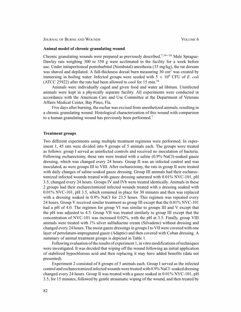
Two different experiments using multiple treatment regimens were performed. In exper-iment 1, 45 rats were divided into 9 groups of 5 animals each. The groups were treatedas follows: group I served as uninfected controls and received no inoculation of bacteria.Following escharectomy, these rats were treated with a saline (0.9% NaCl)-soaked gauzedressing, which was changed every 24 hours. Group II was an infected control and wasinoculated, as were groups III to VIII. After escharectomy, the rats in group II were treatedwith daily changes of saline-soaked gauze dressing. Group III animals had their escharec-tomized infected wounds treated with gauze dressing saturated with 0.01% NVC-101, pH3.5, changed every 24 hours. Groups IV and IVb were treated identically. Animals in these2 groups had their escharectomized infected wounds treated with a dressing soaked with0.01% NVC-101, pH 3.5, which remained in place for 30 minutes and then was replacedwith a dressing soaked in 0.9% NaCl for 23.5 hours. This regimen was repeated every24 hours. Group V received similar treatment as group III except that the 0.01% NVC-101had a pH of 4.0. The regimen for group VI was similar to groups III and V except thatthe pH was adjusted to 4.5. Group VII was treated similarly to group III except that theconcentration of NVC-101 was increased 0.02%, with the pH at 3.5. Finally, group VIIIanimals were treated with 1% silver sulfadiazine cream (Silvadene) without dressing andchanged every 24 hours. The moist gauze dressings in groups I to VII were covered with onelayer of petrolatum-impregnated gauze (Adaptic) and then covered with Coban dressing. Asummary of animal treatment groups is depicted in Table 1.
Following evaluation of the results of experiment 1, in vitro modifications of techniqueswere investigated. It was decided that wiping off the wound following an initial applicationof stabilized hypochlorous acid and then replacing it may have added benefits (data notpresented).
Experiment 2 consisted of 8 groups of 5 animals each. Group I served as the infectedcontrol and escharectomized infected wounds were treated with 0.9% NaCl–soaked dressingchanged every 24 hours. Group II was treated with a gauze soaked in 0.01% NVC-101, pH3.5, for 15 minutes, followed by gentle atraumatic wiping of the wound, and then treated by
82
ROBSON ET AL
Table 1. Summary of treatment groups of rats in experiment 1
Group∗ Treatment
I Uninfected/normal saline
II Infected/normal saline
III Infected/NVC-101, 0.01%, pH 3.5, changed q24 h
IV Infected/NVC-101, 0.01%, pH 3.5, 30 min/23.5 h normal saline
IVb Infected/NVC-101, 0.01%, pH 3.5, 30 min/23.5 h normal saline
V Infected/NVC-101, 0.01%, pH 4.0, changed q24 h
VI Infected/NVC-101, 0.01%, pH 4.5, changed q24 h
VII Infected/NVC-101, 0.02%, pH 3.5, changed q24 h
VIII Infected/Silvadene changed q24 h
∗Five animals per group.
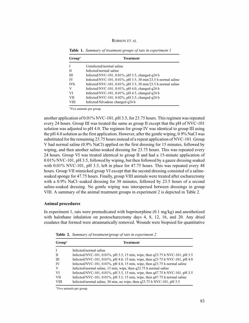
another application of 0.01% NVC-101, pH 3.5, for 23.75 hours. This regimen was repeatedevery 24 hours. Group III was treated the same as group II except that the pH of NVC-101solution was adjusted to pH 4.0. The regimen for group IV was identical to group III usingthe pH 4.0 solution as the first application. However, after the gentle wiping, 0.9% NaCl wassubstituted for the remaining 23.75 hours instead of a repeat application of NVC-101. GroupV had normal saline (0.9% NaCl) applied on the first dressing for 15 minutes, followed bywiping, and then another saline-soaked dressing for 23.75 hours. This was repeated every24 hours. Group VI was treated identical to group II and had a 15-minute application of0.01% NVC-101, pH 3.5, followed by wiping, but then followed by a gauze dressing soakedwith 0.01% NVC-101, pH 3.5, left in place for 47.75 hours. This was repeated every 48hours. Group VII mimicked group VI except that the second dressing consisted of a saline-soaked sponge for 47.75 hours. Finally, group VIII animals were treated after escharectomywith a 0.9% NaCl–soaked dressing for 30 minutes, followed by 23.5 hours of a secondsaline-soaked dressing. No gentle wiping was interspersed between dressings in groupVIII. A summary of the animal treatment groups in experiment 2 is depicted in Table 2.
Animal procedures
In experiment 1, rats were premedicated with buprinorphine (0.1 mg/kg) and anesthetizedwith halothane inhalation on postescharectomy days 4, 8, 12, 16, and 20. Any driedexudates that formed were atraumatically removed. Wounds were biopsied for quantitative
Table 2. Summary of treatment/group of rats in experiment 2
Group∗ Treatment
I Infected/normal saline
II Infected/NVC-101, 0.01%, pH 3.5, 15 min, wipe, then q23.75 h NVC-101, pH 3.5
III Infected/NVC-101, 0.01%, pH 4.0, 15 min, wipe, then q23.75 h NVC-101, pH 4.0
IV Infected/NVC-101, 0.01%, pH 4.0, 15 min, wipe, then q23.75 h normal saline
V Infected/normal saline, 15 min, wipe, then q23.75 h normal saline
VI Infected/NVC-101, 0.01%, pH 3.5, 15 min, wipe, then q47.75 h NVC-101, pH 3.5
VII Infected/NVC-101, 0.01%, pH 3.5, 15 min, wipe, then q47.75 h normal saline
VIII Infected/normal saline, 30 min, no wipe, then q23.75 h NVC-101, pH 3.5
∗Five animals per group.
83
JOURNAL OF BURNS AND WOUNDS VOLUME 6
bacteriology on the day of escharectomy (day 0) and on each of the days of reanesthesia ac-cording to the methods described by Heggers and Robson.5 The wound surface was cleanedwith 70% isopropyl alcohol prior to biopsy to exclude surface contamination. Biopsieswere aseptically weighed, homogenized, serially diluted, and back-plated onto nonselec-tive media. Bacterial counts were completed after 48 hours’ incubation and expressed ascolony-forming units (CFU) per gram of tissue.5
While the rats were anesthetized for the wound biopsies, outlines of the wounds weretraced onto acetate sheets, and area calculations were performed using computerized dig-ital planimetry (Sigma Scan Jandel Scientific, Corte Madera, CA). Care was taken onlyto record the perimeter of the wound that represented the advancing full-thickness marginrather than the edge of any advancing epithelium. This avoided the small component of ad-vancement provided by the smooth, pink, translucent, hairless neoepithelium.16 All animalswere weighed at the time of biopsy and wound measurement.
The animals were sacrificed by Nembutal overdose and bilateral thoracotomies whenthe wound had completely healed or decreased to less than 10% of its original area. Haywoodet al demonstrated that measurement of very small wounds by manual tracing introducedsignificant systematic error and found that wounds followed past this point remained staticfor prolonged periods of time.17
The animals in experiment 2 had the same procedures performed as those in experiment1 except they were performed at different time points, that is, days 0, 2, 4, 7, 9, 11, and14, with the final wound size recorded on day 16. The time points were chosen to captureearlier time points and more frequent changes in the wound size and bacteriology.
Statistical analysis
Mean bacterial counts for each group of animals in both experiments were determined andexpressed as CFU/g of tissue. These values were compared for each experiment using a one-way analysis of variance. Post hoc analyses of differences between groups were carried outusing Tukey’s test (all pairs, multiple-comparison test), with P < .05 considered significant.Sigma Stat statistical software (Jandel Scientific, Corte Madera, CA) was used for dataanalysis.
Serial wound area measurements were plotted against time. For each animal’s data, aGompertz equation was fitted (typical r2 = 0.85).18 Using this approach, a best-fit curvewas generated for each group. Comparison between groups was performed using life tableanalyses and the Wilcoxon rank test. These statistical analyses were performed using SAS19
and BMDP20 packages on a personal computer.
RESULTS
Quantitative bacteriology
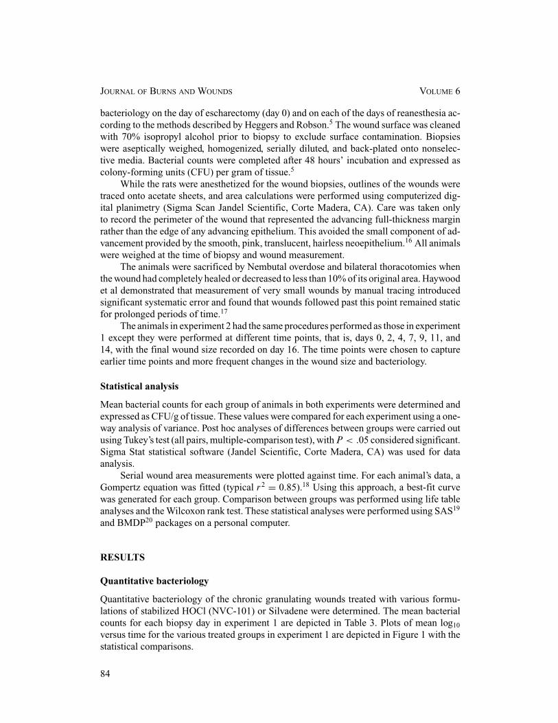
Quantitative bacteriology of the chronic granulating wounds treated with various formu-lations of stabilized HOCl (NVC-101) or Silvadene were determined. The mean bacterialcounts for each biopsy day in experiment 1 are depicted in Table 3. Plots of mean log10
versus time for the various treated groups in experiment 1 are depicted in Figure 1 with thestatistical comparisons.
84
ROBSON ET AL
Table 3. Summary of mean bacterial counts for each treated group in experiment 1
Days of punch biopsy postescharectomy (mean CFU/g)
Group∗ Day 0 Day 4 Day 8 Day 12 Day 16 Day 20
I 8.4 × 10² 2.48 × 10³ 1.49 × 10⁴ 1.63 × 10⁴ 3.71 × 10⁴ 8.04 × 10³
II 2.92 × 10⁷ 8.96 × 10⁶ 4.88 × 10⁵ 1.39 × 10⁵ 1.68 × 10⁵ 2.12 × 10⁴
III 3.12 × 10⁷ 1.39 × 10⁵ 3.6 × 10⁴ 3.24 × 10⁴ 5.28 × 10⁴ 5.16 × 10³
IV 2.42 × 10⁷ 1.72 × 10⁵ 1.31 × 10³ 4.92 × 10³ 8.8 × 10² 1.0 × 101
IVb 9.52 × 10⁷ 3.36 × 10⁷ 1.28 × 10⁵ 1.51 × 10⁴ 2.05 × 10³ 1.5 × 10²
V 1.56 × 10⁸ 1.37 × 10⁵ 8.6 × 10⁴ 2.05 × 10⁴ 6.36 × 10³ 3.12 × 10³
VI 2.45 × 10⁷ 1.44 × 10⁵ 1.25 × 10⁵ 6.14 × 10⁴ 3.56 × 10⁴ 1.38 × 10⁴
VII 2.01 × 10⁸ 1.45 × 10⁵ 5.8 × 10⁴ 9.75 × 10⁴ 3.89 × 10⁴ 9.7 × 10³
VIII 6.52 × 10⁷ 3.75 × 10⁶ 2.4 × 10⁴ NG† NG NG
∗Five animals per group.†NG indicates no growth.
It is clear that Silvadene was the best topical antimicrobial at decreasing the tissuebacterial burden. 0.01% NVC-101, pH 3.5, applied for 30 minutes and then removed fromthe wound proved to be the next most effective regimen for decreasing the bacterial load inexperiment 1. This regimen was used in both groups IV and IVb and the results were similar(Table 3 and Fig 1). The bacterial data from experiment 2 are also depicted in Table 4. Plots of
0
3
6
9
Day 0 Day 4 Day 8 Day 12 Day 16 Day 20
Days of punch biopsy
Me
an
log
10
Uninfected / normal saline Infected / normal saline
Infected / NVC-101, 0.01%, pH 3.5, changed q24 h Infected / NVC-101, 0.01%, pH 3.5, 30 min / 23.5, h normal saline
Infected / NVC-101, 0.01%, pH 3.5, 30 min / 23.5 h normal saline Infected / NVC-101, 0.01 %, pH 4.0, changed q24 h
Infected / NVC-101, 0.01%, pH 4.5, changed q24 h Infected / NVC-101, 0.02%, pH 3.5, changed q24 h
Infected / Silvadene changed q24 h
Figure 1. Depiction of the various groups in experiment 1 demonstrating the superiority of 30 min-utes of NVC-101 application followed by another dressing for 23.5 hours over other regimens ofhypochlorous acid. 1% silver sulfadiazine cream (Silvadene) was the most effective of all agentstested at decreasing the tissue bacterial bioburden.
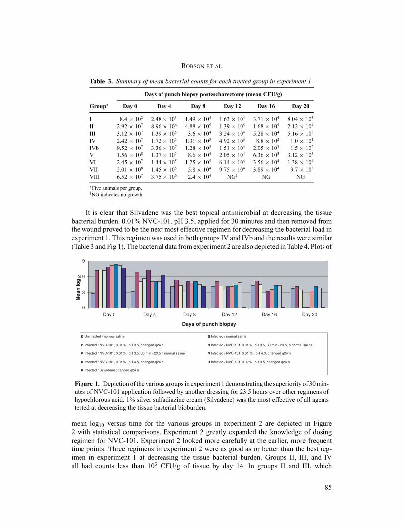
mean log10 versus time for the various groups in experiment 2 are depicted in Figure2 with statistical comparisons. Experiment 2 greatly expanded the knowledge of dosingregimen for NVC-101. Experiment 2 looked more carefully at the earlier, more frequenttime points. Three regimens in experiment 2 were as good as or better than the best reg-imen in experiment 1 at decreasing the tissue bacterial burden. Groups II, III, and IVall had counts less than 103 CFU/g of tissue by day 14. In groups II and III, which
85
JOURNAL OF BURNS AND WOUNDS VOLUME 6
Table 4. Summary of mean bacterial counts for each treated group in experiment 2
Days of punch biopsy postescharectomy (mean CFU/g)
Group∗ Day 0 Day 2 Day 4 Day 7 Day 9 Day 11 Day 14
I 5.0 × 10⁸ 5.0 × 10⁸ 1.27 × 10⁸ 1.4 × 10⁷ 1.85 × 10⁶ 2.32 × 10⁵ 1.41 × 10⁵
II 5.0 × 10⁸ 1.59 × 10⁶ 8.74 × 10⁵ 1.03 × 10⁵ 8.8 × 10³ 3.48 × 10³ 5.0 × 10²
III 5.0 × 10⁸ 3.24 × 10⁶ 2.51 × 10⁶ 3.12 × 10⁴ 2.54 × 10³ 3.48 × 10³ 6.4 × 10²
IV 5.0 × 10⁸ 1.12 × 10⁸ 2.31 × 10⁷ 1.37 × 10⁷ 9.16 × 10⁴ 7.28 × 10³ 4.0 × 10²
V 5.0 × 10⁸ 5.0 × 10⁸ 2.78 × 10⁷ 4.38 × 10⁶ 8.53 × 10⁵ 2.53 × 10⁵ 4.4 × 10⁴
VI 5.0 × 10⁸ 2.01 × 10⁷ 2.60 × 10⁷ 7.74 × 10⁶ 2.24 × 10⁵ 3.68 × 10⁴ 1.60 × 10³
VII 5.0 × 10⁸ 2.17 × 10⁸ 4.06 × 10⁷ 4.06 × 10⁷ 3.48 × 10⁶ 1.95 × 10⁵ 1.56 × 10³
VIII 5.0 × 10⁸ 4.01 × 10⁸ 5.24 × 10⁷ 2.72 × 10⁷ 1.58 × 10⁶ 2.42 × 10⁵ 4.04 × 10⁴
∗Five animals per group.
Figure 2. Depiction of the various groups in experiment 2 demonstratingthe superiority at decreasing bacterial counts of NVC-101 applied for 15 min-utes, followed by gentle atraumatic wiping then 23.75 hours of a seconddressing of NVC-101. There was essentially no difference in the effectswith pH 3.5 and 4.0.
86

ROBSON ET AL
had essentially the same treatment regimens, the bacterial counts decreased more rapidlythan in group IV. For these groups (II and III), the regimen consisted of NVC-101 beingplaced on the wound for 15 minutes, atraumatically wiped off, and then reapplied for 23.75hours. The only difference between the treatments for groups II and III was the pH ofNVC-101. No significant differences were seen between pH 3.5 and pH 4.0 (Table 4 andFig 2).
Body weights
There was an equivalent gain in body weight among all groups during the period of study,with no significant variations among the groups in either experiment 1 or experiment 2
Wound area
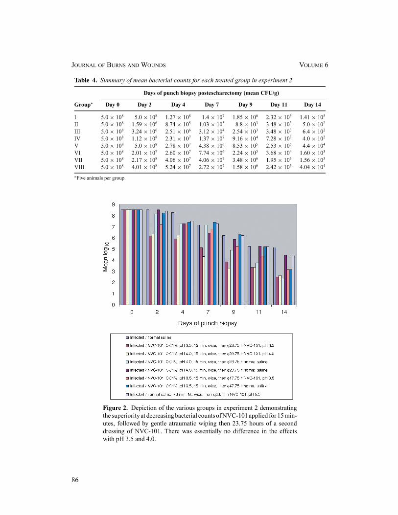
Best-fit healing curves demonstrated that none of the treatment regimens resulted in thearea of the wound increasing in size (Figs 3 and 4).
Infected control animals (group II in experiment 1, group I in experiment 2) retardedhealing as compared to the noninfected controls (group I in experiment 1). Healing curvesfor groups IV and IVb in experiment 1 demonstrated statistically significant increases inreduction in the fraction of open wounds when compared to groups I, III, V, and VIII (P <
.05) and groups II, V, and VII (P < .01) (Fig 3). Groups II, V, and VII demonstrated a slowertrajectory than all other groups, also statistically significant (P < .05) (Fig 3).
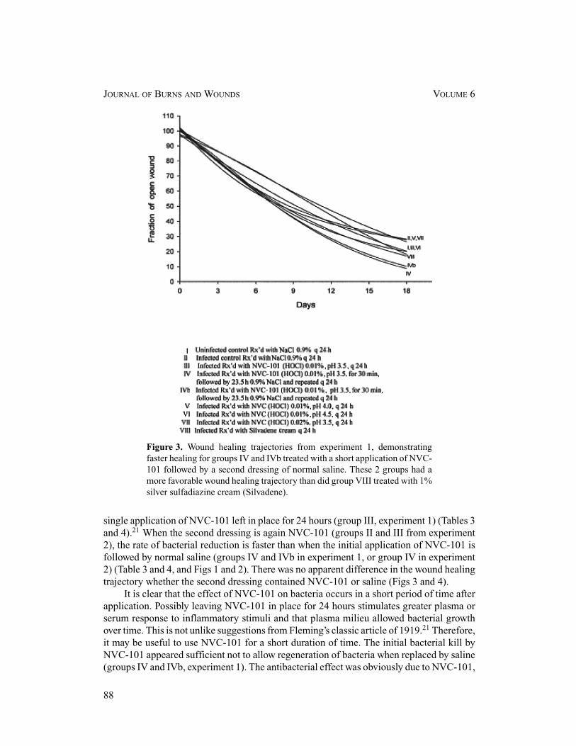
In experiment 2, healing curves for groups II and III demonstrated statistically signif-icant larger reductions in the fraction of open wounds when compared to groups IV, VI,and VII (P < 0.05) and groups I, V, and VIII (P < .01) (Fig 4). Groups I, V, and VIIIdemonstrated a slower healing trajectory than all other groups, which was also statisticallysignificant (P < .05).
DISCUSSION
Because of the deleterious effect of a high tissue bacterial burden on the process of woundhealing, an effectual antimicrobial agent becomes a therapeutic imperative. Such an agentshould be effective as a topical preparation, yet not to be cytotoxic to the cells involved inthe wound healing process.21 Stabilized hypochlorous acid, as tested in the 2 experimentsreported, may prove to be such an agent. Its in vitro antibacterial properties and tissue safetyprofile suggest its potential as a wound care agent.13 However, it is likely rapidly neutralizedin the wound environment.
In experiment 1, Silvadene was, as expected, the most effective antibacterial. However,Silvadene was not as effective at promoting wound closure as were two of the NVC-101regimens (IV and IVb) (Fig 3). The healing that occurred with Silvadene was probably dueto elimination of the tissue bacterial load (Table 3). The reason the wounds did not totallyheal or exceed that with groups IV and IVb is because of the known cytotoxic properties ofSilvadene.11,12
From a review of the quantitative bacteriology data from both experiments, it is clearthat a brief application of NVC-101, followed by a second dressing change is better than a
87
JOURNAL OF BURNS AND WOUNDS VOLUME 6
Figure 3. Wound healing trajectories from experiment 1, demonstratingfaster healing for groups IV and IVb treated with a short application of NVC-101 followed by a second dressing of normal saline. These 2 groups had amore favorable wound healing trajectory than did group VIII treated with 1%silver sulfadiazine cream (Silvadene).
single application of NVC-101 left in place for 24 hours (group III, experiment 1) (Tables 3and 4).21 When the second dressing is again NVC-101 (groups II and III from experiment2), the rate of bacterial reduction is faster than when the initial application of NVC-101 isfollowed by normal saline (groups IV and IVb in experiment 1, or group IV in experiment2) (Table 3 and 4, and Figs 1 and 2). There was no apparent difference in the wound healingtrajectory whether the second dressing contained NVC-101 or saline (Figs 3 and 4).
It is clear that the effect of NVC-101 on bacteria occurs in a short period of time afterapplication. Possibly leaving NVC-101 in place for 24 hours stimulates greater plasma orserum response to inflammatory stimuli and that plasma milieu allowed bacterial growthover time. This is not unlike suggestions from Fleming’s classic article of 1919.21 Therefore,it may be useful to use NVC-101 for a short duration of time. The initial bacterial kill byNVC-101 appeared sufficient not to allow regeneration of bacteria when replaced by saline(groups IV and IVb, experiment 1). The antibacterial effect was obviously due to NVC-101,
88
ROBSON ET AL
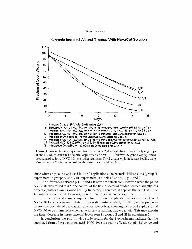
Figure 4. Wound healing trajectories from experiment 2, demonstrating the superiority of groupsII and III, which consisted of a brief application of NVC-101, followed by gentle wiping, and asecond application of NVC-101 over other regimens. The 2 groups with the fastest healing werealso the most effective at controlling the tissue bacterial bioburden.
since when only saline was used in 1 or 2 applications, the bacterial kill was less (group II,experiment 1; groups V and VIII, experiment 2) (Tables 3 and 4, Figs 1 and 2).
The differences between pH 3.5 and 4.0 were not detectable. However, when the pH ofNVC-101 was raised to 4.5, the control of the tissue bacterial burden seemed slightly lesseffective, with a slower wound healing trajectory. Therefore, it appears that a pH of 3.5 or4.0 may be more useful. However, these differences may not be significant.
The role of the atraumatic wiping between dressing applications is not entirely clear. IfNVC-101 kills bacteria immediately or soon after initial contact, then the gentle wiping mayremove the devitalized bacteria and any possible debris, allowing the second application ofNVC-101 to be in immediate contact with any remaining viable bacteria. This may explainthe faster decrease in tissue bacterial levels seen in groups II and III in experiment 2.
In conclusion, the pilot in vivo study results for the 2 experiments indicate that thestabilized form of hypochlorous acid (NVC-101) is equally effective at pH 3.5 or 4.0 and
89

JOURNAL OF BURNS AND WOUNDS VOLUME 6
more effective soon after its application. As opposed to other antimicrobials investigated inthis animal model, NVC-101 controls the tissue bacterial bioburden without inhibiting thewound healing process.
REFERENCES
1. Clark RAF. Biology of dermal wound repairs. Dermatol Clin. 1993;11:647–666.
2. Robson MC, Stenberg BD, Heggers JP. Wound healing alterations caused by infection. Clin Plast Surg.
1990;17:485–492.
3. Payne WG, Wright TE, Ko F, Wheeler C, Wang X, Robson MC. Bacterial degradation of growth factors. JAppl Res. 2003;3:35–40.
4. Robson MC. Wound infection: a failure of wound healing caused by an imbalance of bacteria. Surg Clin NAm. 1997;77:637–650.
5. Heggers JP, Robson MC. Quantitative Bacteriology: Its Role in the Armamentarium of the Surgeon. Boca
Raton, FL: CRC Press; 1991.
6. Serena T, Robson MC, Cooper DM, Ignatious J. Lack of reliability of clinical/visual assessment of chronic
wound infection: the incidence of biopsy-proven infection in venous leg ulcers. Wounds. 2006;18:197–202.
7. Robson MC, Edstrom LE, Krizek TJ, Groskin MG. The efficacy of systematic antibiotics in the treatment
of granulating wounds. J Surg Res. 1974;16:299–306.
8. Robson MC. Management of the contaminated wound—aids in diagnosis and management. In: Krizek TJ,
Houpes JE, eds. Symposium on Basic Science in Plastic Surgery. St. Louis: CV Mosby; 1976.
9. Bergstrom N, Bennett MA, Carlson CE, et al. Treatment of Pressure Ulcers. Clinical Practice Guideline,No. 15. Rockville, MD: U.S. Department of Health and Human Services. Public Health Service, Agency
for Health Care Policy and Research. AHCPR Publication No. 95-0652; December 1994.
10. Wilson JP, Mills JG, Prather ID, Dimitrijerich SD. A toxicity index of skin and wound cleaners used on in
vitro fibroblasts and keratinocytes. Adv Skin Wound Care. 2005;18:373–378.
11. McCauley RL, Linares HA, Herndon DN, Robson MC, Heggers JP. In vitro toxicity of topical antimicrobial
agents to human fibroblasts. J Surg Res. 1989;3:269–274.
12. McCauley RL, Ying YL, Poole B, et al. Differential inhibition of human basal keratinocyte growth to silver
sulfadiazine and mafenide acetate. J Surg Res. 1992;52:276–285.
13. Gerrisen CM, Margerum DW. Non-metal redox kinetics: hypochlorite and hypochlorous acid reactions with
cyanide. Inorg Chem. 1990;29:2758–2762.
14. Robson MC, Krizek TJ. The effect of human amniotic membranes on the bacterial population of infected
rat burns. Ann Surg. 1973;177:144–149.
15. Haywood PG, Robson MC. Animal models of wound contraction. In: Barbul A, Caldwell M, Eaglstain W,
et al., eds. Clinical and Experimental Approaches to Dermal and Epidermal Repair: Normal and ChronicWounds. New York: Alan R. Liss; 1991:305–312.
16. Kuhn MA, Page L, Nguyen K, et al. Basic fibroblast growth factor in a carboxymethylcellulose vehicle
reverses the bacterial retardation of wound contraction. Wounds. 2001;13:73–80.
17. Hayward P, Hokanson J, Heggers J, et al. Fibroblast growth factor reverses the bacterial retardation of wound
contraction. Am J Surg. 1992;163:288–293.
18. Hokanson JA, Hayward PG, Carney DH, Phillips LG, Robson MC. A mathematical model for the analysis
of experimental wound healing data. Wounds. 1992;13:213–220.
19. SAS/STAT Guide for Personal Computers. 6th ed. North Carolina: Cary; 1987:1028.
20. BMDP Statistical Software Manual. Los Angeles: BMDP Statistical Software Inc; 1988.
21. Fleming A. Chemical and physiologic antiseptics, the action of chemical and physiological antiseptics in a
septic wound. Br J Surg. 1919;99–129.
90