inhibition of lung fluid clearance and epithelial … · inhibition of lung fluid clearance and...
TRANSCRIPT
INHIBITION OF LUNG FLUID CLEARANCE AND EPITHELIAL Na+ CHANNELS BY CHLORINE, HYPOCHLOROUS ACID AND CHLORAMINES
Weifeng Song1,2*, Shipeng Wei1,2*, Yongjian Zhou1, Ahmed Lazrak1,2, Gang Liu2,3, James D.
Londino1,2, Giuseppe L. Squadrito2,4,5, and Sadis Matalon1, 2, 4,5
Departments of Anesthesiology1, Medicine3 and Environmental Health Sciences4
Schools of Medicine and Public Health, Centers for Pulmonary Injury and Repair2 and Free Radical Biology5
University of Alabama at Birmingham, Birmingham, AL 35205
* Drs. Song and Wei contributed equally as first authors and are listed in alphabetical
order
Sources of support: PHS grants HL-31197, HL-51173, U01ES015676 and 1U54ES017218
Running Title: Cl2 and its intermediates inhibit ENaC Corresponding author: Sadis Matalon, Ph.D. Department of Anesthesiology, University of Alabama at Birmingham 224 BMR II, 901 19th Street South, Birmingham, AL 35205-3703. email: [email protected]; Fax: (205) 934-7476; Tel: (205) 934-4231 Current address for Dr. Wei: Department of Cardiology, The Fourth Affiliated Hospital of Harbin Medical University, Harbin 150001, China Current address for Dr. Zhou: Department of Gastroenterology, The First People’s Hospital of Guangzhou, Guangzhou, 510180, China ABSTRACT We investigated the mechanisms by which chlorine (Cl2) and its reactive by-products inhibit Na+ dependent alveolar fluid clearance (AFC) in vivo and the activity of amiloride-sensitive epithelial Na+ channels (ENaC) by measuring: AFC in mice exposed to Cl2 (0-500 ppm for 30 min); (2) Na+ and amiloride-sensitive currents (INa and Iamil respectively) across Xenopus oocytes expressing α,β,γ human (h)ENaC incubated with HOCl (1-2000 µM). Both Cl2 and HOCl-derived products decreased AFC in mice and whole cell and single channel INa in a dose dependent manner; these effects were counteracted by serine proteases. Mass spectrometry analysis of the oocyte recording medium identified organic chloramines formed by the interaction of HOCl with HEPES (used as an extracellular buffer). In addition, chloramines formed by the interaction of HOCl with taurine or
glycine, decreased INa in a similar fashion. Pre-incubation of oocytes with serine proteases prevented the decrease of INa by HOCl, whereas perfusion of oocytes with a synthetic 51-mer peptide corresponding to the putative furin and plasmin cleaving segment in the γ-ENaC subunit, restored the ability of HOCl to inhibit INa. Finally, INa of oocytes expressing wild type α and γ ENaC and a mutant form of βENaC (S520K), known to result in ENaC channels locked in the open position, were not altered by HOCl. We concluded that HOCl and its reactive intermediates (such as organic chloramines) inhibit ENaC by affecting channel gating, which could be relieved by proteases cleavage. (237 words) Key words: chloramines; amiloride-sensitive epithelial sodium currents; serine proteases; patch clamp; mass spectrometry.
http://www.jbc.org/cgi/doi/10.1074/jbc.M109.073981The latest version is at JBC Papers in Press. Published on January 27, 2010 as Manuscript M109.073981
Copyright 2010 by The American Society for Biochemistry and Molecular Biology, Inc.
by guest on Novem
ber 11, 2018http://w
ww
.jbc.org/D
ownloaded from

Abbreviations: HOCl: hypochlorous acid; Cl2: chlorine gas; ENaC; amiloride-sensitive epithelial sodium channels; MS=mass spectrometry; AFC= alveolar fluid clearance ND96: oocyte perfusion solution containing (in mM): 100 NaCl, 2 KCl, 1 MgCl2, 1.8 CaCl2, and 10 HEPES at pH 7.6 (osmolarity 200–220 mosm) Note: Throughout this manuscript we use the term hypochlorous acid (HOCl) to refer to the sum of hypochlorous acid and hypochlorite (OCl-) that are present at equilibrium. The actual concentration of these species is governed by the pH and the pKa of HOCl. INTRODUCTION The balance of fluid covering the respiratory and alveolar epithelia is determined in part by the ability of these cells to transport sodium (Na+) and chloride (Cl-) ions in a vectorial fashion. Active Na+ reabsorption across lung epithelia requires
the coordinated entry of Na+ ions through cation and Na+ selective amiloride sensitive channels (ENaC) located at the apical membranes, their extrusion across the basolateral membranes by the electrogenic Na+-K+-ATPase, and the passive movement of K+ ions through basolateral K+ channels. The entry of Na+ ions through apical pathways is thought to be the rate-limiting step in this process (1-3). To preserve neutrality, Cl- ions follow Na+ ions both through transcellular and paracellular pathways(4,5). The coordinated movement of Na+ and Cl- ions creates an oncotic gradient favoring the absorption of alveolar fluid.
Injury to either apical or basolateral pathways by partially reduced intermediates, may lead to impairment of fluid reabsorption, which in turn may result in pulmonary edema, hypoxemia and eventually death from respiratory failure(6-9). One such specie is hypochlorous acid (HOCl), which may be generated either endogenously or exogenously. Millimolar concentrations of HOCl may be generated by activated neutrophils and eosinophils by the catalytic
actions of neutrophil and eosinophil derived myeloperoxidases on chloride (Cl-) and hydrogen peroxide (H2O2) in close proximity of the apical and basolateral membranes of epithelial cells(10,11) The main targets of HOCl and its conjugated base (hypochloride:OCl-) are free functional groups of proteins and amino acids, predominantly sulfhydryl groups (12,13), free amine groups of plasma amino acids (yielding chlorinated amines;(14)) and aromatic amino acids (yielding chlorotyrosine (15-17)). Moreover, the reaction of HOCl with plasma or alveolar nitrite may form reactive intermediates capable of nitrating, chlorinating and dimerizing aromatic amino acids, thus damaging a number of key proteins and altering their functions (18).
In addition to endogenous sources, significant amounts of HOCl are also generated during chlorine (Cl2) inhalation. We have shown that exposure of rats to Cl2 causes extensive injury to the alveolar epithelium as manifested by changes in surfactant function and increased permeability to plasma proteins (19). Furthermore, exposure of surfactant protein A (SP-A), a critical component of innate immunity to HOCl, oxidized and chlorinated amino acids in its carbohydrate recognition domain and decreased its ability to bind mannose residues, an important step in the binding and killing of pathogens (20). However, the effects of Cl2 inhalation on Na+-dependent alveolar fluid clearance (AFC) in vivo, as well as the mechanisms by which Cl2 and HOCl damage amiloride sensitive epithelial Na+ channels (ENaC), have not been investigated. This is an important area of research since damage to ENaC has been associated with abnormal fluid transport in various forms of lung injury(7,21-25).
To address these questions we performed a number of physiological, biophysical and biochemical studies. In our first series of experiments, we exposed mice to Cl2 at concentrations likely to be encountered in industrial accidents or deliberate release of Cl2 into the
by guest on Novem
ber 11, 2018http://w
ww
.jbc.org/D
ownloaded from

atmosphere(19,26,27), and measured Na+-dependent alveolar fluid clearance (AFC) in vivo at various times post exposure, after the mice were returned to room air. Since our results indicated that exposure to Cl2 decreased AFC, we performed additional studies in Xenopus oocytes injected with cRNAs of the human (h)ENaC subunits (α, β and γ ENaC) to identify the cellular and molecular mechanisms by which HOCl (formed by the hydrolysis of Cl2 gas) as well as reactive intermediates formed by the reaction of HOCl with components of the media, decreased Na+ and amiloride sensitive currents (INa and Iamil respectively). Finally we found that when ENaC channels were locked in the open position either by the action of serine proteases(28-30) or the introduction of a point mutation in βENaC (31), HOCl did not decrease INa. We concluded that HOCl inhibits ENaC by altering channel gating and preventing closed channels from opening.
MATERIALS AND METHODS Chemicals: NaOCl, N-acetyl Cysteine, amiloride, HEPES (4-(2-hydroxyethyl)-1-piperazineethanesulfonic acid), BSA (Bovine Serum Albumin) and DTT (Dithiothreitol) were purchased from Sigma-Aldrich Co. (St. Louis, MO). Trypsin elastase and plasmin were purchased from Invitrogen Co. (Carlsbad, CA), Worthington (Lakewood, NJ) and Molecular Innovations (Novi, MI), respectively. Animals: Six to eight weeks old Balb/c and C57BL/6 male mice (20-25 g body weight) were purchased from Charles River Laboratories (Wilmington, MA). They were housed in the animal unit for at least 3 days prior to any experimental procedure where they were provided with mouse chow and water ad libitum. All experimental protocols were approved by the IACUC of the University of Alabama at Birmingham. Exposure of mice to Cl2 gas: Mice were exposed to Cl2 gas (0 to 500 ppm) for 30 minutes as previously described (19). In brief, they were placed inside a cylindrical glass chamber (Specialty Glass, Inc. Houston, TX, part # X02AI99C15A57H5).
Two mass flow controllers (MFC) with Kalrez seals (Scott Specialty Gases, Los Angeles, CA, part # 05236A1V5K) and a microprocessor control unit (Scott Specialty Gases, Los Angeles, CA, part # 05236E4) were used to control the flow rates of compressed air and Cl2 (1000 ppm Cl2 in air; Airgas, Birmingham, AL) to achieve the desired Cl2 concentrations (0 to 500 ppm) in the exposure chamber. A bubble flow meter was used to determine the accuracy of each MFC flow rate on a weekly basis. Air and Cl2 were mixed at a 3 way junction, and they were further mixed by passing through a diffuser located inside the top lid of the exposure chamber. Gases exited the chamber via two large bore diameter ports in its bottom half. The exposure chamber was placed inside a chemical hood located in a negative pressure room. At the end of the exposure period (30 min), the Cl2 gas was turned off, the chamber was vented with compressed air for two to three min, the two halves separated and the mice were removed and returned to room air. Alveolar fluid clearance (AFC) measurements: Alveolar fluid clearance was measured as previously described (7,23). In brief, mice were anesthetized and paralyzed by intraperitoneal injections of diazepam (10 mg/kg body wt; Hospira, Lake Forest, IL), ketamine (200 mg/kg; IVX Animal Health, St. Joseph, MO) and pancuronium bromide (0.04 mg; Gensia Pharmaceuticals, Irvine, CA). They were ventilated with a mouse respirator (model
687; Harvard Apparatus, Holliston, MA) with 100% O2, a tidal volume of 0.2 ml (9-10 ml/kg body wt) and frequency of 160 breathes/min through an 18-gauge intravenous catheter trimmed to about 0.5 inch, inserted into their tracheas. They were then placed in the left decubitus position on a heating pad (Braintree, Cambridge, MA); body temperature was maintained at 37-38°C with a heating lamp. A 0.9% NaCl solution (0.3 ml; approximately 30% of their total lung capacity) containing 5% fatty acid-free bovine serum albumin (BSA; Sigma, St. Louis, MO) was instilled via the tracheal catheter followed by 0.1ml of room
by guest on Novem
ber 11, 2018http://w
ww
.jbc.org/D
ownloaded from
air to clear the dead space and position the fluid in the distal lung (alveolar) space. The osmolarity of the instillate, measured by a Vapor Pressure Osmometer (Wescor, Inc; Logan, UT) was 322 mOsm/kg. In some cases, amiloride (1.5 mM) was added in the instillate to inhibit ENaC. Previous measurements have shown that significantly higher concentrations of amiloride are needed to inhibit ENaC in air filled lungs (7). After 15 or 30 mins of ventilation, the instilled fluid was gently aspirated and kept in a 0.5 ml tube. Samples visually contaminated by blood were excluded. Protein concentrations were measured using the bicinchoninic acid protein assay (Pierce Biotechnology, Rockford, IL). A standard
curve was prepared by assaying known concentrations of BSA in 0.9% NaCl. AFC values were calculated as described before (24,32). In some cases, mice were anesthetized with isoflurane 15 min post Cl2 exposure and 100 μl of saline, containing either trypsin (5 µM) or vehicle were instilled drop-wise in their nostrils. The isoflurane was discontinued, the mice woke up shortly after and their AFC was measured 1 h post exposure, as described above. HOCl preparation: The concentration of HOCl was determined spectrophoto-metrically (Beckman DU-7400, CA) from its absorbance at 292 nm (molar absorption coefficient (ε) = 350 M-1cm-1) at pH 12(33). Stock solutions, prepared fresh every day, were then diluted in oocyte incubation medium (ND96) to the desired final concentration, just before each experiment. Construction of ENaC cRNAs. Human α,β, γ-ENaC cDNAs were sub-cloned into a pCDNA3.1 vector for in vitro transcription. hENaC-α595x was excised by NotI and Xho and cloned into pCDNA3.1 at NotI and XhoI. hENaC βS520K (provided by Dr. Peter Snyder, University of Iowa) were excised by NotI and Acc65I and cloned into pCDNA3 at NotI and XhoI, with Acc65I and XhoI blunt-ended. hENaC-γ575x was excised by NotI and EcoRI and cloned into pCDNA3.1 at NotI and XhoI, with EcoRI and XhoI blunt-ended. All the constructs were verified by DNA sequencing. ENaC
subunit plasmids were linearized and cRNAs were prepared with a cRNA synthesis kit (T7 Message Machine, Ambion Inc, Austin, TX) according to the manufacturer's protocol. cRNAs were dissolved in RNAse free water and their concentrations were determined spectrophotometrically. To facilitate the detection of exogenously expressed α, β, and γ- hENaC in Western blotting studies, in some cases α, β, and γ cDNAs were tagged with Flag, Myc, and V5 epitopes at their C-termini respectively, and tagged-cRNAs were generated as above. All procedures have been described in detail previously (34). Expression of ENaC in Xenopus oocytes: Ovarian tissue containing oocytes from adult female Xenopus laevis toads was dissected under anesthesia, then digested in 2 mg/ml collagenase (type 1A, Roche) in Ca2+-free OR-2 medium, containing (in mM): 82.5 NaCl, 2.0 KCl, 1.0 MgCl2, and 5 HEPES, pH 7.4, under rotation at room temperature for 2 hours as previously described(34). Defolliculated oocytes were washed three times in Ca2+-free OR-2 medium followed by OR-2 medium with 1.0 mM CaCl2. After that, stage VI oocytes were selected and cultured in half-strength Leibovitz-15 medium (Life Technologies, Carlsbad, CA), containing 15 mM HEPES, penicillin (100 U/ml), streptomycin (100 µg/ml), and 5% horse serum, pH 7.6, at 16°C. In all cases, equal amounts (8.3 ng each) of cRNAs were injected into oocytes in a total volume of 50 nl (0.5 µg/µl in RNase free water) per oocyte via a Nanoject microinjector (Drummond, Broomall, PA). Oocytes were then incubated in half-strength L-15 medium for 24 or 48 hr at 16°C until use. The culture medium was changed every other day. Voltage clamp recordings of whole-cell ENaC currents: Whole-cell cation currents were recorded 48-72 hours post-injection across the entire oocyte membrane using the two-electrode voltage clamp technique (35). Briefly, the oocytes were held in a small groove in a chamber of one ml volume at room temperature (21°C). The chamber was filled with ND96 solution, containing (in
by guest on Novem
ber 11, 2018http://w
ww
.jbc.org/D
ownloaded from

mM): 100 NaCl, 2 KCl, 1 MgCl2, 1.8 CaCl2, and 10 HEPES at pH 7.6 (osmolarity 200–220 mosm). The oocytes were impaled with two 3 M KCl-filled electrodes, with resistance 0.4–2.2 MΩ. The recording electrodes were constructed from glass micro-pipettes (Rochester Scientific, Rochester, NY) with a two-stage vertical Narishige PC-10 microelectrode puller (Narishige Scientific Instrument Laboratory, Tokyo, Japan). While in the chamber, oocytes were perfused with ND96 at a flow rate of 3 ml/min using a Warner Six Channel Valve Control Systems apparatus (Warner Instruments, Hamden, CT). Oocytes voltage clamping and currents recordings were accomplished with a Dagan TEV-200A voltage clamp amplifier (Dagan Corporation, Minneapolis, MN). Two reference electrodes were connected to the bath. Oocytes were clamped at a holding potential of -40 mV. Currents were continuously monitored and recorded on a chart recorder and to the hard disk of a personal computer. The currents were elicited by varying oocyte membrane potentials from -140 to +60 mV every 10 seconds following a standard protocol (starting from holding potential for 50 ms, then to -140 mV for 400 ms, subsequently back to -40 mV for 200 ms, and to +60 mV for 400 ms, finally back to -40 mV for 50 ms), generated by pCLAMP 8.0 software (Axon Instruments, Union City, CA). Current-voltage (I-V) relationships were acquired while the membrane potentials were changed from -120 to + 80 mV in 20-mV increments ever 500 ms. In some cases, after obtaining baseline I-V relationships and recording stable currents at -140 mV under control conditions, oocytes were perfused with ND96 mixed with various concentrations of HOCl for 10 min at which time recordings of the I-V relationship were repeated. Amiloride (10 µM) was then added in the bath solution and amiloride-sensitive currents (Iamil) were calculated as the difference currents before and after perfusion with amiloride. In other cases, oocytes were incubated with ND96 with various concentrations of HOCl for 2 to 4 h prior to the recordings of Iamil.
Single-channel ENaC currents recording: Oocytes were incubated for ten min with various concentrations of HOCl in ND96. They were then washed in ND96 and their vitelline membranes were removed mechanically by immerging them into a hypertonic solution containing 200 mM sucrose. Patch-clamp pipettes were prepared from borosilicate glass capillaries (Sutter Instruments Company, Novato, CA) using a two-stage vertical Narishige PC-10 microelectrode puller (Narishige Scientific Instrument Laboratory, Tokyo, Japan) and then fire-polished using a microforge (MF-830, Narishige, Japan). The pipette resistance when filled with ND96 was 7-10 MΩ. Currents were recorded at +100 mV depolarizing potential applied to the on-cell patch at a sampling rate of 5 kHz and filtered at 1 kHz with a low-pass Bessel filter through an Axopatch 200B amplifier (Axon Instruments, Foster City, CA). Data were analyzed using pCLAMP 9 software. Only data from patches with a seal resistance of at least 10 GΩ and stable currents for at least five min of recordings were included in the final analysis. All experiments were carried out at room temperature. Liquid chromatography-mass spectrometry (LC-MS) Analysis: Reactive intermediates formed by the interaction of HOCl with components of the ND96 medium were detected by LC-MS analysis. Briefly, samples were diluted 1:10 in water, then separated with an HPLC system (Shimadzu Class VP, Kyoto, Japan) at 4ºC, consisting of an LC-10AD pump and a SIL-HTC Autosampler, with an injection volume of 20 µl and flow rate of 0.2 ml/min through a C5 column (250 × 2.0 mm, Phenomenex; Torrance, CA). The two mobile phase solvents (A and B) consisted of 0.1% HCOOH (A) and methanol and 0.1% formic acid (B). The column was first equilibrated in solvent A. After sample injection, solvent B was increased from 0–80% within 10 min, then switched back to 0% until flow was stopped at 17 min. The eluted material was passed into the electrospray ionization interface of a
by guest on Novem
ber 11, 2018http://w
ww
.jbc.org/D
ownloaded from

MDS/Sciex Applied Biosystems API 3200 (Foster City, CA) operating in the positive ion mode. Mass spectra were recorded over a 70–500 m/z range. Determination of membrane protease enzymatic activity: Serine protease activity in the oocytes membranes was monitored by measuring the release of a fluorescent probe (AMC) from a peptide substrate Boc-Gln-Ala-Arg-AMC-HCl (Boc: t-Butyl oxy- carbonyl; AMC: 7-Amido-4-Methyl Coumarin; R&D Systems, Minneapolis, MN) using a fluorescence plate reader (FLUOstar OPTIMA, BMG Labtech; Durham, NC) (34). Briefly, five oocytes each, incubated with either HOCl (2 mM) or vehicle in ND96 for ten min were added to a cuvette containing 200 µl of ND96 medium and the reaction was started by adding Boc-Gln-Ala-Arg-AMCHCl (50µM final concentration). The reaction was monitored for up to 20 min by recording fluorescence at 460 nm following excitation at 380 nm. Detection of ENaC protein in oocyte plasma membranes. Xenopus oocyte plasma membranes were purified as previously described (36) with minor modifications. Briefly, oocytes were washed twice in Barth solution (in mM: 90 NaCl, 3 KCl, 0.82 MgSO4, and 5 HEPES, pH 7.6) and homogenized in 0.8 ml of the same solution supplemented with protease inhibitor cocktail (Sigma-Aldrich, St Louis. MO). Homogenates were centrifuged at 250 g for 10 min at 4°C and the supernatant was centrifuged at 16,000 g for 20 min at 4°C to pellet down total membranes. Pellets were lysed in 1% Triton-100 lysis buffer (150 mM NaCl, 50 mM Tris-Cl, pH 7.4, 2 mM EDTA, 1% Triton-X100, protease inhibitor cocktail) and the lysates were centrifuged at 16,000 g for 20 min at 4°C. The supernatants consisted of plasma membrane extracts. Equal amounts of whole oocyte or plasma membrane proteins were separated by 8% polyacrylamide-SDS gels. After transfer onto PVDF membrane, the blots were probed for α, β, and γ- hENaC using antibodies to Flag, Myc, and V5 epitopes (Sigma-Aldrich, St Louis. MO) respectively. The level of GAPDH was also determined to
demonstrate equal loading of the extracts and the purity of the plasma membrane extracts. Construction and application of a protease target peptide in γENaC: A 51-residue peptide between the putative furin and plasmin cleavage sites on the γ-hENaC (amino-eaeswnsvsegkqprfshripllifdqdekgkardfftgrkrkvggsiihk-carboxy) was synthesized by the solid phase method using a Protein Technologies Model PS3 Automated Peptide Synthesizer (Rainin Instrument Company, Inc; Woburn, MA) by the UAB Peptide Synthesis Core facility. The first amino acid from the C-terminus FMOC-Lys (Boc)-OH was derivatised to the solid support using the Wang Resin (100-200 mesh substitution 0.83 mmol/g, Nova Biochem, Damstadt, Germany). The FMOC group was then removed using piperidine in dimethlylformamide. The rest of the amino acids in the sequence were coupled to the first amino acid in the presence of 2-(H-benzotriazole-1-Yl)-1,1,3,3,-tetramethlyuronium hexafluorophosphate (HBTU) and N-methlymorpholine (NMM). After the addition of the final amino acid, the amino protecting FMOC-group was cleaved, and the peptide was released from the resin, with the removal of the side chain protections used for the various amino acids in the sequence using trifluoroacetic acid in the presence of scavengers. The crude peptide was purified using reverse phase high performance liquid chromatography (RP-HPLC). The purity of peptide was determined by HPLC followed by mass spectral analysis. Xenopus oocytes expressing hENaC were perfused for 15 min with either ND96 containing the native peptide (3 µg/ml) or ND96 containing peptide (3 µg/ml) mixed with 2 mM HOCl for ten min. Whole cell inward Na+ currents at -100 mV were continuously recorded and amiloride-sensitive current voltage relationships were obtained at the end of 10 min recordings. Data analysis: All data were shown as means (X) ± one standard error of the mean (SE). Comparisons between data sets were
by guest on Novem
ber 11, 2018http://w
ww
.jbc.org/D
ownloaded from

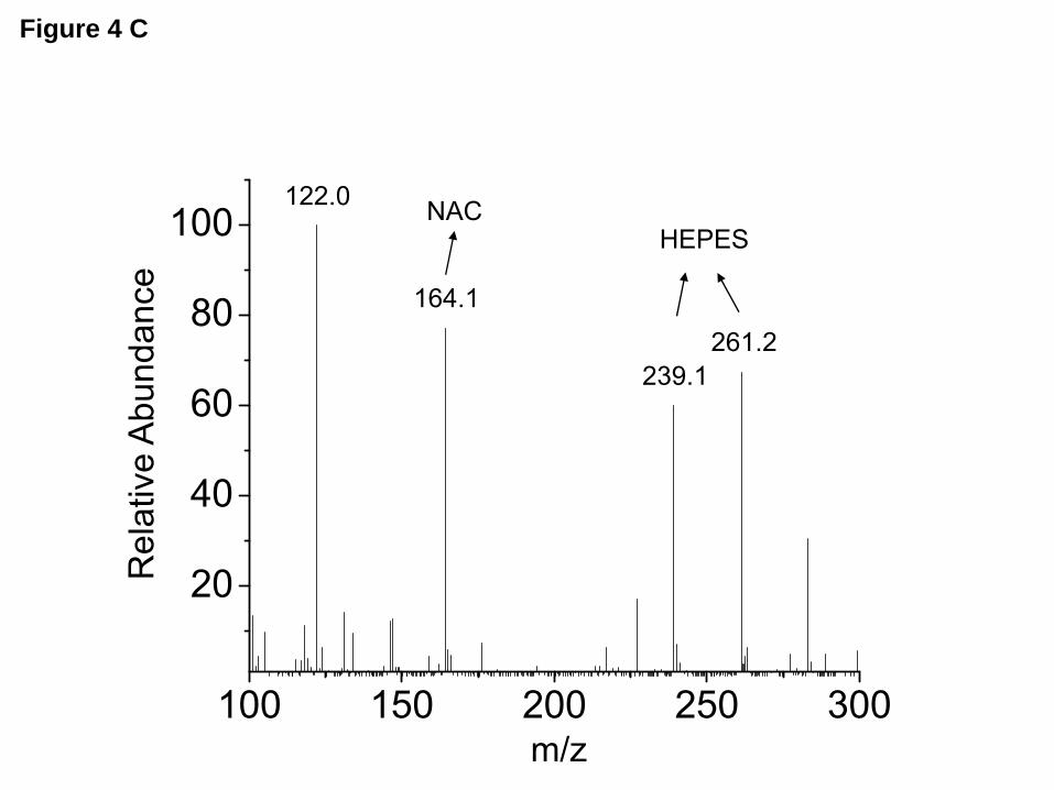
made with unpaired or paired t-test. Multiple comparisons among treatment groups were performed using ANOVA. Differences at P<0.05 were considered significant. RESULTS Exposure of mice to Cl2 decreased their AFC. Previous studies documented the presence of large variations in physiological responses of different species of mice to oxidative stresses (37). For this reason, we exposed both C57BL/6 and BALB/c mice to various concentrations of Cl2 (50-500 ppm for 30 min) and measured AFC at various times post exposure. As shown in Figure 1A, exposure of C57BL/6 mice to increasing concentrations of Cl2 gas (50-500 ppm for 30 min) decreased AFC from 28±0.7% to 15 ±4% (% of instilled volume/30 min, n=3-23, P<0.05), at one hour post exposure. Data shown in Figure 1B (400 ppm, n=4-23) indicate that Cl2 or its reactive intermediates, decreased the amiloride sensitive fraction of AFC, consistent with specific damage to ENaC. Furthermore, as shown in Figure 1C, the AFC values of mice exposed to Cl2 and returned to room air increased towards their baseline values within 90 min post exposure, a time course consistent with the half life of ENaC in epithelial cells (T½ = 1 hr) (38). Similar results were obtained in BALB/c mice (Figure 1D). These data indicate that the observed decrease of AFC was not due to non-specific injury to the alveolar epithelium. Inhibition of INa in h-ENaC expressing oocytes by HOCl. In order to more definitively demonstrate that HOCl damaged ENaC, we recorded whole cell and single channel Na+ currents across hENaC expressing oocytes either during continuous perfusion or incubation with various concentrations of HOCl. For technical reasons, we were unable to expose oocytes to Cl2 gas. As discussed below, our inability to detect HOCl in ND96 (the oocyte recording medium) at the time of perfusion, led us to conclude that HOCl reacted with some of the ND96 components to form intermediates, such as organic chloramines, which may have been responsible for the
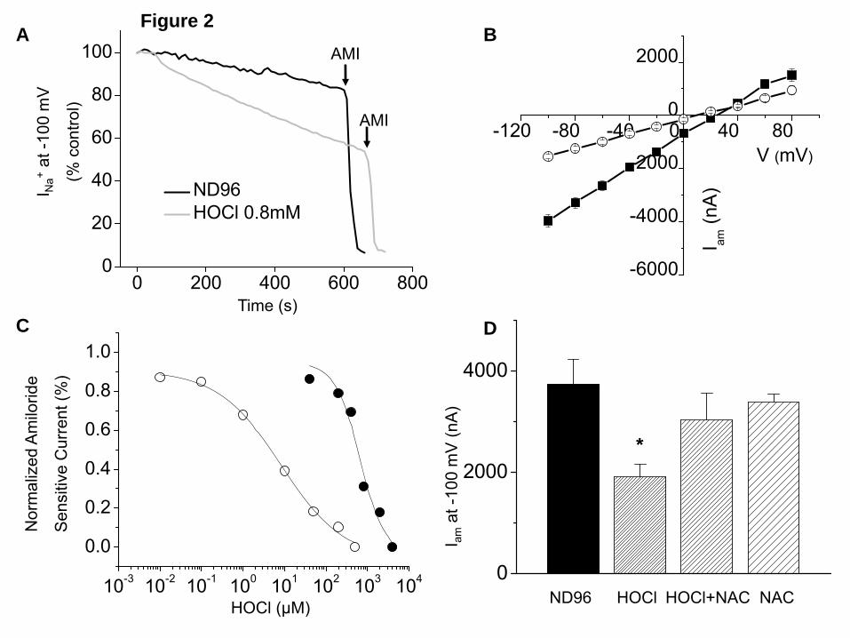
observed effects (39). Thus, in this system, by HOCl-induced injury, we refer to HOCl per se and all formed reactive intermediates. As shown in the various panels of Figure 2, Xenopus oocytes injected with α, β and γ-hENaC expressed significant amount of inwardly rectified INa, more than 90% of which were rapidly inhibited by addition of amiloride (10 μM) into the bath. Perfusion of hENaC-expressing oocytes with ND96 containing HOCl (0.8 mM) for 10 min significantly inhibited both the basal (Figure 2A) and the amiloride-sensitive currents (Figure 2B) with a half-maximum effect at about 600 µM (Figure 2C). When hENaC expressing oocytes were incubated with ND96 containing HOCl for 2h, which better mimics the in vivo situation where activated neutrophils may generate significant amount of HOCl in close proximity of ENaC over long periods of time, significant inhibition of Iamil were noted at a much lower concentrations (half maximum concentration= 4 μM; Figure 2C). Subsequent addition of a variety of reducing agents in the medium (10 mM DTT or 1 mM N-acetylcysteine, NAC) did not reverse Iamil by a significant amount (data not shown), indicating that ENaC inhibition was due to the formation of irreversible oxidation states in key amino acids. On the other hand, perfusion of oocytes with a mixture of ND96 containing HOCl (400 µM) and NAC (1 mM), which scavenged HOCl to non-detectable levels, totally prevented the observed decrease of Iamil (Figure 2D). Addition of HOCl did not significantly alter the pH or the Cl- concentration of the medium, two variables shown to modulate ENaC activity in previous studies (40,41). HOCl inhibits single-channel Na+ currents in h-ENaC expressing oocytes. In our next series of studies, we recorded single-channel currents from hENaC-expressing oocytes using the cell-attached configuration. Forty eight hours post injection with hENaC, oocytes expressed Na+ single channels with unitary conductances of about 4 pS (Figure 3A and C) with long open and closed times, characteristic of ENaC. Perfusion of oocytes
by guest on Novem
ber 11, 2018http://w
ww
.jbc.org/D
ownloaded from

with 2mM HOCl in ND96 for 10 minutes decreased both the number (N) and the open probability (Po) of ENaC (Figure 3B and C). ENaC conductance was not altered. An analysis of the representative control current recording in Figure 3A showed the following values for the open probabilities (Po) of various open levels: level 1, Po1=0.262; level 2, Po2=0.13 and level 3, Po3=0.016; overall NPo=0.57. The values for the representative HOCl treated oocyte recording shown in Figure 3B were: Po1=0.21, Po2=0.017; overall NPo=0.25. Single channel NPo (calculated from all point histograms from recordings of at least ten min) were 0.70±0.10 for control oocytes and 0.21±0.05 after perfusion with 2 mM HOCl for 10 min (Figure 3D; means±SE; n=5; P<0.01). Detection of organic chloramines. As shown in Figure 4A, mass spectrometry analysis of products formed one hour after addition of HOCl (2.5 mM) into ND96, revealed the presence of various chloramine-type compounds. These agents were still present (albeit in smaller concentrations) 6 hr later (Figure 4B). On the other hand, chloramines were not detected when HOCl was added into ND96 containing NAC (Figure 4C) or when HOCl was added in phosphate buffer which did not contain HEPES (data not shown). We concluded that these compounds were most likely formed by the interaction of HOCl with HEPES, which is included traditionally in ND96 as a buffer. This is in agreement with previous reports suggesting that HOCl interacts with HEPES to form chloramines-like compounds (39,42). Chloramines formed by HOCl inhibit INa. We performed a series of studies to test the hypothesis that stable reactive intermediates formed by the interaction of HOCl with ND96 were mainly responsible for the inhibition of ENaC activity in vitro. We synthesized chloramines by mixing excess amounts of either glycine or taurine (10 mM) with HOCl (2mM) in phosphate buffer which lacked HEPES. We chose taurine and glycine because these amino acids exist in high concentrations in human plasma (162±
60 µM and 236±42 µM, (43)) and they react with high rate constants with hypochlorite (4.8±0.1x10⁵ M-1s-1 and 0.65-1.5x10⁵ M-1s-1, respectively (14,44)), suggesting their corresponding chlroramines may be formed in vivo. As shown in Figure 5A, perfusion of hENaC expressing oocytes with the mixture of glycine and HOCl significantly inhibited INa as compare to glycine alone (p<0.05, n=6). Same results were obtained with taurine and HOCl mixture (Figure 5B). Subsequent addition of DTT (10 mM) had no effect on INa indicating the presence of irreversible oxidation states. Serine proteases increased Na+ currents after inhibition by HOCl. Serine proteases, such as trypsin, plasmin and elastase, have been reported to enhance ENaC activity by proteolytic cleavage of the α and γ-ENaC subunits (45-49). We investigate whether extracellular serine proteases reversed the inhibitory effects of HOCl and its reactive products formed by the interactions with components of ND96. We perfused hENaC expressing oocytes with ND96 containing HOCl for 10 min which decreased INa in a time-dependent fashion (Figure 6A). At the end of this period we perfused oocytes with ND96 containing trypsin (100 nM), elastase or plasmin (10 µg/ml each). As shown in Figures 6A-D, perfusion of oocytes with proteases post HOCl rapidly increased INa from about 20% of their control values to nearly normal levels rapidly. In contrast, perfusion with ND96 alone or ND96 containing DTT (up to 10 mM) had no effect on INa (data not shown). Subsequent addition of amiloride (10 µM) in the perfusion medium decreased the current by almost 90%, indicating that these proteases activated ENaC.
In the second set of this series of experiments, we first perfused hENaC expressing oocytes with trypsin, elastase or plasmin and then with HOCl (2 mM). As shown in Figures 7A-D, perfusion of oocytes with serine proteases for 5 minutes dramatically increased INa by 100%; subsequent perfusion with HOCl had no effect on INa (in contrast to more than 60%
by guest on Novem
ber 11, 2018http://w
ww
.jbc.org/D
ownloaded from
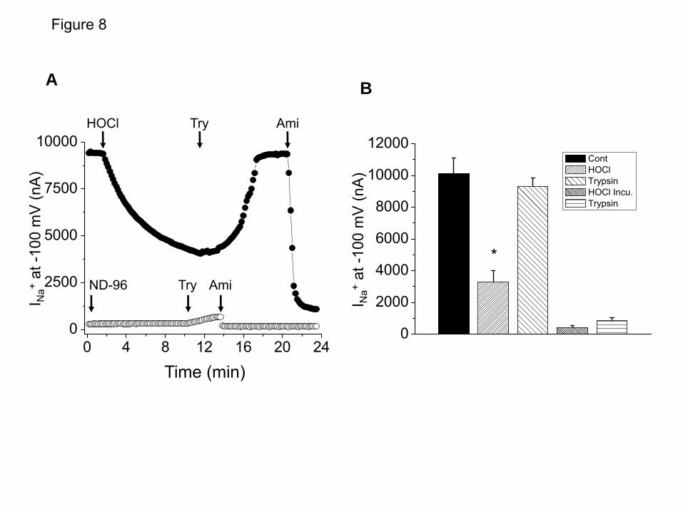
decrease in the absence of protease pre-treatment). As shown in Figure 6, addition of proteases to ENaC expressing oocytes post HOCl exposure did not increase INa to the same extent as in control non-exposed oocytes (Figures 6 and 7), most likely because proteases activate ENaC that have not been altered by HOCl. To test this hypothesis, we incubated ENaC expressing oocytes with HOCl (100 μM) for 2 hours, which decreased INa to almost zero, suggesting that under these conditions all membrane bound ENaC were completely inhibited by HOCl (as shown in Figure 2C). Under these conditions, subsequent perfusion of these oocytes with trypsin increased INa by less than 10% (Figures 8A and B). These data indicated that proteases added after HOCl treatment most likely acted on the subset of ENaC molecules that had not been affected by HOCl.
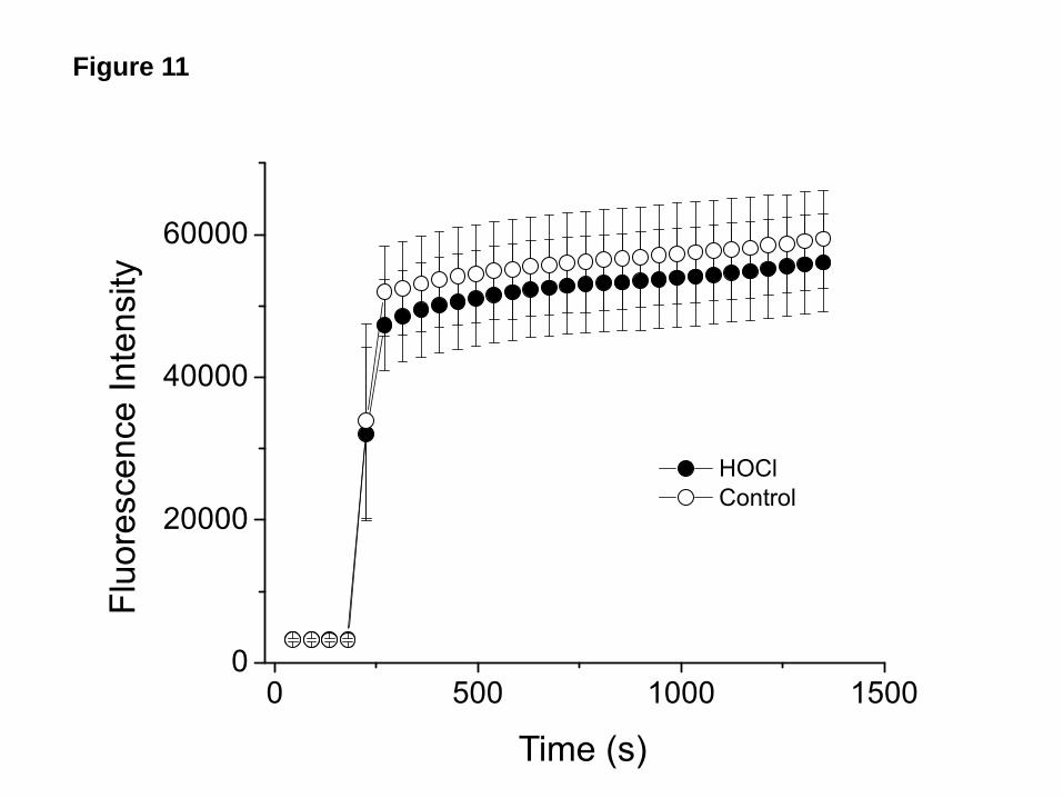
One possible explanation for the lack of effect of HOCl on INa following protease treatment is that proteases increased the number of mature ENaC channels thus decreasing the number of target ENaC molecules for HOCl. To investigate this possibility we treated hENaC expressing oocytes with various concentrations of trypsin, which resulted in a dose dependent increase of INa, and then perfused them with HOCl in ND96 for 10 min. As shown in Figure 9, HOCl had no effect on INa following trypsin treatment, irrespective of the starting value of INa. Membrane ENaC and protease activity levels were not decreased by HOCl. We measured membrane ENaC levels following perfusion of oocytes with HOCl (2 mM) for 10 min. As shown in Figure 10, no significant changes in α, β, or γ-ENaC protein levels were seen. Furthermore, incubation of control and HOCl treated oocytes with the fluorogenic peptide Boc-Gln-Ala-Arg-AMC–HCl (which were cleaved by membrane bound proteases thus increasing fluorescence intensity in the supernatant) resulted in similar levels of fluorescence in the supernatant, indicating that HOCl and its reactive intermediates did not inactivate endogenous membrane
proteases responsible for ENaC activation (Figure 11). Protease cleaved ENaC is insensitive to HOCl due to changes in its gating. Several proteases, including furin, prostasin, plasmin and elastase activate ENaC by cleaving segments in the finger domain within the large extracellular loop of its γ-subunit which locks ENaC in the open state (Figure 12A). Since our data showed that HOCl and its reactive intermediates had no effect on INa following protease activation, we hypothesized that functional injury to ENaC by HOCl required ENaC to be in its closed state. To test this hypothesis, we synthesized a 51-mer peptide mimicking the one released when plasmin cleaves γ-ENaC after it has been first cleaved by furin. As shown in Figure 12B, INa values were increased 4 min after perfusion of hENaC expressing oocytes with ND96 containing plasmin (10 µg/ml); however, subsequent perfusion of these oocytes with ND96 containing the 51-mer peptide (3 µg/ml) returned INa to their control levels. In a second set of experiments, oocytes were first perfused with plasmin (which increased INa) and then with ND96 containing both HOCl (2 mM) and the peptide (3 µg/ml). In this case INa was significantly inhibited by HOCl (Figure 12B). To further investigate the importance of proper ENaC gating in its inhibition by HOCl, we injected oocytes with wild type α and γ-hENaC and a mutant form of βENaC (βS520K), which results in higher amiloride sensitive currents by locking ENaC in their open state (Figure 13A), in agreement with previous reports (31). Addition of trypsin had no effect on INa of these mutant channels; perfusion with 1 mM HOCl for 10 min or incubation with 100 μM HOCl for 2h (which reduced INa of wild type ENaC to zero) had little effect on INa of these channels either (Figures 13A, B). The lack of effect of HOCl on these locked-open channels suggested HOCl inhibited ENaC by affecting channel gating. Sheng et al. (50) also reported that furin cleavage of ENaC relieves its self-inhibition by external [Na+],
by guest on Novem
ber 11, 2018http://w
ww
.jbc.org/D
ownloaded from
a mechanism putatively involving channel gating. Intranasal administration of trypsin post Cl2 exposure increases AFC. In the final set of experiments we assessed whether intranasally instillation of trypsin could reverse the decrease of AFC in mice exposed to Cl2. Mice were exposed to chlorine (300ppm for 30min) and then returned to room air. Fifteen min post exposure, they were briefly anesthetized with isoflurane and trypsin (5 µM; dissolved in 100 μl of saline) was instilled drop-wise in the nostrils. Mice recovered quickly and 1 h post exposure, they were anesthetized, ventilated and AFC was measured. Exposure to Cl2 decreased AFC by about 30% in agreement with the data shown in Figure 1. However, normal values of AFC were seen in mice exposed to Cl2 and instilled with trypsin (Figure 14). Trypsin had no effects on AFC of control mice (in contrast to what was measured in oocytes), most likely because ENaC was maximally stimulated under our experimental conditions. DISCUSSION
Hypochlorous acid/hypochlorite, as the major hydrolysis products of chlorine gas, and predominant oxidant produced by stimulated neutrophils, play an important role in inducing lung edema as well as increasing pulmonary vascular permeability and arterial pressure(51). However, until recently, little was known about the effects of these agents on membrane ion channels, especially ENaC. Herein we demonstrate that reactive intermediates (such as organic chloramines and other reactive intermediates) formed by the action of Cl2 and HOCl, in concentrations likely to be encountered in vivo during pathological conditions (51-53), via interaction with components of the media and lung lining fluid inhibit ENaC and decrease Na+-dependent fluid clearance across the distal lung epithelium. Furthermore, the fact that serine protease both prevent and reverse the effects of these compounds on ENaC and the fact that HOCl has no effect on ENaC channels that remain
constitutively open, indicate that HOCl mainly prevents ENaC channels in the close state from opening, most likely via post translational modifications of key amino acids. To the best of our knowledge this is the first report of this kind.
Cl2 gas is not very soluble in water (Henry’s law constant (KH) = 6.2-9.5 x 10⁻² M/atm or 6.2-9.5 x 10⁻⁸ M/ppm Cl2). The hydrolysis of Cl2 is an equilibrium reaction (K1 = 1 x 10⁻³ M2) that yields the oxidant HOCl, Cl-, and a proton, which combine to form HCl according to the following reaction: Cl2 + H2O ↔ HOCl + H+ +Cl-
(Eq. 1). HOCl is a weak acid (pKa = 7.53) and is therefore partly dissociated to OCl- at physiological pH. Both HOCl and its conjugate base are also powerful oxidants. If one assumes the equilibrium depicted by Eq.1 are established in the epithelial lining fluid (pH 6.92; [Cl-] = 0.103 M), one can calculate that [OCl-]/ [Cl2] = 2 x 10⁴ and [OCl-]/ [HOCl] = 0.25. Thus, in the airway lining fluid the concentration of HOCl will be approximately 80,000 that of Cl2 at equilibrium. HCl damage to components of the alveolar epithelium is probably minimal due to the presence of a significant amount of bicarbonate (11 mM) in the ELF (54) which can neutralize it. Indeed, previous studies have shown that Cl2 gas is much more toxic to the lung than aerosolized HCl (55).
Cl2 per se also reacts with multiple organic compounds with fast kinetics (44), raising the question of which form (Cl2 or HOCl) is responsible for the airway and lung injury during Cl2 exposure (55). It should be stressed that while small concentrations of Cl2 react with cellular components present in the upper airways (56,57), at inhaled concentrations higher than 50 ppm (likely to be present during industrial accidents and terrorist attacks), Cl2 will reach the distal airways and alveolar regions. Of course, low molecular weight scavengers (such as ascorbate and GSH) known to be present in high concentrations in the airway and epithelial lining fluid (reviewed in (58,59)) will react with Cl2 and HOCl reducing their steady state
by guest on Novem
ber 11, 2018http://w
ww
.jbc.org/D
ownloaded from
concentrations. However, our previous measurements, and those of others, show that the antioxidant scavenging capacity in the respiratory tract lining fluid would be relatively limited compared with inhaled Cl2 (19) and continuous exposure to relative high concentration of Cl2 gas (400 ppm for 30 min) should result in high concentration of HOCl intermediates in airway lining fluid.
HOCl is a potent oxidant and will react with multiple biological molecules, including amines, to produce chloramines (k~ 105M-1s-1)(60). Thus, it is well accepted that in a cell culture system, the observed biological effects of HOCl could be at least partially attributed to those of the chloramines, depending on the relative abundance of HOCl and amines, as suggested by some reports(61,62). Thus, organic chloramines, such as chloramines arising from the alpha amino group in free amino acids and the terminal amino group in peptides and proteins, and the side chains of the amino acids histidine, tryptophan and lysine, are likely to be formed in vivo (63-65). In addition, xenobiotic chloramines that may arise, for example, during water treatment using chlorination, may also further contribute to the total exposure to organic chloramines. Our data showed that HOCl reacted with HEPES to form chloramines-type species, which are relatively stable, and most likely responsible for the observed effects on ENaC. The fact that chloramines produced by the reaction of HOCl with glycine or taurine in the absence of HEPES also inhibit ENaC activity in the same manner as chloramines generated by the action of HOCl on HEPES, further supports this conclusion. Of course it is probable that a number of other reactive intermediates may be formed that may also decrease ENaC activity.
The mechanisms by which chloramines inhibit ENaC are complex and poorly understood. Our data showed that plasma membrane ENaC levels were not decreased following exposure of oocytes to HOCl. It has been suggested that at least a protease un-cleaved reserve pool of ENaC with low Po and a furin cleaved pool of ENaC with
intermediate Po exist at the cell membrane, and subsequent cleavage of γ subunit distal to the furin site by protease further activates ENaC by increasing Po (28,29,66,67). Thus the first question to be answered is which pool(s) of ENaC are affected by HOCl? Our data showed that both the inhibitions of amiloride sensitive currents in oocytes after ten minutes exposure to HOCl and of alveolar fluid clearance of mice following exposure to Cl2 were reversed by serine proteases. However, in both cases, significant amounts of currents are still present after HOCl exposure. When oocytes were incubated in HOCl for long periods of time to the point at which amiloride sensitive currents were decreased to almost zero, proteases failed to increase the sodium currents. In addition, INa of oocytes that was activated by exogenous proteases was no longer inhibited by HOCl. These results indicated that HOCl affected at least the ENaC pool with intermediate Po, while ENaC channels in the high Po pool that lacked the γ subunit extracellular segment because they had been further cleaved by proteases, were not sensitive to HOCl. One possible explanation for this insensitivity would be that HOCl damage on ENaC altered accessibility of cleavage sites to protease, similar to the regulation of proteolytic activation of ENaC by intracellular Na+, as reported by Knight et al (31).
It is known that ENaC undergoes complex post-translational modification, and proteases cleavage has been shown to be important for ENaC activation (67). In our oocyte system, the extracellular loops of α and γ ENaC subunits were likely already cleaved by the endogenous protease furin, thus the inhibitory segment in α subunit has been released resulting in the observed single channel activity with intermediate Po. When these oocytes were treated with exogenous proteases (trypsin, elastase or plasmin), the proteolytic cleavage at the site distal to furin released another inhibitory segment in γ subunit, resulting in additional activation of ENaC(48). The 51-mer peptide we synthesized represents the sequence
by guest on Novem
ber 11, 2018http://w
ww
.jbc.org/D
ownloaded from

between the putative furin and plasmin cleavage sites in the finger domain within the extracellular loop of γ-ENaC. Perfusion of oocytes with this peptide decreased the plasmin activated currents, in agreement with what has been reported previously (68). Equally important, perfusion of plasmin treated oocytes with both HOCl and the peptide resulted in a significant decrease of Na+ currents, further demonstrating that the inhibitory effects of HOCl-derived chloramines and other intermediates on ENaC depends on this segment of γ-ENaC. Though the mechanism by which this segment confers the inhibition of ENaC by HOCl is not clear, recent reports have suggested similar relationship between protease cleavage and extracellular or intracellular Na+ inhibition of ENaC(31,50). Thus, both proteases and HOCl regulated ENaC gating through action on the extracellular domain, so the locked-open βS520K mutant is not sensitive to either trypsin or HOCl.
It is not clear how HOCl chloramines affect ENaC gating. The extracellular loop of ENaC has been shown to be the binding sites of ions or peptides modulating ENaC gating(69-71). It has also been suggested that the conformation of the γ subunit segment within the thumb and finger domains in the extracellular loops (28) play a critical role in the regulation of ENaC activity. One possibility is that the amino acid sequence in the 51 mer construct is important for the modulation of ENaC gating by HOCl. Interestingly, there is no cysteine residue in this sequence, which is suggested to be the amino acid most likely to be oxidized by HOCl (12,72). Nevertheless, the possible chemical modifications imparted by HOCl to the 51 mer segment in ENaC can be assessed using the rate constants (60) for the reaction of HOCl with the side chain and the backbone amide nitrogen of the amino acids using a simple competitions kinetics model for pseudo first order conditions (large molar excess of the 51 mer over HOCl), as depicted in Table 1. The competition kinetics analysis indicates that histidines,
modified to chlorohistidine accounts for 83% of the chemical modifications, while modification of lysine residues to chlorolysine accounts for 13% of the chemical modifications. Tryptophan, modified to chlorotryptophan, accounts for 5% of the chemical modifications while the rest of possible modifications occur with negligible yields. These amino acid chloramines may participate in chlorine-transfer reactions or undergo hydrolysis to form carbonyl products.
Several studies have investigated the possible association between partially reduced reactive species and Na+ transport across the alveolar epithelium in both animals and patients with acute lung injury. Re-absorption of isotonic fluid was inhibited during prolonged hemorrhagic shock. Instillation of aminoguanidine, an inhibitor of iNOS, restored fluid re-absorption to normal levels (73). Mycoplasma infection resulted in significant decrease of both Na+-dependent alveolar fluid clearance in Balb/c mice and inhibition of amiloride sensitive Na+ currents across alveolar type II cells isolated from these mice. However, normal levels of AFC were seen when Balb/c mice were pretreated with cyclophosphamide to suppress inflammatory cells and decrease production of reactive intermediates including HOCl(7). Reactive intermediates also inhibit vectorial Na+ transport across ATII cells by decreasing the activity of epithelial Na+ channels via post-translational modifications of either ENaC per se, or of structural proteins (such as actin and fondrin) which are necessary for proper action of ENaC(6,74). Substitution of a single tyrosine in the extracellular loop of αENaC (Y283) with alanine prevented the peroxynitrite induced decrease of amiloride sensitive currents (75). ENaC was also inhibited by sulfhydryl reactive agents added in the cytoplasm and reducing agents reversed the rundown activity of ENaC in excised patches(76). In summary, our combined in vitro and in vivo data demonstrate that chloramines and reactive intermediates formed by the interaction of Cl2 and HOCl with the lung
by guest on Novem
ber 11, 2018http://w
ww
.jbc.org/D
ownloaded from

lining fluid and the incubation medium decrease amiloride sensitive currents (a hallmark of ENaC) by the modulation of ENaC gating and that these effects can be prevented by serine proteases. These findings establish the rational basis for the use of small concentrations of serine proteases to prevent and counteract Cl2 induced injury to epithelial sodium channels thus decreasing pulmonary edema.
Acknowledgments: This work was supported by HL-31197, HL-51173, U01ES015676 and 1U54ES017218. The authors would like to thank Dr. Peter M Snyder for providing the βS520K ENaC mutant; Mr. Steven F. Doran and Ms. Joanne Balanay for their excellent technical assistance with exposing mice to Cl2 gas; Dr. Iles and Postlethwait for many helpful discussions and Ms. Terese Potter for editorial help with this manuscript.
by guest on Novem
ber 11, 2018http://w
ww
.jbc.org/D
ownloaded from

REFERENCES
1. Matalon, S. and O'Brodovich, H. (1999) Annu. Rev. Physiol. 61, 627-661 2. Song, W. and Matalon, S. (2007) Am. J. Physiol Lung Cell Mol. Physiol 293, L855-L858 3. Matthay, M. A., Folkesson, H. G., and Clerici, C. (2002) Physiol Rev. 82, 569-600 4. Nielsen, V. G., Duvall, M. D., Baird, M. S., and Matalon, S. (1998) Am. J. Physiol. 275,
L1127-L1133 5. Fang, X., Song, Y., Hirsch, J., Galietta, L. J., Pedemonte, N., Zemans, R. L., Dolganov,
G., Verkman, A. S., and Matthay, M. A. (2006) Am. J. Physiol Lung Cell Mol. Physiol 290, L242-L249
6. Guo, Y., Duvall, M. D., Crow, J. P., and Matalon, S. (1998) Am. J. Physiol. 274, L369-L377
7. Hickman-Davis, J. M., Nicholas-Bevensee, C., Davis, I. C., Ma, H. P., Davis, G. C., Bosworth, C. A., and Matalon, S. (2006) Am. J. Respir. Crit Care Med. 173, 334-344
8. Dada, L. A., Chandel, N. S., Ridge, K. M., Pedemonte, C., Bertorello, A. M., and Sznajder, J. I. (2003) J. Clin. Invest 111, 1057-1064
9. Vadasz, I., Dada, L. A., Briva, A., Trejo, H. E., Welch, L. C., Chen, J., Toth, P. T., Lecuona, E., Witters, L. A., Schumacker, P. T., Chandel, N. S., Seeger, W., and Sznajder, J. I. (2008) J. Clin. Invest 118, 752-762
10. Weiss, S. J. (1989) N. Engl. J. Med. 320, 365-376 11. Spalteholz, H., Panasenko, O. M., and Arnhold, J. (2006) Arch. Biochem. Biophys. 445,
225-234 12. den Hartog, G. J., Haenen, G. R., Vegt, E., van der Vijgh, W. J., and Bast, A. (2002) Biol.
Chem. 383, 709-713 13. Hawkins, C. L., Pattison, D. I., and Davies, M. J. (2003) Amino. Acids 25, 259-274 14. Weiss, S. J., Klein, R., Slivka, A., and Wei, M. (1982) J. Clin. Invest 70, 598-607 15. Crow, J. P. (1999) Methods Enzymol. 301, 151-160 16. Hazen, S. L., Hsu, F. F., Mueller, D. M., Crowley, J. R., and Heinecke, J. W. (1996) J.
Clin. Invest. 98, 1283-1289 17. Hazen, S. L. and Heinecke, J. W. (1997) J. Clin. Invest. 99, 2075-2081 18. Eiserich, J. P., Cross, C. E., Jones, A. D., Halliwell, B., and van der Vliet, A. (1996) J.
Biol. Chem. 271, 19199-19208 19. Leustik, M., Doran, S., Bracher, A., Williams, S., Squadrito, G. L., Schoeb, T. R.,
Postlethwait, E., and Matalon, S. (2008) Am. J. Physiol Lung Cell Mol. Physiol 295, L733-L743
20. Davis, I. C., Zhu, S., Sampson, J. B., Crow, J. P., and Matalon, S. (2002) Free Radic. Biol. Med. 33, 1703-1713
21. Sartori, C., Allemann, Y., Duplain, H., Lepori, M., Egli, M., Lipp, E., Hutter, D., Turini, P., Hugli, O., Cook, S., Nicod, P., and Scherrer, U. (2002) N. Engl. J. Med. 346, 1631-1636
22. Sartori, C., Matthay, M. A., and Scherrer, U. (2001) Adv. Exp. Med. Biol. 502, 315-338 23. Davis, I. C., Lazarowski, E. R., Chen, F. P., Hickman-Davis, J. M., Sullender, W. M., and
Matalon, S. (2007) Am. J. Respir. Cell Mol. Biol. 37, 379-386 24. Davis, I. C., Xu, A., Gao, Z., Hickman-Davis, J. M., Factor, P., Sullender, W. M., and
Matalon, S. (2007) Am. J. Physiol Lung Cell Mol. Physiol 293, L281-L289 25. Vivona, M. L., Matthay, M., Chabaud, M. B., Friedlander, G., and Clerici, C. (2001) Am.
J. Respir. Cell Mol. Biol. 25, 554-561 26. Batchinsky, A. I., Martini, D. K., Jordan, B. S., Dick, E. J., Fudge, J., Baird, C. A.,
Hardin, D. E., and Cancio, L. C. (2006) J. Trauma 60, 944-956
by guest on Novem
ber 11, 2018http://w
ww
.jbc.org/D
ownloaded from
27. Bell, D. G. (2008) Management of Acute Respiratory Distress Syndrome (ARDS) Following Chlorine Exposure.
28. Kleyman, T. R., Carattino, M. D., and Hughey, R. P. (2009) J. Biol. Chem 284, 20447-20451
29. Maarouf, A. B., Sheng, N., Chen, J., Winarski, K. L., Okumura, S., Carattino, M. D., Boyd, C. R., Kleyman, T. R., and Sheng, S. (2009) J. Biol. Chem 284, 7756-7765
30. Hughey, R. P., Carattino, M. D., and Kleyman, T. R. (2007) Curr. Opin. Nephrol. Hypertens. 16, 444-450
31. Knight, K. K., Wentzlaff, D. M., and Snyder, P. M. (2008) J. Biol. Chem 283, 27477-27482
32. Davis, I. C., Lazarowski, E. R., Hickman-Davis, J. M., Fortenberry, J. A., Chen, F. P., Zhao, X., Sorscher, E., Graves, L. M., Sullender, W. M., and Matalon, S. (2006) Am. J. Respir. Crit Care Med. 173, 673-682
33. Prutz, W. A. (1998) Arch. Biochem. Biophys. 349, 183-191 34. Lazrak, A., Nita, I., Subramaniyam, D., Wei, S., Song, W., Ji, H. L., Janciauskiene, S.,
and Matalon, S. (2009) Am. J. Respir. Cell Mol. Biol. 41, 261-270 35. Lazrak, A., Iles, K. E., Liu, G., Noah, D. L., Noah, J. W., and Matalon, S. (2009) FASEB
J. 23, 3829-3842 36. Leduc-Nadeau, A., Lahjouji, K., Bissonnette, P., Lapointe, J. Y., and Bichet, D. G. (2007)
Am. J. Physiol Cell Physiol 292, C1132-C1136 37. Cho, H. Y. and Kleeberger, S. R. (2007) Free Radic. Biol. Med. 42, 433-445 38. Staub, O., Gautschi, I., Ishikawa, T., Breitschopf, K., Ciechanover, A., Schild, L., and
Rotin, D. (1997) EMBO J. 16, 6325-6336 39. Prutz, W. A. (1996) Arch. Biochem. Biophys. 332, 110-120 40. Collier, D. M. and Snyder, P. M. (2009) J. Biol. Chem 284, 29320-29325 41. Collier, D. M. and Snyder, P. M. (2009) J. Biol. Chem 284, 792-798 42. Prutz, W. A. (1998) Arch. Biochem. Biophys. 357, 265-273 43. Lentner, C. (1984) In Lentner, C., editor. Geigy Scientific Tables, Vol. 3: Physical
Chemistry Composition of Blood Hematoloty Somatometric Data, Ciba-Geigy, Basel 44. Deborde, M. and von, G. U. (2008) Water Res 42, 13-51 45. Kleyman, T. R., Myerburg, M. M., and Hughey, R. P. (2006) Kidney Int. 70, 1391-1392 46. Andreasen, D., Vuagniaux, G., Fowler-Jaeger, N., Hummler, E., and Rossier, B. C. (2006)
J. Am. Soc. Nephrol. 17, 968-976 47. Planes, C., Leyvraz, C., Uchida, T., Angelova, M. A., Vuagniaux, G., Hummler, E.,
Matthay, M., Clerici, C., and Rossier, B. (2005) Am. J. Physiol Lung Cell Mol. Physiol 288, L1099-L1109
48. Rossier, B. C. (2004) Proc. Am. Thorac. Soc. 1, 4-9 49. Donaldson, S. H., Hirsh, A., Li, D. C., Holloway, G., Chao, J., Boucher, R. C., and
Gabriel, S. E. (2002) J. Biol. Chem 277, 8338-8345 50. Sheng, S., Carattino, M. D., Bruns, J. B., Hughey, R. P., and Kleyman, T. R. (2006) Am.
J. Physiol Renal Physiol 290, F1488-F1496 51. Hampton, M. B., Kettle, A. J., and Winterbourn, C. C. (1998) Blood 92, 3007-3017 52. Ghosh, S., Janocha, A. J., Aronica, M. A., Swaidani, S., Comhair, S. A., Xu, W., Zheng,
L., Kaveti, S., Kinter, M., Hazen, S. L., and Erzurum, S. C. (2006) J. Immunol. 176, 5587-5597
53. Zheng, L., Settle, M., Brubaker, G., Schmitt, D., Hazen, S. L., Smith, J. D., and Kinter, M. (2005) J. Biol. Chem 280, 38-47
54. Nielson, D. W. (1986) J. Appl. Physiol. 60, 972-979 55. Martin, J. G., Campbell, H. R., Iijima, H., Gautrin, D., Malo, J. L., Eidelman, D. H.,
Hamid, Q., and Maghni, K. (2003) Am. J. Respir. Crit Care Med. 168, 568-574 56. Nodelman, V. and Ultman, J. S. (1999) J. Appl. Physiol 87, 2073-2080
by guest on Novem
ber 11, 2018http://w
ww
.jbc.org/D
ownloaded from

57. Nodelman, V. and Ultman, J. S. (1999) J. Appl. Physiol 86, 1984-1993 58. Rahman, I. and MacNee, W. (1996) Thorax 51, 348-350 59. Lang, J. D. J., Davis, I., Patel, R., and Matalon, S. (2006) Oxidative and Nitrosative Lung
Injury. Drs. Alfred P. Fishman, Jay A. Fishman, Michael A. Grippi, Larry B. Kaiser and Robert M. Senior, McGraw-Hill
60. Pattison, D. I., Hawkins, C. L., and Davies, M. J. (2007) Biochemistry 46, 9853-9864 61. Peskin, A. V., Midwinter, R. G., Harwood, D. T., and Winterbourn, C. C. (2004) Free
Radic. Biol. Med. 37, 1622-1630 62. Midwinter, R. G., Cheah, F. C., Moskovitz, J., Vissers, M. C., and Winterbourn, C. C.
(2006) Biochem. J. 396, 71-78 63. Winterton, N. (1997) Mutat. Res 373, 293-294 64. Iwase, H., Takahashi, T., Takatori, T., Shimizu, T., Aono, K., Yamada, Y., Iwadate, K.,
and Nagao, M. (1995) Biochem. Biophys. Res Commun. 215, 945-951 65. Heinecke, J. W., Li, W., Mueller, D. M., Bohrer, A., and Turk, J. (1994) Biochemistry 33,
10127-10136 66. Hughey, R. P., Bruns, J. B., Kinlough, C. L., and Kleyman, T. R. (2004) J. Biol. Chem.
279, 48491-48494 67. Hughey, R. P., Bruns, J. B., Kinlough, C. L., Harkleroad, K. L., Tong, Q., Carattino, M.
D., Johnson, J. P., Stockand, J. D., and Kleyman, T. R. (2004) J. Biol. Chem. 279, 18111-18114
68. Bruns, J. B., Carattino, M. D., Sheng, S., Maarouf, A. B., Weisz, O. A., Pilewski, J. M., Hughey, R. P., and Kleyman, T. R. (2007) J. Biol. Chem. 282, 6153-6160
69. Sheng, S., Perry, C. J., and Kleyman, T. R. (2002) J. Biol. Chem. 277, 50098-50111 70. Sheng, S., Perry, C. J., and Kleyman, T. R. (2004) J. Biol. Chem 279, 31687-31696 71. Sheng, S., Bruns, J. B., and Kleyman, T. R. (2004) J. Biol. Chem 279, 9743-9749 72. Pullar, J. M., Winterbourn, C. C., and Vissers, M. C. (1999) Am. J. Physiol 277, H1505-
H1512 73. Pittet, J. F., Lu, L. N., Morris, D. G., Modelska, K., Welch, W. J., Carey, H. V., Roux, J.,
and Matthay, M. A. (2001) J. Immunol. 166, 6301-6310 74. Duvall, M. D., Zhu, S., Fuller, C. M., and Matalon, S. (1998) Am. J. Physiol 274, C1417-
C1423 75. Chen, L., Fuller, C. M., Kleyman, T. R., and Matalon, S. (2004) Am. J. Physiol Renal
Physiol 286, F1202-F1208 76. Kellenberger, S., Gautschi, I., Pfister, Y., and Schild, L. (2005) J. Biol. Chem. 280, 7739-
7747 77. Chen, L., Bosworth, C. A., Pico, T., Collawn, J. F., Varga, K., Gao, Z., Clancy, J. P.,
Fortenberry, J. A., Lancaster, J. R., Jr., and Matalon, S. (2008) Am. J. Respir. Cell Mol. Biol. 39, 150-162
by guest on Novem
ber 11, 2018http://w
ww
.jbc.org/D
ownloaded from

FIGURE LEGENDS Figure 1. Exposure of mice to Cl2 decreases Alveolar Fluid Clearance. (A) C57BL/6 mice were exposed to the indicated concentrations of Cl2 gas for 30 min. Alveolar fluid clearance (expressed as % of instilled volume per 30 min) was measured one hour post exposure in anesthetized and ventilated mice as described in the Methods. (B) C57BL/6 mice were exposed to 400 ppm Cl2 for 30 min. Alveolar fluid clearance was measured one hour post exposure. Amiloride (Am.; 1.5 mM final concentration) or an equivalent volume of vehicle was added in the instilled solution in some animals. (C) Measurements of AFC in Balb/C mice exposed to 400 ppm Cl2 and returned to room air for the indicated intervals. (D) Balb/c mice were exposed to 400 ppm for 30 min. Alveolar fluid clearance was measured in the presence and absence of amiloride. (mean ± SE, *, p<0.05 compared to the corresponding air value; #, p<0.05 compared to the corresponding value without amiloride). Figure 2. HOCl decreases Na+ currents across Xenopus oocytes injected with α, β and γ-hENaC. (A) Oocytes injected with hENaC were perfused with either ND96 or a solution in which 0.8 mM HOCl was added in ND96 (0.8 mM HOCl-ND96). As mentioned in the text and shown in Figure 3 below, the fast reaction of HOCl with HEPES and other compounds in ND96 generated mainly organic chloramines. Inward ENaC currents were measured by pulsing the membrane potential from its resting value (-40 mV) to -140 mV for 500 mS. Amiloride (10 µM) was added into the perfusion medium at the times indicated by the arrows. To better demonstrate the effects of HOCl, the results were expressed as % of their corresponding baseline values prior to addition of HOCl. Shown are characteristic tracings which were repeated using 10 different oocytes from three different isolations. (B) Current-voltage relationships of amiloride-sensitive difference (Iam) currents obtained after perfusion of h-ENaC expressing oocytes with either 0.8 mM HOCl-ND96 (open circles) or ND96 alone (closed squares) for 10 min. (C) Normalized amiloride-sensitive currents across hENaC expressing oocytes which were either incubated (open circles; n≥8 for each data point) or perfused (solid circles; n≥10 for each data point) with the indicated concentrations of HOCl-ND96 for 2 h and 10 min respectively. Normalized amiloride-sensitive currents were calculated as follows as previously described (77): I=1-(Io-Ix)/(Io-Imax) where Io and Ix are the I values in the absence of HOCl and the maximum HOCl added. Inhibition constant (ki) were calculated using the Origin software by fitting the data points with the following equations; I = (1– [1/ (1+Ki/X)]) •Imax, where Imax is the maximum current (i.e. the current in the absence of HOCl) and X is the concentration of HOCl. For clarity only mean values (without SE) are shown. (D) Amiloride sensitive currents (Iam) at -100 mV for hENaC expressing oocytes perfused with HOCl (400 µM), a mixtures of HOCl (400 µM) and N-acetylcysteine (1 mM) or N-acetylcysteine (1 mM) alone. (mean ± SE; N≥ 10 oocytes for each group. *, p<0.05 compared to ND96 alone). Figure 3. HOCl decreases Na+ single channel activity in cell attached patches of hENaC expressing oocytes. Oocytes were patched in the cell-attached mode as described in the text and currents were measured at a pipette holding potential of -100 mV (Vholding=Vapical-Ppipette) and amplitude distribution histograms were generated as described in Methods. (A) Control (B) Ten min post perfusion with 2 mM HOCl-ND96 prior to patching. Open (O) and closed (C) states are indicated to the records. Notice visible decrease of open state of single channels in HOCl perfused oocytes. Typical records from 5 control and 5 HOCl perfused oocytes from two different batches. (C) Amplitude histograms for single channels from control (black) and HOCl perfused (red) oocytes shown in A and B. Typical records which were reproduced as mentioned in panel B. (D) NPo values calculated from all amplitudes histograms as mentioned in Methods. (mean ± SE; n=5; **, p<0.01). Figure 4. Mass spectrometry analysis of reaction products formed by the reaction of HOCl with ND96. 2.5 mM HOCl were added into ND96 for 1 and 6 hours. Samples of medium were
by guest on Novem
ber 11, 2018http://w
ww
.jbc.org/D
ownloaded from

then analyzed with Tandem Mass Spectroscopy as discussed in Materials and Methods. Records show the mass to charge ratios (m/z) for the various fragments formed by the interaction of HOCl with ND96. Possible structures of most abundant compounds (organic chloramines) formed after one (A) and six hours (B) post addition are shown as inserts. Notice the absence of these organic chloramines one hour post HOCl (400 μm) addition into ND96 containing NAC (1 mM) (C). Typical records which were reproduced at least three times with identical results. Figure 5. Inhibition of INa by glycine and taurine chloramines formed in the absence of HEPES. Inward Na+ currents at -100 mV across hENaC expressing oocytes perfused for ten min with: (A) Glycine (10 mM) in phosphate buffer or glycine (10 mM) and 2 mM HOCl in phosphate buffer; and (B) taurine (10 mM) in phosphate buffer or taurine (10 mM) and 2 M HOCl in phosphate buffer. Glycine or taurine were mixed with HOCl in phosphate buffer for one hour prior to perfusion (no HEPES present). Amiloride (10 µM) and DTT (10 mM) were added into the medium 10 min post perfusion. (mean ± SE for the indicated number of oocytes; *, p<0.05). Figure 6. Trypsin, plasmin and elastase increase INa after HOCl inhibition. (A) hENaC expressing oocytes were perfused with 2 mM HOCl-ND96 for 10 min. Inward Na+ currents were measured continuously at -100 mV as described in Methods. After 10 min, oocytes were perfused with ND96 containing 100 nM trypsin which rapidly increased the currents to the control level. Subsequent addition of amiloride (10 µM) into the perfusate decreased the currents to almost zero (recording was discontinued). (B, C, D) Mean values for the experiment shown following perfusion with trypsin (100 nM; B; n=8), elastase (10 µg/ml; C; n=6) or plasmin (10 µg/ml; D; n=6). (mean ± SE, *, p<0.05). Figure 7. Serine proteases renders ENaC insensitive to HOCl. (A) hENaC expressing oocytes were perfused with ND96 containing 100 nM trypsin. Inward Na+ currents were measured continuously at -100 mV as described in Methods. After 5 min, when the INa had reached a new plateau, oocytes were perfused with 2 mM HOCl-ND96 which had no effect on INa. Subsequent addition of amiloride (10 µM) into the perfusate decreased INa to almost zero. (B, C, D) Mean values for the corresponding experiments following perfusion of hENaC expressing oocytes with trypsin (100 nM; B; n=11), elastase (10 µg/ml; C; n=8) or plasmin (10 µg/ml; D; n=6) (means ± SE. * p<0.05). Figure 8. Trypsin has little effect on INa inhibited by HOCl incubation. (A) Representative time course recordings of INa at -100mV from hENaC expressing oocytes perfused by 1 mM HOCl-ND96 (●) or incubated in 100 µM HOCl-ND96 for 2 hours (○), then perfused with trypsin (2 µM) and amiloride (10 µM). (B) Group data showed trypsin increased INa to control levels in oocytes perfused with HOCl-ND96. Incubation with HOCl-ND96 for 2 hours greatly inhibited INa, perfusion with trypsin had little effect on INa of these oocytes (mean ± SE, n=11 for HOCl perfused and 5 for HOCl incubated oocytes, *, p<0.05). Figure 9. Lack of inhibition of INa by chloramines following treatment of oocytes with various concentrations of trypsin. hENaC-expressing oocytes were preincubated with the indicated concentrations of trypsin for 10 min and then perfused with 1mM HOCl-ND96 for 10 min, at which time amiloride (10 µM) was added in the bath (mean ± SE). Figure 10. HOCl does not decrease cell membrane ENaC levels. Xenopus oocytes expressing tagged α, β, and γ- hENaC were incubated with ND96 (Cont) or 2 mM HOCl-ND96 (HOCl) for ten min. In another set of experiments, h-ENaC expressing oocytes were first incubated with trypsin (100 nM) for ten min and then with 2 mM HOCl-ND96 (Trypsin). Equal amount of membrane extracts from each treatment group were resolved by SDS-PAGE and blotted with anti-Flag, Myc, and V5 antibodies to determine the levels of α, β, and γ ENaC on the plasma membrane. Results of a typical experiment which was repeated three times with identical results. Figure 11. Oocyte protease activity is not inhibited by HOCl. hENaC expressing oocytes were incubated with ND96 as control (○), or 2 mM HOCl-ND96 (●) for 10 mins, then moved into a cuvette containing Boc-Gln-Ala-Arg-AMC-HCl (50 μM). Fluorescence was measured
by guest on Novem
ber 11, 2018http://w
ww
.jbc.org/D
ownloaded from
continuously for the next 20 min at 460 nm following excitation at 380 nm (mean ± SE; n=5 for each condition). Figure 12. HOCl inhibits ENaC by interaction with a peptide in the extracellular loop of γ subunit. (A) Partial sequence (136 to 203 aa) within the finger domain in the extracellular loop of γ-ENaC including the putative furin (R143), plasmin (K194) and elastase (A195 and V198) cleavage sites (shown in bold font). A 51-mer peptide was synthesized corresponding to the segment between furin and plasmin cleavage sites (144 to 194, in red color). (B) Left panel (-HOCl): hENaC expressing oocytes were perfused with plasmin (10 µg/ml) followed by ND96 containing the 51-mer peptide and then amiloride (10 µM); Right panel (+HOCl): following activation of hENaC by plasmin perfusion, oocytes were perfused with the mixture of 2mM HOCl-ND96 and the peptide (3 µg/ml) (mean ± SE; n=6 for each group; *, p<0.05 compared with control). Figure 13. INa of βs520k mutant ENaC is not inhibited by HOCl. (A) Representative time-course recordings of INa at -100 mV from oocytes expressing wild-type α, βs520k and wild-type γ ENaC perfused with HOCl-ND96 (1 mM) for 10 mins (●) or incubated with HOCl-ND96 (100 μM) for 2 hours (○), then perfused with trypsin (2 μM) and amiloride (10μM). (B) Group data showed neither perfusion nor incubation with HOCl-ND96 inhibits INa of βs520k mutant ENaC expressing oocytes (mean ± SE, n=14 for HOCl perfused and 5 for HOCl incubated oocytes). Figure 14. Intratracheal trypsin reverses the decrease in AFC in mice exposed to Cl2. Balb/c mice were exposed to Cl2 (300ppm for 30min) and then returned to room air. Fifteen min post exposure, they were briefly anesthetized with isoflurane and trypsin (5 µM; dissolved in 100 μl of saline) was instilled drop-wise in the nostrils. Mice recovered quickly and 1 h post exposure, they were anesthetized, ventilated and AFC was measured. (mean ± SE; control=12; saline=8; trypsin =5. *, p<0.05).
by guest on Novem
ber 11, 2018http://w
ww
.jbc.org/D
ownloaded from

Table 1. Competition Kinetics Assessment of Reactive Centers in The 51 mer Construct That are Subject to Chlorination by HOCl
Amino Acida kb nc nxk Pd Arginine 26 5 1.3x10² 5.4x10⁻⁴ Asparagine 0.03 1 0.03 1.3x10⁻⁷ Cysteine 3.0x10⁷ 0 0 0 Glutamine 0.03 2 0.06 2.5x10⁻⁷ Histidine 1.0x10⁵ 2 2.0x10⁵ 0.83 Lysine 5.0x10³ 6 3.0x10⁴ 0.13 Methionine 3.8x10⁷ 0 0 0 Tryptophan 1.1x10⁴ 1 1.1x10⁴ 4.6x10⁻² Tyrosine 44 0 0 0 Backbone amide 10 50 5x10² 2.1x10⁻³ a. Only those amino acids with k ≥ 0.03 M-1s-1 are listed here. b. Rate constant (M-1s-1) for the reaction of hypochlorite with the side chain of the amino acid or
with the backbone amide nitrogen. c. Number of this amino acid present in the 51 mer or number of backbone amide nitrogens in
the 51 mer. d. Normalized rate for reaction with HOCl calculated from the following equation:
)/(∑=i
iiii knknP
by guest on Novem
ber 11, 2018http://w
ww
.jbc.org/D
ownloaded from

Figure 1A B
0
10
20
30
0
10
20
30
0
10
20
30
(% in
still
ed v
ol./3
0 m
in)
*
0
10
20
# hrs in Air post Cl2
23 3 3 6 12 4
Air 50 200 300 400 500Cl2 (ppm)
(% in
still
ed v
ol./3
0 m
in)
23 8 12 5
Air Cl2Am. Am.
(% in
still
ed v
ol./3
0 m
in)
23 12 4 6 9
Air 1 1.5 4 24
(% in
still
ed v
ol./1
5 m
in)
12 8 7
Air Cl2 AMI
**
#*
*
*
C D
*
by guest on November 11, 2018 http://www.jbc.org/ Downloaded from
0 200 400 600 8000
20
40
60
80
100
ND96 HOCl 0.8mM
Nor
mal
ized
Am
ilorid
e
Sen
sitiv
e C
urre
nt (%
)I N
a+at
-100
mV
(% c
ontro
l)
Time (s)
AMI
AMI
Figure 2A
C
0
2000
4000
-120 -80 -40 0 40 80
-6000
-4000
-2000
0
2000
I am (n
A)
V (mV)
10-3 10-2 10-1 100 101 102 103 104
0.0
0.2
0.4
0.6
0.8
1.0
ND96 HOCl HOCl+NAC NAC
I amat
-100
mV
(nA
)
HOCl (µM)
B
*
D
by guest on November 11, 2018 http://www.jbc.org/ Downloaded from

A
DB
C
1 min0.5 pA
C
OO
CO
OO
Figure 3
0.0
0.2
0.4
0.6
0.8
1.0
ND96 HOCl
**
-0.4 0.0 0.4 0.8 1.2 1.60
5
10
15
NP
oN
umbe
r of E
vent
s
Amplitude (pA)
by guest on November 11, 2018 http://www.jbc.org/ Downloaded from

200 225 250 275 300
20
40
60
80
100
O
HON HN
SOH
O
Cl
O
HONH N
S
OH
O
Cl
247.1
239.0
HEPES
or
249.1
269.0
Rel
ativ
e A
bund
ance
m/z
Figure 4 A
Chloramine
by guest on November 11, 2018 http://www.jbc.org/ Downloaded from

200 225 250 275 300
20
40
60
80
100
O
HON HN
SOH
O
Cl
O
HONH N
S
OH
O
Cl
247.1
239.0
HEPESor
249.1
269.0Rel
ativ
e A
bund
ance
m/z
Figure 4 B
Chloramine
by guest on November 11, 2018 http://www.jbc.org/ Downloaded from
100 150 200 250 300
20
40
60
80
100122.0
164.1
239.1261.2
Rel
ativ
e A
bund
ance
m/z
NACHEPES
Figure 4 C
by guest on November 11, 2018 http://www.jbc.org/ Downloaded from

B
Figure 5
0
2000
4000
6000
8000
0
2000
4000
6000
8000
10000
*
PBS
Gly
+ H
OC
l
Am
.Gly.
I Na+
at -1
00 m
V (n
A)
I Na+
-100
mV
(nA
)
A
*
PBSA
m.
PBS
PBS
Taur
.
Am
.
Taur
.+ H
OC
lD
TTA
m.
by guest on November 11, 2018 http://www.jbc.org/ Downloaded from
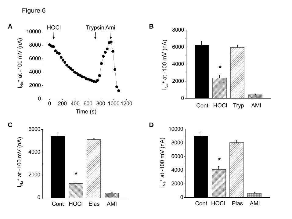
Figure 6
C
0
2000
4000
6000
Cont HOCl Elas AMI
I Na+
at -1
00 m
V (n
A)
*
D
0
2000
4000
6000
8000
10000
Cont HOCl Plas AMI
I Na+
at -1
00 m
V (n
A)
*
A B
0 200 400 600 800 1000 12000
2000
4000
6000
8000
10000
I Na+
at -1
00 m
V (n
A)
Time (s)
HOCl Trypsin Ami
0
2000
4000
6000
8000
Cont HOCl Tryp AMI
I Na+
at -1
00 m
V (n
A)
*
by guest on November 11, 2018 http://www.jbc.org/ Downloaded from

Figure 7
D
0
2000
4000
6000
8000
10000
Cont Plas HOCl AMI
I Na+
at -1
00 m
V (n
A)
C
0
2000
4000
6000
8000
Cont Elas HOCl AMI
I Na+
at -1
00 m
V (n
A)
0
4000
8000
12000
I Na+
at -1
00 m
V (n
A)
Cont Tryp HOCl AMI
BA
0 200 400 600 800 1000 12000
4000
8000
12000
16000
I
(nA
)
Time(s)
Trypsin HOCl Ami
I Na+
at -1
00 m
V (n
A)
Time (s)
by guest on November 11, 2018 http://www.jbc.org/ Downloaded from
0 4 8 12 16 20 240
2500
5000
7500
10000
0
2000
4000
6000
8000
10000
12000
Cont HOCl Trypsin HOCl Incu. Trypsin
I Na+
at -1
00 m
V (n
A)
I Na+
at -1
00 m
V (n
A)
HOCl Try Ami
ND-96 Try Ami
Time (min)
Figure 8
A B
*
by guest on November 11, 2018 http://www.jbc.org/ Downloaded from

0
2000
4000
6000
8000
10000
12000 Cont Trypsin HOCl AMI
10 25 50 100Typsin (nM)
56
5
11
Figure 9I N
a+at
-100
mV
(nA
)
by guest on November 11, 2018 http://www.jbc.org/ Downloaded from
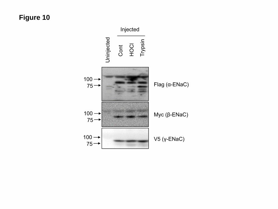
Figure 10
Uni
njec
ted
Con
t
HO
Cl
Tryp
sin
Injected
10075
10075
10075
Flag (α-ENaC)
Myc (β-ENaC)
V5 (γ-ENaC)
by guest on November 11, 2018 http://www.jbc.org/ Downloaded from
0 500 1000 15000
20000
40000
60000
HOCl Control
Time (s)
Fluo
resc
ence
Inte
nsity
Figure 11
by guest on November 11, 2018 http://www.jbc.org/ Downloaded from

136FPESRKRREAESWNSVSEGKQPRFSHRIPLLFDQDEKGKARDFFTGRKRKVGGSIIHKASNVMHIES203
Figure 12
0
2000
4000
6000
8000
10000
12000 Cont Plasmin Peptide AMI
-HOCl +HOCl
A B
I Na+
at -1
00 m
V (n
A)
*
**
by guest on November 11, 2018 http://www.jbc.org/ Downloaded from

0 5 10 15 200
4000
8000
12000
16000
20000
0
4000
8000
12000
16000
I Na+
at -1
00 m
V (n
A)
I Na+
at -1
00 m
V (n
A)
Time (min)
ND96 Try Ami
HOCl Try Ami
Cont HOCl Trypsin HOCl Incubation
Figure 13
A B
by guest on November 11, 2018 http://www.jbc.org/ Downloaded from

Figure 14
0
5
10
15
20
(% in
still
ed v
ol./1
5 m
in)
Air -------------- Cl2 -------------
Saline Trypsin
12 8 5
*
by guest on November 11, 2018 http://www.jbc.org/ Downloaded from
Giuseppe L. Squadrito and Sadis MatalonWeifeng Song, Shipeng Wei, Yongjian Shou, Ahmed Lazrak, Gang Liu, James D. Londino,
hypochlorous acid and chloramines channels by chlorine,+Inhibition of lung fluid clearance and epithelial Na
published online January 27, 2010J. Biol. Chem.
10.1074/jbc.M109.073981Access the most updated version of this article at doi:
Alerts:
When a correction for this article is posted•
When this article is cited•
to choose from all of JBC's e-mail alertsClick here
by guest on Novem
ber 11, 2018http://w
ww
.jbc.org/D
ownloaded from