hydroxy-2-[[2-(4-methoxyphenyl)-1,1-...
TRANSCRIPT

JPET #167007
1
Title Page
Pharmacological Characterization of 6-Hydroxy-8-[(1R)-1-
hydroxy-2-[[2-(4-methoxyphenyl)-1,1-
dimethylethyl]amino]ethyl]-2H-1,4-benzoxazin-3(4H)-one
monohydrochloride, (Olodaterol), a Novel Inhaled ß2-
Adrenoceptor Agonist exerting a 24-hour long Duration of
Action in Preclinical Models
T.Bouyssou, P.Casarosa, E. Naline, S.Pestel, I.Konetzki, P. Devillier and A.Schnapp
Boehringer Ingelheim Pharma GmbH & Co. KG, 88397 Biberach, Germany (T.B.,
P.C., S.P., I.K., A.S.)
2- UPRES EA 220, Hôpital Foch, Université Versailles – Saint Quentin, 11, rue
Guillaume Lenoir, 92150 Suresnes (E.N., P.D.)
JPET Fast Forward. Published on April 6, 2010 as DOI:10.1124/jpet.110.167007
Copyright 2010 by the American Society for Pharmacology and Experimental Therapeutics.
This article has not been copyedited and formatted. The final version may differ from this version.JPET Fast Forward. Published on April 6, 2010 as DOI: 10.1124/jpet.110.167007
at ASPE
T Journals on M
ay 5, 2018jpet.aspetjournals.org
Dow
nloaded from
JPET #167007
2
Running Title Page
Running Title: preclinical pharmacology of olodaterol, a once-daily ß2 agonist
Corresponding author: Dr. Andreas Schnapp
Contact Information: Dept. of Pulmonary Diseases Research, BI Pharma GmbH & Co.
KG, Birkendorferstrasse 65, Biberach an der Riss, Germany
E-mail: [email protected]
Phone: +49-7351-54-5240
Fax: +49-7351-83-5240
Number of text pages: 36
Number of tables: 4
Number of figures: 9
Number of references: 27
Number of words in the abstract: 212
Number of words in the introduction: 714
Number of words in the discussion: 1426
Non-standard abbreviations: human beta adrenoceptor, hß-AR; acetylcholine, ACh;
long acting beta agonist, LABA; chronic obstructive pulmonary disease, COPD;
Chinese hamster ovary, CHO; smooth muscle cells, SMCs.
Section assignment: GI, hepatic, Pulmonary and Renal
This article has not been copyedited and formatted. The final version may differ from this version.JPET Fast Forward. Published on April 6, 2010 as DOI: 10.1124/jpet.110.167007
at ASPE
T Journals on M
ay 5, 2018jpet.aspetjournals.org
Dow
nloaded from

JPET #167007
3
Abstract
The preclinical pharmacological profile of 6-hydroxy-8-[(1R)-1-hydroxy-2-[[2-(4-
methoxyphenyl)-1,1-dimethylethyl]amino]ethyl]-2H-1,4-benzoxazin-3(4H)-one
monohydrochloride, (olodaterol, previously known as BI 1744 CL), a novel,
enantiomeric pure, inhaled human ß2 adrenoceptor (hß2-AR) agonist, was compared
to marketed drugs, like salmeterol and formoterol. In vitro, olodaterol showed a
potent, nearly full agonistic response at the hß2-AR (EC50 = 0.1 nM; intrinsic activity =
88% compared to isoprenaline) and a significant selectivity profile (219-fold and
1622-fold against the hß1- and hß3-ARs, respectively). Similarly, olodaterol was able
to potently reverse contraction induced by different stimuli in isolated human bronchi.
In vivo, antagonistic effects of single doses of olodaterol and formoterol were
measured against acetylcholine challenges in anaesthetized guinea pigs and dogs
for up to 24 hours using the Respimat® Soft MistTM Inhaler. Heart rate and metabolic
parameters (serum potassium, lactate and glucose) were monitored to evaluate
systemic pharmacodynamic effects in the dog model. In both models, olodaterol
provided bronchoprotection over 24 hours. Formoterol applied at an equally effective
dose did not retain efficacy over 24 hours. In both models olodaterol showed a rapid
onset of action comparable to formoterol. Taken together, the preclinical behaviour of
olodaterol suggests that this novel ß2 adrenoceptor agonist has the profile for once-
daily dosing in man concomitant with a fast onset of action and a favourable systemic
pharmacodynamic profile.
This article has not been copyedited and formatted. The final version may differ from this version.JPET Fast Forward. Published on April 6, 2010 as DOI: 10.1124/jpet.110.167007
at ASPE
T Journals on M
ay 5, 2018jpet.aspetjournals.org
Dow
nloaded from

JPET #167007
4
Introduction
Asthma and chronic obstructive pulmonary disease (COPD) are conditions
characterized by airway obstruction, which is variable and reversible in asthma but is
progressive in COPD (Guerra, 2009). Both diseases are very common and their
incidence is increasing globally, placing a growing burden on patients and on health
services in industrialized and developing countries (Braman, 2006;Pauwels and
Rabe, 2004). Beta (ß) adrenoceptor agonists are among the most potent and rapidly
acting bronchodilators currently available for clinical use. In asthma, rapid-acting
inhaled ß2-adrenoceptor agonists are the therapy of choice as a reliever therapy for
episodes of dyspnea and for the pretreatment of exercise-induced
bronchoconstriction (Bateman, et al., 2008). In asthma patients with persistent
symptoms long-acting ß2 adrenoceptor agonists (LABAs), like salmeterol and
formoterol, are administered as an add on controller therapy when the first line
treatment of medium dose inhaled corticosteroids alone fails to achieve control of
asthma (Bateman, et al., 2008). Recently, formoterol has gained some recognition as
a p.r.n. controller therapy because of its fast onset of action, too. In addition, inhaled
ß2 adrenoceptor agonists provide major therapeutic benefits in the treatment of
COPD, such as reduction in symptoms and exacerbations, increases in exercise
capacity and improvements of health-related qualtity of life (Gold, 2009).
ß2 adrenoceptor agonists exert a bronchodilatory effect through activation of ß2
adrenoceptors (ß2-ARs) expressed on airway smooth muscle cells (SMCs).
Additionally, evidence exists that ß2-AR mediated increases in cAMP have an anti-
inflammatory effect in immune cells, e.g. neutrophils and mast cells, providing an
additional rationale for the use of ß2 adrenoceptor agonists in chronic inflammatory
diseases as asthma and COPD.
This article has not been copyedited and formatted. The final version may differ from this version.JPET Fast Forward. Published on April 6, 2010 as DOI: 10.1124/jpet.110.167007
at ASPE
T Journals on M
ay 5, 2018jpet.aspetjournals.org
Dow
nloaded from
JPET #167007
5
However, the utility, convenience and persistence of airflow improvement with short-
acting ß2 adrenoceptor agonists, like salbutamol, is limited by the need of repeated
administration. Furthermore, there is an appreciable increase in efficacy in terms of
patient reported outcomes with long acting bronchodilators (i.e., b.i.d. LABAs and q.d.
anticholinergics) (Jenkins, et al., 2009;Tashkin, et al., 2008), steroids and
combinations (Jenkins, et al., 2009).
Currently, two ß2 adrenoceptor agonists with a twice daily dosing regimen are
marketed, namely formoterol -a full agonist- and salmeterol -a partial agonist. The
clinical significance of the different intrinsic activities between these two agonists is
unclear. However, despite the decrease in dosing frequency with formoterol and
salmeterol, patient compliance is an issue (Ying et al., 1999).
Thus, a once-a-day LABA may have several advantages compared with short-acting
bronchodilators and twice-daily LABAs, including: i) improved convenience and
compliance (COPD and asthma), ii) improved airflow over a complete 24-hour period
(COPD and asthma), iii) a more convenient and stable once-a-day combination
option with a long acting muscarinic antagonist (LAMA), like tiotropium, for patients
for whom more than one bronchodilator is indicated (COPD), iv) a more convenient
and sustained once-a-day free combination option with inhaled steroids (moderate to
severe asthma). Additionally, a higher therapeutic index would be desirable for the
new generation of inhaled ß2 adrenoceptor agonists, as doubling the dose of
currently marketed drugs, e.g. salmeterol and formoterol, causes a significant
increase in the incidence of adverse effects including headache, tremor, palpitations,
muscle cramps and a fall in serum potassium concentration (Sovani, et al., 2004).
To achieve this, improving ligand selectivity for the hß2-AR versus the other family
subtypes hß1-AR (expressed prevalently on cardiac smooth muscle and responsible
for inotropic effects) and hß3-AR (on adipose tissue) is central.
This article has not been copyedited and formatted. The final version may differ from this version.JPET Fast Forward. Published on April 6, 2010 as DOI: 10.1124/jpet.110.167007
at ASPE
T Journals on M
ay 5, 2018jpet.aspetjournals.org
Dow
nloaded from

JPET #167007
6
6-Hydroxy-8-[(1R)-1-hydroxy-2-[[2-(4-methoxyphenyl)-1,1-dimethylethyl]amino]ethyl]-
2H-1,4-benzoxazin-3(4H)-one monohydrochloride, (olodaterol, previously known as
BI 1744 CL), is a novel, enantiopure inhaled ß2 adrenoceptor agonist that was
discovered in a program to identify compounds with a duration of action compatible
with once-daily dosing in humans, a fast onset of action and an increased therapeutic
index compared to the available inhaled ß2 adrenoceptor agonists. Here we describe
the preclinical pharmacological profile of olodaterol, compared to the marketed drugs
formoterol and salmeterol. To understand the behaviour of olodaterol at the
molecular level, interaction with the different ß-AR subtypes was analyzed in binding
and functional assays. Efficacy and duration of bronchoprotection were tested in
pharmacological models of acetylcholine-induced bronchoconstriction in
anaesthetized guinea pigs and dogs over a time period of 24 hours. We report that
olodaterol has an optimized profile of an inhaled LABA combining a high β2-
selectivity, a rapid onset of action and at least a 24-hour duration of action after a
single once-daily administration with minimal systemic pharmacodynamic effects.
This article has not been copyedited and formatted. The final version may differ from this version.JPET Fast Forward. Published on April 6, 2010 as DOI: 10.1124/jpet.110.167007
at ASPE
T Journals on M
ay 5, 2018jpet.aspetjournals.org
Dow
nloaded from

JPET #167007
7
Materials and Methods
Compounds:
Olodaterol hydrochloride (CL), (R,R)/(S,S)-salmeterol xinafoate and (R,R)/(S,S)-
formoterol fumarate were synthesized by the Department of Chemical Research
(Boehringer Ingelheim Pharma GmbH & Co. KG, Biberach, Germany). Acetylcholine
(Acetylcholine ophthalmicum Dispers) was from Dispersa GmbH (Germering,
Germany). Propofol (Propofol-Lipuro 2 %) was obtained from B Braun Melsungen AG,
(D-34209 Melsungen, Germany). Propranolol hydrochloride (Obsidan) was from
Alpharma-Isis GmbH & Co. KG (D-40764 Langenfeld, Germany).
Cell culture techniques
Chinese hamster ovary (CHO) cells transfected with the cDNAs encoding the human
ß1-, ß2- or ß3-adrenoceptors were purchased from Perkin Elmer (Waltham, MD). CHO
cells were grown in Ham’s F12 without glycine, hypoxanthine and thymidine; 10%
dialysed FBS; 100 U/ml penicillin; 100 µg/ml streptomycin. Due to their high level of
receptor expression (see Table 1), these cells were used for performing the affinity
binding studies. A second set of CHO cells stably transfected with human ß1-, ß2-
and ß3-ARs was generated in house and clones harboring low receptor expression
were further selected and used in the functional assays, to avoid high receptor spare
numbers and potential overestimation of agonist potency and intrinsic activity (IA).
This second set of cells, referred to as CHO-hß1-3LOW (see Table 1) were grown in
DMEM supplemented with 1x NEAA and 10% fetal calf serum in the presence of the
selection agent Geneticin (500 µg/ml). Cells were maintained at 37°C in humidified
air containing 5% CO2.
Equilibrium binding experiments
Membrane isolation and purification from CHO-cells stably expressing the human ß1-3
-ARs (high expressing clones) was performed as described previously (Casarosa, et
This article has not been copyedited and formatted. The final version may differ from this version.JPET Fast Forward. Published on April 6, 2010 as DOI: 10.1124/jpet.110.167007
at ASPE
T Journals on M
ay 5, 2018jpet.aspetjournals.org
Dow
nloaded from

JPET #167007
8
al., 2005). In all radioligand experiments the binding buffer consisted of Tris-HCl 50
mM, MgCl2 2 mM, EGTA 1 mM, pH 7.3. After the indicated incubation period, bound
and free [3H]-CGP 12,177 were separated by vacuum filtration using a Brandel
Harvester (Gaithersburg, MD) on GF/B filters presoaked in 0.5% polyethyleneimine,
and washed three times with ice cold binding buffer. Filter disks were added to 3 ml
of scintillation fluid (Ultima Gold from Perkin Elmer) in pony-vials and radioactivity
was quantified using liquid scintillation spectrometry on a Tri-Carb 2900TR Liquid
Scintillation Analyzer (Perkin Elmer). In all experiments, total binding did not reach
10% of the amount added, limiting complications associated with depletion of the free
radioligand concentration. Saturation binding experiments were performed by
incubating membranes (usually 5 to 20 µg/ sample, adjusted according to the Bmax of
the individual cell line) with a range of concentrations of [3H]-CGP 12,177 in a total
volume of 4 ml, to avoid significant ligand depletion at the lower concentrations.
Samples were incubated at room temperature for at least 2 hours under gentle
agitation, before filtration. To obtain affinity estimates of unlabelled agonists,
heterologous competition experiments against [3H]- CGP 12,177 were performed at
equilibrium. Membranes were incubated in the presence of [3H]-CGP 12,177 (final
concentration approximately 0.2 nM), 10 µM GppNHp to ensure an homogeneous
receptor population and different concentrations of agonists, at room temperature
with gentle agitation for at least 2 hours before filtration. Competition displacement
binding data were fitted to the Hill equation and IC50 values obtained from the
inhibition curves were converted to Ki values (Cheng and Prusoff, 1973).
cAMP assay
To determine the functional potency of the different agonists against the different
human ß-ARs, changes in intracellular cAMP levels were determined with CHO-hß1-3
LOW cells in suspension (15.000 cells/well) using Alphascreen technology (Perkin
This article has not been copyedited and formatted. The final version may differ from this version.JPET Fast Forward. Published on April 6, 2010 as DOI: 10.1124/jpet.110.167007
at ASPE
T Journals on M
ay 5, 2018jpet.aspetjournals.org
Dow
nloaded from
JPET #167007
9
Elmer) and a 384 well-plate format (Optiplate, Perkin Elmer), according to the
manufacturer’s protocol. Briefly, cells were stimulated with the respective agonists at
different concentrations in Hanks' buffered saline solution supplemented with 5 mM
HEPES, 0.1% BSA and 500 mM IBMX for 30 minutes at room temperature. Cells
were lysed using Alphascreen reagents. After 2 hours, plates were read on an
Envision plate reader (Perkin Elmer). The concentration of cAMP in the samples was
calculated from a standard curve.
In vitro human bronchial tissue assays:
Human bronchial tissue sampling and tissue preparation was done as described
before (Naline, et al., 1994). The use of human lung tissue for in vitro experiments
was approved by the Regional Ethics Committee. Lung tissue was obtained from 15
patients (12 men, 3 women, mean age = 66 ± 2 years) undergoing surgery for lung
carcinoma. None of the patients had a history of asthma. After the resection a piece
of macroscopically normal tissue obtained at a distance of at least 20 mm from the
malignancy was supplied by the hospital pathologist. Subsegmental bronchi were
dissected free from adhering lung parenchyma and connective tissue, cut in rings and
suspended in parallel on tissue hooks in 10 ml organ baths under an initial load of 3 g
and were equilibrated for 60-90 min with changes in PSS (NaCl 118 mM, KCl 4.7 mM,
CaCl2 2.5 mM, MgSO4 0.6 mM, KH2PO4 1.1 mM, NaHCO3 25.0 mM, glucose 11.7
mM) every 15-20 min prior to any pharmacological intervention. At the end of the
equilibration period, the resting load was stable at 2–4 g. Under these conditions,
responses were optimal and reproducible (Naline, et al., 1994). The total number of
rings used was 157.
Potency and efficacy
A total of 82 rings obtained from 13 patients were used and one concentration–
response curve was recorded with a single ring for each compound. Concentration-
This article has not been copyedited and formatted. The final version may differ from this version.JPET Fast Forward. Published on April 6, 2010 as DOI: 10.1124/jpet.110.167007
at ASPE
T Journals on M
ay 5, 2018jpet.aspetjournals.org
Dow
nloaded from

JPET #167007
10
response curves for olodaterol and formoterol were produced by cumulative addition
of the compounds at intervals of 5-10 min to bronchi at resting tone (to obtain a
relaxation plateau) and to bronchi pre-contracted with either histamine (10 µM,
representing 51% of the maximal contraction induced by 3 mM ACh), or ACh (0.1
mM, representing 80% of ACh max). After the end of the experiment, theophylline 3
mM was added to determine the maximal relaxation.
Electrical field stimulation (EFS)
Experiments were performed as previously described (Naline et al., 2007). A total of
96 rings obtained from 6 patients were used for these experiments. Only one
compound and one concentration were studied in each ring. Each organ bath was
fitted with two platinum plate electrodes (1 cm2) placed alongside the tissue (10 mm
apart) to cause neural release of ACh by transmural EFS (biphasic pulse width 1 ms,
constant current of 320 mA for 10 s at 5 Hz). To obtain the plateau of maximal
contraction, a control response was determined for all bronchi preparations by adding
3 mM ACh, first. After washing, bronchi were allowed to equilibrate for 60 minutes
with a change of the medium every 15 min. For the subsequent duration of the
experiment, 1 µM montelukast and 1 µM indomethacin were present in the buffer to
avoid the influence of leukotrienes and prostaglandins on the neuronal responses,
respectively. After tension had returned to the baseline tone, the preparation was
stimulated every 10 minutes at 5 Hz, pulse width 1 ms and 320 mA current for 10 s
using a stimulator (EMKA Technologies, Mitry Mory, France) where the voltage
output was adjusted to give a constant current and biphasic rectangular pulse of
alternating polarity. These contractions represent 20–50% of the maximal contraction
induced by 3 mM ACh. Compounds (tested at one dose for each ring) or vehicle were
added to the bath for 1 hour in order to reach the relaxation plateau. Magnitude of the
relaxation was expressed as percent of inhibition of EFS-induced contraction
This article has not been copyedited and formatted. The final version may differ from this version.JPET Fast Forward. Published on April 6, 2010 as DOI: 10.1124/jpet.110.167007
at ASPE
T Journals on M
ay 5, 2018jpet.aspetjournals.org
Dow
nloaded from

JPET #167007
11
recorded before drug administration to the organ bath. To determine their respective
potency in preventing EFS-induced contraction (-logIC50), olodaterol and formoterol
were tested at different concentrations (3x10–11M to 3x10⁻⁸M).
Animal Studies:
All animal studies were performed with the approval by the Veternary Authorities
(Regierungspräsidum Tübingen, Germany). For inhaled administration olodaterol and
formoterol were dissolved in a mixture of distilled water and ethanol (40/60, v/v) at
concentrations permitting the administration of the desired dose with three actuations
of the Respimat® Soft MistTM Inhaler connected to the endotracheal tube. For
intraduodenal administration the compounds were dissolved in Natrosol 1% and
applied at a volume of 1 ml/kg.
Bronchoprotection in Guinea pigs
Male Dunkin-Harley guinea pigs (350-400 g, obtained from Harlan Winkelmann,
Germany) fasted over night were used. Anaesthesia was induced by intraperitoneal
injection of 50 mg/kg pentobarbital followed by intravenous infusion of pentobarbital
(15 mg/kg/h) via the jugular vein. A tracheal cannula was introduced after
tracheotomy for artificial ventilation and the internal jugular vein was cannulated for
ACh injection. The animals were ventilated (starling ventilator, Hugo Sachs Elektronik,
Germany) at a stroke volume of 10 ml / 1 kg at a rate of 60 strokes per minute. A
branch of the tracheal cannula was connected to a pressure transducer
(bronchospasm transducer 7020, Ugo Basile, Italy). Bronchospasm (cm H2O) was
recorded using a modified version of the method of Konzett-Roessler (Walland, et al.,
1997). Blood pressure and heart rate were monitored from a carotid artery. All signals
were amplified and measured using a lung and cardiovascular function recording
system (Notocord-hem, Notocord, France). After three stable ACh-induced
This article has not been copyedited and formatted. The final version may differ from this version.JPET Fast Forward. Published on April 6, 2010 as DOI: 10.1124/jpet.110.167007
at ASPE
T Journals on M
ay 5, 2018jpet.aspetjournals.org
Dow
nloaded from

JPET #167007
12
bronchospasms, compounds were administered via the tracheal tube using a
Respimat® Soft MistTM Inhaler. To address the efficacy and duration of action of the
compounds over 5 hours, ACh (10 µg/kg i.v.) was injected every 10 minutes for the
entire study period. To address the onset of action of the compounds,
bronchoconstrictions were induced by ACh (10 µg/kg i.v.) 1, 3, 5, 7 and 20 minutes
after drug inhalation. To address the duration of action, increasing doses of ACh (2-
20 µg/kg i.v.) were injected 6 hours or 24 hours after drug inhalation.
Bronchoprotection in dogs
The bronchoprotective effect of olodaterol and formoterol were investigated in a
model of ACh-induced bronchoconstriction in anesthetized, ventilated beagle dogs
over a period of 3 hours and 24 hours, respectively. The model was essentialy
performed as described before (Casarosa, et al., 2009). In the 3 hour setting
bronchoprotection, cardiovascular and metabolic parameters were evaluated
immediately before and 5, 10, 30, 60, 90, 120, 150 and 180 minutes after
administration of the compounds. To address the duration of action and the systemic
pharmacodynamic effect profile over 24 hours, cardiovascular, metabolic parameters
and bronchospasms were recorded 5 minutes, 30 minutes, 6 hours, 12 hours, and 24
hours after administration of the compounds. In this setting dogs were anaesthetized
30 minutes before ACh-challenge (10 µg/kg i.v.) and regained consciousness 1 hour
later. Concentrations of potassium, glucose and lactate in heparinized venous blood
samples were determined with an ABL 605 analyzer (Radiometer Copenhagen,
Denmark).
Data were analyzed using commercially available software (GraphPad Prism®,
version 5.02, GraphPad Software Inc., San Diego, CA, USA). All results are
expressed as mean ± S.E.M. For the duration of action studies, a two-way ANOVA
This article has not been copyedited and formatted. The final version may differ from this version.JPET Fast Forward. Published on April 6, 2010 as DOI: 10.1124/jpet.110.167007
at ASPE
T Journals on M
ay 5, 2018jpet.aspetjournals.org
Dow
nloaded from

JPET #167007
13
with repeated measures was calculated followed by a Bonferroni multiple comparison
test versus the time-matched vehicle control.
Intraduodenal administration
Beagle dogs of both genders were used (3-5 animals per dose). Animals were
anaesthetized with pentobarbital (30 mg/kg; i.v. bolus) for intubation followed by a
pancuronium bolus (0.5 mg) for muscle relaxation. Maintenance of anaesthesia was
done by i.v. infusion of pentobarbital (10 mg/kg*h) and pancuronium (0.03 mg/kg*h)
into the saphenous vein. While placing the devices, piritramid (10 mg i.v.) and
fentanyl (0.05 mg i.v.) boli were applied, too. Artificial respiration was maintained with
a gas mixture of 70% nitrous oxide and 30% oxygen using a Vivolec respirator
(MEGAMED AG, 6330 Cham, Switzerland). The respiratory parameters were
monitored continuously using a POET (model II, CSI-Europe, Bad Homburg,
Germany).
After the instrumentation was complete, animals were allowed to stabilize for 20-30
min prior to the start of the experiments. Compound administration was done via a
beforehand placed catheter into the duodenum. Blood pressure was measured with a
catheter in the femoral artery, heart rate was derived from blood pressure. Blood
pressure and heart rate were continuously recorded on a computer system after A/D
conversion for further analysis using the NOTOCORD-hem and EXCEL software. At
the end of subsequent 10-minute periods, mean values were calculated from data
over 1 minute. Data were expressed as mean ± S.E.M and were normalized to the
time point just before compound administration (time 0) for graphical presentation.
This article has not been copyedited and formatted. The final version may differ from this version.JPET Fast Forward. Published on April 6, 2010 as DOI: 10.1124/jpet.110.167007
at ASPE
T Journals on M
ay 5, 2018jpet.aspetjournals.org
Dow
nloaded from
JPET #167007
14
Results
In vitro Characterisation of olodaterol (BI 1744 CL)
The in vitro pharmacology of olodaterol (Figure 1) was determined in CHO-K1 cell-
lines selectively and stably expressing either of the human ß1-, ß2- or ß3-ARs, to
ensure that measurements were made at a single receptor subtype. Different clones
bearing high or low levels of receptors were selected (Table 1) and used in binding
and functional assays, respectively.
The agonists’ affinities for the different ß-adrenoceptor subtypes were determined in
heterologous competitive binding experiments against [3H]-CGP 12177 in the
presence of 5′-Guanylyl-imidodiphosphate (Gpp(NH)p), a non-hydrolyzable analog of
GTP, to ensure monophasic binding curves. Results are summarized in Table 2.
Olodaterol had a subnanomolar affinity for the ß2-AR (pKi of 9.14) and was selective
for this receptor in comparision to the ß1-AR and ß3-AR subtypes.
Given the Gαs coupling of ß-ARs, the agonist-induced accumulation of cAMP was
used as a functional readout (Figure 2). CHO cell lines stably expressing low levels of
ß-ARs were selected (Table 1), to avoid high receptor spare numbers and potential
overestimation of agonist potency and intrinsic activity (IA). The agonists’ potencies
(pEC50) and intrinsic activities (reported as percentage of the maximal effect of
isoprenaline) are summarized in Table 3. In line with the binding data, olodaterol
shows the highest potency for the hß2-AR among the tested drugs (EC50 = 0.1 nM),
and the profile of an almost full agonist with an IA of 88%, not statistically different
from the reference full agonist isoprenaline and formoterol. However, in contrast to
formoterol, olodaterol is only a partial agonist for the hß1 adrenoceptor (IA at hß1AR
are 52% and 91% for olodaterol and formoterol, respectively) and shows an
increased functional selectivity versus the ß1 and ß3 adrenoceptors (Table 3).
This article has not been copyedited and formatted. The final version may differ from this version.JPET Fast Forward. Published on April 6, 2010 as DOI: 10.1124/jpet.110.167007
at ASPE
T Journals on M
ay 5, 2018jpet.aspetjournals.org
Dow
nloaded from

JPET #167007
15
In vitro pharmacological profile of olodaterol on human bronchi: Potency and
efficacy
The pharmacological behaviour of olodaterol, in comparison to formoterol, was next
assessed in human bronchial strips in the presence of different contractile agents
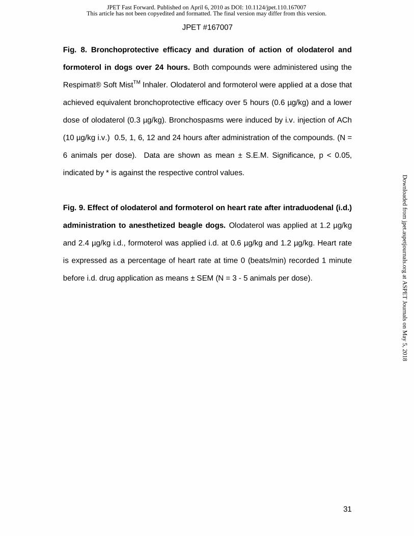
(Table 4). On basal tone preparations, olodaterol and formoterol potently relaxed the
bronchi with non-significant differences in potency and efficacy (Table 4, Figure 3A)
(two-tailed t test). Similarly, the potency and efficacy of olodaterol and formoterol
were not statistically different when histamine was used as a contractile agent (Table
4, Figure 3B). To mimic the cholinergic tone typical of COPD, ACh and electric field
stimulation (EFS; to induce neural-mediated release of ACh) were used, too. EFS-
induced contractions were potently inhibited in a concentration-dependent manner by
olodaterol (pIC50 = 9.49) and formoterol (pIC50 = 9.73) (Table 4, Figure 3D), with
formoterol causing a slightly higher maximal inhibition of EFS-induced contraction
(97%) compared to BI 1744 CL (86%).
Conversely, pre-contraction with ACh (100 µM) decreased the potencies and
maximal relaxant effects of BI 1744 CL and formoterol (p<0.05), with no significant
difference between the two ß2 adrenoceptor agonists (Table 4, Figure 3D).
In vivo profile of olodaterol
The in vivo efficacy and systemic pharmacodynamic profile of olodaterol and
formoterol were determined in bronchoconstriction models in guinea pigs and dogs.
In these models, the compounds were applied intra-tracheally to anesthetized
animals using the Respimat® Soft MistTM Inhaler and bronchoconstriction was
induced by intravenous application of acetylcholine at various time-points after
administration of the compounds.
Dose Response in Guinea Pigs. After administration of different doses of each
compound, bronchoprotection, heart rate and blood pressure were recorded over a
This article has not been copyedited and formatted. The final version may differ from this version.JPET Fast Forward. Published on April 6, 2010 as DOI: 10.1124/jpet.110.167007
at ASPE
T Journals on M
ay 5, 2018jpet.aspetjournals.org
Dow
nloaded from

JPET #167007
16
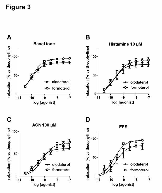
period of 5 hours. As shown in Figure 4A, olodaterol induced a dose-dependent
bronchoprotection when applied at doses from 0.1 µg/kg to 3 µg/kg. A full
bronchoprotection of 100% was achieved at the dose of 3 µg/kg. Olodaterol
demonstrated at all efficacious doses a bronchoprotection lasting over the entire
study period of 5 hours. For formoterol the maximal bronchoprotection of 100% was
achieved at a dose of 1 µg/kg and 3 µg/kg (Figure 4B). In contrast to olodaterol,
formoterol demonstrated an increased duration of action with increased doses. A
decline in bronchoprotection was observed after 30 minutes and 150 minutes at
doses of 0.3 µg/kg and 1 µg/kg, respectively. Formoterol retained a full
bronchoprotection over 5 hours at a dose of 3 µg/kg (Figure 4B). Both compounds
did not show an increase in heart rate and blood pressure over the entire study
period at all doses tested (data not shown).
Duration of Action in Guinea Pigs. To address the duration of action of olodaterol
and formoterol, both compounds were applied via intra-tracheal instillation to guinea
pigs and bronchoconstrictions were induced by increasing ACh doses from 2 µg/kg to
20 µg/kg after 6 or 24 hours, respectively. Olodaterol and formoterol were applied at
a dose that achieved equivalent bronchoprotective efficacy over 5 hours (3 µg/kg). In
addition, two lower doses of olodaterol (1 µg/kg and 0.1 µg/kg) were tested in this
setting. As shown in Figure 5A, olodaterol and formoterol administered at a dose of 3
µg/kg retained a strong efficacy after 6 hours. However, only olodaterol still protected
the animals against ACh-induced bronchospasms when administered at a dose of 3
µg/kg and a lower dose (1 µg/kg) after 24 hours. In contrast, formoterol applied at the
initially equal effective dose, retained no activity after 24 hours (Figure 5B).
Onset of Action in Guinea Pigs. The onset of action of olodaterol in comparison to
formoterol was determined in the guinea pig model described above. Both
compounds were administered at three different doses using the Respimat® Soft
This article has not been copyedited and formatted. The final version may differ from this version.JPET Fast Forward. Published on April 6, 2010 as DOI: 10.1124/jpet.110.167007
at ASPE
T Journals on M
ay 5, 2018jpet.aspetjournals.org
Dow
nloaded from

JPET #167007
17
MistTM Inhaler and bronchospasms were induced by ACh 1, 3, 5, 7 and 20 minutes
after drug inhalation. As shown in Figure 6A and 6B, both compounds exerted a rapid
onset of action and achieved a full bronchoprotection within 3-6 minutes after
inhalation.
Dose Response and Systemic Pharmacodynamic Effect Profile in Dogs. The
efficacy and duration of bronchoprotection induced by olodaterol was investigated in
a second species, namely anesthetized ventilated beagle dogs. Again, test
compounds were administered by inhalation using the Respimat® Soft MistTM Inhaler
and bronchoconstriction was induced by repeated intravenous injections of
acetylcholine at different time points after compound administration. This model was
also used to study the systemic pharmacodynamic effects of the compounds in
further detail, since beagle dogs are very sensitive to the cardiovascular (e.g. heart
rate increase) and metabolic (e.g. increase in serum potassium, glucose and lactate)
effects mediated by systemic stimulation of β-adrenoceptors (Greaves, 1998).
Olodaterol inhibited the ACh-induced bronchoconstriction in dogs in a dose-
dependent manner (Figure 7A). A maximal bronchoprotective effect of about 60%
was reached after 10 minutes at a dose of 0.3 µg/kg olodaterol. At this dose,
bronchoprotection was approximately 20% after 3 hours. At the inhaled dose of 0.6
µg/kg olodaterol exerted the same maximal efficacy but maintained a
bronchoprotection of about 40% after 3 hours. At the highest dose tested (1.2 µg/kg),
a profile comparable to the 0.6 µg/kg dose was observed (data not shown). However,
at this dose cardiovascular effects (e.g. increase in heart rate above 50%, see Figure
7B) were observed. Administration of olodaterol did not result in changes in serum
potassium (Figure 7C), or serum lactate (Figure 7D) and serum glucose (data not
shown) at any dose tested. From this experiment, the maximum-effective dose of
olodaterol in the dog was determined as 0.6 µg/kg. The maximum-effective dose of
This article has not been copyedited and formatted. The final version may differ from this version.JPET Fast Forward. Published on April 6, 2010 as DOI: 10.1124/jpet.110.167007
at ASPE
T Journals on M
ay 5, 2018jpet.aspetjournals.org
Dow
nloaded from

JPET #167007
18
formoterol was determined as 0.6 µg/kg, too (see Figure 7A). At this dose, formoterol
showed -compared to olodaterol- a more pronounced and longer lasting tachycardia
(Figure 7B). In addition and in contrast to olodaterol a long lasting decrease in serum
potassium levels (Figure 7C) and a significant increase in serum lactate (Figure 7D)
was observed for formoterol at both doses used. Formoterol was devoid of effects on
serum glucose (data not shown).
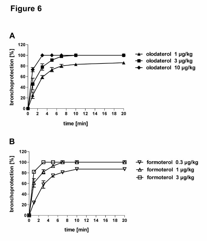
Duration of Action in Dogs. The bronchoprotection of olodaterol and formoterol was
determined 0.1, 0.5, 6, 12 and 24 hours after inhalation of a single dose of each
compound. Both compounds were administered at the respective maximum-effective
dose (0.6 µg/kg). Olodaterol was also used at a 2-fold lower dose. As shown in
Figure 8, olodaterol (0.6 µg/kg) induced a maximal bronchoprotection of about 60%
after 0.5 hour, consistent with the 3 hour study described above. 24 hours after
administration, animals treated with 0.6 µg/kg olodaterol retained a bronchoprotection
of approximately 20%. In contrast, formoterol tested at its maximum-effective dose
was completely inactive after 12 hours (Figure 8). When administered at a 2-fold
lower dose olodaterol exerted no bronchoprotection after 12 hours. The two doses of
olodaterol tested were devoid of heart rate effects and metabolic effects (data not
shown).
Systemic Pharmacodynamic Effects of Olodaterol after intra-duodenal
Administration. In man a significant proportion of the dose inhaled via the
Respimat® Soft MistTM Inhaler is swallowed (Dalby, et al., 2004). In the animal
experiments described above, swallowing did not occur, since the compounds were
applied either by intra-tracheal (i.t.) administration or by the Respimat® Soft MistTM
inhaler connected to the endotracheal tube. To mimic the systemic
pharmacodynamic effects after complete swallowing of the entire dose, olodaterol
This article has not been copyedited and formatted. The final version may differ from this version.JPET Fast Forward. Published on April 6, 2010 as DOI: 10.1124/jpet.110.167007
at ASPE
T Journals on M
ay 5, 2018jpet.aspetjournals.org
Dow
nloaded from

JPET #167007
19
was applied intraduodenally (i.d.) to anesthetized dogs at 1.2 µg/kg and 2.4 µg/kg
corresponding to doses 2-fold and 4-fold above its maximum-effective dose.
Cardiovascular and metabolic parameters were recorded over a period of 3 hours.
For comparison, formoterol was applied in the same experimental setting at its
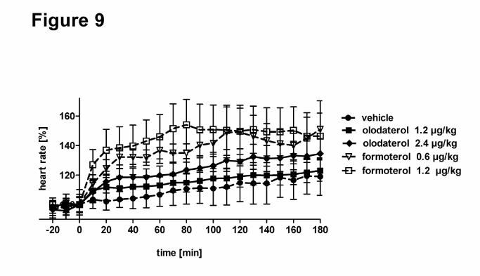
maximum-effective dose (0.6 µg/kg) and 2-fold above (1.2 µg/kg). As shown in Figure
9, i.d. administration of olodaterol induced only a small increase in heart rate of
maximally 10% and 20% when given 2-fold and 4-fold above the maximum-effective
dose, respectively. Systolic and diastolic blood pressure were decreased by
maximally 10% up to 40 minutes post olodaterol administration and returned to
normal (data not shown). Formoterol administered i.d. at its maximum-effective dose
and 2-fold above induced, compared to olodaterol, more pronounced and stronger
dose-dependent effects on heart rate (Figure 9) and systolic blood pressure (data not
shown). Blood pressure was initially decreased up to 25% with the 2-fold fully
effective dose. This decrease persisted for diastolic blood pressure, while systolic
blood pressure increased by about 10% starting at about 40 min post administration.
This article has not been copyedited and formatted. The final version may differ from this version.JPET Fast Forward. Published on April 6, 2010 as DOI: 10.1124/jpet.110.167007
at ASPE
T Journals on M
ay 5, 2018jpet.aspetjournals.org
Dow
nloaded from

JPET #167007
20
Discussion
With chronic diseases such as COPD and asthma, patient adherence to medication
plans is a major obstacle to successful management (Bender, 2002). One factor
contributing to poor adherence is a complicated or a multiple treatment regimen, and
simplified dosing regimens are known to improve compliance (Bender, 2002).
Therefore, long duration of action (preferably 24 hours) is an important feature of
drugs intended to treat chronic diseases, enabling both prolonged efficacy and a
simple, once-daily dosing regime that improves patient compliance (Tamura and
Ohta, 2007). This strategy, which has been proven successful with the long acting
muscarinic antagonist (LAMA) tiotropium (Spiriva®) (Tashkin, et al., 2008), is
currently being pursued also within a second class of bronchodilators, namely the ß2
adrenoceptor agonists (Cazzola and Matera, 2008).
Here, we describe a comprehensive preclinical characterization of olodaterol
(previously known as BI 1744 CL), which was identified as part of a program aimed at
the discovery of selective ß2 adrenoceptor agonists with potential for once-daily
administration. In vitro data indicate that olodaterol possesses a high, subnanomolar
affinity for the hß2-AR and an excellent selectivity against the other adrenoceptor
subtypes. In line with the binding data, olodaterol was the most potent agonist for the
ß2-AR mediated stimulation of cAMP and exerted an excellent selectivity profile. In
the evaluation of new ß2 adrenoceptor agonists under development, their intrinsic
activity needs to be taken into consideration, as partial agonists may act as a ß2
antagonist in the presence of a full ß-agonist (Lipworth and Grove, 1997). In fact, a
partial ß adrenoceptor agonist exhibits opposite agonist and antagonist activity
depending on the prevailing degree of adrenergic tone or the presence of a ß
adrenoceptor agonist with higher intrinsic activity (e.g. rescue therapies). To this end,
we took particular care in testing the functional response of olodaterol in a cell line
This article has not been copyedited and formatted. The final version may differ from this version.JPET Fast Forward. Published on April 6, 2010 as DOI: 10.1124/jpet.110.167007
at ASPE
T Journals on M
ay 5, 2018jpet.aspetjournals.org
Dow
nloaded from

JPET #167007
21
with moderate levels of ß2-AR expression (Table 1), similar to airway SMCs
(expression levels reported to be 100 fmol/mg, (Mak, et al., 1994) to avoid an
overestimation of the agonist efficacy, as it is known for systems with high receptor
spare numbers (Kenakin, 2004). In this setting, olodaterol offered the profile of an
almost full agonist, with an intrinsic activity of 88%. These results were further
translated into a more physiologically relevant model, i.e. human lung parenchyma.
Here, olodaterol dose-dependently reversed the constriction induced by different
stimuli, like histamine, ACh and EFS, with an efficacy not statistically different from
the full agonist formoterol under all conditions. Taken together, the in vitro data
indicate that olodaterol, similarly to formoterol and salmeterol, shows high selectivity
for the hß2-AR in terms of affinity and potency. However, differently from the currently
marketed ß2 adrenoceptor agonists, olodaterol has a differential efficacy profile
towards the different ß-ARs, with a full agonist-like profile on the hß2-AR and a partial
agonism against the hß1-AR, whereas formoterol and salmeterol exert either a full-
agonistic or a partial agonistic profile for all ß-ARs, respectively. This profile could
translate in an efficacious bronchodilatory effect with reduced cardiovascular side
effects.
In order to obtain information regarding the functional in vivo bronchoprotective
profile, taking into account both pharmacodynamic and pharmacokinetic properties,
olodaterol was tested in pharmacological models of acetylcholine (ACh)-induced
bronchoconstriction in anaesthetized guinea pigs and dogs. ACh-induced
bronchoconstriction models are widely used to test the in vivo efficacy, potency, and
duration of action of bronchodilators -such as ß-agonists and anticholinergics- and
are a good predictor for the efficacy of compounds in human airway diseases such as
COPD, since an increase in the vagal cholinergic tone is discussed as the major
reversible component in COPD (Barnes, 2004).
This article has not been copyedited and formatted. The final version may differ from this version.JPET Fast Forward. Published on April 6, 2010 as DOI: 10.1124/jpet.110.167007
at ASPE
T Journals on M
ay 5, 2018jpet.aspetjournals.org
Dow
nloaded from

JPET #167007
22
To mimic the clinical situation further, the Respimat® Soft MistTM Inhaler was used for
the administration of olodaterol, and for better comparision, for formoterol, too. The
Respimat® Inhaler is a novel device which creates a soft mist aerosol without the use
of propellants. In our in vivo studies the drugs were provided in water ethanol (40/60,
v/v) solutions dissolved at concentrations permitting the administration of the desired
dose with three actuations.
In both models, olodaterol provided bronchoprotection over 24 hours, whereas
formoterol applied at an equally effective dose did not retain efficacy over 24 hours. It
is noteworthy to mention that according to our observations the bronchoprotection
mediated by ß2 adrenoceptor agonists in the ACh-induced bronchoconstriction model
in dogs is significantly less efficacious than in guinea pigs. Whereas most ß2-
adrenocpeptor agonists studied during our research project easily exerted a 100%
bronchoprotection in guinea pigs we did not identify a ß adrenoceptor agonist
capable of a 100% bronchoprotection in the dog model. The reason behind this
discrepancy is not understood by us, but the different sensitivities of the two models
may explain why in the dog model -in constrast to guinea pigs- olodaterol showed
only at its maximum-effective dose a duration of action over 24 hours.
Formoterol is known as a ß2 adrenoceptor agonist with a fast onset of action in
humans and has gained recognition as a p.r.n. controller therapy because of this fast
onset of action. Most interestingly, in the two species studied, also olodaterol offered
a quick onset of action, similar to formoterol. The maximal bronchoprotection after
inhalation of a single dose of olodaterol or formoterol was reached in guinea pigs
within 3-6 minutes and 10 minutes in dogs, suggesting that olodaterol may have an
rapid onset of action in humans, too.
This article has not been copyedited and formatted. The final version may differ from this version.JPET Fast Forward. Published on April 6, 2010 as DOI: 10.1124/jpet.110.167007
at ASPE
T Journals on M
ay 5, 2018jpet.aspetjournals.org
Dow
nloaded from

JPET #167007
23
Since beagle dogs are very sensitive to the cardiovascular (e.g. heart rate increase)
and metabolic (e.g. increase in serum potassium, glucose and lactate) effects
mediated by the systemic stimulation of ß adrenoceptors (Greaves, 2000), the
systemic pharmacodynamic effects of olodaterol were studied in this species, too. In
the first experimental setting we determined the systemic effects after inhaled
(intratracheal) administration of the compound in the same animals used for for the
efficacy studies. In the second setting we applied the compounds by intra-duodenal
administration to mimic swallowing of the entire dose. Our preclinical data obtained in
the two settings show that, for a given degree of bronchodilator activity, olodaterol
has a greater cardiovascular (as assessed by heart rate) and metabolic (as assessed
serum potassium, serum, glucose and serum lactate) safety margin than formoterol.
Furthermore, in the two preclinical species analyzed, olodaterol was devoid of
systemic pharmacodynamic effects at doses achieving a duration of action of 24
hours, suggesting a sufficient therapeutic window for its use in man. Since the
sytemic effects of beta agonists on serum potassium, serum lactate, or serum
glucose are caused by the activitation of ß2 adrenoceptors in skeletal muscle and
liver, we speculate that the larger safety margin we observed for olodaterol in
comparison to formoterol in the dog model reflects differences in the pharmacokinetic
profile and thus the systemic exposure of the two compounds.
The preclinical data presented here were confirmed in clinical studies both in asthma
(O´Byrne, 2008) and COPD (van Noord, 2008, van Noord, 2009) patients. In all
studies olodaterol showed a 24-hour duration of action after once-a day dosing
concomitant with a good safety profile. The 24-hour bronchodilator efficacy of once
daily dosing with olodaterol in patients with COPD was confirmed in a 4 week study,
with all doses of olodaterol showing statistically significant increases in the primary
endpoint, trough FEV1, compared to placebo after 28 days of treatment, again with
This article has not been copyedited and formatted. The final version may differ from this version.JPET Fast Forward. Published on April 6, 2010 as DOI: 10.1124/jpet.110.167007
at ASPE
T Journals on M
ay 5, 2018jpet.aspetjournals.org
Dow
nloaded from

JPET #167007
24
an excellent safety profile (van Noord, 2009). Furthermore, in the 4 week study no
differences in the FEV1 profile after the first dose (day 1) and after 4 weeks treatment
(day 29) were observed, implying the absence of clinical desensitization noted in
some clinical studies after regular use of ß2 adrenoceptor agonists (Larj and Bleecker,
2002).
Therefore, a once-daily ß2 adrenoceptor agonist, like olodaterol, offers -compared
with short-acting bronchodilators and b.i.d. LABAs - an improved convenience and
compliance for asthma and COPD patients and has the potential to be combined with
either a once-daily anticholinergic, like tiotropium (Tashkin, et al., 2008;Casarosa, et
al., 2009) or upcoming new compounds within this class (Cazzola and Matera, 2008),
once daily corticosteroids, or both, presented to the patients either as free or fixed-
dose combinations. Besides the improved convenience, these combinations may
offer beneficial long-term outcomes for the patients.
In summary, our preclinical data demonstate that olodaterol is an enantiomeric pure,
selective and potent agonist of the human ß2 adrenoceptor. This molecule combines
a novel efficacy profile towards the different ß-ARs - by exerting almost full intrinsic
activity at ß2-AR and a weak partial agonism at ß1-AR - together with a long duration
of action, allowing a once-daily administration in humans, a rapid onset of action and
an improved systemic pharmacodynamic effect profile.
This article has not been copyedited and formatted. The final version may differ from this version.JPET Fast Forward. Published on April 6, 2010 as DOI: 10.1124/jpet.110.167007
at ASPE
T Journals on M
ay 5, 2018jpet.aspetjournals.org
Dow
nloaded from

JPET #167007
25
References
Barnes PJ (2004) The role of anticholinergics in chronic obstructive pulmonary
disease
2. Am J Med 117 Suppl 12A:24S-32S.
Bateman ED, Hurd SS, Barnes PJ, Bousquet J, Drazen JM, FitzGerald M, Gibson P,
Ohta K, O'Byrne P, Pedersen SE, Pizzichini E, Sullivan SD, Wenzel SE and Zar HJ
(2008) Global strategy for asthma management and prevention: GINA executive
summary. Eur Respir J 31:143-178.
Bender BG (2002) Overcoming barriers to nonadherence in asthma treatment. J
Allergy Clin Immunol 109:S554-S559.
Braman SS (2006) The global burden of asthma. Chest 130:4S-12S.
Casarosa P, Bouyssou T, Germeyer S, Schnapp A, Gantner F and Pieper M (2009)
Preclinical evaluation of long-acting muscarinic antagonists: comparison of tiotropium
and investigational drugs. J Pharmacol Exp Ther 330:660-668.
Casarosa P, Waldhoer M, LiWang PJ, Vischer HF, Kledal T, Timmerman H,
Schwartz TW, Smit MJ and Leurs R (2005) CC and CX3C chemokines differentially
interact with the N terminus of the human cytomegalovirus-encoded US28 receptor
3. J Biol Chem 280:3275-3285.
Cazzola M and Matera MG (2008) Novel long-acting bronchodilators for COPD and
asthma. Br J Pharmacol 155:291-299.
This article has not been copyedited and formatted. The final version may differ from this version.JPET Fast Forward. Published on April 6, 2010 as DOI: 10.1124/jpet.110.167007
at ASPE
T Journals on M
ay 5, 2018jpet.aspetjournals.org
Dow
nloaded from

JPET #167007
26
Cheng Y and Prusoff WH (1973) Relationship between the inhibition constant (K1)
and the concentration of inhibitor which causes 50 per cent inhibition (I50) of an
enzymatic reaction. Biochem Pharmacol 22:3099-3108.
Dalby R, Spallek M and Voshaar T (2004) A review of the development of Respimat
Soft Mist Inhaler. Int J Pharm 283:1-9.
Gold PM (2009) The 2007 GOLD Guidelines: a comprehensive care framework.
Respir Care 54:1040-1049.
Greaves P (1998) Patterns of drug-induced cardiovascular pathology in the beagle
dog: relevance for humans. Exp Toxicol Pathol 50:283-293.
Greaves P (2000) Patterns of cardiovascular pathology induced by diverse
cardioactive drugs. Toxicol Lett 112-113:547-552.
Guerra S (2009) Asthma and chronic obstructive pulmonary disease. Curr Opin
Allergy Clin Immunol 9:409-416.
Jenkins CR, Jones PW, Calverley PM, Celli B, Anderson JA, Ferguson GT, Yates JC,
Willits LR and Vestbo J (2009) Efficacy of salmeterol/fluticasone propionate by GOLD
stage of chronic obstructive pulmonary disease: analysis from the randomised,
placebo-controlled TORCH study. Respir Res 10:59.
Kenakin T (2004) Principles: receptor theory in pharmacology. Trends Pharmacol Sci
25:186-192.
Larj MJ and Bleecker ER (2002) Effects of beta2-agonists on airway tone and
bronchial responsiveness. J Allergy Clin Immunol 110:S304-S312.
This article has not been copyedited and formatted. The final version may differ from this version.JPET Fast Forward. Published on April 6, 2010 as DOI: 10.1124/jpet.110.167007
at ASPE
T Journals on M
ay 5, 2018jpet.aspetjournals.org
Dow
nloaded from

JPET #167007
27
Lipworth BJ and Grove A (1997) Evaluation of partial beta-adrenoceptor agonist
activity. Br J Clin Pharmacol 43:9-14.
Mak JC, Grandordy B and Barnes PJ (1994) High affinity [3H]formoterol binding sites
in lung: characterization and autoradiographic mapping. Eur J Pharmacol 269:35-41.
Naline E, Zhang Y, Qian Y, Mairon N, Anderson GP, Grandordy B and Advenier C
(1994) Relaxant effects and durations of action of formoterol and salmeterol on the
isolated human bronchus. Eur Respir J 7:914-920.
Naline E, Trifilieff A, Fairhurst RA, Advenier C and Molimard C (2007) Effect of
indacaterol, a novel long-acting ß2-agonist, on isolated human bronchi. Eur Respir J
29:575-581.
O´Byrne P, Van der Linde J, Boulet L-P, Cockroft D, Fitzgerald M, Hart L, Korducki L,
Brannan JD, and Hamilton A (2008) Single doses of BI 1744 CL, a novel LABA, are
effective for up to 32 hours in asthmatic patients. Am J Respir Crit Care Med 177:
A932
Pauwels RA and Rabe KF (2004) Burden and clinical features of chronic obstructive
pulmonary disease (COPD). Lancet 364:613-620.
Sovani MP, Whale CI and Tattersfield AE (2004) A benefit-risk assessment of inhaled
long-acting beta2-agonists in the management of obstructive pulmonary disease.
Drug Saf 27:689-715.
Tamura G and Ohta K (2007) Adherence to treatment by patients with asthma or
COPD: comparison between inhaled drugs and transdermal patch. Respir Med
101:1895-1902.
This article has not been copyedited and formatted. The final version may differ from this version.JPET Fast Forward. Published on April 6, 2010 as DOI: 10.1124/jpet.110.167007
at ASPE
T Journals on M
ay 5, 2018jpet.aspetjournals.org
Dow
nloaded from

JPET #167007
28
Tashkin DP, Celli B, Senn S, Burkhart D, Kesten S, Menjoge S and Decramer M
(2008) A 4-year trial of tiotropium in chronic obstructive pulmonary disease
7. N Engl J Med 359:1543-1554.
Van Noord JA, Smeets JJ, Drenth BM, Pivovarova A, Hamilton AL and Cornelissen
PJG (2008) Single doses of BI 1744 CL, a novel β2-agonist, are effective for up to 24
hrs in COPD patients. Am J Respir Crit Care Med 177: A961.
Van Noord JA, Korducki L, Hamilton AL and Koker P (2009) Four weeks once daily
treatment with BI 1744 CL, a novel long-acting β2-agonist, is effective in COPD
patients. Am J Respir Crit Care Med 179: A6183.
Walland A, Palluk R, Burkard S and Hammer R (1997) Compensation of muscarinic
bronchial effects of talsaclidine by concomitant sympathetic activation in guinea pigs.
Eur J Pharmacol 330:213-219.
Ying Y, Leung KM, Berkbigler D, Halbert RJ, and Legoretta AP (1999) Compliance
with US asthma management guidelines and specialty care: a regional variation or
national concern? J Eval Clin Pract 5: 213-221
This article has not been copyedited and formatted. The final version may differ from this version.JPET Fast Forward. Published on April 6, 2010 as DOI: 10.1124/jpet.110.167007
at ASPE
T Journals on M
ay 5, 2018jpet.aspetjournals.org
Dow
nloaded from

JPET #167007
29
Legends for Figures
Fig. 1. Chemical structures of olodaterol (A), formoterol (B) and salmeterol (C).
Fig. 2. Functional selectivity of ß2-adrenoceptor agonists against the different
ß-AR subtypes. A, B, C: CHO cells selectively expressing the hß1 (A), hß2 (B) or
hß3 (C) adrenoceptors were stimulated with increasing concentrations of agonists
and cAMP levels were quantified. Data are presented as percentage of maximal
isoprenaline-induced cAMP accumulation and the mean of three independent
experiments (± S.E.M.) is shown.
Fig. 3. Effect of olodaterol and formoterol on isolated human bronchi. A) at
resting tone and on bronchi pre-contracted with B) 10 µM histamine (51%
acetylcholine (ACh) maximum (max)), C) with 0.1 mM ACh (80% ACh max). and D)
electrical field stimulaton (EFS). Data are shown as % of theophylline-induced
relaxation and are expressed as mean + S.E.M of a number of experiments indicated
in table 4 for each condition.
Fig. 4. Bronchoprotective efficacy of olodaterol and formoterol in guinea pigs.
The bronchoprotection of olodaterol (A) and formoterol (B) was determined in a
model of ACh-induced bronchoconstriction in anaesthetized guinea pigs.
Submaximal bronchospasms were induced by i.v. injections of ACh (10 - 12 µg/kg)
every 10 minutes. Bronchoprotection is expressed as the percentage of inhibition of
the increase in pulmonary resistance induced by ACh. Both compounds were
administered with the Respimat® Soft MistTM Inhaler. The time course of the
bronchoprotection was recorded for 5 hours (N = 2 animals per dose).
This article has not been copyedited and formatted. The final version may differ from this version.JPET Fast Forward. Published on April 6, 2010 as DOI: 10.1124/jpet.110.167007
at ASPE
T Journals on M
ay 5, 2018jpet.aspetjournals.org
Dow
nloaded from

JPET #167007
30
Fig. 5. Duration of action of olodaterol and formoterol in guinea pigs. The
bronchoprotective efficacy of olodaterol and formoterol was determined 6 hours (A)
and 24 hours (B) after intra-tracheal instillation. Bronchospasms were induced by
increasing ACh doses from 2 µg/kg to 20 µg/kg with a progression of 2 µg/kg per i.v.
injection. N = 5–6 animals per dose. Data are shown as mean ± S.E.M. Significance,
p < 0.05, indicated by * is against the respective control values.
Fig. 6. Onset of action of olodaterol and formoterol in guinea pigs. Olodaterol
(A) or formoterol (B) were administered with the Respimat® Soft MistTM Inhaler at
three different doses. Bronchospasms were induced by ACh (10 µg/kg) 1, 3, 5, 7 and
20 minutes after drug inhalation. Bronchoprotection is expressed as the percentage
of inhibition of the increase in pulmonary resistance induced by ACh. (N = 6 animals
per dose).
Fig. 7. Bronchoprotective efficacy and systemic pharmacodynamic effects of
olodaterol and formoterol in dogs over 3 hours. A, B, C, D: Both compounds
were administered to anaesthetized ventilated dogs at doses of 0.15 µg/kg, 0.3 µg/kg,
0.6 µg/kg and 1.2 µg/kg using the Respimat® Soft MistTM Inhaler. Bronchospasms
were induced by i.v. injection of ACh (10 µg/kg i.v.) after 10, 30, 60, 120 and 180
minutes. Bronchoprotection (A) is expressed as the percentage of inhibition of the
increase in pulmonary resistance induced by ACh. Systemic pharmacodynamic
effects including heart rate (B), serum potassium (C) and serum lactate (D) were
recorded at the same time points. (N = 6 animals per dose).
This article has not been copyedited and formatted. The final version may differ from this version.JPET Fast Forward. Published on April 6, 2010 as DOI: 10.1124/jpet.110.167007
at ASPE
T Journals on M
ay 5, 2018jpet.aspetjournals.org
Dow
nloaded from
JPET #167007
31
Fig. 8. Bronchoprotective efficacy and duration of action of olodaterol and
formoterol in dogs over 24 hours. Both compounds were administered using the
Respimat® Soft MistTM Inhaler. Olodaterol and formoterol were applied at a dose that
achieved equivalent bronchoprotective efficacy over 5 hours (0.6 µg/kg) and a lower
dose of olodaterol (0.3 µg/kg). Bronchospasms were induced by i.v. injection of ACh
(10 µg/kg i.v.) 0.5, 1, 6, 12 and 24 hours after administration of the compounds. (N =
6 animals per dose). Data are shown as mean ± S.E.M. Significance, p < 0.05,
indicated by * is against the respective control values.
Fig. 9. Effect of olodaterol and formoterol on heart rate after intraduodenal (i.d.)
administration to anesthetized beagle dogs. Olodaterol was applied at 1.2 µg/kg
and 2.4 µg/kg i.d., formoterol was applied i.d. at 0.6 µg/kg and 1.2 µg/kg. Heart rate
is expressed as a percentage of heart rate at time 0 (beats/min) recorded 1 minute
before i.d. drug application as means ± SEM (N = 3 - 5 animals per dose).
This article has not been copyedited and formatted. The final version may differ from this version.JPET Fast Forward. Published on April 6, 2010 as DOI: 10.1124/jpet.110.167007
at ASPE
T Journals on M
ay 5, 2018jpet.aspetjournals.org
Dow
nloaded from

JPET #167007
32
Table 1. Pharmacological characterization of the different CHO cell lines stably
expressing the human ß1-3 adrenoceptors. Levels of receptor expression
(expressed in pmol/mg of protein content in membrane preparations) were
determined in saturation binding assays, as described in the material and methods
section. The average of at least three independent experiments performed in
triplicate is shown.
Cell line Bmax
(pmol/mg) Use
CHO-hß1-HIGH 12.4 ± 0.3
Binding experiments CHO-hß2-HIGH 1.6 ± 0.1
CHO-hß3-HIGH 22.0 ± 1.6
CHO-hß1-LOW 0.52 ± 0.04 Functional experiments
(cAMP) CHO-hß2-LOW 0.056 ± 0.009
CHO-hß3-LOW 0.23 ± 0.06
This article has not been copyedited and formatted. The final version may differ from this version.JPET Fast Forward. Published on April 6, 2010 as DOI: 10.1124/jpet.110.167007
at ASPE
T Journals on M
ay 5, 2018jpet.aspetjournals.org
Dow
nloaded from

JPET #167007
33
Table 2. Binding affinities of different ß2-AR agonists against the three human
ß-adrenoceptor subtypes. The equilibrium dissociation constants (pKi) of the
different ligands were determined in heterologous competition experiments against
[3H]-CGP 12,177 in the presence of a GTP analog. The pKi values (± SEM) shown
are the average of at least three independent experiments performed in triplicate.
Agonist pKi hß1 pKi hß2 pKi hß3 Ratio
ß1/ß2
Ratio
ß3/ß2
Isoprenaline 6.49 ± 0.01 6.54 ± 0.03 5.57 ± 0.07 1 9
Olodaterol 7.33 ± 0.05 9.14 ± 0.04 5.26 ± 0.14 65 7586
Formoterol 6.07 ± 0.04 8.29 ± 0.03 5.58 ± 0.12 166 513
Salmeterol 6.14 ± 0.02 9.24 ± 0.08 5.43 ± 0.01 1259 6457
This article has not been copyedited and formatted. The final version may differ from this version.JPET Fast Forward. Published on April 6, 2010 as DOI: 10.1124/jpet.110.167007
at ASPE
T Journals on M
ay 5, 2018jpet.aspetjournals.org
Dow
nloaded from

JPET #167007
34
Table 3. Functional properties of different ß2-AR agonists against the three
human ß-adrenoceptor subtypes. Potency (pEC50 values) and intrinsic activity (I.A.,
expressed as percentage of isoprenaline-induced maximal response) of the different
ß2-adrenoceptor agonists were determined in CHO cell lines selectively expressing
the ß-AR subtype of interest, with the cAMP accumulation assay. Values shown are
the average (± SEM) of at least 3 independent experiments, with each point
determined in triplicate. The statistical significance of differences in intrinsic activity
among the agonists for each receptor subtype were determined using one-way
analysis of variance with Dunnett's post test for multiple comparisons. Statistical
significance is denoted compared with the reference agonist isoprenaline (*: p < 0.05).
hß1 hß2 hß3 Ratio
ß1/ß2
Ratio
ß3/ß2 pEC50 I.A. pEC50 I.A. pEC50 I.A.
Isoprenaline 9.27 ± 0.08 100% 8.58 ± 0.08 100% 7.86 ± 0.07 100% 0.004 10
Olodaterol 7.55 ± 0.08 52 ± 8 % * 9.93 ± 0.07 88 ± 2% 6.57 ± 0.08 81 ± 2% * 240 1698
Formoterol 7.83 ± 0.06 91 ± 4% 9.73 ± 0.10 97 ± 3% 7.60 ± 0.07 100 ± 2% 87 155
Salmeterol 6.08 ± 0.07 40 ± 6% * 9.90 ± 0.04 54 ± 7% * 6.15 ± 0.17 56 ± 3% * 6607 5623
*: p < 0.05
This article has not been copyedited and formatted. The final version may differ from this version.JPET Fast Forward. Published on April 6, 2010 as DOI: 10.1124/jpet.110.167007
at ASPE
T Journals on M
ay 5, 2018jpet.aspetjournals.org
Dow
nloaded from

JPET #167007
35
Table 4. Potency and maximal efficacy (Emax) of olodaterol and formoterol on
human isolated bronchi at resting tone or after a pre-contraction with 10 µM
histamine or 0.1 mM acetylcholine. Potency values (pEC50, mean ± S.E.M.) represent
the average of n independent experiments. Maximal efficacy (Emax, mean ± S.E.M.) is
reported as percentage of the maximal effect of theophilline. The maximal efficacies
obtained with either olodaterol or formoterol in the presence of the different stimuli
were analyzed by using one-way ANOVA followed by Dunnet's multiple-comparison
test. Statistical significance is denoted compared versus resting tone (*: p < 0.05).
Stimulus:
Olodaterol Formoterol
pEC50 Emax (%) EC50 Emax (%)
Resting tone 9.97±0.10 (11) 87±3 9.96±0.10 (10) 96±2
Histamine [10 µM] 9.70±0.13 (7) 80±6 9.49±0.20 (6) 92±7
ACh[100 µM] 9.44±0.08 (13) 65±4* 9.13±0.10 (11) 74±5*
EFS 9.49±0.24 (5) 86±5 9.73±0.13 (6) 97±1
This article has not been copyedited and formatted. The final version may differ from this version.JPET Fast Forward. Published on April 6, 2010 as DOI: 10.1124/jpet.110.167007
at ASPE
T Journals on M
ay 5, 2018jpet.aspetjournals.org
Dow
nloaded from

This article has not been copyedited and formatted. The final version may differ from this version.JPET Fast Forward. Published on April 6, 2010 as DOI: 10.1124/jpet.110.167007
at ASPE
T Journals on M
ay 5, 2018jpet.aspetjournals.org
Dow
nloaded from

This article has not been copyedited and formatted. The final version may differ from this version.JPET Fast Forward. Published on April 6, 2010 as DOI: 10.1124/jpet.110.167007
at ASPE
T Journals on M
ay 5, 2018jpet.aspetjournals.org
Dow
nloaded from
This article has not been copyedited and formatted. The final version may differ from this version.JPET Fast Forward. Published on April 6, 2010 as DOI: 10.1124/jpet.110.167007
at ASPE
T Journals on M
ay 5, 2018jpet.aspetjournals.org
Dow
nloaded from

This article has not been copyedited and formatted. The final version may differ from this version.JPET Fast Forward. Published on April 6, 2010 as DOI: 10.1124/jpet.110.167007
at ASPE
T Journals on M
ay 5, 2018jpet.aspetjournals.org
Dow
nloaded from

This article has not been copyedited and formatted. The final version may differ from this version.JPET Fast Forward. Published on April 6, 2010 as DOI: 10.1124/jpet.110.167007
at ASPE
T Journals on M
ay 5, 2018jpet.aspetjournals.org
Dow
nloaded from
This article has not been copyedited and formatted. The final version may differ from this version.JPET Fast Forward. Published on April 6, 2010 as DOI: 10.1124/jpet.110.167007
at ASPE
T Journals on M
ay 5, 2018jpet.aspetjournals.org
Dow
nloaded from

This article has not been copyedited and formatted. The final version may differ from this version.JPET Fast Forward. Published on April 6, 2010 as DOI: 10.1124/jpet.110.167007
at ASPE
T Journals on M
ay 5, 2018jpet.aspetjournals.org
Dow
nloaded from
This article has not been copyedited and formatted. The final version may differ from this version.JPET Fast Forward. Published on April 6, 2010 as DOI: 10.1124/jpet.110.167007
at ASPE
T Journals on M
ay 5, 2018jpet.aspetjournals.org
Dow
nloaded from
This article has not been copyedited and formatted. The final version may differ from this version.JPET Fast Forward. Published on April 6, 2010 as DOI: 10.1124/jpet.110.167007
at ASPE
T Journals on M
ay 5, 2018jpet.aspetjournals.org
Dow
nloaded from