human vegf immunoassay quantikine · human vegf immunoassay ... principle of the assay ... • this...
TRANSCRIPT

Human VEGF Immunoassay
Quantikine® ELISA
This package insert must be read in its entirety before using this product. For research use only. Not for use in diagnostic procedures.
Catalog Number DVE00 Catalog Number SVE00 Catalog Number PDVE00
For the quantitative determination of human Vascular Endothelial Growth Factor (VEGF) concentrations in cell culture supernates, serum, and plasma.

MANUFACTURED AND DISTRIBUTED BY:
USA & Canada | R&D Systems, Inc. 614 McKinley Place NE, Minneapolis, MN 55413, USATEL: (800) 343-7475 (612) 379-2956 FAX: (612) 656-4400E-MAIL: [email protected]
DISTRIBUTED BY:
UK & Europe | R&D Systems Europe, Ltd.19 Barton Lane, Abingdon Science Park, Abingdon OX14 3NB, UKTEL: +44 (0)1235 529449 FAX: +44 (0)1235 533420E-MAIL: [email protected]
China | R&D Systems China Co., Ltd.24A1 Hua Min Empire Plaza, 726 West Yan An Road, Shanghai PRC 200050TEL: +86 (21) 52380373 FAX: +86 (21) 52371001E-MAIL: [email protected]
TABLE OF CONTENTS
SECTION PAGE
INTRODUCTION ....................................................................................................................................................................1
PRINCIPLE OF THE ASSAY ..................................................................................................................................................2
LIMITATIONS OF THE PROCEDURE ................................................................................................................................2
TECHNICAL HINTS ................................................................................................................................................................2
MATERIALS PROVIDED & STORAGE CONDITIONS ..................................................................................................3
OTHER SUPPLIES REQUIRED ............................................................................................................................................4
PRECAUTIONS ........................................................................................................................................................................4
SAMPLE COLLECTION & STORAGE ................................................................................................................................4
REAGENT PREPARATION ....................................................................................................................................................5
ASSAY PROCEDURE ............................................................................................................................................................6
CALCULATION OF RESULTS ..............................................................................................................................................7
TYPICAL DATA ........................................................................................................................................................................7
PRECISION ...............................................................................................................................................................................8
RECOVERY................................................................................................................................................................................8
SENSITIVITY ............................................................................................................................................................................8
LINEARITY ................................................................................................................................................................................9
CALIBRATION .........................................................................................................................................................................9
SAMPLE VALUES ................................................................................................................................................................. 10
SPECIFICITY .......................................................................................................................................................................... 11
REFERENCES ........................................................................................................................................................................ 12
PLATE LAYOUT .................................................................................................................................................................... 13

www.RnDSystems.com 1
INTRODUCTIONVascular endothelial growth factor (VEGF or VEGF-A), also known as vascular permeability factor (VPF), is a potent mediator of both angiogenesis and vasculogenesis in the fetus and adult (1-3). It is a member of the PDGF family that is characterized by the presence of eight conserved cysteine residues in a cystine knot structure and the formation of antiparallel disulfide-linked dimers (4). Humans express alternately spliced isoforms of 121, 145, 165, 183, 189, and 206 amino acids (aa) in length (4). VEGF165 appears to be the most abundant and potent isoform, followed by VEGF121 and VEGF189 (3, 4). Isoforms other than VEGF121 contain basic heparin-binding regions and are not freely diffusible (4). Human VEGF165 shares 88% aa sequence identity with corresponding regions of mouse and rat VEGF. VEGF is expressed in multiple cells and tissues including skeletal and cardiac muscle (5, 6), hepatocytes (7), osteoblasts (8), neutrophils (9), macrophages (10), keratinocytes (11), brown adipose tissue (12), CD34+ stem cells (13), endothelial cells (14), fibroblasts, and vascular smooth muscle cells (15). VEGF expression is induced by hypoxia and cytokines such as IL-1, IL-6, IL-8, oncostatin M, and TNF-α (3, 4, 9, 16). VEGF isoforms are differentially expressed during development and in the adult (3).
VEGF dimers bind to two related receptor tyrosine kinases, VEGF R1 (also called Flt-1) and VEGF R2 (Flk-1/KDR), and induce their homodimerization and autophosphorylation (3, 4, 7, 17, 18). These receptors have seven extracellular immunoglobulin-like domains and an intracellular split tyrosine kinase domain. They are expressed on vascular endothelial cells and a range of non-endothelial cells. Although VEGF affinity is highest for binding to VEGF R1, VEGF R2 appears to be the primary mediator of VEGF angiogenic activity (3, 4). VEGF165 also binds the semaphorin receptor, neuropilin-1, which promotes complex formation with VEGF R2 (19).
VEGF is best known for its role in vasculogenesis. During embryogenesis, VEGF regulates the proliferation, migration, and survival of endothelial cells (3, 4), thus regulating blood vessel density and size, but playing no role in determining vascular patterns. VEGF promotes bone formation through osteoblast and chondroblast recruitment and is also a monocyte chemoattractant (20-22). After birth, VEGF maintains endothelial cell integrity and is a potent mitogen for micro- and macro-vascular endothelial cells. In adults, VEGF functions mainly in wound healing and the female reproductive cycle (3). In diseased tissues, VEGF promotes vascular permeability. It is thus thought to contribute to tumor metastasis by promoting both extravasation and tumor angiogenesis (23, 24). Various strategies have been employed therapeutically to antagonize VEGF-mediated tumor angiogenesis (25). Circulating VEGF levels correlate with disease activity in autoimmune diseases such as rheumatoid arthritis, multiple sclerosis and systemic lupus erythematosus (26).
The Quantikine Human VEGF Immunoassay is a 4.5 hour solid phase ELISA designed to measure VEGF165 levels in cell culture supernates, serum, and plasma. It contains Sf 21-expressed recombinant human VEGF165 and antibodies raised against the recombinant protein. Results obtained for naturally occurring human VEGF and recombinant human VEGF121 showed linear curves that were parallel to the standard curves obtained using the Quantikine Human VEGF Immunoassay standards. These results indicate that this kit can be used to determine relative mass values for natural human VEGF.
For research use only. Not for use in diagnostic procedures.2
PRINCIPLE OF THE ASSAYThis assay employs the quantitative sandwich enzyme immunoassay technique. A monoclonal antibody specific for human VEGF has been pre-coated onto a microplate. Standards and samples are pipetted into the wells and any VEGF present is bound by the immobilized antibody. After washing away any unbound substances, an enzyme-linked polyclonal antibody specific for human VEGF is added to the wells. Following a wash to remove any unbound antibody-enzyme reagent, a substrate solution is added to the wells and color develops in proportion to the amount of VEGF bound in the initial step. The color development is stopped and the intensity of the color is measured.
LIMITATIONS OF THE PROCEDURE• FOR RESEARCH USE ONLY. NOT FOR USE IN DIAGNOSTIC PROCEDURES.
• The kit should not be used beyond the expiration date on the kit label.
• Do not mix or substitute reagents with those from other lots or sources.
• It is important that the Calibrator Diluent selected for the standard curve be consistent with the samples being assayed.
• If samples generate values higher than the highest standard, dilute the samples with the appropriate Calibrator Diluent and repeat the assay.
• Any variation in standard diluent, operator, pipetting technique, washing technique, incubation time or temperature, and kit age can cause variation in binding.
• Variations in sample collection, processing, and storage may cause sample value differences.
• This assay is designed to eliminate interference by other factors present in biological samples. Until all factors have been tested in the Quantikine Immunoassay, the possibility of interference cannot be excluded.
TECHNICAL HINTS• When mixing or reconstituting protein solutions, always avoid foaming.
• To avoid cross-contamination, change pipette tips between additions of each standard level, between sample additions, and between reagent additions. Also, use separate reservoirs for each reagent.
• To ensure accurate results, proper adhesion of plate sealers during incubation steps is necessary.
• When using an automated plate washer, adding a 30 second soak period following the addition of Wash Buffer, and/or rotating the plate 180 degrees between wash steps may improve assay precision.
• Substrate Solution should remain colorless until added to the plate. Keep Substrate Solution protected from light. Substrate Solution should change from colorless to gradations of blue.
• Stop Solution should be added to the plate in the same order as the Substrate Solution. The color developed in the wells will turn from blue to yellow upon addition of the Stop Solution. Wells that are green in color indicate that the Stop Solution has not mixed thoroughly with the Substrate Solution.

www.RnDSystems.com 3
MATERIALS PROVIDED & STORAGE CONDITIONSStore the unopened kit at 2-8 °C. Do not use past kit expiration date.
PART PART #CATALOG # DVE00
CATALOG # SVE00 DESCRIPTION
STORAGE OF OPENED/ RECONSTITUTED MATERIAL
Human VEGF Microplate
890218 1 plate 6 plates 96 well polystyrene microplate (12 strips of 8 wells) coated with a monoclonal antibody specific for human VEGF.
Return unused wells to the foil pouch containing the desiccant pack. Reseal along entire edge of zip-seal. May be stored for up to 1 month at 2-8 °C.*
Human VEGF Standard
890220 2 vials 12 vials Recombinant VEGF165 in a buffered protein base with preservatives; lyophilized. Refer to the vial label for reconstitution volume.
Discard the VEGF stock solution and dilutions after 4 hours. Use a fresh standard for each assay.
Human VEGF Conjugate
890219 1 vial 6 vials 21 mL/vial of a polyclonal antibody specific for human VEGF conjugated to horseradish peroxidase with preservatives.
May be stored for up to 1 month at 2-8 °C.*
Assay Diluent RD1W
895117 1 vial 6 vials 11 mL/vial of a buffered protein base with preservatives.
Calibrator Diluent RD5K
895119 1 vial 6 vials 21 mL/vial of a buffered protein base with preservatives. For cell culture supernate samples.
Calibrator Diluent RD6U
895148 1 vial 6 vials 21 mL/vial of animal serum with preservatives. For serum/plasma samples. May contain a precipitate.
Wash Buffer Concentrate
895003 1 vial 6 vials 21 mL/vial of a 25-fold concentrated solution of buffered surfactant with preservative. May turn yellow over time.
Color Reagent A 895000 1 vial 6 vials 12 mL/vial of stabilized hydrogen peroxide.
Color Reagent B 895001 1 vial 6 vials 12 mL/vial of stabilized chromogen (tetramethylbenzidine).
Stop Solution 895032 1 vial 6 vials 6 mL/vial of 2 N sulfuric acid.
Plate Sealers N/A 4 strips 24 strips Adhesive strips.
* Provided this is within the expiration date of the kit.
DVE00 contains sufficient materials to run an ELISA on one 96 well plate. SVE00 (SixPak) contains sufficient materials to run ELISAs on six 96 well plates.
This kit is also available in a PharmPak (R&D Systems, Catalog # PDVE00). PharmPaks contain sufficient materials to run ELISAs on 50 microplates. Specific vial counts of each component may vary. Please refer to the literature accompanying your order for specific vial counts.
For research use only. Not for use in diagnostic procedures.4
OTHER SUPPLIES REQUIRED• Microplate reader capable of measuring absorbance at 450 nm, with the correction
wavelength set at 540 nm or 570 nm.
• Pipettes and pipette tips.
• Deionized or distilled water.
• Squirt bottle, manifold dispenser, or automated microplate washer.
• 500 mL graduated cylinder.
• Polypropylene test tubes for dilution of standards.
• Human VEGF Controls (optional; R&D Systems, Catalog # QC01-1).
PRECAUTIONSCalibrator Diluent RD6U contains sodium azide which may react with lead and copper plumbing to form explosive metallic azides. Flush with large volumes of water during disposal.
VEGF is detectable in saliva. Take precautionary measures to prevent contamination of the kit reagents while running the assay.
The Stop Solution provided with this kit is an acid solution.
Some components in this kit contain a preservative which may cause an allergic skin reaction. Avoid breathing mist.
Color Reagent B may cause skin, eye, and respiratory irritation. Avoid breathing fumes.
Wear protective gloves, clothing, eye, and face protection. Wash hands thoroughly after handling. Please refer to the MSDS on our website prior to use.
SAMPLE COLLECTION & STORAGEThe sample collection and storage conditions listed below are intended as general guidelines. Sample stability has not been evaluated.
Cell Culture Supernates - Cell culture supernates should contain at least 1% fetal calf serum for stability of the VEGF. Remove particulates by centrifugation and assay immediately or aliquot and store samples at ≤ -20 °C. Avoid repeated freeze-thaw cycles.
Serum - Use a serum separator tube (SST) and allow samples to clot for 30 minutes before centrifugation for 15 minutes at 1000 x g. Remove serum and assay immediately or aliquot and store samples at ≤ -20 °C. Avoid repeated freeze-thaw cycles.
Plasma - Collect plasma using EDTA, heparin, or citrate as an anticoagulant. Centrifuge for 15 minutes at 1000 x g within 30 minutes of collection. Assay immediately or aliquot and store samples at ≤ -20 °C. Avoid repeated freeze-thaw cycles.

www.RnDSystems.com 5
REAGENT PREPARATIONBring all reagents to room temperature before use.
Wash Buffer - If crystals have formed in the concentrate, warm to room temperature and mix gently until the crystals have completely dissolved. Add 20 mL of Wash Buffer Concentrate to deionized or distilled water to prepare 500 mL of Wash Buffer.
Substrate Solution - Color Reagents A and B should be mixed together in equal volumes within 15 minutes of use. Protect from light. 200 μL of the resultant mixture is required per well.
Human VEGF Standard - Refer to the vial label for reconstitution volume. Reconstitute the Human VEGF Standard with Calibrator Diluent RD5K (for cell culture supernate samples) or Calibrator Diluent RD6U (for serum/plasma samples). Calibrator Diluent RD6U may contain a precipitate. Mix well before and during use. This reconstitution produces a stock solution of 2000 pg/mL. Allow the standard to sit for a minimum of 15 minutes with gentle agitation prior to making dilutions.
For Cell Culture Supernate Samples: Use polypropylene tubes. Pipette 500 μL of Calibrator Diluent RD5K into each tube. Use the stock solution to produce a dilution series (below). Mix each tube thoroughly before the next transfer. The 1000 pg/mL dilution serves as the high standard. Calibrator Diluent RD5K serves as the zero standard (0 pg/mL).
500 µL Std.
2000 pg/mL 1000 pg/mL 500 pg/mL 250 pg/mL 125 pg/mL 62.5 pg/mL 31.3 pg/mL 15.6 pg/mL
500 µL 500 µL 500 µL 500 µL 500 µL 500 µL
500 µL Std.
2000 pg/mL 1000 pg/mL 500 pg/mL 250 pg/mL 125 pg/mL 62.5 pg/mL 31.3 pg/mL
500 µL 500 µL 500 µL 500 µL 500 µL
For Serum/Plasma Samples: Use polypropylene tubes. Pipette 500 μL of Calibrator Diluent RD6U into each tube. Use the stock solution to produce a dilution series (below). Mix each tube thoroughly before the next transfer. The 2000 pg/mL standard serves as the high standard. Calibrator Diluent RD6U serves as the zero standard (0 pg/mL)

For research use only. Not for use in diagnostic procedures.6
ASSAY PROCEDURE Bring all reagents and samples to room temperature before use. It is recommended that all standards, samples, and controls be assayed in duplicate.
1. Prepare all reagents, working standards, and samples as directed in the previous sections.
2. Remove excess microplate strips from the plate frame, return them to the foil pouch containing the desiccant pack, and reseal.
3. For Cell Culture Supernate Samples: Add 50 μL of Assay Diluent RD1W to each well. For Serum/Plasma Samples: Add 100 μL of Assay Diluent RD1W to each well.
4. For Cell Culture Supernate Samples: Add 200 μL of Standard, control, or sample per well. For Serum/Plasma Samples: Add 100 μL of Standard, control, or sample per well. Cover with the adhesive strip provided and incubate for 2 hours at room temperature. A plate layout is provided to record the standards and samples assayed.
5. Aspirate each well and wash, repeating the process twice for a total of three washes. Wash by filling each well with Wash Buffer (400 μL) using a squirt bottle, manifold dispenser, or autowasher. Complete removal of liquid at each step is essential to good performance. After the last wash, remove any remaining Wash Buffer by aspirating or decanting. Invert the plate and blot it against clean paper towels.
6. Add 200 μL of Human VEGF Conjugate to each well. Cover with a new adhesive strip. Incubate for 2 hours at room temperature.
7. Repeat the aspiration/wash as in step 5.
8. Add 200 μL of Substrate Solution to each well. Protect from light. For Cell Culture Supernate Samples: Incubate for 20 minutes at room temperature. For Serum/Plasma Samples: Incubate for 25 minutes at room temperature.
9. Add 50 μL of Stop Solution to each well. If color change does not appear uniform, gently tap the plate to ensure thorough mixing. If the color in the wells is green or the color change does not appear uniform, gently tap the plate to ensure thorough mixing.
10. Determine the optical density of each well within 30 minutes, using a microplate reader set to 450 nm. If wavelength correction is available, set to 540 nm or 570 nm. If wavelength correction is not available, subtract readings at 540 nm or 570 nm from the readings at 450 nm. This subtraction will correct for optical imperfections in the plate. Readings made directly at 450 nm without correction may be higher and less accurate.
www.RnDSystems.com 7
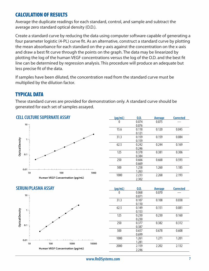
CALCULATION OF RESULTSAverage the duplicate readings for each standard, control, and sample and subtract the average zero standard optical density (O.D.).
Create a standard curve by reducing the data using computer software capable of generating a four parameter logistic (4-PL) curve fit. As an alternative, construct a standard curve by plotting the mean absorbance for each standard on the y-axis against the concentration on the x-axis and draw a best fit curve through the points on the graph. The data may be linearized by plotting the log of the human VEGF concentrations versus the log of the O.D. and the best fit line can be determined by regression analysis. This procedure will produce an adequate but less precise fit of the data.
If samples have been diluted, the concentration read from the standard curve must be multiplied by the dilution factor.
TYPICAL DATAThese standard curves are provided for demonstration only. A standard curve should be generated for each set of samples assayed.
(pg/mL) O.D. Average Corrected0 0.074 0.075 —
0.07615.6 0.118 0.120 0.045
0.12131.3 0.159 0.159 0.084
0.15962.5 0.242 0.244 0.169
0.246125 0.378 0.381 0.306
0.384250 0.666 0.668 0.593
0.669500 1.258 1.260 1.185
1.2631000 2.233 2.268 2.193
2.302
(pg/mL) O.D. Average Corrected0 0.068 0.070 —
0.07131.3 0.107 0.108 0.038
0.11062.5 0.149 0.151 0.081
0.153125 0.230 0.230 0.160
0.230250 0.377 0.382 0.312
0.387500 0.657 0.678 0.608
0.6991000 1.261 1.271 1.201
1.2812000 2.159 2.202 2.132
2.246
CELL CULTURE SUPERNATE ASSAY
SERUM/PLASMA ASSAY

For research use only. Not for use in diagnostic procedures.8
PRECISIONIntra-assay Precision (Precision within an assay) Three samples of known concentration were tested twenty times on one plate to assess intra-assay precision.
Inter-assay Precision (Precision between assays) Three samples of known concentration were tested in forty separate assays to assess inter-assay precision. Assays were performed by at least three technicians using two lots of components.
CELL CULTURE SUPERNATE ASSAY
Intra-Assay Precision Inter-Assay Precision
Sample 1 2 3 1 2 3
n 20 20 20 40 40 40
Mean (pg/mL) 29.1 123 531 32.8 128 495
Standard deviation 1.9 5.0 18.4 2.8 6.4 33.0
CV (%) 6.5 4.1 3.5 8.5 5.0 6.7
SERUM/PLASMA ASSAY
Intra-Assay Precision Inter-Assay Precision
Sample 1 2 3 1 2 3
n 20 20 20 40 40 40
Mean (pg/mL) 53.7 235 910 64.5 250 1003
Standard deviation 3.6 10.6 46.2 5.7 17.4 61.7
CV (%) 6.7 4.5 5.1 8.8 7.0 6.2
RECOVERYThe recovery of human VEGF spiked to three different levels throughout the range of the assay in various matrices was evaluated.
Sample Type Average % Recovery Range
Cell culture media (n=5) 102 95-111%
Serum (n=5) 102 92-115%
EDTA plasma (n=5) 97 82-113%
Heparin plasma (n=5) 93 82-102%
Citrate plasma (n=5) 100 88-113%
SENSITIVITYUsing Calibrator Diluent RD5K the minimum detectable dose (MDD) of human VEGF is typically less than 5.0 pg/mL. Using Calibrator Diluent RD6U the MDD is typically less than 9.0 pg/mL.
The MDD was determined by adding two standard deviations to the mean optical density value of twenty zero standard replicates and calculating the corresponding concentration.
www.RnDSystems.com 9
LINEARITYTo assess linearity of the assay, samples were spiked with high concentrations of human VEGF and diluted with the appropriate Calibrator Diluent to produce samples with values within the dynamic range of the assay.
Cell culture media (n=5)
Serum (n=5)
EDTA plasma (n=5)
Heparin plasma (n=5)
Citrate plasma (n=5)
1:2Average % of Expected 98 97 97 94 95
Range (%) 94-100 91-103 82-107 87-99 90-100
1:4Average % of Expected 96 97 98 93 94
Range (%) 93-99 93-104 91-106 85-98 89-99
1:8Average % of Expected 93 96 96 92 92
Range (%) 88-102 93-103 89-106 85-101 85-97
1:16Average % of Expected 93 94 94 94 92
Range (%) 88-105 91-101 84-106 83-103 85-98
CALIBRATIONThis immunoassay is calibrated against a highly purified Sf 21-expressed recombinant human VEGF165 produced at R&D Systems.
The NIBSC/WHO VEGF165 1st WHO Reference Reagent 02/286 (recombinant human DNA) was evaluated in this kit. The dose response curve of the reference reagent 02/286 parallels the Quantikine standard curve. To convert sample values obtained with the Quantikine Human VEGF kit to approximate NIBSC/WHO 02/286 Units, use the equation below.
NIBSC/WHO (02/286) approximate value (U/mL) = 0.002 x Quantikine VEGF value (pg/mL)
Note: Based on data generated in April 2011.

For research use only. Not for use in diagnostic procedures.10
SAMPLE VALUESSerum/Plasma - Samples from apparently healthy volunteers were evaluated for the presence of human VEGF in this assay. No medical histories were available for the donors used in this study.
Sample Type Mean of Detectable (pg/mL) % Detectable Range (pg/mL)
Serum (n=37) 220 100 62-707
EDTA plasma (n=37) 61 24 ND-115
Heparin plasma (n=37) 41 22 ND-55
Citrate plasma (n=37) ___ 0 ND
ND=Non-detectable
Cell Culture Supernates: Human peripheral blood mononuclear cells (10 x 106 cells/mL) were cultured in RPMI supplemented with 5% fetal bovine serum, 50 μM β-mercaptoethanol, 2 mM L-glutamine, 100 U/mL penicillin, and 100 μg/mL streptomycin sulfate. The cells were cultured unstimulated or stimulated with 10 μg/mL PHA for 5 days. Aliquots of the cell culture supernates were removed, assayed for levels of human VEGF, and measured 87.1 pg/mL and 1041 pg/mL, respectively.
PC-3 human prostate cancer cells were cultured in RPMI supplemented with 10% fetal bovine serum, 2 mM L-glutamine, 100 U/mL penicillin, and 100 μg/mL streptomycin sulfate. The cells were cultured unstimulated or stimulated with 60 nM of PMA for 24 hours. Aliquots of the cell culture supernates were removed, assayed for levels of human VEGF, and measured 3373 pg/mL and 3652 pg/mL, respectively.
JEG-3 human epithelial choriocarcinoma cells were cultured in MEM + NEAA supplemented with 10% fetal bovine serum, 2 mM L-glutamine, 100 U/mL penicillin, and 100 μg/mL streptomycin sulfate. The cells were cultured unstimulated or stimulated with 100 ng/mL of LPS for 1 day. Aliquots of the cell culture supernates were removed, assayed for levels of human VEGF, and measured 1538 pg/mL and 1938 pg/mL, respectively.

www.RnDSystems.com 11
SPECIFICITYThis assay recognizes natural and recombinant human VEGF. This assay also recognizes recombinant human VEGF165b.
The factors listed below were prepared at 50 ng/mL in Calibrator Diluent and assayed for cross-reactivity. Preparations of the following factors at 50 ng/mL in a mid-range VEGF control were assayed for interference. The following factors showed no cross-reactivity or interference.
Recombinant human:PDGF-AAPDGF-ABPDGF-BBPDGF-CCPDGF-DDPlGFPlGF-2VEGF165/PlGFVEGF-B167
VEGF-CVEGF-DVEGF R3
Recombinant mouse:PDGF-CCPlGF-2VEGF120
VEGF164
VEGF R3
Recombinant rat:PDGF-AAPDGF-ABPDGF-BBVEGF164
Recombinant zebrafish:VEGF
Natural proteins:human PDGFporcine PDGF
Recombinant BovineVEGF
164
VEGF-related factors showing cross-reactivity or interference.
Recombinant human VEGF R1/Flt-1 Interference at levels ≥ 500 pg/mL
Recombinant human VEGF R2/KDR Interference at levels ≥ 2000 pg/mL
Recombinant mouse VEGF R1/Flk-1 Interference at levels ≥ 500 pg/mL
Recombinant mouse VEGF R2/KDR Interference at levels ≥ 4000 pg/mL
Recombinant canine VEGF Cross-reacts approximately 67%
Recombinant feline VEGF Cross-reacts approximately 82%
The factors listed below were prepared at 10 ng/mL in Calibrator Diluent and assayed for cross-reactivity. Preparations of the following factors at 10 ng/mL in a mid-range VEGF control were assayed for interference. The following factors showed no cross-reactivity or interference.
Recombinant bovine:VEGF
164

For research use only. Not for use in diagnostic procedures.12
REFERENCES1. Leung, D.W. et al. (1989) Science 246:1306.
2. Keck, P.J. et al. (1989) Science 246:1309.
3. Byrne, A.M. et al. (2005) J. Cell. Mol. Med. 9:777.
4. Robinson, C.J. and S.E. Stringer (2001) J. Cell. Sci. 114:853.
5. Richardson, R.S. et al. (1999) Am. J. Physiol. 277:H2247.
6. Sugishita, Y. et al. (2000) Biochem. Biophys. Res. Commun. 268:657.
7. Yamane, A. et al. (1994) Oncogene 9:2683.
8. Goad, D.L. et al. (1996) Endocrinology 137:2262.
9. Gaudry, M. et al. (1997) Blood 90:4153.
10. Mclaren, J. et al. (1996) J. Clin. Invest. 98:482.
11. Diaz, B.V. et al. (2000) J. Biol. Chem. 275:642.
12. Asano, A. et al. (1997) Biochem. J. 328:179.
13. Bautz, F. et al. (2000) Exp. Hematol. 28:700.
14. Namiki, A. et al. (1995) J. Biol. Chem. 270:31189.
15. Nauck, M. et al. (1997) Am. J. Respir. Cell. Mol. Biol. 16:398.
16. Angelo, L.S. and R. Kurzrock (2007) Clin. Cancer Res. 13:2825.
17. Neufeld, G. et al. (1999) FASEB. J. 13:9.
18. Kowalewski, M.P. et al. (2005) Accession #ABB82619.
19. Pan, Q. et al. (2007) J. Biol. Chem. 282:24049.
20. Dai, J. and A.B. Rabie (2007) J. Dent. Res. 86:937.
21. Breier, G. (2000) Semin. Thromb. Hemost. 26:553.
22. Barleon, B. et al. (1996) Blood 87:3336.
23. Weis, S.M. and D.A. Cheresh (2005) Nature 437:497.
24. Thurston, G. (2002) J. Anat. 200:575.
25. Grothey, A. and E. Galanis (2009) Nat. Rev. Clin. Oncol. 6:507.
26. Carvalho, J.F. et al. (2007) J. Clin. Immunol. 27:246.

www.RnDSystems.com 13
PLATE LAYOUTUse this plate layout to record standards and samples assayed.

For research use only. Not for use in diagnostic procedures.14
NOTES
04.95 750134.24 5/16
©2016 R&D Systems, Inc.