human pancreatic cancer stem cells are sensitive to dual
TRANSCRIPT
Urtasun et al. BMC Cancer (2015) 15:223 DOI 10.1186/s12885-015-1249-2
RESEARCH ARTICLE Open Access
Human pancreatic cancer stem cells are sensitiveto dual inhibition of IGF-IR and ErbB receptorsNerea Urtasun1,2*†, Anna Vidal-Pla1†, Sandra Pérez-Torras1,2,3 and Adela Mazo1,2,3
Abstract
Background: Pancreatic ductal adenocarcinoma is a particularly challenging malignancy characterized by poorresponsiveness to conventional chemotherapy. Although this tumor frequently overexpresses or possessesconstitutively activated variants of IGF-IR and EGFR/Her-2, clinical trials using inhibitors of these receptors havefailed. ErbB receptors have been proposed as one mechanism involved in the resistance to IGF-IR inhibitors. There-fore, combined treatment with inhibitors of both IGF-IR and ErbB receptors would appear to be a good strategy forovercoming the emergence of resistance.
Methods: Sensitivity of cells to NVP-AEW541 and lapatinib in single or combination treatment was assessed by MTTor WST-8 assays in a panel of human pancreatic cancer cell lines and cancer stem cells. Tumorspheres enriched incancer stem cells were obtained from cultures growing in non-adherent cell plates. The effects on cell signallingpathways were analyzed by Western blot.
Results: We found that combined treatment with the IGF-IR and EGFR/Her-2 inhibitors NVP-AEW541 and lapatinib,respectively, synergistically inhibited pancreatic cancer cell growth. Analysis at molecular level argued in favor ofcross-talk between IGF-IR and ErbBs pathways at IRS-1 level and indicated that the synergistic effect is associatedwith the total abolishment of Akt, Erk and IRS-1 phosphorylation. Moreover, these inhibitors acted synergistically intumorsphere cultures to eliminate cancer stem cells, in contrast to their resistance to gemcitabine.
Conclusions: Taken together, these data indicate that simultaneous blockade of IGF-IR and EGFR/Her-2 usingNVP-AEW541 and lapatinib may overcome resistance in pancreatic cancer. Thus, the synergy observed with thiscombined treatment indicates that it may be possible to maximize patient benefit with the appropriate combination ofcurrently known anticancer agents.
Keywords: Pancreatic ductal adenocarcinoma, Cancer stem cells, IGF-IR, EGFR, Her-2
BackgroundPancreatic ductal adenocarcinoma (PDAC) is one of thefive most common causes of cancer death, owing to its latediagnosis, high dissemination at early stages, and poor re-sponsiveness to both radio- and chemotherapy [1]. Gemci-tabine remains the current standard first-line treatment[2]. However, chemotherapy in advanced disease confersonly modest survival advantage and symptoms palliation.Recent clinical trials of gemcitabine combination therapies
* Correspondence: [email protected]†Equal contributors1Departament de Bioquímica i Biologia Molecular, Universitat de Barcelona,Barcelona, Spain2Institut de Biomedicina de la Universitat de Barcelona (IBUB), Barcelona,SpainFull list of author information is available at the end of the article
© 2015 Urtasun et al.; licensee BioMed CentraCommons Attribution License (http://creativecreproduction in any medium, provided the orDedication waiver (http://creativecommons.orunless otherwise stated.
have produced significant, but low, response rates in ad-vanced pancreatic cancer, underscoring the need for newtherapeutic approaches [3-5].An important consideration in these strategies is the
heterogeneity of pancreatic tumors. In this context, sev-eral studies investigating pancreatic cancer biology haveidentified a subpopulation of cells termed pancreaticcancer stem cells (PCSCs) [6-8]. This subpopulation mayplay a critical role in the resistance to chemotherapy andradiation, suggesting that such cells may be the sourceof some cases of pancreatic cancer relapse [9,10]. There-fore, therapeutic modalities that lead to the eliminationof CSCs could improve clinical outcome in patients withpancreatic cancer.
l. This is an Open Access article distributed under the terms of the Creativeommons.org/licenses/by/4.0), which permits unrestricted use, distribution, andiginal work is properly credited. The Creative Commons Public Domaing/publicdomain/zero/1.0/) applies to the data made available in this article,
Urtasun et al. BMC Cancer (2015) 15:223 Page 2 of 8
Receptor tyrosine kinases are currently among themost promising therapeutic targets in a wide range oftumors. Inhibition of receptor tyrosine kinases of theErbB family has been approved for the treatment of differ-ent tumors and is used extensively to treat breast cancer[11]. There has also been growing research interest ininsulin-like growth factor-1 receptor (IGF-IR) as a targetfor antitumor therapy [12-14], given the demonstrated abil-ity of IGF-IR to potently contribute to a variety of onco-genic effects, including cell proliferation, cell survival, andcell differentiation [15-17]. IGF-IR is frequently overex-pressed or activated in pancreatic cancer, a factor that mostlikely contributes to the aggressive growth characteristicsand poor prognosis of these tumors [18-20]. Moreover,molecular mechanisms that lead to autocrine activation ofthe IGF-IR and stimulation of downstream signalingthrough phosphorylation (activation) of Akt have beenidentified and could further substantially contribute totumor progression and invasion [21,22].On the basis of these findings, IGF-IR has come to be
viewed as a rational therapeutic target in pancreatic can-cer, prompting clinical investigations of IGF-IR inhibitors.However, recent clinical trials of anti-IGF-IR compoundsin combination with gemcitabine have failed to demon-strate improved patient survival [23,24], a failure attribut-able, at least in part, to the development of resistance.One mechanism proposed to account for resistance is ac-tivation of alternative survival pathways [25]. Among thesecandidate alternative pathways are those activated bymembers of the ErbB receptor family, which are import-ant in regulating cell survival [26-28] and are frequentlyoverexpressed in pancreatic carcinomas [29,30]. Import-antly, activation of mitogen-activated protein kinases(MAPKs) by ErbB receptors signaling may counterbalancethe decrease in phosphorylated Akt induced by IGF-IR in-hibitors. This compensatory mechanism could explain thefailure of treatments based on individual inhibition ofIGF-IR or ErbB [14,28,31], and suggests that therapeuticstrategies based on combined inhibition of IGF-IR andErbB receptors could overcome this resistance.In the current study, we tested this hypothesis, investi-
gating the impact of concurrent inhibition of IGF-IRsand epidermal growth factor receptors (EGFR/Her-2) byNVP-AEW541 and lapatinib tyrosine kinase inhibitors,respectively, on pancreatic cancer cell lines and particu-larly on PCSCs.
MethodsReagents and immunochemicalsLapatinib was kindly provided by GlaxoSmithKline(Brentford, UK) and NVP-AEW541 was a kind gift ofNovartis Pharma (Basel, Switzerland). Stock solutions ofdrugs were prepared in dimethyl sulfoxide and storedat −20°C, and diluted in fresh media before each
experiment. Insulin-like growth factor (IGF-I) and Epider-mal growth factor (EGF) (Peprotech, Rocky Hill, NJ, USA)were dissolved in phosphate-buffered saline containing0.1% bovine serum albumin (BSA). Cells were immuno-stained using antibodies against EGFR (1005), Her-2 (C-18), Her-3 (C-17), IGF-IRβ (C-20) and Akt-1 (C-20) (SantaCruz Biotechnology, Santa Cruz, CA, USA); phospho-Akt (Ser473), phospho-p44/42 (Thr202/Tyr204) andp44/42 (137 F5) (Cell Signaling Technology, Danvers,MA, USA); phospho-IRS-1 (Tyr612) and phospho-IRS-1 (Tyr896) (Invitrogen, Camarillo, CA, USA); and β-actin (Sigma-Aldrich, St. Louis, MO, USA).
Cell cultureNP-9, NP-18, and NP-29 cell lines (kindly provided byDr Capella from Hospital de la Santa Creu i Sant Pau,Barcelona, Spain) were derived from human pancreaticadenocarcinomas xenografted in nude mice [32]. TheBxPC3 cell line was obtained from the American TypeCulture Collection (Manassas, VA, USA). CP15T andCP15A cell lines were also derived from a human pan-creatic adenocarcinoma xenografted in nude mice byour group [33]. The research protocol complied with theethical guidelines of the 1975 Declaration of Helsinkiand was approved by the ethics committee of Universitatde Barcelona. All participants provided written informedconsent. NP-9, NP-29, CP15T and CP15A cells weregrown in a 1:1 mixture of Dulbecco’s modified Eagle’smedium (DMEM) and F12 medium; BxPC3 cells weregrown in DMEM; and NP-18 cell were grown in RPMI-1640 medium (Gibco, Grand Island, NY, USA). All mediawere supplemented with 5% fetal bovine serum and anti-biotics (penicillin/streptomycin). Cells were maintained ina humidified atmosphere of 5% CO2 at 37°C and subcul-tured every 3–4 days.For tumorsphere cultures, cells were grown in ultra-
low attachment plates (Corning, Gendale, AZ, USA) usingserum-free DMEM:F12 (1:1) supplemented with B-27, N2,antibiotic-antimycotic (Invitrogen), 20 ng/ml human EGF,and 20 ng/ml human basic fibroblast growth factor (bFGF;Peprotech). Tumorspheres were dissociated weekly usingtrypsin and maintained for several passages. Experimentswere performed between the fourth and seventh passage[34].
Dose–response assaysDose–response assays were performed by seeding 2–5 × 10³ cells/well in 96-well culture plates. Cultureswere exposed to increasing concentrations of lapatiniband/or NVP-AEW541 for 72 h, at which time cell via-bility was determined by MTT (3-[4,5-dimethylthiazol-2-yl]-2,5 diphenyl tetrazolium bromide) assay.Assays comparing monolayers and tumorspheres were
performed by seeding single-cell suspensions at a density
Urtasun et al. BMC Cancer (2015) 15:223 Page 3 of 8
of 1.5 × 10³ cells/well in standard or ultra-low-adhesion96-well culture plates, respectively, with increasing con-centrations of lapatinib and/or NVP-AEW541. Cell viabil-ity was determined 72 h post-treatment using a WST-8assay (Sigma-Aldrich), as described by the manufacturer.Data were fitted to a dose–response curve using stand-
ard nonlinear regression, adapting a Hill equation withGrafit software (Erithacus Software, Ltd., Horley, UK) toobtain 50% inhibitory concentration (IC50) values. Cellsurvival for all experiments is expressed as the percent-age of viable cells relative to that in untreated cells (de-fined as 100%).The coefficient of drug interaction (CDI) was used to
analyze the effect of drug combination. CDI was calcu-lated based on the absorbance in each group, as CDI =AB/(A × B), where AB is the ratio for the combinationgroup relative to the control group, and A and B are theratios of each single agent group relative to the controlgroup. Thus, a CDI value < 1 indicates synergy, a CDIvalue = 1 indicates additive effects, and a CDI value > 1indicates antagonism. CDIs less than 0.7 indicate a sig-nificant synergistic effect.
Protein extraction and Western blotCells were lysed in ice-cold lysis buffer containing20 mM Tris (pH 8), 150 mM NaCl, 10 mM EDTA,10 mM Na4P2O7, 2 mM VO4
3−, 100 mM NaF, 1 mM β-glycerophosphate, 1% NP40, and protease and phos-phatase inhibitor cocktails (Roche Applied Sciences,Penzberg, Germany). Lysates containing equal amounts
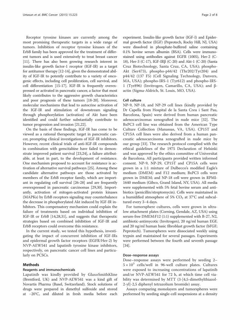
Figure 1 Inhibition of IGF-I and ErbB receptors with NVP-AEW541 andand ErbB receptors and their signaling pathway components. Cells cultured toanalyzed by Western blot. (B) Dose–response curves and IC50 values for NVP-after seeding with increasing concentrations of NVP-AEW541 or lapatinib, andData are presented as means ± standard deviation of a representative experim
of protein (20 μg for monolayer experiments and 30 μg forexperiments comparing monolayers and tumorspheres),assessed by Bradford assay (Bio-Rad, Hercules, CA, USA),were electrophoretically separated on 8% polyacrylamide-sodium dodecyl sulfate gels and transferred to nitrocellu-lose membranes (Schleicher and Schuell, Dassel, Germany).Membranes were immunoblotted with the indicated pri-mary antibodies. Antibody labeling was detected using anenhanced chemiluminescence detection kit (Biological In-dustries, Kibbutz Beit Haemek, Israel).
ResultsSensitivity of human pancreatic cancer cell lines toNVP-AEW541 and lapatinibExpression levels of IGF-IR and ErbB family receptorswere examined in a panel of human pancreatic cancercell lines. IGF-IR expression levels varied, with highlevels detected in NP-29 and CP15A cell lines. Notably,the highest levels of EGFR expression were also found inNP-29 cells, whereas EGFR expression was negligible inCP15T and CP15A cells. In contrast, Her-2, which wasobserved in all cell lines, showed marked expression inCP15T and CP15A cells. Her-3 expression was onlyclearly detectable in NP-29, CP15T, and CP15A cells.Intracellular signaling pathways were assessed by evalu-
ating Akt and Erk (extracellular signal-regulated kinase)phosphorylation. These experiments revealed a range ofactivation levels, with NP-9 cells showing the highestlevels of Akt phosphorylation and CP15A cells showingthe lowest levels of Erk phosphorylation (Figure 1A).
lapatinib in pancreatic cancer cell lines. (A) Basal levels of IGF-Iapproximately 90% confluence were lysed and proteins in lysates were
AEW541 and lapatinib in the panel of cell lines. Cells were treated 24 hcell viability was measured by MTT assay 72 h after the start of treatment.ent (n = 3). ● NP-9, ♦ NP-18, ■ NP-29, CP15T,▲ CP15A.
Urtasun et al. BMC Cancer (2015) 15:223 Page 4 of 8
The effects of the IGF-IR inhibitor, NVP-AEW541,and the EGFR and Her-2 inhibitor, lapatinib were thenexamined in all five cell lines. NVP-AEW541 induced aconcentration-dependent inhibition of growth in all celllines. IC50 values ranged from 4.4 to 17.6 μM, with themost potent effect observed in NP-18 cells. Lapatinibalso induced concentration-dependent growth inhibitionin all cell lines. Again, NP-18 cells showed the highestsensitivity, and IC50 values ranged from 8.0 to 41.2 μM(Figure 1B).
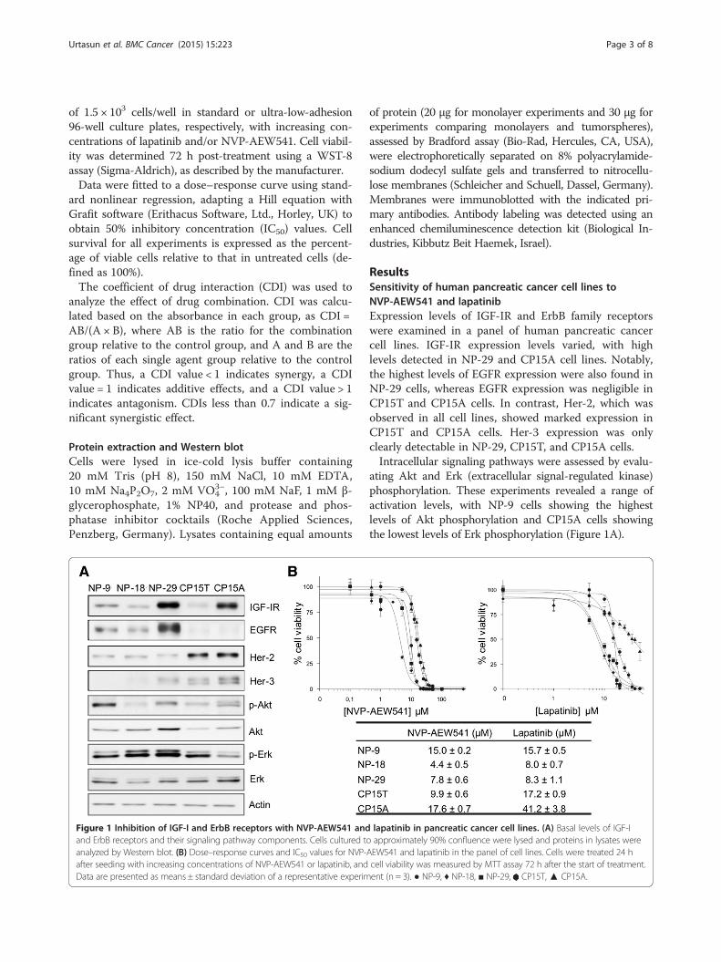
Response of pancreatic cancer cells to combined IGF-IRand EGFR/Her-2 inhibitionResistance to individual treatment with the IGF-IR andEGFR/Her-2 inhibitors NVP-AEW541 and lapatinib, re-spectively, has been reported, reflecting the operation ofcompensatory mechanisms between the two pathways. Toevaluate whether the individual effects of these drugs arepotentiated by concurrent inhibition of both pathways, weassayed these two drugs in combination in the five celllines. Increasing concentrations of lapatinib were com-bined with a fixed (IC20) concentration of NVP-AEW541.When used in combination, these drugs exhibited very po-tent synergy in all cell lines, with coefficients of drug inter-action (CDIs) clearly < 0.7; remarkably, in some cases, CDIvalues were < 0.1 (Figure 2A).To evaluate the effects of these drugs on the intracel-
lular signaling activity of both pathways, we selectedthe NP-29 cell line, which exhibited the lowest CDI. Incontrol cells, an IGF-I stimulus promoted substantialAkt and IRS-1 (Y612) phosphorylation and a small in-crease in IRS-1 (Y896) phosphorylation, but did notaffect Erk1/2 phosphorylation. This suggests that theactivity of the Ras-MAPK pathway is independent ofIGF-I in these cells. Conversely, EGF stimulation re-sulted in elevated phosphorylation of Erk1/2, IRS-1(Y612), and IRS-1 Y896 (Figure 2B). Inhibition of IGF-IRby NVP-AEW541 decreased IGF-I-induced phosphoryl-ation of Akt and IRS-1 (Y612). In cells stimulated withEGF or IGF-I + EGF, NVP-AEW541 treatment increasedEGFR pathway activation to a greater degree than in con-trol cells, enhancing phosphorylation of Erk1/2, IRS1(Y612), and IRS1 Y896 (Figure 2B). Whereas treatmentwith lapatinib diminished EGF-stimulated activation ofErk1/2 and IRS-1 (Y896), it did not significantly attenuateIGF-I- or IGF-I + EGF-induced activation of Akt and IRS-1 (Y612) (Figure 2B). Interestingly, simultaneous inhib-ition of both IGF-IR and EGFR/Her-2 by NVP-AEW541and lapatinib completely abrogated IGF-I-, EGF-, andIGF-I + EGF-stimulated phosphorylation of Akt, Erk1/2,IRS-1 (Y612) and IRS-1 (Y896), confirming at the molecu-lar level the strong synergy observed in cytotoxicity exper-iments (Figure 2B,C).
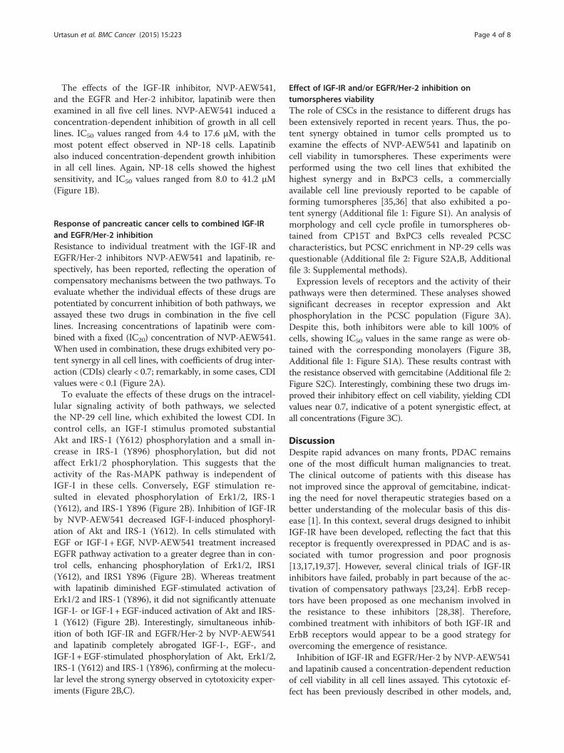
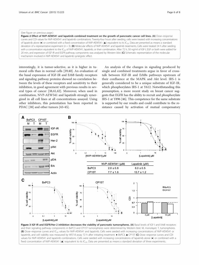
Effect of IGF-IR and/or EGFR/Her-2 inhibition ontumorspheres viabilityThe role of CSCs in the resistance to different drugs hasbeen extensively reported in recent years. Thus, the po-tent synergy obtained in tumor cells prompted us toexamine the effects of NVP-AEW541 and lapatinib oncell viability in tumorspheres. These experiments wereperformed using the two cell lines that exhibited thehighest synergy and in BxPC3 cells, a commerciallyavailable cell line previously reported to be capable offorming tumorspheres [35,36] that also exhibited a po-tent synergy (Additional file 1: Figure S1). An analysis ofmorphology and cell cycle profile in tumorspheres ob-tained from CP15T and BxPC3 cells revealed PCSCcharacteristics, but PCSC enrichment in NP-29 cells wasquestionable (Additional file 2: Figure S2A,B, Additionalfile 3: Supplemental methods).Expression levels of receptors and the activity of their
pathways were then determined. These analyses showedsignificant decreases in receptor expression and Aktphosphorylation in the PCSC population (Figure 3A).Despite this, both inhibitors were able to kill 100% ofcells, showing IC50 values in the same range as were ob-tained with the corresponding monolayers (Figure 3B,Additional file 1: Figure S1A). These results contrast withthe resistance observed with gemcitabine (Additional file 2:Figure S2C). Interestingly, combining these two drugs im-proved their inhibitory effect on cell viability, yielding CDIvalues near 0.7, indicative of a potent synergistic effect, atall concentrations (Figure 3C).
DiscussionDespite rapid advances on many fronts, PDAC remainsone of the most difficult human malignancies to treat.The clinical outcome of patients with this disease hasnot improved since the approval of gemcitabine, indicat-ing the need for novel therapeutic strategies based on abetter understanding of the molecular basis of this dis-ease [1]. In this context, several drugs designed to inhibitIGF-IR have been developed, reflecting the fact that thisreceptor is frequently overexpressed in PDAC and is as-sociated with tumor progression and poor prognosis[13,17,19,37]. However, several clinical trials of IGF-IRinhibitors have failed, probably in part because of the ac-tivation of compensatory pathways [23,24]. ErbB recep-tors have been proposed as one mechanism involved inthe resistance to these inhibitors [28,38]. Therefore,combined treatment with inhibitors of both IGF-IR andErbB receptors would appear to be a good strategy forovercoming the emergence of resistance.Inhibition of IGF-IR and EGFR/Her-2 by NVP-AEW541
and lapatinib caused a concentration-dependent reductionof cell viability in all cell lines assayed. This cytotoxic ef-fect has been previously described in other models, and,
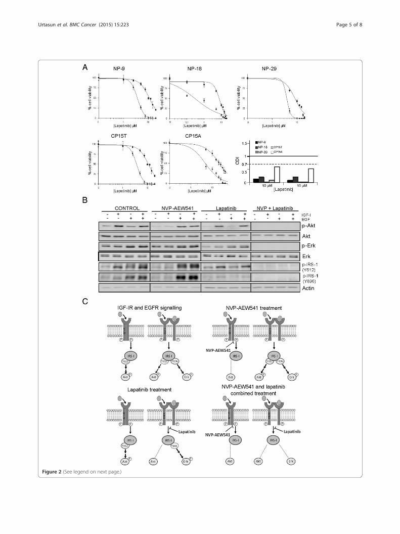
Figure 2 (See legend on next page.)
Urtasun et al. BMC Cancer (2015) 15:223 Page 5 of 8
(See figure on previous page.)Figure 2 Effect of NVP-AEW541 and lapatinib combined treatment on the growth of pancreatic cancer cell lines. (A) Dose–responsecurves and CDI values for NVP-AEW541 and lapatinib combinations. Twenty-four hours after seeding, cells were treated with increasing concentrationsof lapatinib alone (●) or combined with a fixed concentration of NVP-AEW541 (▲) equivalent to its IC20. Data are presented as means ± standarddeviation of a representative experiment (n = 3). (B) Molecular effects of NVP-AEW541 and lapatinib treatments. Cells were treated 24 h after seedingwith a concentration equivalent to the IC20 of NVP-AEW541, lapatinib, or their combination. After 72 h, 50 ng/ml of IGF-I, EGF or both were added for20 min, and expression of IGF-IR and EGFR pathway components was analyzed by Western blot. (C) Schematic representation of the molecularmechanism involved in NVP-AEW541 and lapatinib synergistic effect.
Urtasun et al. BMC Cancer (2015) 15:223 Page 6 of 8
interestingly, it is tumor-selective, as it is higher in tu-moral cells than in normal cells [39,40]. An evaluation ofthe basal expression of IGF-IR and ErbB family receptorsand signaling pathway proteins showed no correlation be-tween the levels of these receptors and sensitivity to theirinhibition, in good agreement with previous results in sev-eral types of cancer [38,41,42]. Moreover, when used incombination, NVP-AEW541 and lapatinib strongly syner-gized in all cell lines at all concentrations assayed. Usingother inhibitors, this potentiation has been reported inPDAC [38] and other tumors [43-45].
Figure 3 IGF-IR and EGFR/Her-2 inhibition decreases the viability of pand their signaling pathway components in BxPC3 and CP15T tumorsphere(B) Dose–response curves and IC50 values for NVP-AEW541 and lapatinib. Clapatinib, and cell viability was measured by WST-8 assay 72 h after initiatinvalues for NVP-AEW541 and lapatinib combinations. Cells were seeded withfixed concentration of NVP-AEW541 (▲) equivalent to its IC20. Data are pre
An analysis of the changes in signaling produced bysingle and combined treatments argue in favor of cross-talk between IGF-IR and ErbBs pathways upstream oftheir confluence at the MAPK and Akt level. IRS-1 isgenerally considered to be a unique substrate of IGF-IR,which phosphorylates IRS-1 at Y612. Notwithstanding thispresumption, a more recent study on breast cancer sug-gests that EGFR has the ability to recruit and phosphorylateIRS-1 at Y896 [46]. This competence for the same substrateis supported by our results and could contribute to the re-sistance caused by activation of mutual compensatory
ancreatic tumorspheres. (A) Basal levels of IGF-I and ErbB receptorss were determined by Western blot. M, monolayer; T, tumorspheres.ells were seeded with increasing concentrations of NVP-AEW541 org treatment. ● BxPC3, ■ CP15T (C) Dose–response curves and CDIincreasing concentrations of lapatinib alone (●) or combined with a
sented as means ± standard deviation of three experiments.
Urtasun et al. BMC Cancer (2015) 15:223 Page 7 of 8
pathways. The IRS-1 phosphorylation pattern clearly indi-cated that blocking IGF-IR signaling strongly induced phos-phorylation of IRS-1 at Y896. This increase in IRS-1phosphorylation highlights the crucial influence of this newmechanism—activation of MAPK and especially Akt phos-phorylation—in the resistance to IGF-IR inhibitors, andpoints to preferential channeling of ErbB receptor signalingto IRS-I (Y896) phosphorylation via phosphorylated Akt.Interestingly, when both receptors were inhibited, IRS-1,Akt and MAPK phosphorylation were completely abol-ished, reinforcing the utility of combined inhibition of bothpathways in averting the resistance induced by individualtreatments.Despite these good in vitro results, the outcome in pa-
tients has been disappointing. One possible reason for thefailure of these targeted drugs could be the role of PCSCsin resistance [47,48]. The importance of the IGF-IR path-way in treatments targeting PCSCs has not been previ-ously described, although several recent reports havedemonstrated an association of this receptor with cellstemness in some tumors [49,50]. Our results showed thatpancreatic cancer tumorspheres were sensitive to treat-ment with either NVP-AEW541 or lapatinib, in contrastto their high resistance to gemcitabine. Remarkably, com-bining both drugs again produced a synergistic effect simi-lar to that observed in monolayers. This synergy intumorspheres, which has not been previously described,indicates that inhibition of both pathways in PCSCs canalso overcome the resistance caused by these compensa-tory pathways in this subpopulation.
ConclusionsSimultaneous inhibition of IGF-IR and ErbB receptors byNVP-AEW541 and lapatinib circumvented the resistanceobserved at the molecular level with individual treatments.Interestingly, these inhibitors were also able to eliminatePCSCs, overcoming their resistance to conventionalchemotherapy. Thus, the synergy observed with this com-bined treatment indicates that it may be possible tomaximize patient benefit with the appropriate combin-ation of currently known anticancer agents.
Additional files
Additional file 1: Figure S1. Effect of NVP-AEW541 and lapatinib in theBxPC3 monolayers. (A) Dose–response curves and IC50 values for NVP-AEW541 and lapatinib. Cells were seeded with increasing concentrations ofNVP-AEW541 or lapatinib, and cell viability was measured by WST-8 assay72 h after starting treatment. Data are presented as means ± standarddeviation of three experiments. (B) Dose–response curve and CDI values forNVP-AEW541 and lapatinib combination. Twenty-four hours after seeding,cells were treated with increasing concentrations of lapatinib alone (●) orcombined with a fixed concentration of NVP-AEW541 (▲) equivalent to itsIC20. Data are presented as means ± standard deviation of three experiments.
Additional file 2: Figure S2. Characterization of tumorspheresobtained from different human pancreatic cancer cell lines. (A)
Morphology of BxPC3, CP15T, and NP-29 tumorspheres. Cells weremaintained under standard culture conditions (monolayers) or in stemcell medium on ultra-low-adhesion plates (tumorspheres). Scale bar =5 μm. (B) Cell cycle profiles of monolayers and tumorspheres. S-phaserepresented in light grey, G2/M-phase in dark grey, and G0/G1-phase inblack. (C) Dose–response curve and IC50 values of gemcitabine for mono-layers and tumorspheres. Cells were seeded with increasingconcentrations of gemcitabine, and cell viability was measured by WST-8assay 72 h after starting treatment. Data are presented as means ±standard deviation of three experiments. ■BxPC3 monolayer, □BxPC3tumorspheres, ●CP15T monolayer, ○CP15T tumorspheres.
Additional file 3: Analysis of cell cycle by flow cytometry.
AbbreviationsCDI: Coefficient of drug interaction; CSC: Cancer stem cells; EGF: Epidermalgrowth factor; EGFR: Epidermal growth factor receptor; Erk: Extracellular signal-regulated kinase; IC50: 50% inhibitory concentration; IGF: Insulin-like growthfactor; IGF-IR: Insulin-like growth factor-1 receptor; IRS-1: Insulin receptor substrate1; MAPKs: Mitogen-activated protein kinases; pAkt: Phosphorylated Akt;PCSC: Pancreatic cancer stem cells; PDAC: Pancreatic ductal adenocarcinoma.
Competing interestsThe authors declare that they have no competing interests.
Authors’ contributionsNU carried out the experiments related to tumorspheres and helped to draftthe manuscript. AVP carried out the experiments related to monolayers andhelped to draft the manuscript. SPT participated in the design of the studyand helped to draft the manuscript. AM participated in the design of thestudy and helped to draft the manuscript. All authors read and approved thefinal manuscript.
AcknowledgementsThis work has been supported by grants BIO2008-04692-C03-03 and SAF2011-23660 (Ministerio de Economia y Competitividad) and receives partial supportof the Generalitat de Catalunya (2009SGR624). The group belongs to theNational Biomedical Research Institute on Liver and Gastrointestinal Diseases(CIBERehd) and SPT is a CIBER researcher. CIBER is an initiative of the Institutode Salud Carlos III (ISCIII, Ministerio de Economia y Competitividad). AVP hasbeen the recipient of a FI fellow from the Generalitat de Catalunya. We aregrateful to GlaxoSmithKline and Novartis Pharma for kindly provided lapatiniband NVP-AEW541, respectively.In memoriam of Dr. Adela Mazo, who passed away on March 24th 2015.
Author details1Departament de Bioquímica i Biologia Molecular, Universitat de Barcelona,Barcelona, Spain. 2Institut de Biomedicina de la Universitat de Barcelona(IBUB), Barcelona, Spain. 3CIBERehd, Madrid, Spain.
Received: 28 August 2014 Accepted: 24 March 2015
References1. Siegel R, Naishadham D, Jemal A. Cancer statistics, 2012. CA Cancer J Clin.
2012;62(1):10–29.2. Burris 3rd HA, Moore MJ, Andersen J, Green MR, Rothenberg ML, Modiano
MR, et al. Improvements in survival and clinical benefit with gemcitabine asfirst-line therapy for patients with advanced pancreas cancer: a randomizedtrial. J Clin Oncol. 1997;15(6):2403–13.
3. Di Marco M, Di Cicilia R, Macchini M, Nobili E, Vecchiarelli S, Brandi G, et al.Metastatic pancreatic cancer: is gemcitabine still the best standardtreatment? (Review). Oncol Rep. 2010;23(5):1183–92.
4. Saif MW, Lee Y, Kim R. Harnessing gemcitabine metabolism: a step towardspersonalized medicine for pancreatic cancer. Ther Adv Med Oncol.2012;4(6):341–6.
5. Yang ZY, Yuan JQ, Di MY, Zheng DY, Chen JZ, Ding H, et al. Gemcitabineplus erlotinib for advanced pancreatic cancer: a systematic review withmeta-analysis. PLoS One. 2013;8(3):e57528.
6. Hermann PC, Huber SL, Herrler T, Aicher A, Ellwart JW, Guba M, et al.Distinct populations of cancer stem cells determine tumor growth and
Urtasun et al. BMC Cancer (2015) 15:223 Page 8 of 8
metastatic activity in human pancreatic cancer. Cell Stem Cell.2007;1(3):313–23.
7. Li C, Heidt DG, Dalerba P, Burant CF, Zhang L, Adsay V, et al. Identificationof pancreatic cancer stem cells. Cancer Res. 2007;67(3):1030–7.
8. Li C, Wu JJ, Hynes M, Dosch J, Sarkar B, Welling TH, et al. c-Met is a markerof pancreatic cancer stem cells and therapeutic target. Gastroenterology.2011;141(6):2218–27. e2215.
9. Balic A, Dorado J, Alonso-Gomez M, Heeschen C. Stem cells as the root ofpancreatic ductal adenocarcinoma. Exp Cell Res. 2012;318(6):691–704.
10. Bednar F, Simeone DM. Pancreatic cancer stem cell biology and itstherapeutic implications. J Gastroenterol. 2011;46(12):1345–52.
11. Higgins MJ, Baselga J. Targeted therapies for breast cancer. J Clin Invest.2011;121(10):3797–803.
12. Sachdev D, Yee D. Disrupting insulin-like growth factor signaling as a potentialcancer therapy. Mol Cancer Ther. 2007;6(1):1–12.
13. Pollak M. Insulin and insulin-like growth factor signalling in neoplasia. NatRev Cancer. 2008;8(12):915–28.
14. Ioannou N, Dalgleish AG, Seddon AM, Mackintosh D, Guertler U, Solca F,et al. Anti-tumour activity of afatinib, an irreversible ErbB family blocker, inhuman pancreatic tumour cells. Br J Cancer. 2011;105(10):1554–62.
15. Samani AA, Yakar S, LeRoith D, Brodt P. The role of the IGF system in cancergrowth and metastasis: overview and recent insights. Endocr Rev.2007;28(1):20–47.
16. Werner H, Bruchim I. The insulin-like growth factor-I receptor as anoncogene. Arch Physiol Biochem. 2009;115(2):58–71.
17. Subramani R, Lopez-Valdez R, Arumugam A, Nandy S, Boopalan T,Lakshmanaswamy R. Targeting insulin-like growth factor 1 receptor inhibitspancreatic cancer growth and metastasis. PLoS One. 2014;9(5):e97016.
18. Karna E, Surazynski A, Orlowski K, Laszkiewicz J, Puchalski Z, Nawrat P, et al.Serum and tissue level of insulin-like growth factor-I (IGF-I) and IGF-I bindingproteins as an index of pancreatitis and pancreatic cancer. Int J Exp Pathol.2002;83(5):239–45.
19. Bardeesy N, DePinho RA. Pancreatic cancer biology and genetics. Nat RevCancer. 2002;2(12):897–909.
20. Ioannou N, Seddon AM, Dalgleish A, Mackintosh D, Modjtahedi H.Expression pattern and targeting of HER family members and IGF-IR inpancreatic cancer. Front Biosci (Landmark Ed). 2012;17:2698–724.
21. Bergmann U, Funatomi H, Yokoyama M, Beger HG, Korc M. Insulin-likegrowth factor I overexpression in human pancreatic cancer: evidence forautocrine and paracrine roles. Cancer Res. 1995;55(10):2007–11.
22. Zhang D, Brodt P. Type 1 insulin-like growth factor regulates MT1-MMPsynthesis and tumor invasion via PI 3-kinase/Akt signaling. Oncogene.2003;22(7):974–82.
23. Kindler HL, Richards DA, Garbo LE, Garon EB, Stephenson Jr JJ, Rocha-Lima CM,et al. A randomized, placebo-controlled phase 2 study of ganitumab (AMG479) or conatumumab (AMG 655) in combination with gemcitabine in patientswith metastatic pancreatic cancer. Ann Oncol. 2012;23(11):2834–42.
24. von Mehren M, Britten CD, Pieslor P, Saville W, Vassos A, Harris S, et al.Phase I, dose-escalation study of BIIB022 (anti-IGF-1R antibody) in advancedsolid tumors. J Clin Oncol. 2010;28(Suppl.). Abstract 2612.
25. Huang F, Hurlburt W, Greer A, Reeves KA, Hillerman S, Chang H, et al.Differential mechanisms of acquired resistance to insulin-like growth factor-ireceptor antibody therapy or to a small-molecule inhibitor, BMS-754807, ina human rhabdomyosarcoma model. Cancer Res. 2010;70(18):7221–31.
26. Scaltriti M, Baselga J. The epidermal growth factor receptor pathway: amodel for targeted therapy. Clin Cancer Res. 2006;12(18):5268–72.
27. Buck E, Eyzaguirre A, Rosenfeld-Franklin M, Thomson S, Mulvihill M, Barr S,et al. Feedback mechanisms promote cooperativity for small moleculeinhibitors of epidermal and insulin-like growth factor receptors. Cancer Res.2008;68(20):8322–32.
28. Haluska P, Carboni JM, TenEyck C, Attar RM, Hou X, Yu C, et al. HER receptorsignaling confers resistance to the insulin-like growth factor-I receptorinhibitor, BMS-536924. Mol Cancer Ther. 2008;7(9):2589–98.
29. Ueda S, Hatsuse K, Tsuda H, Ogata S, Kawarabayashi N, Takigawa T, et al.Potential crosstalk between insulin-like growth factor receptor type 1 andepidermal growth factor receptor in progression and metastasis of pancreaticcancer. Mod Pathol. 2006;19(6):788–96.
30. Papageorgio C, Perry MC. Epidermal growth factor receptor-targetedtherapy for pancreatic cancer. Cancer Invest. 2007;25(7):647–57.
31. Lu Y, Zi X, Zhao Y, Pollak M. Overexpression of ErbB2 receptor inhibits IGF-I-induced Shc-MAPK signaling pathway in breast cancer cells. BiochemBiophys Res Commun. 2004;313(3):709–15.
32. Capella G, Farre L, Villanueva A, Reyes G, Garcia C, Tarafa G, et al. Orthotopicmodels of human pancreatic cancer. Ann N Y Acad Sci. 1999;880:103–9.
33. Perez-Torras S, Vidal-Pla A, Miquel R, Almendro V, Fernandez-Cruz L, NavarroS, et al. Characterization of human pancreatic orthotopic tumor xenograftssuitable for drug screening. Cell Oncol (Dordr). 2011;34(6):511–21.
34. Cao L, Zhou Y, Zhai B, Liao J, Xu W, Zhang R, et al. Sphere-forming cellsubpopulations with cancer stem cell properties in human hepatoma celllines. BMC Gastroenterol. 2011;11:71.
35. Ji Q, Hao X, Zhang M, Tang W, Yang M, Li L, et al. MicroRNA miR-34 inhibitshuman pancreatic cancer tumor-initiating cells. PLoS One. 2009;4(8):e6816.
36. Wei HJ, Yin T, Zhu Z, Shi PF, Tian Y, Wang CY. Expression of CD44, CD24and ESA in pancreatic adenocarcinoma cell lines varies with localmicroenvironment. Hepatobiliary Pancreat Dis Int. 2011;10(4):428–34.
37. Tognon CE, Sorensen PH. Targeting the insulin-like growth factor 1 receptor(IGF1R) signaling pathway for cancer therapy. Expert Opin Ther Targets.2012;16(1):33–48.
38. Ioannou N, Seddon AM, Dalgleish A, Mackintosh D, Modjtahedi H.Treatment with a combination of the ErbB (HER) family blocker afatinib andthe IGF-IR inhibitor, NVP-AEW541 induces synergistic growth inhibition ofhuman pancreatic cancer cells. BMC Cancer. 2013;13:41.
39. Howes AL, Richardson RD, Finlay D, Vuori K. 3-Dimensional culture systemsfor anti-cancer compound profiling and high-throughput screening revealincreases in EGFR inhibitor-mediated cytotoxicity compared to monolayerculture systems. PLoS One. 2014;9(9):e108283.
40. Premkumar DR, Jane EP, Pollack IF. Co-administration of NVP-AEW541 anddasatinib induces mitochondrial-mediated apoptosis through Bax activationin malignant human glioma cell lines. Int J Oncol. 2010;37(3):633–43.
41. Cunningham MP, Thomas H, Marks C, Green M, Fan Z, Modjtahedi H. Co-targeting the EGFR and IGF-IR with anti-EGFR monoclonal antibody ICR62and the IGF-IR tyrosine kinase inhibitor NVP-AEW541 in colorectal cancercells. Int J Oncol. 2008;33(5):1107–13.
42. Mukohara T, Shimada H, Ogasawara N, Wanikawa R, Shimomura M,Nakatsura T, et al. Sensitivity of breast cancer cell lines to the novel insulin-like growth factor-1 receptor (IGF-1R) inhibitor NVP-AEW541 is dependenton the level of IRS-1 expression. Cancer Lett. 2009;282(1):14–24.
43. Browne BC, Crown J, Venkatesan N, Duffy MJ, Clynes M, Slamon D, et al.Inhibition of IGF1R activity enhances response to trastuzumab in HER-2-positive breast cancer cells. Ann Oncol. 2011;22(1):68–73.
44. Esparis-Ogando A, Ocana A, Rodriguez-Barrueco R, Ferreira L, Borges J,Pandiella A. Synergic antitumoral effect of an IGF-IR inhibitor andtrastuzumab on HER2-overexpressing breast cancer cells. Ann Oncol.2008;19(11):1860–9.
45. Kaulfuss S, Burfeind P, Gaedcke J, Scharf JG. Dual silencing of insulin-likegrowth factor-I receptor and epidermal growth factor receptor in colorectalcancer cells is associated with decreased proliferation and enhancedapoptosis. Mol Cancer Ther. 2009;8(4):821–33.
46. Knowlden JM, Jones HE, Barrow D, Gee JM, Nicholson RI, Hutcheson IR.Insulin receptor substrate-1 involvement in epidermal growth factorreceptor and insulin-like growth factor receptor signalling: implication forGefitinib (‘Iressa’) response and resistance. Breast Cancer Res Treat.2008;111(1):79–91.
47. Abel EV, Simeone DM. Biology and clinical applications of pancreatic cancerstem cells. Gastroenterology. 2013;144(6):1241–8.
48. Clarke MF, Dick JE, Dirks PB, Eaves CJ, Jamieson CH, Jones DL, et al. Cancerstem cells–perspectives on current status and future directions: AACRWorkshop on cancer stem cells. Cancer Res. 2006;66(19):9339–44.
49. Dallas NA, Xia L, Fan F, Gray MJ, Gaur P, Van Buren 2nd G, et al.Chemoresistant colorectal cancer cells, the cancer stem cell phenotype, andincreased sensitivity to insulin-like growth factor-I receptor inhibition. CancerRes. 2009;69(5):1951–7.
50. Xu C, Xie D, Yu SC, Yang XJ, He LR, Yang J, et al. beta-Catenin/POU5F1/SOX2 transcription factor complex mediates IGF-I receptor signaling andpredicts poor prognosis in lung adenocarcinoma. Cancer Res.2013;73(10):3181–9.