human il-6 quantikine · sample values ... trans-signaling enables a wider range of ... measure...
TRANSCRIPT
Human IL-6 Immunoassay
Quantikine® ELISA
This package insert must be read in its entirety before using this product. For research use only. Not for use in diagnostic procedures.
Catalog Number D6050 Catalog Number S6050 Catalog Number PD6050
For the quantitative determination of human Interleukin 6 (IL-6) concentrations in cell culture supernates, serum, and plasma.

MANUFACTURED AND DISTRIBUTED BY:
USA & Canada | R&D Systems, Inc. 614 McKinley Place NE, Minneapolis, MN 55413, USATEL: (800) 343-7475 (612) 379-2956 FAX: (612) 656-4400E-MAIL: [email protected]
DISTRIBUTED BY:
UK & Europe | R&D Systems Europe, Ltd.19 Barton Lane, Abingdon Science Park, Abingdon OX14 3NB, UKTEL: +44 (0)1235 529449 FAX: +44 (0)1235 533420E-MAIL: [email protected]
China | R&D Systems China Co., Ltd.24A1 Hua Min Empire Plaza, 726 West Yan An Road, Shanghai PRC 200050TEL: +86 (21) 52380373 FAX: +86 (21) 52371001E-MAIL: [email protected]
TABLE OF CONTENTS
SECTION PAGE
INTRODUCTION ....................................................................................................................................................................1
PRINCIPLE OF THE ASSAY ..................................................................................................................................................2
LIMITATIONS OF THE PROCEDURE ................................................................................................................................2
TECHNICAL HINTS ................................................................................................................................................................2
MATERIALS PROVIDED & STORAGE CONDITIONS ..................................................................................................3
OTHER SUPPLIES REQUIRED ............................................................................................................................................4
PRECAUTIONS ........................................................................................................................................................................4
SAMPLE COLLECTION & STORAGE ................................................................................................................................4
REAGENT PREPARATION ....................................................................................................................................................5
ASSAY PROCEDURE ............................................................................................................................................................6
CALCULATION OF RESULTS ..............................................................................................................................................7
TYPICAL DATA ........................................................................................................................................................................7
PRECISION ...............................................................................................................................................................................8
RECOVERY................................................................................................................................................................................8
SENSITIVITY ............................................................................................................................................................................8
LINEARITY ................................................................................................................................................................................9
CALIBRATION .........................................................................................................................................................................9
SAMPLE VALUES ....................................................................................................................................................................9
SPECIFICITY .......................................................................................................................................................................... 10
REFERENCES ........................................................................................................................................................................ 11
PLATE LAYOUT .................................................................................................................................................................... 12
www.RnDSystems.com 1
INTRODUCTIONInterleukin 6 (IL-6) is a pleiotropic, α-helical, 22-28 kDa phosphorylated and variably glycosylated cytokine that plays important roles in the acute phase reaction, inflammation, hematopoiesis, bone metabolism, and cancer progression (1-5). Mature human IL-6 is 183 amino acids (aa) in length and shares 39% aa sequence identity with mouse and rat IL-6 (6). Alternative splicing generates several isoforms with internal deletions, some of which exhibit antagonistic properties (7-10). Cells known to express IL-6 include CD8+ T cells, fibroblasts, synoviocytes, adipocytes, osteoblasts, megakaryocytes, endothelial cells (under the influence of endothelins), sympathetic neurons, cerebral cortex neurons, adrenal medulla chromaffin cells, retinal pigment cells, mast cells, keratinocytes, Langerhans cells, fetal and adult astrocytes, neutrophils, monocytes, eosinophils, colonic epithelial cells, B1 B cells and pancreatic islet beta cells (2, 11-33). IL-6 production is generally correlated with cell activation and is normally kept in control by glucocorticoids, catecholamines, and secondary sex steroids (2). Normal human circulating IL-6 is in the 1 pg/mL range, with slight elevations during the menstrual cycle, modest elevations in certain cancers, and large elevations after surgery (34-38).
IL-6 induces signaling through a cell surface heterodimeric receptor complex composed of a ligand binding subunit (IL-6 R alpha) and a signal transducing subunit (gp130). IL-6 binds to IL-6 Rα, triggering IL-6 Rα association with gp130 and gp130 dimerization (39). gp130 is also a component of the receptors for CLC, CNTF, CT-1, IL-11, IL-27, LIF, and OSM (40). Soluble forms of IL-6 Rα are generated by both alternative splicing and proteolytic cleavage (5). In a mechanism known as trans-signaling, complexes of soluble IL-6 and IL-6 Rα elicit responses from gp130-expressing cells that lack cell surface IL-6 Rα (5). Trans-signaling enables a wider range of cell types to respond to IL-6, as the expression of gp130 is ubiquitous, while that of IL-6 Rα is predominantly restricted to hepatocytes, monocytes, and resting lymphocytes (2, 5). Soluble splice forms of gp130 block trans-signaling from IL-6/IL-6 Rα but not from other cytokines that use gp130 as a co-receptor (5, 41).
IL-6, along with TNF-α and IL-1, drives the acute inflammatory response. IL-6 is almost solely responsible for fever and the acute phase response in the liver, and it is important in the transition from acute inflammation to either acquired immunity or chronic inflammatory disease (1-5). When dysregulated, it contributes to chronic inflammation in conditions such as obesity, insulin resistance, inflammatory bowel disease, arthritis, and sepsis (2, 5). IL-6 modulates bone resorption and is a major effector of inflammatory joint destruction in rheumatoid arthritis through its promotion of Th17 cell development and activity (1). It contributes to atherosclerotic plaque development and destabilization as well as the development of inflammation-associated carcinogenesis (1, 2). IL-6 can also function as an anti-inflammatory molecule, as in skeletal muscle where it is secreted in response to exercise (2). In addition, it enhances hematopoietic stem cell proliferation and the differentiation of memory B cells and plasma cells (42).
The Quantikine® Human IL-6 Immunoassay is a 4.5 hour solid phase immunoassay designed to measure human IL-6 in cell culture supernates, serum, and plasma. It contains E. coli-expressed recombinant human IL-6, and antibodies raised against the recombinant protein. Natural human IL-6 showed dose-response curves that were parallel to the standard curves obtained using the Quantikine® kit standards, indicating that this kit can be used to determine relative levels of natural human IL-6.

For research use only. Not for use in diagnostic procedures.2
It has been observed in our laboratories that the measurement of IL-6 is insensitive to the addition of the recombinant form of the IL-6 soluble receptor. Therefore it is probable that experimental sample measurements reflect the total amount of IL-6 present, i.e., the total amount of free IL-6 plus the amount of IL-6 initially bound to soluble receptors, if any are present in the samples. High levels of high-affinity autoantibodies to IL-6 in the serum of some blood donors have been reported (36, 37). Such autoantibodies have the potential to interfere with the measurement of IL-6 by ELISA immunoassays.
PRINCIPLE OF THE ASSAYThis assay employs the quantitative sandwich enzyme immunoassay technique. A monoclonal antibody specific for human IL-6 has been pre-coated onto a microplate. Standards and samples are pipetted into the wells and any IL-6 present is bound by the immobilized antibody. After washing away any unbound substances, an enzyme-linked polyclonal antibody specific for human IL-6 is added to the wells. Following a wash to remove any unbound antibody-enzyme reagent, a substrate solution is added to the wells and color develops in proportion to the amount of IL-6 bound in the initial step. The color development is stopped and the intensity of the color is measured.
LIMITATIONS OF THE PROCEDURE• FOR RESEARCH USE ONLY. NOT FOR USE IN DIAGNOSTIC PROCEDURES.• The kit should not be used beyond the expiration date on the kit label.• Do not mix or substitute reagents with those from other lots or sources.• It is important that the calibrator diluent selected for the standard curve be consistent with the
samples being assayed.• If samples generate values higher than the highest standard, dilute the samples with the
appropriate calibrator diluent and repeat the assay. If cell culture supernate samples require larger dilutions, perform an intermediate dilution with culture media and the final dilution with the appropriate calibrator diluent.
• Any variation in standard diluent, operator, pipetting technique, washing technique, incubation time or temperature, and kit age can cause variation in binding.
• Variations in sample collection, processing, and storage may cause sample value differences.• This assay is designed to eliminate interference by other factors present in biological samples.
Until all factors have been tested in the Quantikine® Immunoassay, the possibility of interference cannot be excluded.
TECHNICAL HINTS• When mixing or reconstituting protein solutions, always avoid foaming.• To avoid cross-contamination, change pipette tips between additions of each standard level,
between sample additions, and between reagent additions. Also, use separate reservoirs for each reagent.
• To ensure accurate results, proper adhesion of plate sealers during incubation steps is necessary.• When using an automated plate washer, adding a 30 second soak period following the addition
of Wash Buffer, and/or rotating the plate 180 degrees between wash steps may improve assay precision.
• Substrate Solution should remain colorless until added to the plate. Keep Substrate Solution protected from light. Substrate Solution should change from colorless to gradations of blue.
• Stop Solution should be added to the plate in the same order as the Substrate Solution. The color developed in the wells will turn from blue to yellow upon addition of the Stop Solution. Wells that are green in color indicate that the Stop Solution has not mixed thoroughly with the Substrate Solution.

www.RnDSystems.com 3
MATERIALS PROVIDED & STORAGE CONDITIONSStore the unopened kit at 2-8 °C. Do not use past kit expiration date.
PART PART #CATALOG # D6050
CATALOG # S6050 DESCRIPTION
STORAGE OF OPENED/ RECONSTITUTED MATERIAL
Human IL-6 Microplate
890045 1 plate 6 plates 96 well polystyrene microplate (12 strips of 8 wells) coated with a monoclonal antibody specific for human IL-6.
Return unused wells to the foil pouch containing the desiccant pack. Reseal along entire edge of zip-seal. May be stored for up to 1 month at 2-8 °C.*
Human IL-6 Standard
890047 1 vial 6 vials Recombinant human IL-6 in a buffered protein base with preservatives; lyophilized. Refer to the vial label for reconstitution volume.
Aliquot and store for up to 1 month at ≤ -20 °C in a manual defrost freezer.* Avoid repeated freeze-thaw cycles.
Human IL-6 Conjugate
890046 1 vial 6 vials 21 mL/vial of a polyclonal antibody specific for human IL-6 conjugated to horseradish peroxidase with preservatives.
May be stored for up to 1 month at 2-8 °C.*
Assay Diluent RD1W
895117 1 vial 6 vials 11 mL/vial of a buffered protein base with preservatives.
Calibrator Diluent RD5T
895175 1 vial 6 vials 21 mL/vial of a buffered protein base with preservatives. For cell culture supernate samples.
Calibrator Diluent RD6F
895018 1 vial 6 vials 21 mL/vial of animal serum with preservatives. For serum/plasma samples.
Wash Buffer Concentrate
895003 1 vial 6 vials 21 mL/vial of a 25-fold concentrated solution of buffered surfactant with preservative. May turn yellow over time.
Color Reagent A 895000 1 vial 6 vials 12 mL/vial of stabilized hydrogen peroxide.
Color Reagent B 895001 1 vial 6 vials 12 mL/vial of stabilized chromogen (tetramethylbenzidine).
Stop Solution 895032 1 vial 6 vials 6 mL/vial of 2 N sulfuric acid.
Plate Sealers N/A 4 strips 24 strips Adhesive strips.
* Provided this is within the expiration date of the kit.
D6050 contains sufficient materials to run an ELISA on one 96 well plate. S6050 (SixPak) contains sufficient materials to run ELISAs on six 96 well plates.
This kit is also available in a PharmPak (R&D Systems®, Catalog # PD6050). PharmPaks contain sufficient materials to run ELISAs on 50 microplates. Specific vial counts of each component may vary. Refer to the literature accompanying your order for specific vial counts.

For research use only. Not for use in diagnostic procedures.4
OTHER SUPPLIES REQUIRED• Microplate reader capable of measuring absorbance at 450 nm, with the correction
wavelength set at 540 nm or 570 nm.
• Pipettes and pipette tips.
• Deionized or distilled water.
• 500 mL graduated cylinder.
• Squirt bottle, manifold dispenser, or automated microplate washer.
• Test tubes for dilution of standards.
• Human IL-6 Controls (optional; R&D Systems®, Catalog # QC01-1).
PRECAUTIONSCalibrator Diluent RD6F contains sodium azide which may react with lead and copper plumbing to form explosive metallic azides. Flush with large volumes of water during disposal.
The Stop Solution provided with this kit is an acid solution.
Some components in this kit contain a preservative which may cause an allergic skin reaction. Avoid breathing mist.
Color Reagent B may cause skin, eye, and respiratory irritation. Avoid breathing fumes.
Wear protective gloves, clothing, eye, and face protection. Wash hands thoroughly after handling. Refer to the MSDS on our website prior to use
SAMPLE COLLECTION & STORAGEThe sample collection and storage conditions listed below are intended as general guidelines. Sample stability has not been evaluated.
Cell Culture Supernates - Remove particulates by centrifugation and assay immediately or aliquot and store samples at ≤ -20 °C. Avoid repeated freeze-thaw cycles.
Serum - Use a serum separator tube (SST) and allow samples to clot for 30 minutes at room temperature before centrifugation for 15 minutes at 1000 x g. Remove serum and assay immediately or aliquot and store samples at ≤ -20 °C. Avoid repeated freeze-thaw cycles.
Plasma - Collect plasma using EDTA, heparin, or citrate as an anticoagulant. Centrifuge for 15 minutes at 1000 x g within 30 minutes of collection. Assay immediately or aliquot and store samples at ≤ -20 °C. Avoid repeated freeze-thaw cycles.

www.RnDSystems.com 5
REAGENT PREPARATIONBring all reagents to room temperature before use.
Wash Buffer - If crystals have formed in the concentrate, warm to room temperature and mix gently until the crystals have completely dissolved. Add 20 mL of Wash Buffer Concentrate to deionized or distilled water to prepare 500 mL of Wash Buffer.
Substrate Solution - Color Reagents A and B should be mixed together in equal volumes within 15 minutes of use. Protect from light. 200 μL of the resultant mixture is required per well.
Human IL-6 Standard - Refer to the vial label for reconstitution volume. Reconstitute the Human IL-6 Standard with Calibrator Diluent RD5T (for cell culture supernate samples) or Calibrator Diluent RD6F (for serum/plasma samples). This reconstitution produces a stock solution of 300 pg/mL. Allow the standard to sit for a minimum of 15 minutes with gentle agitation prior to making dilutions.
Pipette 667 μL of Calibrator Diluent RD5T (for cell culture supernate samples) or Calibrator Diluent RD6F (for serum/plasma samples) into the 100 pg/mL tube. Pipette 500 μL of the appropriate calibrator diluent into each remaining tube. Use the stock solution to produce a dilution series (below). Mix each tube thoroughly before the next transfer. The undiluted Human IL-6 Standard (300 pg/mL) serves as the high standard. The appropriate calibrator diluent serves as the zero standard (0 pg/mL).
333 µL Std.
300 pg/mL 100 pg/mL 50 pg/mL 25 pg/mL 12.5 pg/mL 6.25 pg/mL 3.13 pg/mL
500 µL 500 µL 500 µL 500 µL 500 µL
For research use only. Not for use in diagnostic procedures.6
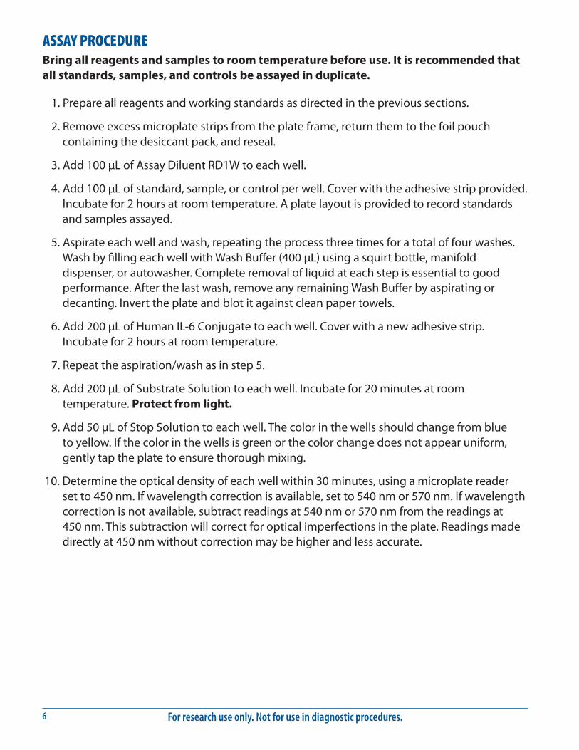
ASSAY PROCEDURE Bring all reagents and samples to room temperature before use. It is recommended that all standards, samples, and controls be assayed in duplicate.
1. Prepare all reagents and working standards as directed in the previous sections.
2. Remove excess microplate strips from the plate frame, return them to the foil pouch containing the desiccant pack, and reseal.
3. Add 100 μL of Assay Diluent RD1W to each well.
4. Add 100 μL of standard, sample, or control per well. Cover with the adhesive strip provided. Incubate for 2 hours at room temperature. A plate layout is provided to record standards and samples assayed.
5. Aspirate each well and wash, repeating the process three times for a total of four washes. Wash by filling each well with Wash Buffer (400 μL) using a squirt bottle, manifold dispenser, or autowasher. Complete removal of liquid at each step is essential to good performance. After the last wash, remove any remaining Wash Buffer by aspirating or decanting. Invert the plate and blot it against clean paper towels.
6. Add 200 μL of Human IL-6 Conjugate to each well. Cover with a new adhesive strip. Incubate for 2 hours at room temperature.
7. Repeat the aspiration/wash as in step 5.
8. Add 200 μL of Substrate Solution to each well. Incubate for 20 minutes at room temperature. Protect from light.
9. Add 50 μL of Stop Solution to each well. The color in the wells should change from blue to yellow. If the color in the wells is green or the color change does not appear uniform, gently tap the plate to ensure thorough mixing.
10. Determine the optical density of each well within 30 minutes, using a microplate reader set to 450 nm. If wavelength correction is available, set to 540 nm or 570 nm. If wavelength correction is not available, subtract readings at 540 nm or 570 nm from the readings at 450 nm. This subtraction will correct for optical imperfections in the plate. Readings made directly at 450 nm without correction may be higher and less accurate.

www.RnDSystems.com 7
CALCULATION OF RESULTSAverage the duplicate readings for each standard, control, and sample and subtract the average zero standard optical density (O.D.).
Create a standard curve by reducing the data using computer software capable of generating a four parameter logistic (4-PL) curve fit. As an alternative, construct a standard curve by plotting the mean absorbance for each standard on the y-axis against the concentration on the x-axis and draw a best fit curve through the points on the graph. The data may be linearized by plotting the log of the human IL-6 concentrations versus the log of the O.D. and the best fit line can be determined by regression analysis. This procedure will produce an adequate but less precise fit of the data.
If samples have been diluted, the concentration read from the standard curve must be multiplied by the dilution factor.
TYPICAL DATAThese standard curves are provided for demonstration only. A standard curve should be generated for each set of samples assayed.
(pg/mL) O.D. Average Corrected0 0.022 0.025 —
0.0283.13 0.050 0.051 0.026
0.0526.25 0.078 0.078 0.053
0.07812.5 0.134 0.135 0.110
0.13625 0.247 0.246 0.221
0.24550 0.472 0.468 0.443
0.465100 0.865 0.850 0.825
0.836300 2.524 2.520 2.495
2.515
(pg/mL) O.D. Average Corrected0 0.025 0.027 —
0.0293.13 0.049 0.050 0.023
0.0516.25 0.078 0.078 0.051
0.07712.5 0.127 0.128 0.101
0.12925 0.236 0.236 0.209
0.23650 0.438 0.440 0.413
0.442100 0.780 0.776 0.749
0.773300 2.176 2.198 2.171
2.221
CELL CULTURE SUPERNATE ASSAY
SERUM/PLASMA ASSAY
For research use only. Not for use in diagnostic procedures.8
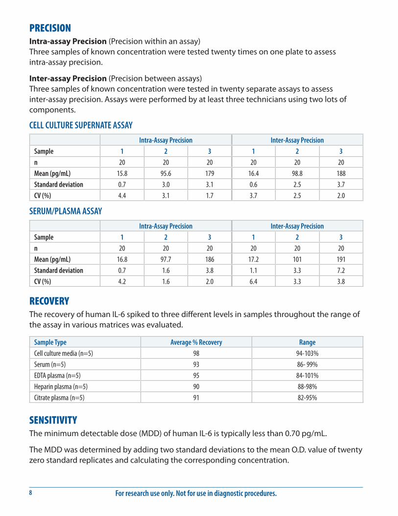
PRECISIONIntra-assay Precision (Precision within an assay) Three samples of known concentration were tested twenty times on one plate to assess intra-assay precision.
Inter-assay Precision (Precision between assays) Three samples of known concentration were tested in twenty separate assays to assess inter-assay precision. Assays were performed by at least three technicians using two lots of components.
CELL CULTURE SUPERNATE ASSAY
Intra-Assay Precision Inter-Assay Precision
Sample 1 2 3 1 2 3
n 20 20 20 20 20 20
Mean (pg/mL) 15.8 95.6 179 16.4 98.8 188
Standard deviation 0.7 3.0 3.1 0.6 2.5 3.7
CV (%) 4.4 3.1 1.7 3.7 2.5 2.0
SERUM/PLASMA ASSAY
Intra-Assay Precision Inter-Assay Precision
Sample 1 2 3 1 2 3
n 20 20 20 20 20 20
Mean (pg/mL) 16.8 97.7 186 17.2 101 191
Standard deviation 0.7 1.6 3.8 1.1 3.3 7.2
CV (%) 4.2 1.6 2.0 6.4 3.3 3.8
RECOVERYThe recovery of human IL-6 spiked to three different levels in samples throughout the range of the assay in various matrices was evaluated.
Sample Type Average % Recovery Range
Cell culture media (n=5) 98 94-103%
Serum (n=5) 93 86- 99%
EDTA plasma (n=5) 95 84-101%
Heparin plasma (n=5) 90 88-98%
Citrate plasma (n=5) 91 82-95%
SENSITIVITYThe minimum detectable dose (MDD) of human IL-6 is typically less than 0.70 pg/mL.
The MDD was determined by adding two standard deviations to the mean O.D. value of twenty zero standard replicates and calculating the corresponding concentration.

www.RnDSystems.com 9
LINEARITYTo assess the linearity of the assay, samples were spiked with high concentrations of human IL-6 in various matrices and diluted with the appropriate calibrator diluent to produce samples with values within the dynamic range of the assay.
Cell culture media (n=4)
Serum (n=4)
EDTA plasma (n=4)
Heparin plasma (n=4)
Citrate plasma (n=4)
1:2Average % of Expected 99 97 101 103 101
Range (%) 96-101 92-100 98-105 96-109 96-106
1:4Average % of Expected 100 101 104 106 105
Range (%) 93-110 93-107 97-110 97-113 101-109
1:8Average % of Expected 96 102 100 104 106
Range (%) 92-100 96-108 86-112 93-111 101-111
1:16Average % of Expected 94 103 99 105 101
Range (%) 83-108 93-111 90-110 99-107 90-114
CALIBRATIONThis immunoassay is calibrated against highly purified E. coli-expressed recombinant human IL-6 produced at R&D Systems®. The NIBSC/WHO 1st International Standard for IL-6 (89/548), which was intended as a potency standard, was evaluated in this kit. The NIBSC/WHO standard is a CHO cell-derived recombinant human IL-6.
The dose response curve of the International Standard (89/548) parallels the Quantikine® standard curve. To convert sample values obtained with the Quantikine® Human IL-6 kit to approximate NIBSC 89/548 units, use the equation below.
NIBSC (89/548) approximate value (IU/mL)=0.131 x Quantikine® Human IL-6 value (pg/mL)
SAMPLE VALUESSerum/Plasma - Forty serum and plasma samples from apparently healthy volunteers were evaluated for the presence of human IL-6 in this assay. Thirty-three samples measured less than the lowest standard, 3.13 pg/mL. Seven samples measured between 3.13 and 12.5 pg/mL. No medical histories were available for the donors used in this study.
Cell Culture Supernates - Human peripheral blood mononuclear cells (1 x 10⁶ cells/mL) were cultured in RPMI supplemented with 10% fetal bovine serum, 50 μM β-mercaptoethanol, 2 mM L-glutamine, 100 U/mL penicillin, and 100 μg/mL streptomycin sulfate and stimulated for 1, 3, and 5 days with 10 μg/mL PHA. Aliquots of the culture supernates were removed on days 1, 3, and 5 and assayed for levels of human IL-6.
Condition Day 1 (pg/mL) Day 3 (pg/mL) Day 5 (pg/mL)
Unstimulated 575 311 660
Stimulated 17,130 17,520 16,340

For research use only. Not for use in diagnostic procedures.10
SPECIFICITYThis assay recognizes natural and recombinant human IL-6.
The factors listed below were prepared at 50 ng/mL in Calibrator Diluent RD5T and at 100 ng/mL in Calibrator Diluent RD6F and assayed for cross-reactivity. Preparations of the following factors at 50 ng/mL in a mid-range recombinant human IL-6 control prepared in Calibrator Diluent RD5T and 100 ng/mL in a mid-range IL-6 control prepared in Calibrator Diluent RD6F were assayed for interference. No significant cross-reactivity or interference was observed.
Recombinant human:CNTFG-CSFGM-CSFgp130IL-1αIL-1βIL-2IL-3IL-4IL-6 RαIL-6 Rα/gp130
IL-7IL-8IL-11IL-12LIFLIF ROSMTNF-αTNF-β
Recombinant mouse:GM-CSFIL-2IL-3IL-4IL-5IL-6IL-7IL-11IL-12
Recombinant rat:CNTF
Natural proteins:bovine FGF acidicbovine FGF basichuman PDGFporcine PDGFhuman TGF-β1porcine TGF-β1.2porcine TGF-β2
IL-6
Untre
ated
LPS
OA Anti-
TNF-α
+ OA
H7 H7 +
OA
HU21
1
HU21
1 +
OA
150
100
75
50
37
25
20
kDa
2000
Untreated LP
S OATN
F-α +
OA H7H7 +
OA
HU211HU211 +
OA
400060008000
10000120001400016000
pg/m
L
Monocytes were prepared from human PBMCs by adherence to plastic. Adherent monocytes were washed, replated, and allowed to rest for 24 hours. Pretreatments were for 30 minutes: neutralizing anti-human TNF-α (R&D Systems®, Catalog # MAB610) at 5 μg/mL, H7 serine kinase inhibitor (Tocris, Catalog # 0542) at 10 μM, or HU211 NFκB inhibitor (Tocris, Catalog # 2861) at 10 μM. Following the pretreatment, 500 ng/mL LPS or 30 ng/mL okadaic acid (OA, Tocris, Catalog # 1136) was added for 20 hours as indicated. Conditioned media was tested in the Quantikine® ELISA, resolved by SDS-PAGE, transferred to a PVDF membrane, and immunoblotted with the detection antibody used in this kit. The immunoprecipitation/Western Blot shows direct correlation with the ELISA value for these samples.

www.RnDSystems.com 11
REFERENCES1. Mansell, A. and B.J. Jenkins (2013) Cytokine Growth Factor Rev. 24:249.2. Schuett, H. et al. (2009) Thromb. Haemost. 102:215.3. Erta, M. et al. (2012) Int. J. Biol. Sci. 8:1254.4. Garbers, C. et al. (2012) Cytokine Growth Factor Rev. 23:85.5. Mihara, M. et al. (2012) Clin. Sci. (Lond.) 122:143.6. Hirano, T. et al. (1986) Nature 324:73.7. Kestler, D.P. et al. (1995) Blood 86:4559.8. Kestler, D.P. et al. (1999) Am. J. Hematol. 61:169.9. Bihl, M.P. et al. (2002) Am. J. Respir. Cell Mol. Biol. 27:48.
10. Alberti, L. et al. (2005) Cancer Res. 65:2.11. May, L.T. et al. (1986) Proc. Natl. Acad. Sci. USA 83:8957.12. Sad, S. et al. (1995) Immunity 2:271.13. Cichy, J. et al. (1996) Biochem. Biophys. Res. Commun. 227:318.14. Miyazawa, K. et al. (1998) Am. J. Pathol. 152:793.15. Fried, S.K. et al. (1998) Endocrinology 83:847.16. Ishimi, Y. et al. (1990) J. Immunol. 145:3297.17. Jiang, S. et al. (1994) Blood 84:4151.18. Xin, X. et al. (1995) Endocrinology 136:132.19. Marz, P. et al. (1998) Proc. Natl. Acad. Sci. USA 95:3251.20. Ringheim, G.E. et al. (1995) J. Neuroimmunol. 63:113.21. Gadient, R.A. et al. (1995) Neurosci. Lett. 194:17.22. Kuppner, M.C. et al. (1995) Immunology 84:265.23. Gagari, E. et al. (1997) Blood 89:2654.24. Cumberbatch, M. et al. (1996) Immunology 87:513. 25. Fujisawa, H. et al. (1997) J. Interferon Cytokine Res. 17:347.26. Lee, S.C. et al. (1993) J. Immunol. 150:2659.27. Lafortune, L. et al. (1996) J. Neuropathol. Exp. Neurol. 55:515.28. Ericson, S.G. et al. (1998) Blood 91:2099.29. Melani, C. et al. (1993) Blood 81:2744.30. Lacy, P. et al. (1998) Blood 91:2508.31. Jung, H.C. et al. (1995) J. Clin. Invest. 95:55.32. Spencer, N.F.L. and R.A. Daynes (1997) Int. Immunol. 9:745.33. Campbell, I.L. et al. (1989) J. Immunol. 143:1188.34. D’Auria, L. et al. (1997) Eur. Cytokine Netw. 8:383.35. Yamamura, M. et al. (1998) Br. J. Haematol. 100:129.36. Angstwurm, M.W.A. et al. (1997) Cytokine 9:370.37. Mouawad, R. et al. (1996) Clin. Cancer Res. 2:1405.38. Sakamoto, K. et al. (1994) Cytokine 6:181.39. Murakami, M. et al. (1993) Science 260:1808.40. Muller-Newen, G. (2003) Sci. STKE 2003:PE40.41. Mitsuyama, K. et al. (2006) Clin. Exp. Immunol. 143:125.42. Cerutti, A. et al. (1998) J. Immunol. 160:2145.

For research use only. Not for use in diagnostic procedures.12
PLATE LAYOUTUse this plate layout to record standards and samples assayed.

www.RnDSystems.com 13
NOTES

For research use only. Not for use in diagnostic procedures.14
NOTES
01.06 749909.8 3/17
©2017 R&D Systems®, Inc.
All trademarks and registered trademarks are the property of their respective owners.