mouse c-reactive protein/crp quantikine · the quantikine® mouse c-reactive protein/crp...
TRANSCRIPT
Mouse C-Reactive Protein/CRP Immunoassay
Quantikine® ELISA
This package insert must be read in its entirety before using this product. For research use only. Not for use in diagnostic procedures.
Catalog Number MCRP00
For the quantitative determination of mouse C-Reactive Protein (CRP) concentrations in cell culture supernates, tissue lysates, serum, and plasma.
MANUFACTURED AND DISTRIBUTED BY:
USA & Canada | R&D Systems, Inc. 614 McKinley Place NE, Minneapolis, MN 55413, USATEL: (800) 343-7475 (612) 379-2956 FAX: (612) 656-4400E-MAIL: [email protected]
DISTRIBUTED BY:
UK & Europe | R&D Systems Europe, Ltd.19 Barton Lane, Abingdon Science Park, Abingdon OX14 3NB, UKTEL: +44 (0)1235 529449 FAX: +44 (0)1235 533420E-MAIL: [email protected]
China | R&D Systems China Co., Ltd.24A1 Hua Min Empire Plaza, 726 West Yan An Road, Shanghai PRC 200050TEL: +86 (21) 52380373 FAX: +86 (21) 52371001E-MAIL: [email protected]
TABLE OF CONTENTS
SECTION PAGEINTRODUCTION .....................................................................................................................................................................1
PRINCIPLE OF THE ASSAY ...................................................................................................................................................1
LIMITATIONS OF THE PROCEDURE .................................................................................................................................2
TECHNICAL HINTS .................................................................................................................................................................2
PRECAUTIONS .........................................................................................................................................................................2
MATERIALS PROVIDED & STORAGE CONDITIONS ...................................................................................................3
OTHER SUPPLIES REQUIRED .............................................................................................................................................4
OTHER SUPPLIES REQUIRED FOR TISSUE LYSATE SAMPLES ................................................................................4
SAMPLE COLLECTION & STORAGE .................................................................................................................................4
SAMPLE PREPARATION........................................................................................................................................................4
REAGENT PREPARATION .....................................................................................................................................................5
ASSAY PROCEDURE .............................................................................................................................................................6
CALCULATION OF RESULTS ...............................................................................................................................................7
TYPICAL DATA .........................................................................................................................................................................7
PRECISION ................................................................................................................................................................................8
RECOVERY.................................................................................................................................................................................8
LINEARITY .................................................................................................................................................................................8
SENSITIVITY .............................................................................................................................................................................9
CALIBRATION ..........................................................................................................................................................................9
SAMPLE VALUES .....................................................................................................................................................................9
SPECIFICITY ........................................................................................................................................................................... 10
REFERENCES ......................................................................................................................................................................... 11
PLATE LAYOUT ..................................................................................................................................................................... 12

www.RnDSystems.com 1
INTRODUCTIONC-Reactive Protein (CRP), also known as Pentraxin 1, is a non-glycosylated protein in the Pentraxin family that also includes Pentraxin 2/SAP and Pentraxin 3/TSG-14. CRP functions as a sensor and activator of the innate immune response (1). In humans, it is a major acute-phase protein; its circulating concentration is dramatically elevated at the onset of inflammation (2). In mice, however, serum CRP levels increase only slightly during inflammation, and the analogous acute phase role is filled by Pentraxin 2 (3). CRP assembles non-covalently into a 110-120 kDa cyclical pentamer (4). Mature mouse CRP shares 71% amino acid sequence identity with human and rat CRP (5).
CRP binds and opsonizes apoptotic cells (6-8) as well as bacteria such as S. pneumoniae (9, 10). It subsequently enhances the phagocytosis of these opsonized cells (6, 8-10). CRP additionally binds several proteins in the complement cascade including C1q, C4BP, and Factor H (8, 11-13). It enhances activation of the classical complement pathway and the deposition of C3b (9). In later stages of the response, CRP inhibits complement-mediated cell lysis through its binding to C4BP and Factor H (8, 12). These interactions induce the upregulation of complement inhibitory proteins CD46, CD59, and CD55/DAF, and inhibit assembly of the membrane attack complex (MAC) (8, 14).
CRP binds to FcγRI, FcγRIIA, and FcγRIIB on macrophages and dendritic cells (15-17), and Fc receptors are required for the phagocytosis of CRP-opsonized target cells (6, 10, 18). CRP binding to FcγRI induces Src activation which subsequently triggers the inhibitory FcγRIIb and dampens the inflammatory response (15, 19). CRP additionally promotes dendritic cell maturation and humoral immunity (10). In cardiovascular disease, CRP binds to oxidized LDL, exacerbates tissue damage in coronary artery infarction, and inhibits the repair of injured vascular endothelium (7, 19, 20).
The Quantikine® Mouse C-Reactive Protein/CRP Immunoassay is a 4.5 hour solid-phase ELISA designed to measure mouse CRP in cell culture supernates, tissue lysates, serum, and plasma. It contains NS0-expressed recombinant mouse CRP and antibodies raised against the recombinant factor. This immunoassay has been shown to accurately quantitate recombinant mouse CRP. Results obtained using natural mouse CRP showed dose response curves that were parallel to the standard curves obtained using the Quantikine® kit standards. These results indicate that this kit can be used to determine relative mass values for mouse natural CRP.
PRINCIPLE OF THE ASSAYThis assay employs the quantitative sandwich enzyme immunoassay technique. A monoclonal antibody specific for mouse CRP has been pre-coated onto a microplate. Standards, control, and samples are pipetted into the wells and any CRP present is bound by the immobilized antibody. After washing away any unbound substances, an enzyme-linked polyclonal antibody specific for mouse CRP is added to the wells. Following a wash to remove any unbound antibody-enzyme reagent, a substrate solution is added to the wells. The enzyme reaction yields a blue product that turns yellow when the Stop Solution is added. The intensity of the color measured is in proportion to the amount of CRP bound in the initial step. The sample values are then read off the standard curve.
For research use only. Not for use in diagnostic procedures.2
LIMITATIONS OF THE PROCEDURE• FOR RESEARCH USE ONLY. NOT FOR USE IN DIAGNOSTIC PROCEDURES.
• The kit should not be used beyond the expiration date on the kit label.
• Do not mix or substitute reagents with those from other lots or sources.
• If samples generate values higher than the highest standard, further dilute samples with calibrator diluent and repeat the assay.
• Any variation in standard diluent, operator, pipetting technique, washing technique, incubation time or temperature, and kit age can cause variation in binding.
• Variations in sample collection, processing, and storage may cause sample value differences.
• This assay is designed to eliminate interference by other factors present in biological samples. Until all factors have been tested in the Quantikine® Immunoassay, the possibility of interference cannot be excluded.
TECHNICAL HINTS• When mixing or reconstituting protein solutions, always avoid foaming.
• To avoid cross-contamination, change pipette tips between additions of each standard level, between sample additions, and between reagent additions. Also, use separate reservoirs for each reagent.
• To ensure accurate results, proper adhesion of plate sealers during incubation steps is necessary.
• Substrate Solution should remain colorless until added to the plate. Keep Substrate Solution protected from light. Substrate Solution should change from colorless to gradations of blue.
• Stop Solution should be added to the plate in the same order as the Substrate Solution. The color developed in the wells will turn from blue to yellow upon addition of the Stop Solution.
PRECAUTIONSThe Stop Solution provided with this kit is an acid solution.
Some components in this kit contain a preservative which may cause an allergic skin reaction. Avoid breathing mist.
Color Reagent B may cause skin, eye, and respiratory irritation. Avoid breathing fumes.
Wear protective gloves, clothing, eye, and face protection. Wash hands thoroughly after handling. Refer to the MSDS on our website prior to use.
www.RnDSystems.com 3
MATERIALS PROVIDED & STORAGE CONDITIONSStore the unopened kit at 2-8 °C. Do not use past kit expiration date.
PART PART # DESCRIPTIONSTORAGE OF OPENED/ RECONSTITUTED MATERIAL
Mouse CRP Microplate
894713 96 well polystyrene microplate (12 strips of 8 wells) coated with a monoclonal antibody specific for mouse CRP.
Return unused wells to the foil pouch containing the desiccant pack. Reseal along entire edge of the zip-seal. May be stored for up to 1 month at 2-8 °C.*
Mouse CRP Standard
894715 2 vials of recombinant mouse CRP in a buffered protein base with preservatives; lyophilized. Refer to the vial label for reconstitution volume.
Discard after use. Use a fresh standard and control for each assay.Mouse CRP
Control894716 2 vials of recombinant mouse CRP in a
buffered protein base with preservatives; lyophilized. The assay value of the control should be within the range specified on the label.
Mouse CRP Conjugate
894714 12 mL of a polyclonal antibody specific for mouse CRP conjugated to horseradish peroxidase with preservatives.
May be stored for up to 1 month at 2-8 °C.*
Assay Diluent RD1W
895117 11 mL of a buffered protein solution with preservatives.
Calibrator Diluent RD5P Concentrate
895151 21 mL of a concentrated buffered protein base with preservatives. Use diluted 1:5 in this assay.
Wash Buffer Concentrate
895003 21 mL of a 25-fold concentrated solution of buffered surfactant with preservative. May turn yellow over time.
Color Reagent A 895000 12 mL of stabilized hydrogen peroxide.
Color Reagent B 895001 12 mL of stabilized chromogen (tetramethylbenzidine).
Stop Solution 895174 23 mL of diluted hydrochloric acid.
Plate Sealers N/A 4 adhesive strips.
* Provided this is within the expiration date of the kit.
For research use only. Not for use in diagnostic procedures.4
OTHER SUPPLIES REQUIRED• Microplate reader capable of measuring absorbance at 450 nm, with the correction
wavelength set at 540 nm or 570 nm.
• Pipettes and pipette tips.
• Deionized or distilled water.
• Squirt bottle, manifold dispenser, or automated microplate washer.
• 100 mL and 500 mL graduated cylinders.
• Polypropylene test tubes for dilution of standards and samples.
OTHER SUPPLIES REQUIRED FOR TISSUE LYSATE SAMPLES• Cell Lysis Buffer 2 (R&D Systems®, Catalog # 895347)
• PBS
SAMPLE COLLECTION & STORAGEThe sample collection and storage conditions listed below are intended as general guidelines. Sample stability has not been evaluated.
Cell Culture Supernates - Remove particulates by centrifugation. Assay immediately or aliquot and store samples at ≤ -20 °C. Avoid repeated freeze-thaw cycles.
Tissue Lysates - Cell must be lysed prior to assay as directed in the Sample Values section.
Serum - Allow blood samples to clot for 2 hours at room temperature before centrifuging for 20 minutes at 2000 x g. Remove serum and assay immediately or aliquot and store samples at ≤ -20 °C. Avoid repeated freeze-thaw cycles.
Plasma - Collect plasma using EDTA or heparin as an anticoagulant. Centrifuge for 20 minutes at 2000 x g within 30 minutes of collection. Assay immediately or aliquot and store samples at ≤ -20 °C. Avoid repeated freeze-thaw cycles.
Note: Citrate plasma has not been validated for use in this assay. Repeated freeze-thaw cycles will cause sample variation.
SAMPLE PREPARATIONSerum and plasma samples require a 2000-fold dilution. A 2000-fold dilution can be achieved by adding 10 μL of sample to 490 μL of Calibrator Diluent RD5P (diluted 1:5).* Complete the 2000-fold dilution by adding 10 μL of the diluted sample to 390 μL Calibrator Diluent RD5P (diluted 1:5).
*See Reagent Preparation section.
www.RnDSystems.com 5
REAGENT PREPARATIONBring all reagents to room temperature before use.
Mouse CRP Control - Reconstitute the control with 1.0 mL of deionized or distilled water. Mix thoroughly. Assay the control undiluted.
Wash Buffer - If crystals have formed in the concentrate, warm to room temperature and mix gently until the crystals have completely dissolved. Add 20 mL of Wash Buffer Concentrate to deionized or distilled water to prepare 500 mL of Wash Buffer.
Substrate Solution - Color Reagents A and B should be mixed together in equal volumes within 15 minutes of use. Protect from light. 100 μL of the resultant mixture is required per well.
Calibrator Diluent RD5P (diluted 1:5) - Add 20 mL of Calibrator Diluent RD5P Concentrate to 80 mL of deionized or distilled water to prepare 100 mL of Calibrator Diluent RD5P (diluted 1:5).
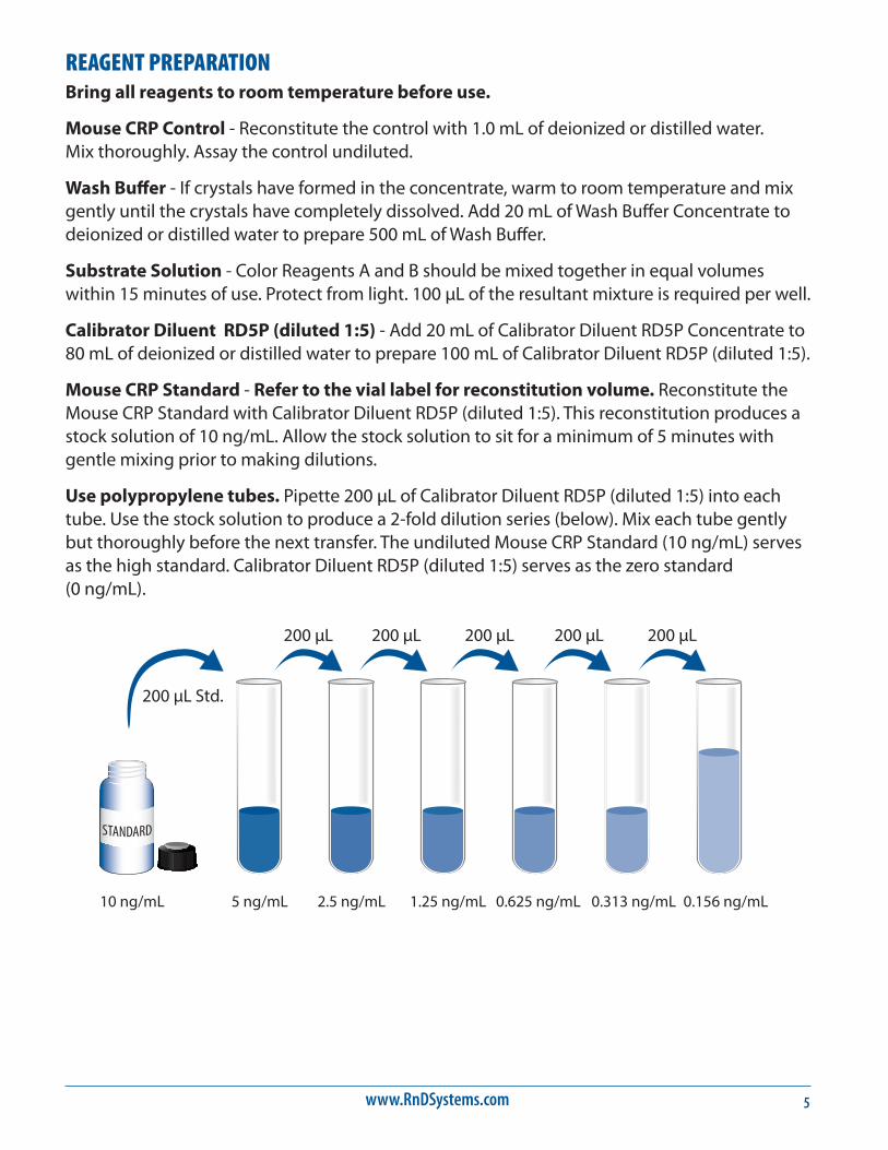
Mouse CRP Standard - Refer to the vial label for reconstitution volume. Reconstitute the Mouse CRP Standard with Calibrator Diluent RD5P (diluted 1:5). This reconstitution produces a stock solution of 10 ng/mL. Allow the stock solution to sit for a minimum of 5 minutes with gentle mixing prior to making dilutions.
Use polypropylene tubes. Pipette 200 μL of Calibrator Diluent RD5P (diluted 1:5) into each tube. Use the stock solution to produce a 2-fold dilution series (below). Mix each tube gently but thoroughly before the next transfer. The undiluted Mouse CRP Standard (10 ng/mL) serves as the high standard. Calibrator Diluent RD5P (diluted 1:5) serves as the zero standard (0 ng/mL).
200 µL Std.
10 ng/mL 5 ng/mL 2.5 ng/mL 1.25 ng/mL 0.625 ng/mL 0.313 ng/mL 0.156 ng/mL
200 µL 200 µL 200 µL 200 µL 200 µL

For research use only. Not for use in diagnostic procedures.6
ASSAY PROCEDURE Bring all reagents and samples to room temperature before use. It is recommended that all standards, control, and samples be assayed in duplicate.
1. Prepare all reagents, standard dilutions, control, and samples as directed in the previous sections.
2. Remove excess microplate strips from the plate frame, return them to the foil pouch containing the desiccant pack, and reseal.
3. Add 50 μL of Assay Diluent RD1W to each well.
4. Add 50 μL of standard, control, or sample* per well. Cover with the adhesive strip provided. Incubate for 2 hours at room temperature. A plate layout is provided to record standards and samples assayed.
5. Aspirate each well and wash, repeating the process three times for a total of four washes. Wash by filling each well with Wash Buffer (400 μL) using a squirt bottle, manifold dispenser, or autowasher. Complete removal of liquid at each step is essential to good performance. After the last wash, remove any remaining Wash Buffer by aspirating or decanting. Invert the plate and blot it against clean paper towels.
6. Add 100 μL of Mouse CRP Conjugate to each well. Cover with a new adhesive strip. Incubate for 2 hours at room temperature.
7. Repeat the aspiration/wash as in step 5.
8. Add 100 μL of Substrate Solution to each well. Incubate for 30 minutes at room temperature on the benchtop. Protect from light.
9. Add 100 μL of Stop Solution to each well. Gently tap the plate to ensure thorough mixing.
10. Determine the optical density of each well within 30 minutes, using a microplate reader set to 450 nm. If wavelength correction is available, set to 540 nm or 570 nm. If wavelength correction is not available, subtract readings at 540 nm or 570 nm from the readings at 450 nm. This subtraction will correct for optical imperfections in the plate. Readings made directly at 450 nm without correction may be higher and less accurate.
*Samples may require dilution. See Sample Preparation section.
www.RnDSystems.com 7
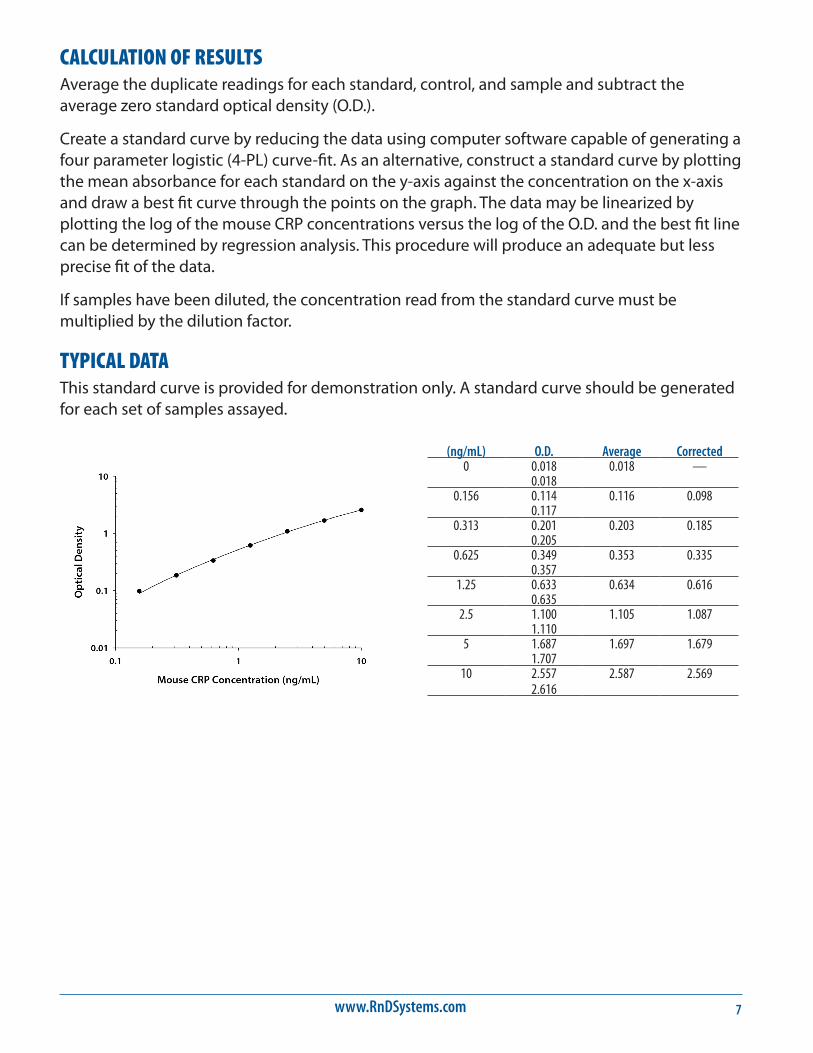
CALCULATION OF RESULTSAverage the duplicate readings for each standard, control, and sample and subtract the average zero standard optical density (O.D.).
Create a standard curve by reducing the data using computer software capable of generating a four parameter logistic (4-PL) curve-fit. As an alternative, construct a standard curve by plotting the mean absorbance for each standard on the y-axis against the concentration on the x-axis and draw a best fit curve through the points on the graph. The data may be linearized by plotting the log of the mouse CRP concentrations versus the log of the O.D. and the best fit line can be determined by regression analysis. This procedure will produce an adequate but less precise fit of the data.
If samples have been diluted, the concentration read from the standard curve must be multiplied by the dilution factor.
TYPICAL DATAThis standard curve is provided for demonstration only. A standard curve should be generated for each set of samples assayed.
(ng/mL) O.D. Average Corrected0 0.018 0.018 —
0.0180.156 0.114 0.116 0.098
0.1170.313 0.201 0.203 0.185
0.2050.625 0.349 0.353 0.335
0.3571.25 0.633 0.634 0.616
0.6352.5 1.100 1.105 1.087
1.1105 1.687 1.697 1.679
1.70710 2.557 2.587 2.569
2.616
For research use only. Not for use in diagnostic procedures.8
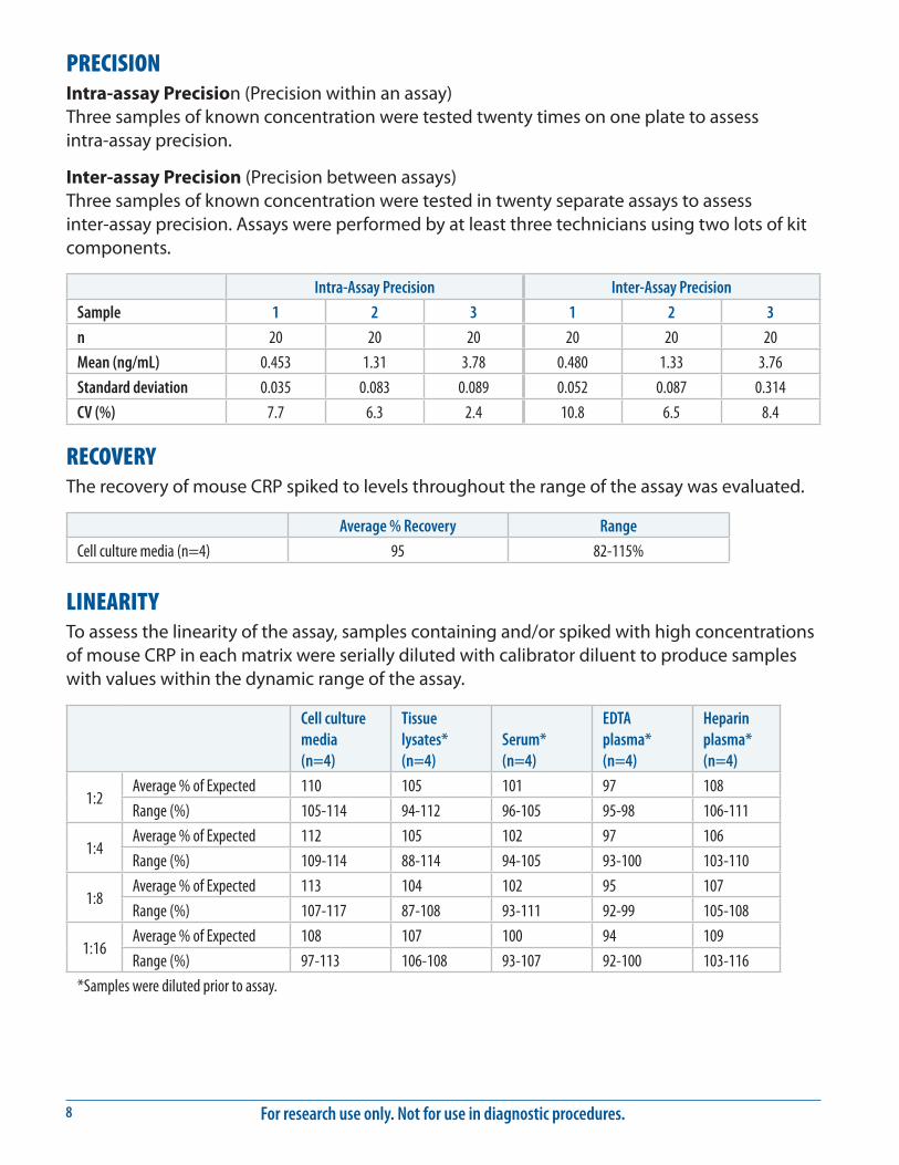
PRECISIONIntra-assay Precision (Precision within an assay) Three samples of known concentration were tested twenty times on one plate to assess intra-assay precision.
Inter-assay Precision (Precision between assays) Three samples of known concentration were tested in twenty separate assays to assess inter-assay precision. Assays were performed by at least three technicians using two lots of kit components.
Intra-Assay Precision Inter-Assay Precision
Sample 1 2 3 1 2 3
n 20 20 20 20 20 20
Mean (ng/mL) 0.453 1.31 3.78 0.480 1.33 3.76
Standard deviation 0.035 0.083 0.089 0.052 0.087 0.314
CV (%) 7.7 6.3 2.4 10.8 6.5 8.4
RECOVERYThe recovery of mouse CRP spiked to levels throughout the range of the assay was evaluated.
Average % Recovery Range
Cell culture media (n=4) 95 82-115%
LINEARITYTo assess the linearity of the assay, samples containing and/or spiked with high concentrations of mouse CRP in each matrix were serially diluted with calibrator diluent to produce samples with values within the dynamic range of the assay.
Cell culture media (n=4)
Tissue lysates* (n=4)
Serum* (n=4)
EDTA plasma* (n=4)
Heparin plasma* (n=4)
1:2Average % of Expected 110 105 101 97 108
Range (%) 105-114 94-112 96-105 95-98 106-111
1:4Average % of Expected 112 105 102 97 106
Range (%) 109-114 88-114 94-105 93-100 103-110
1:8Average % of Expected 113 104 102 95 107
Range (%) 107-117 87-108 93-111 92-99 105-108
1:16Average % of Expected 108 107 100 94 109
Range (%) 97-113 106-108 93-107 92-100 103-116
*Samples were diluted prior to assay.

www.RnDSystems.com 9
SENSITIVITYTwenty-nine assays were evaluated and the minimum detectable dose (MDD) of mouse CRP ranged from 0.002-0.015 ng/mL. The mean MDD was 0.006 ng/mL.
The MDD was determined by adding two standard deviations to the mean O.D. value of twenty zero standard replicates and calculating the corresponding concentration.
CALIBRATIONThis immunoassay is calibrated against a highly purified NS0-expressed recombinant mouse C-Reactive Protein/CRP produced at R&D Systems®.
SAMPLE VALUESSerum/Plasma - Samples were evaluated for the presence of mouse CRP in this assay.
Samples Mean (ng/mL) Range (ng/mL) Standard Deviation (ng/mL)
Serum (n=10) 8351 3976-12,792 2814
EDTA plasma (n=5) 4395 3168-5360 948
Heparin plasma (n=5) 5545 4404-7218 1027
Cell Culture Supernates - Organs from mice were rinsed with PBS then homogenized with a tissue homogenizer and cultured in RPMI 1640 supplemented with 10% fetal bovine serum, 2 mM L-glutamine, 100 U/mL penicillin, and 100 μg/mL streptomycin sulfate. Cells were cultured unstimulated for 1 or 3 days. Aliquots of the cell culture supernates were removed and assayed for mouse CRP.
Tissue Value (ng/mL)
Heart (1 day) 4.26
Kidney (3 days) 2.88
Tissue Lysates - Organs from mice were rinsed with PBS, cut into 1-2 mm pieces, and homogenized with a tissue homogenizer in PBS. An equal volume of Cell Lysis Buffer 2 was added and tissues were lysed at room temperature for 30 minutes with gentle agitation. Debris was then removed by centrifugation. Aliquots of the lysates were removed and assayed for mouse CRP.
Tissue Value (ng/mg of cell lysate)
Heart (1 day) 9.04
Kidney (3 days) 8.25

For research use only. Not for use in diagnostic procedures.10
SPECIFICITYThis assay recognizes natural and recombinant mouse CRP.
The factors listed below were prepared at 1 μg/mL in calibrator diluent and assayed for cross-reactivity. Preparations of the following factors at 1 μg/mL in a mid-range mouse CRP control were assayed for interference. No significant cross-reactivity or interference was observed.
Recombinant mouse:Pentraxin-2TSGTSG-6TSG-14Fcγ RIFcγ RIIBFcγ RIII
Recombinant rat:C-Reactive Protein
Recombinant human:C-Reactive Protein
CRP
Untre
ated
Case
in24
hour
Case
in48
hour
75
50
37
25
20
15
10
kDa
0
2
4
6
8
10
Untreated Casein 24 hour Casein 48 hour
CRP
(μg/
mL)
Mouse serum samples from untreated C57/Bl6 mice or mice treated with Casein for 24 or 48 hours were analyzed by Western Blot and this Quantikine® ELISA kit. Samples were resolved under reducing SDS-PAGE conditions, transferred to a PVDF membrane, and immunoblotted with the detection antibody supplied in this kit. The Western Blot and ELISA values for these samples correlate. While Mouse CRP was present in untreated mice as detected using the Quantikine® ELISA, the concentration was below the threshold of detection by Western Blot.
www.RnDSystems.com 11
REFERENCES1. Du Clos, T.W. and C. Mold (2011) Curr. Opin. Organ Transplant. 16:15.
2. Ahmed, M.S et al. (2012) ISRN Inflamm. 2012:953461.
3. Pepys, M.B. et al. (1979) Nature 278:259.
4. Shrive, A.K. et al. (1996) Nat. Struct. Biol. 3:346.
5. Whitehead, A.S. et al. (1990) Biochem. J. 266:283.
6. Mold, C. et al. (2002) J. Autoimmun. 19:147.
7. Chang, M-K. et al. (2002) Proc. Natl. Acad. Sci. USA 99:13043.
8. Gershov, D. et al. (2000) J. Exp. Med. 192:1353.
9. Mukerji, R. et al. (2012) J. Immunol. 189:5327.
10. Thomas-Rudolph, D. et al. (2007) J. Immunol. 178:7283.
11. McGrath, F.D.G. et al. (2006) J. Immunol. 176:2950.
12. Sjoberg, A.P. et al. (2006) J. Immunol. 176:7612.
13. Okemefuna, A.I. et al. (2010) J. Biol. Chem. 285:1053.
14. Li, S-H. et al. (2004) Circulation 109:833.
15. Marjon, K.D. et al. (2009) J. Immunol. 182:1397.
16. Manolov, D.E. et al. (2004) Arterioscler. Thromb. Vasc. Biol. 24:2372.
17. Stein, M.P. et al. (2000) J. Immunol. 164:1514.
18. Bodman-Smith, K.B. et al. (2004) J. Leukoc. Biol. 75:1029.
19. Sundgren, N.C. et al. (2011) Circ. Res. 109:1132.
20. Griselli, M. et al. (1999) J. Exp. Med. 190:1733.
For research use only. Not for use in diagnostic procedures.12
PLATE LAYOUTUse this plate layout to record standards and samples assayed.

www.RnDSystems.com 13
NOTES
For research use only. Not for use in diagnostic procedures.14
NOTES
01/14 752882.2 10/16
©2016 R&D Systems®, Inc.
All trademarks and registered trademarks are the property of their respective owners.