hplc separation of glucosinolates from leaves and...
TRANSCRIPT

Journal of Chromatographic Science, Vol. 26, November 1988
HPLC Separation of Glucosinolates from Leaves and Seeds of Arabidopsis thaliana and Their Identification Using Thermospray Liquid Chramatography/Mass Spectrometry* L.R. Hogge, D.W. Reed, a n d E.W. Underhill Plant B i o t e c h n o l o g y Inst i tu te , Na t iona l R e s e a r c h C o u n c i l , 1110 G y m n a s i u m R d . , Saska toon ,
S a s k a t c h e w a n , S 7 N O W 9 C a n a d a
G.W. Haughn D e p a r t m e n t of B io logy , Un ivers i t y of S a s k a t c h e w a n , S a s k a t o o n , S a s k a t c h e w a n , S7N O W O C a n a d a
Abstract
Leaf and seed extracts of Arabidopsis thaliana var. Columbia contain a large number of glucosinolates, representing close to 2 5 % of those known to occur in nature. The glucosinolates, in the form of their desulphated analogs, are separated by reversed-phase, high-performance liquid chromatography (HPLC). Seventeen are identified using thermospray liquid chromatography/mass spectrometry (TSP LC/MS). Additional glucosinolates, present in trace amounts, are identified as isothiocyanates by electron impact and chemical ionization gas chromatography/MS (GC/MS). In total, 23 glucosinolates are detected and these include four series of homologs and analogs. Fifteen possess aliphatic side chains, of which six contain ω-methylthioalkyl and six contain ω-methylsulphinylalkyl side chains; eight possess aromatic side chains, of which four constitute an homologous series of benzoic acid esters and three possess 3-indolylmethyl-based structures. Sixteen of the glucosinolates are detected in Arabidopsis thaliana for the first time and three of these, 4-hydroxybutyl glucosinolate, 5-benzoyloxypentyl glucosinolate, and 6-benzoyloxyhexyl glucosinolate, represent novel plant constituents.
In radish, esterification of the thioglucosyl moiety by sinapic acid is also known to occur (1,2) . A l l can be hydrolyzed by the enzyme myrosinase to give D-glucose, sulphate, and a variety o f products derived from the aglycone moiety, including isothiocyanates, nitriles, and other products (3) . Glucosinolates have been reported in 11 plant families and within the Cruciferae
* NRCC Communication No. 29493
they have been found in all species examined (4 ) . Glucosinolates, as a class o f plant constituents, are o f major
importance to several vegetable crops due to the flavor components (isothiocyanates or mustard oils) generated by hydrolysis when the plant tissues are crushed. The hydrolytic products of some glucosinolates exhibit toxic effects in mammals and, thus, have limited the usefulness of some Cruciferae species as fodder (1 ) . Information concerning the biosynthetic pathway of glucosinolates and the genes that encode the enzymes involved in their synthesis could facilitate the genetic engineering of altered glucosinolate levels in crops. One approach to obtaining such information is through the analysis of mutants. As part of a program on the genetic manipulation of glucosinolate levels in crop plants, the present authors plan to take advantage of the speed and versatility of genetic analysis in the small crucifer Arabidopsis thaliana ( L . ) Heynh to isolate and characterize mutants deficient in glucosinolate biosynthesis (5 ) . A necessary first step was to determine the natural composition of glucosinolates in leaf and seed tissue of wild type plants and to adopt a method that would be amenable for their analysis in individual plants.
Only fragmentary reports are available on the glucosinolate composition of seed and leaf tissue of Arabidopsis. In one study, allylglucosinolate and isopropylglucosinolate were identified in leaf tissue as enzyme-derived isothiocyanates and nitriles (6 ) . Seven different glucosinolates have been detected in extracts of seeds by employing negative ion fast atom bombardment (FAB) mass spectrometry (7) and chemical ionization/ mass spectrometry ( C I / M S ) (8,9) . In a preliminary high-performance liquid chromatographic ( H P L C ) analysis o f seed and leaf extracts of Arabidopsis, the present authors found evidence of an even greater number o f glucosinolates, and only a few exhibited retention times that corresponded with available reference standards.
The separation of glucosinolates present in wild type Arabidopsis thaliana leaves and seeds by reversed-phase H P L C of their desulphated analogs is reported here. The technique is rapid and allows detection of the predominant glucosinolates using quantities o f leaf and seed tissue that can be obtained from a single plant. Glucosinolates were identified using the methods of thermospray liquid chromatography/MS ( T S P L C / M S ) (10,11) and gas chromatography ( G Q / M S of their myrosinase-derived isothiocyanates.
Reproduction (photocopying) of editorial content of this journal is prohibited without publisher's permission. 551
Introduction
More than 100 glucosinolates are known. They are represented by the general structure:

Journal of Chromatographic Science, Vol. 26, November 1988
Experimental
Arabidopsis thaliana ( L . ) Heynh. used in this study were descended from the Columbia wild type. The methods employed for growing the plants and harvesting seed were as described by Somerville and Ogren (12), except an 18-h photoperiod rather than continuous illumination was employed.
Glucosinolates were recovered from leaves and seeds separately by extraction into 20 times their fresh weight of boiling 80% ethanol. In the case of leaves, the tissue was rapidly weighed following removal from the plant and immediately placed into boiling solvent. Seeds and leaves were extracted for ca. 5 min at this temperature and ground using a Polytron tissue homogenizer; then, the mixtures were filtered. The residues were extracted again in a similar volume of boiling 80% ethanol. Ethanol was removed from each combined extract by rotary evaporation at 35 °C , and the aqueous mixture was filtered through a Celite pad to give a clear solution.
A n aliquot of the glucosinolate solution was adjusted to pH 7 with phosphate buffer and extracted three times, each time with two volumes of C H 2 C 1 2 . After removing traces of solvent, the aqueous solution was incubated with myrosinase for 2 h at 20°C, and the liberated isothiocyanates were recovered by three extractions, each time with two volumes of C H 2 C 1 2 . The solvent was dried ( M g S 0 4 ) and concentrated by rotary evaporation at 35°C.
Another portion of the glucosinolate solution was applied to DEAE-Sephadex A-25 (pyridine acetate form), and the glucosinolates were converted into their desulpho analogs by overnight treatment with aryl sulphatase (Sigma Chemical, Type H - l ) . The procedure used was similar to that described by Mitchinton et al. (13). The column eluate containing the de-sulphated products was placed in a boiling water bath for 3 min to inactivate any β-galactosidase activity, and then it was concentrated in a Savant Speed Vac concentrator. Ortho-nitro-phenyl-β-D-galactopyranoside was added as an internal standard when required, and the samples were passed through a 0.22-/µm filter.
H P L C of desulfoglucosinolates was carried out using a Spectra Physics Model 8700 X R system equipped with a Model 8780 X R autosampler and a Spectroflow 773 U V detector at a wavelength setting of 226 nm, 0.1 A U F S . Chromatograms were recorded using a Hewlett-Packard Model 3392A plotter-integrator. Samples, 50 μL· each, were separated using a Whatman Partisphere C18 column (110x4.7 mm i.d., 5-μm particle size). Mobile phase at 1 m L / m i n was programmed as follows: 1.25% acetonitrile in water for 5 min, a linear gradient from 1.25 to 22.5% acetonitrile over the next 15 min, constant at 22.5% acetonitrile for 5 min, and a linear gradient to 100% acetonitrile for 15 min. The column was reconditioned by a linear gradient to 1.25% acetonitrile over 5 min and washed with this concentration for 15 min prior to an injection.
L C separation of desulphoglucosinolates in TSP L C / M S was performed using a Waters system consisting of two pumps, Models 590 and 510, controlled by a Model 680 gradient controller. Samples, 20 each, were injected, and separation of the desulfoglucosinolates was achieved on a C ) 8 column similar to the one indicated above, but using a linear gradient elution with 0.1 Μ ammonium acetate and acetonitrile (0 to 60% acetonitrile over 30 min) at a flow rate of 0.9 m L / m i n . TSP L C / M S was performed as described (11) employing a Model 3300 Finnigan mass spectrometer retrofitted with a Vestec thermospray interface. This mass spectrometer and the Model
4500 used for G C were interfaced to an Incos 2300 data acquisition system (Finnigan). The thermospray source was maintained at 265 °C , and spectra were acquired by scanning from m/z 115 to 450 every 2.0 s.
G C / M S data for isothiocyanates were obtained using a Finnigan Model 4500 GC-MS. CI (methane reagent and helium carrier gas) and electron impact ( E I ) (70 eV ionization energy) mass spectra were acquired every second over the mass range of 40 to 400. Samples were separated using a DB-5 (J&W Scientific) phenyl methyl silicone fused-silica column (60 m χ 0.32 mm i .d . ) . After on-column injection with the injector cooled to 38°C, the oven temperature was raised with maximum heating from 40° to 100°C and then programmed at 4° /min to 310°C.
Results and Discussion
Although glucosinolates themselves constitute a uniform class of natural compounds, they do not all yield similar myrosinase hydrolysis products that can be readily analyzed; therefore, the method of reversed-phase H P L C (13) was utilized for their analysis in Arabidopsis seed and leaf tissues. The H P L C profiles of desulfoglucosinolates obtained (Figure 1) indicated the presence of a large number of components. Their identities are listed in Table I and discussed below; each is designated numerically, and these numbers are employed in all figures and tables. Both the composition and relative amounts of the glucosinolates in the two tissue extracts differed appreciably. That the major peaks in the U V trace of the H P L C effluents were, in fact, desulfoglucosinolates was confirmed in a parallel experiment. Extracts were treated in the same way as those initially employed, except myrosinase was added to the crude glucosinolate solution prior to its treatment with sulfatase and recovery on the Sephadex column. Apart from the presence of one or two peaks eluting within the first 1.6 min of the chromatogram, none of the major peaks recorded in the initial chromatograms were found in the myrosinase-treated seed and leaf extracts.
Recent successes in qualitative analyses of glucosinolates as their desulphated analogs using TSP L C / M S (10,11) led to the use of this method for the identification of the glucosinolates detected in the leaf and seed extracts. Positive ion TSP spectra
Figure 1. Reversed-phase HPLC separation of desulphoglucosinolates in leaf (upper) and seed (lower) extracts of Arabidopsis thaliana (Columbia). Numbers refer to glucosinolates listed in Table I. See text for conditions of the analysis.
552

Journal of Chromatographic Science, Vol. 26, November 1988
Table I. Glucosinolates Identified in Arabidopsis thaliana Leaf and Seed Tissues*
of desulphoglucosinolates contained prominent diagnostic ions f rom which the molecular structures of the glucosinolate side chains, R, can be deduced. I t should be noted that structural assignments for these diagnostic ions have not been confirmed experimentally; however, the following structures are consistent with ions which have been observed (10,11):
Ions of high intensities, often constituting the base peaks and corresponding to [R] + , are found in the spectra of 3-indolyl-methyldesulphoglucosinolate and its hydroxylated and meth-oxylated analogs (11, and unpublished data), making these compounds particularly suited for analysis using TSP L C / M S . Additionally, T S P spectra of desulphoglucosinolates are characterized by the presence of ions at m/z 180 and 214, which are related to the thioglucosyl portion of the molecules (10,11).
Seventeen glucosinolates were identified as their desulphated analogs by positive ion TSP L C / M S . The reconstructed ion current ( R I C ) chromatograms of leaf and seed extracts are presented in Figure 2. The differences in the elution times of the components in the two analyses were due to the employment of two different C18 columns. Further, the apparent low abundance of 6 in the seed T S P L C / M S trace (compare 6 in Figures 1 and 2) was an anomaly caused by an increase in the thermospray probe tip temperature as the organic content of the mobile phase increased during the gradient elution, which, in turn, decreased detection sensitivity.
Present in each desulphoglucosinolate spectrum were ions at m/z 180 and 214, indicative of the thioglucosyl moiety (Figures 3 through 5). Three indole-containing glucosinolates were readily identified by the complete correspondence of their spectra and retention times with those previously reported (11); these included 3-indolylmethylglucosinolate (16) and its 4-methoxy- and
Figure 2. RIC of thermospray LC/MS analysis of desulphoglucosinolates derived from leaf (upper) and seed (lower) extracts of Arabidopsis thaliana (Columbia). Numbers refer to glucosinolates listed in Table I. See text for details of analysis.
1-methoxy-substituted analogs (17,18). A n homologous series of five ω-methylthioalkylglucosinolates (2 to 6) was identified by diagnostic fragment ions and the appearance of protonated molecular ions, D , at m/z 342, 356, 370, 384, and 400; the spectra of these components are given in Figure 3. Ions corresponding to A , A + 1, A + 2, Β, Β + 1, and C were present in the spectrum of each component. Further, the homologous nature of this series was indicated by their elution times (Figures 1 and 2) The identification of three other desulphoglucosinolates (14, 19, and 20) also was based on the appearance of a similar series of diagnostic fragmentation and molecular ions (Figure 4) . The spectrum of 4-hydroxybutyl desulphoglucosinolate (14) was dominated by the ion at m/z 137 corresponding to structure A + l , and although no ion was observed for A , ions representing Β, B + 1, and C were present.
The remaining group of components, whose structures were initially deduced from their TSP spectra, constituted another
553
Aliphatic glucosinolates Structure of R group Aromatic glucosinolates
Structure of R group Aliphatic glucosinolates
Structure of R group Aromatic glucosinolates
Structure of R group
With Methylthioalkyl side chains With heterocyclic side chains
1 Η
1. 3-Methylthiopropyl CH3-S-(CH2)3- 16. 3-lndolylmethyl 1 Η
2. 4-Methylthiobutyl CH3-S-(CH2)4- 1 Η 3. 5-Methylthiopentyl CH3-S-(CH2)5-1 Η
4. 6-Methylthiohexyl CH3-S-(CH2)6-
1 Η
5. 7-Methylthioheptyl CH3-S-(CH2)7-
1 Η
6. 8-Methylthiooctyl CH3-S-(CH2)8- 17. 4-Methoxy-3-indolylmethyl O C H 3
I Η
With Methylsulphinylalkyl side chains
O C H 3
I Η
7. 3-Methylsulphinylpropyl CH3-S0-(CH2)3-
O C H 3
I Η 8. 4-Methylsulphinylbutyl CH3-S0-(CH2)4-
O C H 3
I Η
9. 5-Methylsulphinylpentyl CH3-S0-(CH2)5-
O C H 3
I Η
10. 6-Methylsulphinylhexyl CH3-SO-(CH2)6- 18. 1 -Methoxy-3-indolylmethyl
I OCH3
11. 7-Methylsulphinylheptyl CH3-S0-(CH2)7-18. 1 -Methoxy-3-indolylmethyl
I OCH3
12. 8-Methylsulphinyloctyl CH3-S0-(CH2)8- I OCH3 With other side chains I OCH3
13. 3-Hydroxypropyl H0-(CH2)3-14. 4-Hydroxybutyl HO-(CH2)4- With non-heterocyclic side chains
C6H5C00-(CH2)3-C6H5C00-(CH2)4-C6H5C00-(CH2)5-C6H5C00-(CH2)6-C 6Hs-(CH 2) 2-
15. 3-Butenyl CH2-CH-(CH2)2- 19. 3-Benzoyloxypropyl C6H5C00-(CH2)3-C6H5C00-(CH2)4-C6H5C00-(CH2)5-C6H5C00-(CH2)6-C 6Hs-(CH 2) 2-
15. 3-Butenyl CH2-CH-(CH2)2-20. 4-Benzoyloxybutyl 21. 5-Benzoyloxypentyl 22. 6-Benzoyloxyhexyi 23. 2-Phenylethyl
C6H5C00-(CH2)3-C6H5C00-(CH2)4-C6H5C00-(CH2)5-C6H5C00-(CH2)6-C 6Hs-(CH 2) 2-
* Numbers assigned to glucosinolates are used in tables, figures, and text.

Journal of Chromatographic Science, Vol. 26, November 1988
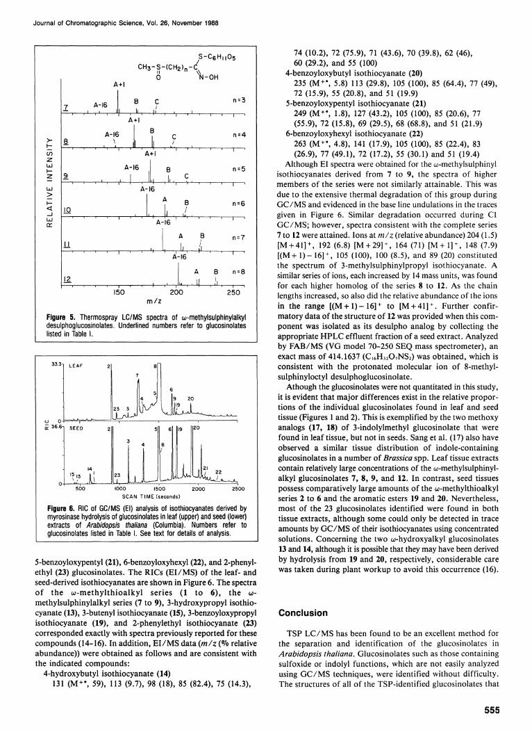
homologous series, ω-methylsulphinylalkylglucosinolates (7 to 12). Although members of this group were the more abundant components in leaf and seed extracts (Figure 2) , protonated molecular ions were not detected (Figure 5) . The spectrum of 3-methylsulphinylpropyl desulphoglucosinolate (7, n = 3) contains ions corresponding to each of the three fragmentation groups ( A through C ) . In addition, the spectrum contains two ions, both of low intensity at 26 and 16 mass units, less than that assigned fragmentation ion A . As the length of the side chain increased from n = 3 to n = 8, the ion intensity of A + 1 decreased; A increased to a maximum at n = 5 and then decreased; A-16 increased to the point where it represented the
base peak in the spectra of 10, 11 , and 12; fragmentation ions Β and C decreased to the point where C was no longer observed in spectra where n > 5 .
Structures of glucosinolates identified by TSP L C / M S , except for the 3 indole-containing group, were reconfirmed by El or CI G C / M S of isothiocyanates formed by treating the glucosides with myrosinase. (Note the same numbers that have been assigned to designate glucosinolate structures apply also to their derived isothiocyanates.) Six other glucosinolates not in sufficient concentration in either the leaf or seed extracts to be detectable by TSP L C / M S were identified. The six were 3-methylthiopropyl (1), 3-hydroxypropyl (13), 3-butenyl (15),
Figure 4. Thermospray LC/MS spectra of desulphoglucosinolates. Underlined numbers refer to glucosinolates listed in Table I.
554
Figure 3. Thermospray LC/MS spectra of ω-methylthioalkyl de-sulphoglucosinolates. Underlined numbers refer to glucosinolates listed in Table I.
Journal of Chromatographic Science, Vol. 26, November 1988
Figure 5. Thermospray LC/MS spectra of ω-methylsulphinylalkyl desulphoglucosinolates. Underlined numbers refer to glucosinolates listed in Table I.
Figure 6. RIC of (El) analysis of isothiocyanates derived by myrosinase hydrolysis of glucosinolates in leaf (upper) and seed (lower) extracts of Arabidopsis thaliana (Columbia). Numbers refer to glucosinolates listed in Table I. See text for details of analysis.
5-benzoyloxypentyl (21), 6-benzoyloxyhexyl (22), and 2-phenyl-ethyl (23) glucosinolates. The RICs ( Ε Ι / M S ) o f the leaf- and seed-derived isothiocyanates are shown in Figure 6. The spectra o f the ω-methy l th ioa lky l series ( 1 to 6 ) , the ω-methylsulphinylalkyl series (7 to 9 ) , 3-hydroxypropyl isothio-cyanate (13), 3-butenyl isothiocyanate (15), 3-benzoyloxypropyl isothiocyanate (19), and 2-phenylethyl isothiocyanate (23) corresponded exactly with spectra previously reported for these compounds (14-16). In addition, E I / M S data (m/z (°7o relative abundance)) were obtained as follows and are consistent with the indicated compounds:
4-hydroxybutyl isothiocyanate (14) 131 ( Μ + · , 59), 113 (9.7) , 98 (18), 85 (82.4), 75 (14.3),
74 (10.2), 72 (75.9), 71 (43.6), 70 (39.8), 62 (46), 60 (29.2), and 55 (100)
4-benzoyloxybutyl isothiocyanate (20) 235 ( M + · , 5.8) 113 (29.8), 105 (100), 85 (64.4), 77 (49), 72 (15.9), 55 (20.8), and 51 (19.9)
5-benzoyloxypentyl isothiocyanate (21) 249 ( M + · , 1.8), 127 (43.2), 105 (100), 85 (20.6), 77 (55.9), 72 (15.8), 69 (29.5), 68 (68.8), and 51 (21.9)
6-benzoyloxyhexyl isothiocyanate (22) 263 ( M + · , 4.8), 141 (17.9), 105 (100), 85 (22.4), 83 (26.9), 77 (49.1), 72 (17.2), 55 (30.1) and 51 (19.4)
Although EI spectra were obtained for the ω-methylsulphinyl isothiocyanates derived from 7 to 9, the spectra of higher members of the series were not similarly attainable. This was due to the extensive thermal degradation of this group during G C / M S and evidenced in the base line undulations in the traces given in Figure 6. Similar degradation occurred during CI G C / M S ; however, spectra consistent with the complete series 7 to 12 were attained. Ions at m/z (relative abundance) 204 (1.5) [ M + 41] + , 192 (6.8) [ M + 29] + , 164 (71) [ M + l ] + , 148 (7.9) [ ( M + 1 ) - 1 6 ] + , 105 (100), 100 (8.5), and 89 (20) constituted the spectrum of 3-methylsulphinylpropyl isothiocyanate. A similar series of ions, each increased by 14 mass units, was found for each higher homolog of the series 8 to 12. As the chain lengths increased, so also did the relative abundance of the ions in the range [ ( M + l ) - 1 6 ] + to [ M + 41] + . Further confirmatory data of the structure of 12 was provided when this component was isolated as its desulpho analog by collecting the appropriate H P L C effluent fraction of a seed extract. Analyzed by F A B / M S ( V G model 70-250 SEQ mass spectrometer), an exact mass of 414.1637 (C 1 6H 3 2O 7NS 2) was obtained, which is consistent with the protonated molecular ion of 8-methyl-sulphinyloctyl desulphoglucosinolate.
Athough the glucosinolates were not quantitated in this study, it is evident that major differences exist in the relative proportions o f the individual glucosinolates found in leaf and seed tissue (Figures 1 and 2) . This is exemplified by the two methoxy analogs (17, 18) of 3-indolylmethyl glucosinolate that were found in leaf tissue, but not in seeds. Sang et al. (17) also have observed a similar tissue distribution of indole-containing glucosinolates in a number of Brassica spp. Leaf tissue extracts contain relatively large concentrations of the ω-methylsulphinylalkyl glucosinolates 7, 8, 9 , and 12. In contrast, seed tissues possess comparatively large amounts of the ω-methylthioalkyl series 2 to 6 and the aromatic esters 19 and 20. Nevertheless, most of the 23 glucosinolates identified were found in both tissue extracts, although some could only be detected in trace amounts by G C / M S of their isothiocyanates using concentrated solutions. Concerning the two ω-hydroxyalkyl glucosinolates 13 and 14, although it is possible that they may have been derived by hydrolysis from 19 and 20, respectively, considerable care was taken during plant workup to avoid this occurrence (16).
Conclusion
TSP L C / M S has been found to be an excellent method for the separation and identification of the glucosinolates in Arabidopsis thaliana. Glucosinolates such as those containing sulfoxide or indolyl functions, which are not easily analyzed using G C / M S techniques, were identified without difficulty. The structures of all of the TSP-identified glucosinolates that
555

Journal of Chromatographic Science, Vol. 26, November 1988
yield isothiocyanates as products of myrosinase hydrolysis were confirmed by G C / M S . G C / M S analysis of isothiocyanates, although not applicable to all glucosinolates, was more sensitive than T S P L C / M S , allowing for the detection of trace amounts of glucosinolates. O f the 23 glucosinolates detected in this study, 16 constitute newly identified components of Arabidopsis thaliana, and of these, three glucosinolates, 14, 2 1 , and 22, have not been reported to occur in plants. The presence of seven glucosinolates previously indicated (7-9) as constituents of seeds were confirmed; these were glucosinolates 2 , 5, 6, 1 1 , 12, 19, and 20. The presence of three, allylglucosinolate, isopropyl-glucosinolate (6 ) , and 6-heptenylglucosinolate (7) , whose occurrence had been noted earlier, were unverified. Indeed, the lack of detection of these compounds could be due to varietal differences within this species.
The number and diversity in structure of the glucosinolates found in Arabidopsis thaliana provide an unusual opportunity for studies o f the biochemical genetics o f this class of sulphur-containing glucosides.
Acknowledgments
The authors thank C .R . Somerville, M S U - D O E Plant Research Laboratory, Michigan State University, East Lansing, M I , for his generous gift o f Arabidopsis thaliana seeds; D . I . McGregor , Agriculture Canada, Research Station, Saskatoon, Sask., for helpful suggestions on H P L C analysis of glucosinolates; P . Brooks, Institute for Geological and Petroleum Research, Calgary, Al ta . , for F A B M S ; and E . M . Giblin and D.J .H . Olson for expert technical assistance.
References
1. G.R. Fenwick, R.K. Heaney, and W.J. Mullin. Glucosinolates and their breakdown products in food and food plants. CRC Crit Rev. Food Sci. Nutr. 18: 123-201 (1983).
2. M. Linscheid, D. Wendisch, and D. Straek. The structures of sinapic acid esters and their metabolism in cotyledons of Raphanus sativus. Z. Naturforsch. 35C, 907-22 (1980).
3. M. Benn. Glucosinolates. PureAppl. Chem. 49:197-210(1977).
4. A. Kjaer. The natural distribution of glucosinolates: a uniform group of sulfur-containing glucosides. In, Chemistry in Botanical Classification, G. Bendz and J. Santesson, Eds. Academic Press, New York, 1974, pp. 229-34.
5. M. Estelle and C.R. Somerville. The mutants of Arabidopsis thaliana. Trends in Genet. 1 1 : 189-93 (1986).
6. R.A. Cole. Isothiocyanates, nitriles, and thiocyanates as products of autolysis of glucosinolates in Cruciferae. Phytochemistry 15: 759-62 (1976).
7. G.R. Fenwick, J. Eagles, and R. Self. The fast atom bombardment mass spectra of glucosinolates and glucosinolate mixtures. Org. Mass Spectrom. 17: 544-46 (1982).
8. J. Eagles, G.R. Fenwick, R. Gmelin, and D. Rakow. The chemical ionization mass spectra of glucosinolates (mustard oil glycosides) and desulphoglucosinolates. A useful aid for structural analysis. Biomed. Mass Spectrom. 8: 265-69 (1981).
9. J. Eagles, G.R. Fenwick, and R.K. Heaney. Gas chromatography chemical ionization mass spectrometry of glucosinolate derivatives. Biomed. Mass Spectrom. 8: 278-82 (1981).
10. F.A. Mellon, J.R. Chapman, and J.A.E. Pratt. Thermospray liquid chromatography/mass spectrometry in food and agricultural research. J. Chromatogr. 394: 209-22 (1987).
11. L.R. Hogge, D.W. Reed, and E.W. Underhill. The identification of desulfoglucosinolates using thermospray LC/MS. J. Chromatogr. Sci. 26: 348-51 (1988).
12. C.R. Somerville and W.L. Oren. Isolation of photorespiration mutants in Arabidopsis thaliana. In, Methods in Chloroplast Molecular Biology. M. Edelman, R.B. Hallick and N.H. Chua (Eds). Elsevier Biomedical Press, New York, 1982, pp. 129-39.
13. I. Minchinton, J. Sang, D. Burke, and R.J.W. Truscott. Separation of desulphoglucosinolates by reversed-phase high-performance liquid chromatography. J. Chromatogr. 247: 141-48 (1982).
14. A. Kjaer, M. Ohashi, J.M. Wilson, and C. Djerassi. Mass spectra of isothiocyanates. Acta Chem. Scand. 17: 2143-54 (1963).
15. G.F Spencer and M.E. Daxenbichler. Gas chromatography/mass spectrometry of nitriles, isothiocyanates, and oxazolidinethiones derived from cruciferous glucosinolates. J. Sci. Food Agric. 3 1 : 359-67 (1980).
16. M.E. Daxenbichler, G.F. Spencer, and W.P. Schroeder. 3-Hydroxy-propylglucosinolate, a new glucosinolate in seeds of Erysimum hieracifolium and Malcolmia maritima. Phytochemistry 19:813-15 (1980).
17. J.P. Sang, I.R. Minchinton, P.K. Johnstone, and R.J.W. Truscott. Glucosinolate profiles in the seed, root, and leaf tissue of cabbage, mustard, rapeseed radish, and swede. Can. J. Plant Sci. 64: 77-93 (1984).
Manuscript received April 21, 1988.
556