high-nacl intake impairs dynamic autoregulation of renal ... · saeed a, dibona gf, marcussen n,...
TRANSCRIPT

High-NaCl intake impairs dynamic autoregulation of renal blood flow inANG II-infused rats
Aso Saeed,1 Gerald F. DiBona,2 Niels Marcussen,3 and Gregor Guron1
1Department of Molecular and Clinical Medicine/Nephrology, Institute of Medicine, The Sahlgrenska Academy at theUniversity of Gothenburg, Gothenburg, Sweden; 2Departments of Internal Medicine and Molecular Physiology andBiophysics, Department of Veterans Affairs Medical Center and University of Iowa Carver College of Medicine, Iowa City,Iowa; and 3Department of Pathology, Odense University Hospital, Odense, Denmark
Submitted 17 May 2010; accepted in final form 12 August 2010
Saeed A, DiBona GF, Marcussen N, Guron G. High-NaCl intakeimpairs dynamic autoregulation of renal blood flow in ANG II-infusedrats. Am J Physiol Regul Integr Comp Physiol 299: R1142–R1149, 2010.First published August 18, 2010; doi:10.1152/ajpregu.00326.2010.—Theaim of this study was to investigate dynamic autoregulation of renalblood flow (RBF) in ANG II-infused rats and the influence ofhigh-NaCl intake. Sprague-Dawley rats received ANG II (250ng·kg�1·min�1 sc) or saline vehicle (sham) for 14 days after whichacute renal clearance experiments were performed during thiobutabar-bital anesthesia. Rats (n � 8–10 per group) were either on a normal(NNa; 0.4% NaCl)- or high (HNa; 8% NaCl)-NaCl diet. Separategroups were treated with 4-hydroxy-2,2,6,6-tetramethylpiperidine-1-oxyl (tempol; 1 M in drinking water). Transfer function analysis fromarterial pressure to RBF in the frequency domain was used to examinethe myogenic response (MR; 0.06–0.09 Hz) and the tubuloglomerularfeedback mechanism (TGF; 0.03–0.06 Hz). MAP was elevated inANG II-infused rats compared with sham groups (P � 0.05). RBF inANG II HNa was reduced vs. sham NNa and sham HNa (6.0 � 0.3vs. 7.9 � 0.3 and 9.1 � 0.3 ml·min�1·g kidney wt�1, P � 0.05).transfer function gain in ANG II HNa was significantly elevated in thefrequency range of the MR (1.26 � 0.50 dB, P � 0.05 vs. all othergroups) and in the frequency range of the TGF (�0.02 � 0.50 dB,P � 0.05 vs. sham NNa and sham HNa). Gain values in thefrequency range of the MR and TGF were significantly reduced bytempol in ANG II-infused rats on HNa diet. In summary, the MR andTGF components of RBF autoregulation were impaired in ANG IIHNa, and these abnormalities were attenuated by tempol, suggestinga pathogenetic role for superoxide in the impaired RBF autoregulatoryresponse.
tubuloglomerular feedback; superoxide; myogenic response
HYPERTENSION IS A COMMON CAUSE of kidney injury and end-stagerenal disease and accelerates loss of kidney function in patientswith chronic kidney disease, regardless of the underlyingetiology (19). However, the risk of renal injury is variable, andthe pathophysiological mechanisms by which hypertensioncauses renal parenchymal injury are complex and incompletelyunderstood (5, 13, 27, 28, 34). The renal blood flow (RBF)autoregulatory response, mediated mainly by the myogenicresponse (MR) and the tubuloglomerular feedback mechanism(TGF), stabilizes RBF and glomerular filtration rate (GFR),despite wide variations in arterial blood pressure (AP) (9, 23).RBF autoregulation (RBFA) may also serve a protective func-tion, particularly in hypertension, by preventing transmission
of systemic AP to the glomerular capillaries (2, 28). A role forautoregulatory capacity as a determinant of vulnerability torenal injury has been suggested in the 5/6 renal ablation modelin rats (4) and in genetic models characterized by impairedRBFA (37, 38). In addition, treatment with dihydropyridinecalcium channel blockers, which interfere with RBFA byimpairing the MR, has been shown to increase the susceptibil-ity to hypertensive glomerular injury in rats subjected to 5/6nephrectomy (17).
Increased dietary NaCl has been shown to accelerate renalinjury in hypertension (1, 5, 32). However, the pathophysio-logical mechanisms by which increased NaCl intake enhancesrenal injury in hypertension are multiple and not fully eluci-dated. Takenaka et al. (35) showed that preglomerular arte-rioles in Dahl salt-sensitive rats, on a high-NaCl diet, exhibitedreduced myogenic responsiveness to increased AP, suggestinga role for impaired RBFA as a cause of renal injury in thismodel (35). In addition, several studies indicate that reactiveoxygen species contribute to renal injury in NaCl-sensitiveforms of hypertension (18, 36). In the present study, wehypothesized that the combined influence of ANG II, and ahigh-NaCl intake, could lead to impaired autoregulation ofRBF in this hypertensive model. In addition, we speculated thatsuperoxide (O2
·�), a reactive oxygen species whose productionis stimulated by ANG II (15, 27), could be involved in theabnormal RBF autoregulatory response. To examine this hy-pothesis we used transfer function (TF) analysis that enabled usto analyze the separate contributions of the MR and the TGFmechanism to dynamic autoregulation of RBF in chronicallyANG II-infused Sprague-Dawley rats on either a normal or ahigh-NaCl diet.
METHODS
General Procedures
Male Sprague-Dawley rats (Harlan, Horst, The Netherlands)weighing �300 g were used. All experiments were approved by theregional ethics committee in Gothenburg, Sweden. Rats had freeaccess to rat chow and tap water and were kept in rooms with acontrolled temperature of 24–26°C and a 12:12-h dark-light cycle.Chemicals were from Sigma (St. Louis, MO), if not stated otherwise.
Protocol
Rats received ANG II (250 ng·kg�1·min�1 sc) or isotonic salinevehicle (sham) via osmotic minipumps (Alzet model 2002) for 14days, after which acute experiments were performed. Rats were eitheron a normal (NNa, 0.4% NaCl)- or high (HNa, 8% NaCl)-NaCl diet(Lantmännen, Sweden), creating the following groups: 1) sham NNa(n � 10); 2) sham HNa (n � 9); 3) ANG II NNa (n � 9); and (4) ANG
Address for reprint requests and other correspondence: A. Saeed, Dept. ofMolecular and Clinical Medicine/Nephrology, Institute of Medicine, TheSahlgrenska Academy at the Univ. of Gothenburg, Vita Straket 12, SahlgrenskaUniv. Hospital, S-413 45 Gothenburg, Sweden (e-mail: [email protected]).
Am J Physiol Regul Integr Comp Physiol 299: R1142–R1149, 2010.First published August 18, 2010; doi:10.1152/ajpregu.00326.2010.
http://www.ajpregu.orgR1142
by 10.220.33.4 on January 15, 2017http://ajpregu.physiology.org/
Dow
nloaded from
II HNa (n � 8). In separate groups, the membrane-permeable super-oxide dismutase mimetic 4-hydroxy-2,2,6,6-tetramethylpiperidine-1-oxyl (tempol) was administered in the drinking water (1 M ) through-out the 14-day period: 5) sham NNa�tempol (n � 8); 6) shamHNa�tempol (n � 10); 7) ANG II NNa�tempol (n � 10); and 8)ANG II HNa�tempol (n � 10). Tempol administered by drinkingwater in this concentration has been demonstrated to significantlyreduce AP and markers of oxidative stress in several hypertensive ratmodels (15, 18, 36) including in ANG II HNa (30).
Surgical Preparation and Measurements
Rats were anaesthetized with thiobutabarbital (Inactin; 120 mg/kgip), placed on a heating table, and surgically prepared for renalclearance experiments as previously described (29). An AP catheterwas inserted via the femoral artery and was connected to a pressuretransducer (Smiths Medical, Kirchseeon, Germany) for monitoring ofarterial pressure [AP; pulsatile and mean (MAP)] and heart rate usinga data acquisition program (MP 150; Biopac Systems, Santa Barbara,CA). The left kidney was exposed by a flank incision and immobilizedin a plastic cup. The left ureter was catheterized for urine collection.Rectal and kidney temperatures were kept at 37°C. A perivascularultrasonic transit-time flow probe (0.7 VB) was placed around the leftrenal artery and connected to a flowmeter (model T206, filter 100 Hz)for measurement of RBF (Transonic Systems, Ithaca, NY). GFR wasdetermined by measuring renal 51Cr-EDTA clearance (AmershamLaboratories, Buckinghamshire, UK) as described (29). Blood wassampled at the start and completion of two consecutive 20-min urinaryclearance periods, and mean values of plasma radioactivity were usedto calculate GFR. Arterial blood samples (0.3 ml) were replaced byequivalent volumes of 4% BSA in isotonic saline. Rats were infusedwith 10 ml·kg�1·h�1 of isotonic saline throughout. Rats were killedby an overdose of pentobarbital sodium, and the kidneys wereexcised and weighed. Kidneys were immersion-fixed in parafor-maldehyde and prepared for histological analyses. Renal vascularresistance (RVR) was calculated as MAP (mmHg)/RBF (ml·min�1·gkidney wt�1), and filtration fraction (FF) was estimated as GFR/RBF.
TF Analysis
Data used to examine the dynamic relationship between AP andRBF, i.e., dynamic autoregulation of RBF, were sampled at 62.5 Hzyielding 75,000 data points for each 20-min period (150,000 datapoints for the two 20-min periods). Processing of AP and RBF datawas performed off-line by using previously developed software rou-tines written for Matlab 7.14 (The MathWorks, Natick, MA). Aftersubtracting the mean value from the data files, they were digitallylow-pass filtered (3.0 Hz cut-off frequency, finite-impulse response,order 50) and then resampled to a rate of 6.25 Hz. These 6.25-Hz datafiles were split into blocks of 2,048 data points, yielding a frequencydiscrimination of 0.003 Hz. Power spectral density (PSD) of AP andRBF was calculated, as described (10, 11). The TF spectra werecalculated from AP (input) and RBF (output). The TF gain was takenas the quotient of the cross spectrum of input and output divided bythe power spectrum of the input (10, 11). Coherence is a frequencydomain estimate of a linear correlation (i.e., squared coherence, akinto coefficient of determination) between two signals indicating thedegree to which the variance in one signal can be explained by a linearoperation on the other signal (10, 11). The coherence spectra werecalculated from AP (input) and RBF (output). The coherence functionwas taken as the quotient of the square of the cross spectrum of inputand output divided by the product of the power spectral densities ofAP and RBF (10, 11). These algorithms involved a Hanning windowwith 50% overlap of the blocks (12 blocks in the two 20-minrecording periods). To permit comparison among rats, the TF gain(magnitude) values over the frequency range have been normalized tothe mean value of the renal vascular conductance for the entire dataset. After conversion of the normalized TF gain values into decibels
[20 log (gain)], a mean spectrum was calculated from the consecutivespectra in each rat, and these were subsequently averaged for all rats.The TF gain corresponds to the ratio of the amplitude of normalizedfluctuations in RBF divided by those of AP. In the presence of RBFA,fluctuations of RBF are attenuated vs. those of AP causing the TF gainto be negative. Thus, positive TF gain values indicate impaired RBFA(9, 23). Phase and coherence spectra were similarly calculated andaveraged. Data over the range of frequencies for the MR (0.08–0.18)and the TGF mechanism (0.03–0.06) were analyzed (9, 23). The slopeof gain reduction in the frequency range of the MR was determined byleast squares fitting of the linear region of gain reduction, and thephase peak was estimated as the average phase value within the samefrequency interval. In addition, to assess the contribution of the MR toRBFA, mean gain values in the frequency range of 0.06 -0.09 Hz wereused to minimize corruption by TGF (�0.06 Hz) and myogenictransients (�0.09 Hz) (38). To assess the effect of filtering at either 30Hz or 100 Hz, we made two consecutive recording periods in a singlerat with 30 Hz filter/62.5 Hz sampling followed by 100-Hz filter/62.5-Hz sampling. Each of these data sets was subjected to the sameprocessing and analysis as described above. The results showed thatover the frequency range of interest (0.01–1.0 Hz) there were nosignificant differences in PSD for AP and RBF or in TF gain, phase,or coherence in the TGF (0.03–0.06 Hz) or MR (0.08–0.18 Hz)frequency ranges. To determine the threshold for coherence abovewhich it exceeds zero with a certain significance level, we used themethod described by Koopmans (26), which depends on the totalnumber of samples, the total number of blocks, and the nature of thetapering window. In this study with large sample numbers, coherencevalues � 0.1 are significantly different from zero at P � 0.001.
Kidney Histology
Kidneys were processed using routine techniques, and 3-�m thicktransverse sections through the hilar area were prepared and stainedwith hematoxylin and eosin, periodic acid-Schiff, and elastin-vanG-ieson’s. Histopathological changes were scored semiquantitatively(0–3) by an investigator (N. Marcussen) blinded to treatment group.The scores: 0, when no pathologic changes were present; 1, when fewof the structures showed changes and the changes were mild; 2, whenmoderate changes were present; and 3, when severe changes werepresent in the structures under investigation. For glomerular parame-ters, the percentage of pathologically altered glomeruli was estimated.Cortical arteries (i.e., interlobar, arcuate, and interlobular arteries) andarterioles (i.e., afferent and efferent arterioles) were scored separately.
Statistical Analysis
All values are means � SE. Analyses were performed usingone-way ANOVA. Normality was tested with the Shapiro-Wilk test,and equality of variances was assessed with the Levene’s test. If datawere not normally distributed or had unequal variances, Kruskal-Wallis one-way ANOVA on ranks was used. An unpaired t-test andMann-Whitney U-test was used when appropriate. Bonferroni correc-tions were made for multiple comparisons. To reduce the number ofcomparisons, no statistical analyses were made between ANG II NNaand sham HNa. Group ANG II NNa�tempol was compared withANG II NNa and sham NNa, and group ANG II HNa�tempol wascompared with ANG II HNa and sham HNa. In all cases, a P value �0.05 was considered statistically significant. The statistical softwareSPSS 17.0 (SPSS, Chicago, IL) was used.
RESULTS
Kidney Function and Renal Hemodynamics
Effects of NaCl intake and ANG II. Group sham HNashowed elevated RBF and reduced FF compared with shamrats on a normal NaCl diet, sham NNa (Table 1). In addition,
R1143DYNAMIC RENAL AUTOREGULATION IN ANG II-INFUSED RATS
AJP-Regul Integr Comp Physiol • VOL 299 • NOVEMBER 2010 • www.ajpregu.org
by 10.220.33.4 on January 15, 2017http://ajpregu.physiology.org/
Dow
nloaded from
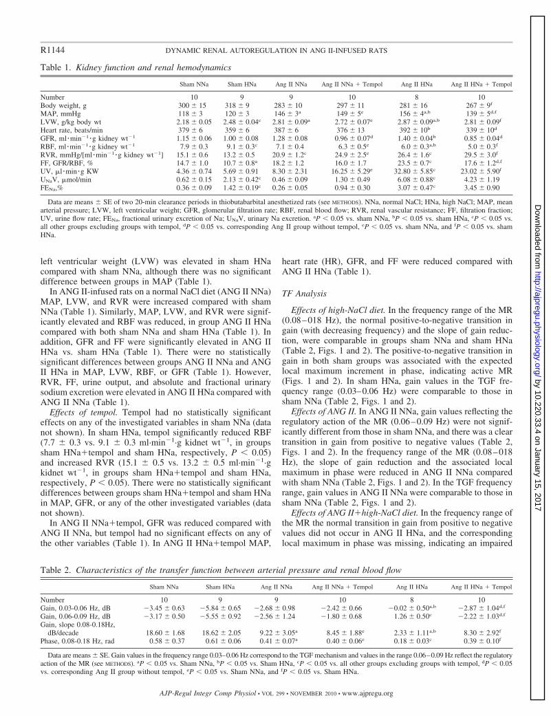
left ventricular weight (LVW) was elevated in sham HNacompared with sham NNa, although there was no significantdifference between groups in MAP (Table 1).
In ANG II-infused rats on a normal NaCl diet (ANG II NNa)MAP, LVW, and RVR were increased compared with shamNNa (Table 1). Similarly, MAP, LVW, and RVR were signif-icantly elevated and RBF was reduced, in group ANG II HNacompared with both sham NNa and sham HNa (Table 1). Inaddition, GFR and FF were significantly elevated in ANG IIHNa vs. sham HNa (Table 1). There were no statisticallysignificant differences between groups ANG II NNa and ANGII HNa in MAP, LVW, RBF, or GFR (Table 1). However,RVR, FF, urine output, and absolute and fractional urinarysodium excretion were elevated in ANG II HNa compared withANG II NNa (Table 1).
Effects of tempol. Tempol had no statistically significanteffects on any of the investigated variables in sham NNa (datanot shown). In sham HNa, tempol significantly reduced RBF(7.7 � 0.3 vs. 9.1 � 0.3 ml·min�1·g kidnet wt�1, in groupssham HNa�tempol and sham HNa, respectively, P � 0.05)and increased RVR (15.1 � 0.5 vs. 13.2 � 0.5 ml·min�1·gkidnet wt�1, in groups sham HNa�tempol and sham HNa,respectively, P � 0.05). There were no statistically significantdifferences between groups sham HNa�tempol and sham HNain MAP, GFR, or any of the other investigated variables (datanot shown).
In ANG II NNa�tempol, GFR was reduced compared withANG II NNa, but tempol had no significant effects on any ofthe other variables (Table 1). In ANG II HNa�tempol MAP,
heart rate (HR), GFR, and FF were reduced compared withANG II HNa (Table 1).
TF Analysis
Effects of high-NaCl diet. In the frequency range of the MR(0.08–018 Hz), the normal positive-to-negative transition ingain (with decreasing frequency) and the slope of gain reduc-tion, were comparable in groups sham NNa and sham HNa(Table 2, Figs. 1 and 2). The positive-to-negative transition ingain in both sham groups was associated with the expectedlocal maximum increment in phase, indicating active MR(Figs. 1 and 2). In sham HNa, gain values in the TGF fre-quency range (0.03–0.06 Hz) were comparable to those insham NNa (Table 2, Figs. 1 and 2).
Effects of ANG II. In ANG II NNa, gain values reflecting theregulatory action of the MR (0.06–0.09 Hz) were not signif-icantly different from those in sham NNa, and there was a cleartransition in gain from positive to negative values (Table 2,Figs. 1 and 2). In the frequency range of the MR (0.08–018Hz), the slope of gain reduction and the associated localmaximum in phase were reduced in ANG II NNa comparedwith sham NNa (Table 2, Figs. 1 and 2). In the TGF frequencyrange, gain values in ANG II NNa were comparable to those insham NNa (Table 2, Figs. 1 and 2).
Effects of ANG II�high-NaCl diet. In the frequency range ofthe MR the normal transition in gain from positive to negativevalues did not occur in ANG II HNa, and the correspondinglocal maximum in phase was missing, indicating an impaired
Table 1. Kidney function and renal hemodynamics
Sham NNa Sham HNa Ang II NNa Ang II NNa � Tempol Ang II HNa Ang II HNa � Tempol
Number 10 9 9 10 8 10Body weight, g 300 � 15 318 � 9 283 � 10 297 � 11 281 � 16 267 � 9f
MAP, mmHg 118 � 3 120 � 3 146 � 3a 149 � 5e 156 � 4a,b 139 � 5d,f
LVW, g/kg body wt 2.18 � 0.05 2.48 � 0.04c 2.81 � 0.09a 2.72 � 0.07e 2.87 � 0.09a,b 2.81 � 0.09f
Heart rate, beats/min 379 � 6 359 � 6 387 � 6 376 � 13 392 � 10b 339 � 10d
GFR, ml �min�1 �g kidney wt�1 1.15 � 0.06 1.00 � 0.08 1.28 � 0.08 0.96 � 0.07d 1.40 � 0.04b 0.85 � 0.04d
RBF, ml �min�1 �g kidney wt�1 7.9 � 0.3 9.1 � 0.3c 7.1 � 0.4 6.3 � 0.5e 6.0 � 0.3a,b 5.0 � 0.3f
RVR, mmHg/[ml �min�1 �g kidney wt�1] 15.1 � 0.6 13.2 � 0.5 20.9 � 1.2c 24.9 � 2.5e 26.4 � 1.6c 29.5 � 3.0f
FF, GFR/RBF, % 14.7 � 1.0 10.7 � 0.8a 18.2 � 1.2 16.0 � 1.7 23.5 � 0.7c 17.6 � 1.2d,f
UV, �l �min �g KW 4.36 � 0.74 5.69 � 0.91 8.30 � 2.31 16.25 � 5.29e 32.80 � 5.85c 23.02 � 5.90f
UNaV, �mol/min 0.62 � 0.15 2.13 � 0.42c 0.46 � 0.09 1.30 � 0.49 6.08 � 0.88c 4.23 � 1.19FENa,% 0.36 � 0.09 1.42 � 0.19c 0.26 � 0.05 0.94 � 0.30 3.07 � 0.47c 3.45 � 0.90
Data are means � SE of two 20-min clearance periods in thiobutabarbital anesthetized rats (see METHODS). NNa, normal NaCl; HNa, high NaCl; MAP, meanarterial pressure; LVW, left ventricular weight; GFR, glomerular filtration rate; RBF, renal blood flow; RVR, renal vascular resistance; FF, filtration fraction;UV, urine flow rate; FENa, fractional urinary excretion of Na; UNaV, urinary Na excretion. aP � 0.05 vs. sham NNa, bP � 0.05 vs. sham HNa, cP � 0.05 vs.all other groups excluding groups with tempol, dP � 0.05 vs. corresponding Ang II group without tempol, eP � 0.05 vs. sham NNa, and fP � 0.05 vs. shamHNa.
Table 2. Characteristics of the transfer function between arterial pressure and renal blood flow
Sham NNa Sham HNa Ang II NNa Ang II NNa � Tempol Ang II HNa Ang II HNa � Tempol
Number 10 9 9 10 8 10Gain, 0.03-0.06 Hz, dB �3.45 � 0.63 �5.84 � 0.65 �2.68 � 0.98 �2.42 � 0.66 �0.02 � 0.50a,b �2.87 � 1.04d,f
Gain, 0.06-0.09 Hz, dB �3.17 � 0.50 �5.55 � 0.92 �2.56 � 1.24 �1.80 � 0.68 1.26 � 0.50c �2.22 � 1.03d,f
Gain, slope 0.08-0.18Hz,dB/decade 18.60 � 1.68 18.62 � 2.05 9.22 � 3.05a 8.45 � 1.88e 2.33 � 1.11a,b 8.30 � 2.92f
Phase, 0.08-0.18 Hz, rad 0.58 � 0.37 0.61 � 0.06 0.41 � 0.07a 0.40 � 0.06e 0.18 � 0.03c 0.39 � 0.10f
Data are means � SE. Gain values in the frequency range 0.03–0.06 Hz correspond to the TGF mechanism and values in the range 0.06–0.09 Hz reflect the regulatoryaction of the MR (see METHODS). aP � 0.05 vs. Sham NNa, bP � 0.05 vs. Sham HNa, cP � 0.05 vs. all other groups excluding groups with tempol, dP � 0.05vs. corresponding Ang II group without tempol, eP � 0.05 vs. Sham NNa, and fP � 0.05 vs. Sham HNa.
R1144 DYNAMIC RENAL AUTOREGULATION IN ANG II-INFUSED RATS
AJP-Regul Integr Comp Physiol • VOL 299 • NOVEMBER 2010 • www.ajpregu.org
by 10.220.33.4 on January 15, 2017http://ajpregu.physiology.org/
Dow
nloaded from

MR (Figs. 1 and 2). Gain values in the frequency range of0.06–0.09 Hz were significantly elevated in ANG II HNacompared with sham NNa, sham HNa, and ANG II NNa (Table2). In the TGF frequency range, gain values in ANG II HNawere significantly elevated compared with sham NNa andsham HNa (Table 2, Figs. 1 and 2).
Effects of tempol. Tempol had no statistically significanteffects on dynamic RBFA assessed by TF analysis in groupssham NNa and sham HNa (data not shown). Similarly, in theANG II NNa�tempol group, tempol did not significantlyaffect TF variables, including the slope of gain reduction andphase values, in the frequency range of the MR (Table 2, Fig. 3).However, in ANG II HNa�tempol, there was a clear transitionin gain from positive to negative values in the frequency rangeof the MR, and this was associated with a local maximum inphase indicating an active MR (Fig. 4). Consequently, gainvalues were significantly reduced in ANG II HNa�tempolcompared with ANG II HNa in the frequency range of both theMR (0.06–0.09 Hz) and the TGF (0.03–0.06 Hz), (Table 2,Fig. 4). Nevertheless, TF analysis variables remained signifi-cantly different in ANG II HNa�tempol compared with shamHNa in the frequency range of both the MR and the TGF(Table 2).
Kidney Histology
There were no statistically significant differences betweengroups in tubulointerstitial changes (i.e., inflammation, fibro-sis, and tubular dilatation) or glomerular abnormalities (i.e.,glomerular size variation, ischemia, focal segmental glomeru-losclerosis, and global glomerulosclerosis) (data not shown).There was significant thickening of intimal and medial layersand hyaline deposition/necrosis of cortical arteries in bothANG II NNa and ANG II HNa compared with sham NNa (Fig.5, Table 3). In addition, cortical arterioles (i.e., afferent andefferent arterioles) showed significant hyperplasia and hyali-nosis compared with sham (Fig. 5, Table 3). However, themagnitude of arterial and arteriolar changes in ANG II NNaand ANG II HNa were not significantly different, and tempolhad no significant effects on vascular abnormalities (Fig. 5,Table 3). Notably, vascular changes in hypertensive animalswere generally mild and showed a focal distribution.
DISCUSSION
The main finding of the present study was that a high-NaClintake in chronically ANG II-infused rats resulted in a markedimpairment of the MR of RBFA. This abnormality was notseen in sham HNa and was significantly more pronounced thanin ANG II NNa, indicating a synergistic effect of high-NaClintake and elevated ANG II. In addition, this abnormality inRBFA in ANG II HNa was corrected by tempol, suggesting arole for O2
·� in the impaired autoregulatory response.In the frequency range of the MR, TF analysis showed a
reduced slope of gain reduction and a diminished phase peak inANG II NNa compared with sham NNa, indicating an impairedMR. This result differs from a previous study in which dy-namic RBFA was assessed in conscious dogs infused with
Fig. 2. Enlarged transfer function gain (top) and phase (bottom) in anesthetizedSprague-Dawley rats after 14 days of ANG II (250 ng·kg�1·min�1 sc) or salinevehicle (sham) infusion. Rats were on either an NNa or HNa diet (seeMETHODS). In the frequency range of the mypgenic response (MR; 0.08 – 018Hz), gain values showed a positive-to-negative transition in groups shamNNa, sham HNa, and ANG II NNa. The positive-to-negative transition ingain in these groups was associated with the expected local maximum inphase, indicating active MR. In ANG II HNa, the positive-to-negativetransition in gain in the frequency range of the MR remained above 0 dB,and the local maximum in phase was missing, indicating impaired MR.Values are means � SE.
Fig. 1. Power spectral density (PSD) for arterial pressure (AP) and renal bloodflow (RBF) and transfer function gain, phase, and coherence in anesthetizedSprague-Dawley rats after 14 days of ANG II (250 ng·kg�1·min�1 sc) or salinevehicle (sham) infusion. Rats were on either a normal (NNa)- or a high(HNa)-NaCl diet (see METHODS). Values are means � SE.
R1145DYNAMIC RENAL AUTOREGULATION IN ANG II-INFUSED RATS
AJP-Regul Integr Comp Physiol • VOL 299 • NOVEMBER 2010 • www.ajpregu.org
by 10.220.33.4 on January 15, 2017http://ajpregu.physiology.org/
Dow
nloaded from

ANG II to produce plasma ANG II levels within the physio-logical range (24). These investigators found that ANG II didnot affect the efficiency of RBFA or the relative contribution ofthe MR and TGF components (24). The discrepancy betweenstudies could be explained by marked differences in the exper-imental protocols, as the potential difference between con-scious and anesthetized animals. In addition, in our studyanimals were hypertensive and infused with much higher ANGII concentrations and for a longer duration. However, severalstudies have demonstrated in experimental models closelyresembling ours that RBFA is impaired in chronically ANGII-infused hypertensive animals (8, 21, 40) as well as in thenonstenotic kidney of Goldblatt hypertensive rats (31). Most ofthe studies on chronically ANG II-infused rats have beenperformed using the in vitro blood-perfused juxtamedullarynephron model in which responses of afferent arterioles tostep-wise changes in perfusion pressure were analyzed (8, 21,40). In contrast, our study presents data on dynamic autoreg-ulation of whole kidney RBF during spontaneous fluctuationsin AP in intact ANG II hypertensive animals. In addition, weextend previous findings by suggesting that chronic ANG IIinfusion selectively affects the MR component of RBFA inanimals on a normal NaCl intake.
The major finding in the present study was the much morepronounced impairment in dynamic RBFA in ANG II HNa
compared with ANG II NNa. This abnormality in ANG II HNawas characterized by an almost complete absence of a MR.This synergistic effect of ANG II and high-NaCl intake ondynamic RBFA represents a novel finding. One could hypoth-esize that the impairment in RBFA, mainly in group ANG IIHNa, would make kidneys vulnerable primarily to reductionsin AP and not to glomerular hypertension, as RVR was mark-edly elevated and RBF was reduced in these rats. However,despite the reduction in RBF, GFR tended to be elevated inANG II HNa, and FF was significantly increased vs. the othergroups. These results indicate that the increase in resistancepredominantly occurred at the efferent glomerular arteriolesand that glomerular capillary pressure was maintained or ele-vated despite reductions in RBF. In this situation it is reason-able to speculate that the aforementioned abnormalities inRBFA in ANG II HNa could eventually cause pressure-in-duced glomerular injury. In addition, it may seem contradictorythat RVR was clearly increased in group ANG II HNa in viewof the impaired MR demonstrated by TF analysis. However, itis important to recognize that impaired dynamic RBFA doesnot indicate a certain level of RBF at a certain level of AP, butmore likely predicts whether the response of RBF following adynamic change in arterial pressure will be normal. In line with
Fig. 4. PSD for AP and RBF, and transfer function gain, phase, and coherencein anesthetized Sprague-Dawley rats after 14 days of ANG II-infusion (250ng·kg�1·min�1 sc) with or without tempol in drinking water (1 M). Rats wereon an HNa diet (see METHODS). Contrary to in ANG II HNa, there was, in groupANG II HNa�tempol, a clear transition in gain from positive to negativevalues in the frequency range of the MR, and this was associated with a localmaximum in phase, indicating an active MR. Values are means � SE.
Fig. 3. PSD for AP and RBF, and transfer function gain, phase, and coherencein anesthetized Sprague-Dawley rats after 14 days of ANG II-infusion (250ng·kg�1·min�1 sc) with or without tempol in drinking water (1 M). Rats wereon an NNa diet (see METHODS). Tempol did not significantly affect transferfunction variables. Values are means � SE.
R1146 DYNAMIC RENAL AUTOREGULATION IN ANG II-INFUSED RATS
AJP-Regul Integr Comp Physiol • VOL 299 • NOVEMBER 2010 • www.ajpregu.org
by 10.220.33.4 on January 15, 2017http://ajpregu.physiology.org/
Dow
nloaded from

our results, Elmarakby et al. (13) showed that autoregulatoryresponses of afferent arterioles to step-wise increases in per-fusion pressure were blunted in ANG II-infused rats on highdietary NaCl (13). However, in that study no comparison wasmade to ANG II-infused animals on a normal NaCl intake.Interestingly, Zhao et al. (41) investigated the effects of ANG
II and high-NaCl diet on afferent arteriolar responses to ace-tylcholine and sodium nitroprusside by using the in vitroblood-perfused juxtamedullary nephron preparation and foundthat vasodilator responses were significantly more attenuated inANG II-infused rats on a high-NaCl diet compared with thoseon a normal NaCl diet (41). Although RBFA was not assessed,
Table 3. Arterial and arteriolar changes in kidney cortex
Sham NNa Ang II NNa Ang II HNa Ang II NNa � Tempol Ang II HNa � Tempol
Arteriolar hyperplasia 0 1.25 � 0.16* 0.88 � 0.30* 0.63 � 0.18 0.50 � 0.19Arteriolar hyalinosis 0 0.38 � 0.18 1.00 � 0.13* 0.50 � 0.27 0.62 � 0.33Thickening of the intimal layer of arteries 0 0.13 � 0.13 0.13 � 0.13 0.38 � 0.18 0.13 � 0.13Thickening of the media layer of arteries 0.12 � 0.13 1.50 � 0.27* 1.63 � 0.18* 1.38 � 0.38 1.25 � 0.25Hyaline deposition/necrosis of arteries 0 1.00 � 0.27* 1.50 � 0.27* 1.38 � 0.42 0.88 � 0.30
Data are means � SE. Semiquantitative assessment of cortical arteries and arterioles (i.e., afferent and efferent arterioles) was performed by using a scale from0 to 3 (see METHODS). Kruskal-Wallis test was used to compare the groups: Sham NNa, Ang II NNa, and Ang II HNa. Mann-Whitney U-test was used to comparegroup Ang II NNa vs. Ang II NNa�Tempol and group Ang II HNa vs. Ang II HNa�Tempol. Values are means � SE. *P � 0.05 vs. Sham NNa.
Fig. 5. Renal cortical arteries and arteriolesin Sprague-Dawley rats after 14 days ofANG II (250 ng·kg�1·min�1 sc) or salinevehicle (sham) infusion with or without tem-pol in drinking water (1 M). Rats were oneither an NNa- or HNa diet (see METHODS).Top, left: sham NNa; top, right: ANG II NNa;bottom, left: ANG II HNa; and bottom, right:ANG II HNa�tempol. Black arrows indicatearterioles, and green arrows indicate arteries.Sections were stained with periodic acid-Schiff. Magnification, 200. All ANG II-infused groups showed mild thickening ofintimal and medial layers and hyaline depo-sition/necrosis of arteries. In addition, affer-ent and efferent arterioles showed mild hy-perplasia and hyalinosis.
R1147DYNAMIC RENAL AUTOREGULATION IN ANG II-INFUSED RATS
AJP-Regul Integr Comp Physiol • VOL 299 • NOVEMBER 2010 • www.ajpregu.org
by 10.220.33.4 on January 15, 2017http://ajpregu.physiology.org/
Dow
nloaded from

these results demonstrate that the combined influence of ANGII and high-NaCl intake produces more pronounced abnormal-ities in afferent arterioles, the main site of RBFA, than eitherstimulus alone.
In the present study, tempol attenuated abnormalities in TFgain values in the frequency ranges of the MR in ANG II HNa,indicating an important role for O2
·� in the impaired autoreg-ulatory response specifically under the circumstance of bothelevated ANG II and high-NaCl intake. However, tempol didnot restore gain values in group ANG II HNa�tempol back tothe normal levels seen in sham groups, but only to the levelsobserved in group ANG II NNa. In addition, tempol reducedAP only in ANG II HNa. In line with our results, previousstudies have demonstrated that in ANG II-infused animals,high-NaCl intake further increased O2
·� generation and lipidperoxidation compared with animals on a normal NaCl intake(6, 22). The mechanisms by which tempol attenuated abnor-malities in RBFA in ANG II HNa in the present study remainto be elucidated. One might speculate that the AP reduction,per se, could play a role as indicated by Inscho et al. (20) inANG II-infused rats on a normal NaCl diet. However, in thepresent study, tempol clearly did not return AP to normal,suggesting that other, pressure-independent mechanisms mightbe involved. Interestingly, Sharma et al. (33) showed thattransforming growth factor-1 markedly attenuated RBFA inrats using the blood-perfused juxtamedullary nephron tech-nique and that this was associated with increased arteriolarO2
·� production (33). In addition, pretreatment with tempolprevented the impairment in RBFA induced by transforminggrowth factor-1, indicating an important role for O2
·� (33)similar to in our study.
Alternatively, one could speculate that tempol improveddynamic RBFA in ANG II HNa in the present study byreducing sympathetic nerve activity as suggested by a reducedAP and heart rate in group ANG II HNa�tempol. Previousstudies have shown that chronic ANG II infusion increasesO2
·� formation in the rostral ventrolateral medulla (6) andoverall sympathetic activity (25) in rats on a high-NaCl dietcompared with rats on a normal NaCl diet. In addition, tempoladministered intracerebroventricularly prevents increases inAP and renal sympathetic nerve activity (RSNA) in response toANG II injected intracerebroventricularly (7). Since it haspreviously been demonstrated that vasoconstrictor intensitiesof RSNA impair dynamic RBFA (12), it is possible that tempolimproved RBFA in the present study by reducing RSNA.However, arguing against this hypothesis, Yoshimoto et al.(39) have shown that although whole-body sympathetic activ-ity is elevated in ANG II-infused rats on a high-NaCl diet,RSNA is not.
In the present study, histological analyses showed modestbut significant arterial and arteriolar changes in the renal cortexof both ANG II NNa and ANG II HNa. However, vascularchanges were of similar extent in both groups and wereunaffected by tempol. Although, dynamic RBFA was signifi-cantly more impaired in ANG II HNa compared with ANG IINNa, and attenuated by tempol, there was no correlationbetween structural vascular changes and autoregulatory capac-ity. Taken together, these findings indicate that abnormalitiesin dynamic RBFA could not be explained by structural alter-ations of the renal vasculature. Although impaired RBFA,assessed at steady-state using step changes in renal perfusion
pressure has been shown to increase the susceptibility tohypertensive glomerular injury (2, 17, 37, 38), impaired dy-namic RBFA in ANG II HNa did not result in increasedglomerular injury as evidenced by semiquantitative analyses.Possibly, the short duration of the experiment could explain thelimited glomerular changes. However, it is also possible thatsteady-state and dynamic analyses of RBFA provide importantbut different insights into the autoregulatory behavior of therenal vascular bed (3, 16).
Noteworthy, ANG II-infused rats on a high-NaCl dietshowed significantly higher urinary water and Na� excretionrates compared with group sham HNa. Food and water intakewere not measured in the present study. However, as ANG IIis a very potent dipsogenic factor and also increases Na�
appetite (14), it is reasonable to speculate that these effects ofANG II could contribute to the observed increases in urinarywater and Na� excretion in group ANG II HNa.
In conclusion, ANG II-infused rats on a high-NaCl dietdeveloped marked impairments in dynamic RBFA, mainlyaffecting the MR, that were significantly more pronouncedthan in rats on a normal NaCl diet. In addition, these abnor-malities were attenuated by tempol, suggesting a pathogeneticrole for O2
·� in the impaired RBF autoregulatory response.
Perspectives and Significance
Hypertension is an important cause of kidney injury andend-stage renal disease (19). However, the pathophysiologicalmechanisms by which hypertension causes renal injury aremultiple and incompletely understood (13, 27, 28, 34). Theresults of the present study suggest that a very high-NaClintake in ANG II-dependent forms of hypertension, in additionto elevating AP further, could increase the susceptibility tohypertensive glomerular injury by impairing RBFA. For in-stance, in patients with renovascular hypertension and unilat-eral renal artery stenosis, the avoidance of a high-NaCl intakemight protect the nonstenotic kidney from progressive glomer-ular injury. Alternatively, pharmacological agents that targetO2
·� may exert renoprotective effects by attenuating abnor-malities in RBFA in the same situation. It remains to beinvestigated whether the effect of high-NaCl intake on RBFAin ANG II-infused rats is specific for this model or relevantalso in other hypertensive states.
ACKNOWLEDGMENTS
The technical assistance of Elisabeth Grimberg is acknowledged.
GRANTS
This study was supported by grants from the Swedish Heart-Lung Foun-dation, the Göteborg Medical Society, the Swedish Medical Society, theSwedish Association for Kidney Patients, Swedish Society of Nephrology, andBrit Wennerström’s Research Foundation.
DISCLOSURES
No conflicts of interest, financial or otherwise, are declared by the author(s).
REFERENCES
1. Abu-Amarah I, Bidani AK, Hacioglu R, Williamson GA, Griffin KA.Differential effects of salt on renal hemodynamics and potential pressuretransmission in stroke-prone and stroke-resistant spontaneously hyperten-sive rats. Am J Physiol Renal Physiol 289: F305–F313, 2005.
R1148 DYNAMIC RENAL AUTOREGULATION IN ANG II-INFUSED RATS
AJP-Regul Integr Comp Physiol • VOL 299 • NOVEMBER 2010 • www.ajpregu.org
by 10.220.33.4 on January 15, 2017http://ajpregu.physiology.org/
Dow
nloaded from

2. Bidani AK, Griffin KA, Williamson G, Wang X, Loutzenhiser R.Protective importance of the myogenic response in the renal circulation.Hypertension 54: 393–398, 2009.
3. Bidani AK, Hacioglu R, Abu-Amarah I, Williamson GA, Loutzen-hiser R, Griffin KA. “Step” vs. “dynamic” autoregulation: implicationsfor susceptibility to hypertensive injury. Am J Physiol Renal Physiol 285:F113–F120, 2003.
4. Bidani AK, Schwartz MM, Lewis EJ. Renal autoregulation and vulner-ability to hypertensive injury in remnant kidney. Am J Physiol Renal FluidElectrolyte Physiol 252: F1003–F1010, 1987.
5. Bigazzi R, Bianchi S, Baldari D, Sgherri G, Baldari G, Campese VM.Microalbuminuria in salt-sensitive patients. A marker for renal and car-diovascular risk factors. Hypertension 23: 195–199, 1994.
6. Braga VA. Dietary salt enhances angiotensin-II-induced superoxide for-mation in the rostral ventrolateral medulla. Auton Neurosci 155: 14–18,2010.
7. Campese VM, Shaohua Y, Huiquin Z. Oxidative stress mediates angio-tensin II-dependent stimulation of sympathetic nerve activity. Hyperten-sion 46: 533–539, 2005.
8. Casellas D, Bouriquet N, Moore LC. Branching patterns and autoregu-latory responses of juxtamedullary afferent arterioles. Am J Physiol RenalPhysiol 272: F416–F421, 1997.
9. Cupples WA, Braam B. Assessment of renal autoregulation. Am JPhysiol Renal Physiol 292: F1105–F1123, 2007.
10. DiBona GF. Dynamic analysis of patterns of renal sympathetic nerveactivity: implications for renal function. Exp Physiol 90: 159–161, 2005.
11. DiBona GF, Sawin LL. Effect of endogenous angiotensin II on thefrequency response of the renal vasculature. Am J Physiol Renal Physiol287: F1171–F1178, 2004.
12. DiBona GF, Sawin LL. Effect of renal denervation on dynamic autoreg-ulation of renal blood flow. Am J Physiol Renal Physiol 286: F1209–F1218, 2004.
13. Elmarakby AA, Quigley JE, Olearczyk JJ, Sridhar A, Cook AK,Inscho EW, Pollock DM, Imig JD. Chemokine receptor 2b inhibitionprovides renal protection in angiotensin II - salt hypertension. Hyperten-sion 50: 1069–1076, 2007.
14. Fitzsimons JT. Angiotensin, thirst, and sodium appetite. Physiol Rev 78:583–686, 1998.
15. Griendling KK, Ushio-Fukai M. Reactive oxygen species as mediatorsof angiotensin II signaling. Regul Pept 91: 21–27, 2000.
16. Griffin KA, Hacioglu R, Abu-Amarah I, Loutzenhiser R, WilliamsonGA, Bidani AK. Effects of calcium channel blockers on “dynamic” and“steady-state step” renal autoregulation. Am J Physiol Renal Physiol 286:F1136–F1143, 2004.
17. Griffin KA, Picken MM, Bidani AK. Deleterious effects of calciumchannel blockade on pressure transmission and glomerular injury in ratremnant kidneys. J Clin Invest 96: 793–800, 1995.
18. Hisaki R, Fujita H, Saito F, Kushiro T. Tempol attenuates the devel-opment of hypertensive renal injury in Dahl salt-sensitive rats. Am JHypertens 18: 707–713, 2005.
19. Hsu CY, McCulloch CE, Darbinian J, Go AS, Iribarren C. Elevatedblood pressure and risk of end-stage renal disease in subjects withoutbaseline kidney disease. Arch Intern Med 165: 923–928, 2005.
20. Inscho EW, Cook AK, Murzynowski JB, Imig JD. Elevated arterialpressure impairs autoregulation independently of AT1 receptor activation.J Hypertens 22: 811–818, 2004.
21. Inscho EW, Imig JD, Deichmann PC, Cook AK. Candesartan cilexetilprotects against loss of autoregulatory efficiency in angiotensin II-infusedrats. J Am Soc Nephrol 10, Suppl 11: S178–S183, 1999.
22. Johansson ME, Bernberg E, Andersson IJ, Bie P, Skott O, Gan LM,Bergstrom G. High-salt diet combined with elevated angiotensin IIaccelerates atherosclerosis in apolipoprotein E-deficient mice. J Hypertens27: 41–47, 2009.
23. Just A. Mechanisms of renal blood flow autoregulation: dynamics andcontributions. Am J Physiol Regul Integr Comp Physiol 292: R1–R17,2007.
24. Just A, Ehmke H, Wittmann U, Kirchheim HR. Role of angiotensin IIin dynamic renal blood flow autoregulation of the conscious dog. J Physiol538: 167–177, 2002.
25. King AJ, Novotny M, Swain GM, Fink GD. Whole body norepinephrinekinetics in ANG II-salt hypertension in the rat. Am J Physiol Regul IntegrComp Physiol 294: R1262–R1267, 2008.
26. Koopmans LH. Preface. In: The Spectral Analysis of Time Series (2nded.). San Diego, CA: Academic, 1995.
27. Laursen JB, Rajagopalan S, Galis Z, Tarpey M, Freeman BA, Har-rison DG. Role of superoxide in angiotensin II-induced but not cate-cholamine-induced hypertension. Circulation 95: 588–593, 1997.
28. Loutzenhiser R, Griffin K, Williamson G, Bidani A. Renal autoregu-lation: new perspectives regarding the protective and regulatory roles ofthe underlying mechanisms. Am J Physiol Regul Integr Comp Physiol 290:R1153–R1167, 2006.
29. Nitescu N, Grimberg E, Guron G. Low-dose candesartan improves renalblood flow and kidney oxygen tension in rats with endotoxin-inducedacute kidney dysfunction. Shock 30: 166–172, 2008.
30. Ogihara T, Asano T, Ando K, Chiba Y, Sakoda H, Anai M, ShojimaN, Ono H, Onishi Y, Fujishiro M, Katagiri H, Fukushima Y, KikuchiM, Noguchi N, Aburatani H, Komuro I, Fujita T. Angiotensin II-induced insulin resistance is associated with enhanced insulin signaling.Hypertension 40: 872–879, 2002.
31. Ploth DW. Angiotensin-dependent renal mechanisms in two-kidney, one-clip renal vascular hypertension. Am J Physiol Renal Fluid ElectrolytePhysiol 245: F131–F141, 1983.
32. Sasser JM, Pollock JS, Pollock DM. Renal endothelin in chronic angio-tensin II hypertension. Am J Physiol Regul Integr Comp Physiol 283:R243–R248, 2002.
33. Sharma K, Cook A, Smith M, Valancius C, Inscho EW. TGF- impairsrenal autoregulation via generation of ROS. Am J Physiol Renal Physiol288: F1069–F1077, 2005.
34. Susic D, Zhou X, Frohlich ED. Angiotensin blockade prevents salt-induced injury of the renal circulation in spontaneously hypertensive rats.Am J Nephrol 29: 639–645, 2009.
35. Takenaka T, Forster H, De Micheli A, Epstein M. Impaired myogenicresponsiveness of renal microvessels in Dahl salt-sensitive rats. Circ Res71: 471–480, 1992.
36. Trolliet MR, Rudd MA, Loscalzo J. Oxidative stress and renal dysfunc-tion in salt-sensitive hypertension. Kidney Blood Press Res 24: 116–123,2001.
37. Van Dokkum RP, Alonso-Galicia M, Provoost AP, Jacob HJ, RomanRJ. Impaired autoregulation of renal blood flow in the fawn-hooded rat.Am J Physiol Regul Integr Comp Physiol 276: R189–R196, 1999.
38. Wang X, Ajikobi DO, Salevsky FC, Cupples WA. Impaired myogenicautoregulation in kidneys of Brown Norway rats. Am J Physiol RenalPhysiol 278: F962–F969, 2000.
39. Yoshimoto M, Miki K, Fink GD, King A, Osborn JW. Chronicangiotensin II infusion causes differential responses in regional sympathe-tic nerve activity in rats. Hypertension 55: 664–651.
40. Zhao X, Cook AK, Field M, Edwards B, Zhang S, Zhang Z, PollockJS, Imig JD, Inscho EW. Impaired Ca2� signaling attenuates P2Xreceptor-mediated vasoconstriction of afferent arterioles in angiotensin IIhypertension. Hypertension 46: 562–568, 2005.
41. Zhao X, Pollock DM, Inscho EW, Zeldin DC, Imig JD. Decreased renalcytochrome P450 2C enzymes and impaired vasodilation are associatedwith angiotensin salt-sensitive hypertension. Hypertension 41: 709–714,2003.
R1149DYNAMIC RENAL AUTOREGULATION IN ANG II-INFUSED RATS
AJP-Regul Integr Comp Physiol • VOL 299 • NOVEMBER 2010 • www.ajpregu.org
by 10.220.33.4 on January 15, 2017http://ajpregu.physiology.org/
Dow
nloaded from