high-extinction virtually imaged phased array-based...
TRANSCRIPT
High-extinction virtually imaged phased array-based Brillouin spectroscopy of turbidbiological mediaAntonio Fiore, Jitao Zhang, Peng Shao, Seok Hyun Yun, and Giuliano Scarcelli Citation: Applied Physics Letters 108, 203701 (2016); doi: 10.1063/1.4948353 View online: http://dx.doi.org/10.1063/1.4948353 View Table of Contents: http://scitation.aip.org/content/aip/journal/apl/108/20?ver=pdfcov Published by the AIP Publishing Articles you may be interested in A GPU based high-resolution multilevel biomechanical head and neck model for validating deformable imageregistration Med. Phys. 42, 232 (2015); 10.1118/1.4903504 High-resolution Brillouin spectroscopy with angular dispersion-type Fabry-Perot interferometer and its applicationto a quartz crystal Rev. Sci. Instrum. 78, 076104 (2007); 10.1063/1.2753593 Light beating spectroscopy of Brillouin scattering in gases and solids J. Appl. Phys. 100, 023505 (2006); 10.1063/1.2214220 Nonscanning Brillouin spectroscopy applied to solid materials Rev. Sci. Instrum. 73, 4390 (2002); 10.1063/1.1516847 High pressure angle-dispersive Brillouin spectroscopy: A technique for determining acoustic velocities andattenuations in liquids and solids Rev. Sci. Instrum. 73, 1235 (2002); 10.1063/1.1445869
Reuse of AIP Publishing content is subject to the terms at: https://publishing.aip.org/authors/rights-and-permissions. Download to IP: 128.8.142.64 On: Tue, 17 May 2016
22:04:38
High-extinction virtually imaged phased array-based Brillouin spectroscopyof turbid biological media
Antonio Fiore,1 Jitao Zhang,1 Peng Shao,2 Seok Hyun Yun,2 and Giuliano Scarcelli1,a)
1Fishell Department of Bioengineering, University of Maryland, College Park, College Park, Maryland 20742,USA2Harvard Medical School and Wellman Center for Photomedicine, Massachusetts General Hospital,50 Blossom St., Boston, Massachusetts 02114, USA
(Received 8 March 2016; accepted 14 April 2016; published online 17 May 2016)
Brillouin microscopy has recently emerged as a powerful technique to characterize the mechanical
properties of biological tissue, cell, and biomaterials. However, the potential of Brillouin microscopy
is currently limited to transparent samples, because Brillouin spectrometers do not have sufficient
spectral extinction to reject the predominant non-Brillouin scattered light of turbid media. To over-
come this issue, we combined a multi-pass Fabry-Perot interferometer with a two-stage virtually
imaged phased array spectrometer. The Fabry-Perot etalon acts as an ultra-narrow band-pass filter for
Brillouin light with high spectral extinction and low loss. We report background-free Brillouin spec-
tra from Intralipid solutions and up to 100 lm deep within chicken muscle tissue. VC 2016 Author(s).All article content, except where otherwise noted, is licensed under a Creative CommonsAttribution (CC BY) license (http://creativecommons.org/licenses/by/4.0/).[http://dx.doi.org/10.1063/1.4948353]
Brillouin light scattering spectroscopy has been a power-
ful technique in applied physics and material science for sev-
eral decades by enabling the noninvasive characterization of
material properties through the measurement of acoustic pho-
nons.1 From a measurement standpoint, Brillouin scattering
spectroscopy is challenging because it requires both high
spectral resolution to resolve optical frequency shifts on the
order of 1–10 GHz (i.e., <0.001 nm) and high spectral extinc-
tion to detect weak spontaneous Brillouin signatures next to
the much stronger (>107) non-shifted optical signals. To
address these challenges, historically, Brillouin spectroscopy
has relied on multi-pass scanning Fabry-Perot (FP) interfer-
ometers.2 Although these instruments have been continuously
refined over the years,3 the scanning-based approach and low-
throughput generally result in long data acquisition times of
minutes to hours per spectrum.4
In the past few years, a new approach to Brillouin spec-
troscopy has emerged, which dramatically enhanced mea-
surement throughput. Using virtually imaged phased array
(VIPA) etalons,5 parallel spectral detection enabled collect-
ing the entire Brillouin spectrum in one shot with sub-GHz
resolution and high throughput efficiency. This advancement
has allowed performing Brillouin spectral characterizations
within milliseconds and low power levels, thus extending the
reach of Brillouin spectroscopy to biomaterials, biological
cells, and ocular tissue in vivo.5,6 However, VIPA-based
spectrometers have not yet achieved the spectral extinction
ratio of FP interferometers, and this has limited the interrog-
ation of turbid media such as biological tissue. In this con-
text, significant effort to increase the spectrometer’s ability
to reject non-Brillouin scattered light has been put forward
in the past few years.7,8
Here, we report a spectrometer configuration featuring a
tunable, high-throughput and narrow-bandpass filter based
on a low-finesse Fabry Perot etalon. Thanks to this innova-
tion, we increased the overall spectral extinction by more
than 10 fold with respect to state-of-the-art spectrometers
with less than �2 dB insertion loss. This enabled us to per-
form rapid Brillouin spectral characterization deep into non-
transparent biological tissue without any limitation due to
elastic scattering background.
The spectrometer consists of a triple-pass Fabry-Perot
(3PFP) bandpass filter and a two-stage VIPA spectrometer
(Fig. 1(a)). The 3PFP filter was placed on the collimated
beam path, before a two-stage VIPA spectrometer featuring
two VIPA etalons of 17 GHz Free Spectral Range (FSR). To
build the 3PFP, we used a fused silica etalon of 3.37 mm
thickness (i.e., 30 GHz FSR) coated for 60% reflectivity,
resulting in low finesse (6). Using low reflectivity and low
finesse, the etalon in single-pass configuration has a
�5 GHz bandwidth with <10% loss. However, at low fi-
nesse, the spectral extinction is limited to 11 dB. Using a
triple-pass configuration, the filter featured a 3 GHz band-
width and �40% insertion loss. The throughput of the filter
is less than the ideal case (<15%) because of secondary
resonant cavities formed between the mirrors and the
etalon due to the multi-pass configuration. Nevertheless, for
equal rejection performances, the multi-pass low-finesse
approach is twice as efficient as a high-finesse single pass
configuration.9
By changing the angle between the etalon and the incom-
ing beam, the filter bandpass can be tuned with high precision
to isolate the desired Brillouin scattering signal (Stokes peak
from a water sample in this case), as shown in Figure 1(b).
This allows to suppress the stray laser light due to reflections
within the spectrometer as well as the background noise due to
elastic scattering within the sample. Note that, in Figure 1(b),
a)Author to whom correspondence should be addressed. Electronic mail:
0003-6951/2016/108(20)/203701/3 VC Author(s) 2016.108, 203701-1
APPLIED PHYSICS LETTERS 108, 203701 (2016)
Reuse of AIP Publishing content is subject to the terms at: https://publishing.aip.org/authors/rights-and-permissions. Download to IP: 128.8.142.64 On: Tue, 17 May 2016
22:04:38
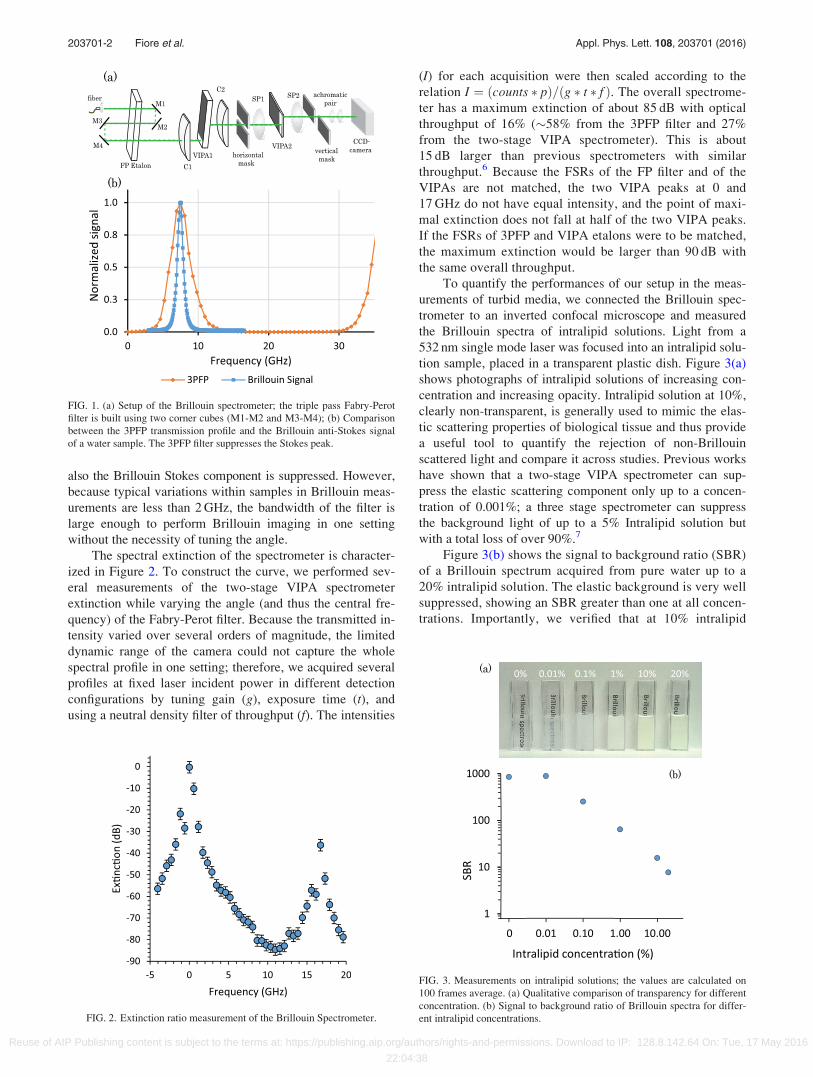
also the Brillouin Stokes component is suppressed. However,
because typical variations within samples in Brillouin meas-
urements are less than 2 GHz, the bandwidth of the filter is
large enough to perform Brillouin imaging in one setting
without the necessity of tuning the angle.
The spectral extinction of the spectrometer is character-
ized in Figure 2. To construct the curve, we performed sev-
eral measurements of the two-stage VIPA spectrometer
extinction while varying the angle (and thus the central fre-
quency) of the Fabry-Perot filter. Because the transmitted in-
tensity varied over several orders of magnitude, the limited
dynamic range of the camera could not capture the whole
spectral profile in one setting; therefore, we acquired several
profiles at fixed laser incident power in different detection
configurations by tuning gain (g), exposure time (t), and
using a neutral density filter of throughput (f). The intensities
(I) for each acquisition were then scaled according to the
relation I ¼ ðcounts � pÞ=ðg � t � f Þ. The overall spectrome-
ter has a maximum extinction of about 85 dB with optical
throughput of 16% (�58% from the 3PFP filter and 27%
from the two-stage VIPA spectrometer). This is about
15 dB larger than previous spectrometers with similar
throughput.6 Because the FSRs of the FP filter and of the
VIPAs are not matched, the two VIPA peaks at 0 and
17 GHz do not have equal intensity, and the point of maxi-
mal extinction does not fall at half of the two VIPA peaks.
If the FSRs of 3PFP and VIPA etalons were to be matched,
the maximum extinction would be larger than 90 dB with
the same overall throughput.
To quantify the performances of our setup in the meas-
urements of turbid media, we connected the Brillouin spec-
trometer to an inverted confocal microscope and measured
the Brillouin spectra of intralipid solutions. Light from a
532 nm single mode laser was focused into an intralipid solu-
tion sample, placed in a transparent plastic dish. Figure 3(a)
shows photographs of intralipid solutions of increasing con-
centration and increasing opacity. Intralipid solution at 10%,
clearly non-transparent, is generally used to mimic the elas-
tic scattering properties of biological tissue and thus provide
a useful tool to quantify the rejection of non-Brillouin
scattered light and compare it across studies. Previous works
have shown that a two-stage VIPA spectrometer can sup-
press the elastic scattering component only up to a concen-
tration of 0.001%; a three stage spectrometer can suppress
the background light of up to a 5% Intralipid solution but
with a total loss of over 90%.7
Figure 3(b) shows the signal to background ratio (SBR)
of a Brillouin spectrum acquired from pure water up to a
20% intralipid solution. The elastic background is very well
suppressed, showing an SBR greater than one at all concen-
trations. Importantly, we verified that at 10% intralipid
FIG. 1. (a) Setup of the Brillouin spectrometer; the triple pass Fabry-Perot
filter is built using two corner cubes (M1-M2 and M3-M4); (b) Comparison
between the 3PFP transmission profile and the Brillouin anti-Stokes signal
of a water sample. The 3PFP filter suppresses the Stokes peak.
FIG. 2. Extinction ratio measurement of the Brillouin Spectrometer.
FIG. 3. Measurements on intralipid solutions; the values are calculated on
100 frames average. (a) Qualitative comparison of transparency for different
concentration. (b) Signal to background ratio of Brillouin spectra for differ-
ent intralipid concentrations.
203701-2 Fiore et al. Appl. Phys. Lett. 108, 203701 (2016)
Reuse of AIP Publishing content is subject to the terms at: https://publishing.aip.org/authors/rights-and-permissions. Download to IP: 128.8.142.64 On: Tue, 17 May 2016
22:04:38

solution, our Brillouin measurement is shot-noise limited,
thus unaffected by elastic background.
Next, we demonstrated the ability to detect background-
free Brillouin signal from biological tissue. The tissue sam-
ple was a thin slice of chicken breast of area of about 1 cm2,
placed on the bottom of glass dish plate. The laser light was
focused into the tissue through the glass cover-slip at a
power of 6 mW and exposure time of 300 ms. Fig. 4(a)
shows the Brillouin signal intensity as a function of tissue
depth reported as average and standard deviation of 100
frames. The focusing depth was varied by translating the
objective lens along the z-axis of the microscope. For com-
parison, the background signal at each depth is reported,
showing the excellent suppression of the elastic scattering
provided by the instrument. Figure 4(b) shows the signal to
noise ratio (SNR) of this measurement as a function of tissue
depth. Brillouin spectra with SNR greater than one were
obtained from the tissue at depths up to 100 lm. We
observed an exponential decay of SNR yielding a mean free
path of �150 lm, consistent with the intensity loss due to
elastic scattering in chicken breast tissue10 and thus similar
to other optical modalities that do not suffer from back-
ground issues.
Importantly, other methods of rejecting elastically scat-
tered light that use absorption lines of gas cells require
experimentalists to perform Brillouin measurements with
specific laser wavelengths and are prone to degradations due
to laser frequency drift. In contrast, our spectrometer main-
tains its performance over a spectral band only limited by
the coatings of the optical elements (Fabry-Perot, VIPA, mir-
rors, and lenses). Therefore, the spectrometer can accommo-
date Brillouin experiments with a wide variety of excitation
wavelengths; moreover, operating at low-finesse and with a
tunable etalon, laser frequency drifts can be effectively
handled.
Several improvements can be implemented to improve
the practical performances of our spectrometer. As mentioned
before, the FSR of the Fabry-Perot etalon and the VIPAs
should be the same to reach maximal extinction. Moreover,
using the same principles demonstrated here, a reflection con-
figuration of the 3PFP would allow to build a notch-filter
rather than a bandpass filter, with similar performances of
throughput and extinction but larger transmitted bandwidth,
which could be useful for Brillouin measurements exhibiting
large shift variations of non-homogenous sample.
In conclusion, we have developed a two-stage VIPA
Brillouin spectrometer with confocal sampling and a triple
pass Fabry-Perot bandpass filter. We demonstrated rapid
Brillouin measurement of intralipid solutions and turbid bio-
logical tissue with effective suppression of elastic scattering
background. This investigation may expand the reach of
Brillouin technology beyond ocular tissue to important areas
such as the mechanical characterizations of tumors and athe-
rosclerotic plaques.
This work was supported in part by the National
Institutes of Health (K25EB015885, R21EY023043, and
R01EY025454); National Science Foundation (CMMI-
1537027 and CBET-0853773); Human Frontier Science
Program (Young Investigator Grant) and the Ministry of
Science of Korea, under the “ICT Consilience Creative
Program” (IITP-2015-R0346-15-1007).
1R. D. Hartschuh, A. Kisliuk, V. Novikov, A. P. Sokolov, P. R. Heyliger,
C. M. Flannery, W. L. Johnson, C. L. Soles, and W.-L. Wu, Appl. Phys.
Lett. 87(17), 173121 (2005); J. G. Dil, Rep. Prog. Phys. 45(3), 285 (1982);
A. A. Serga, T. Schneider, B. Hillebrands, S. O. Demokritov, and M. P.
Kostylev, Appl. Phys. Lett. 89(6), 063506 (2006); T. Still, R. Sainidou, M.
Retsch, U. Jonas, P. Spahn, G. P. Hellmann, and G. Fytas, Nano Lett.
8(10), 3194 (2008); Y. Minami and K. Sakai, Rev. Sci. Instrum. 80(1),
014902 (2009).2J. R. Sandercock, RCA Rev. 36(1), 89 (1975).3K. J. Koski and J. L. Yarger, Appl. Phys. Lett. 87(6), 061903 (2005); S.-i.
Itoh, Jpn. J. Appl. Phys., Part 1 37(5S), 3134 (1998); Y. Minami, T. Yogi,
and K. Sakai, Appl. Phys. Lett. 93(16), 161107 (2008).4M. Ahart, M. Somayazulu, S. A. Gramsch, R. Boehler, H.-k. Mao, and R.
J. Hemley, J. Chem. Phys. 134(12), 124517 (2011); A. V. Svanidze, V. P.
Romanov, and S. G. Lushnikov, JETP Lett. 93(7), 409 (2011).5G. Scarcelli and S. H. Yun, Nat. Photonics 2(1), 39 (2008).6G. Scarcelli, W. J. Polacheck, H. T. Nia, K. Patel, A. J. Grodzinsky, R. D.
Kamm, and S. H. Yun, Nat. Methods 12(12), 1132 (2015).7G. Scarcelli and S. H. Yun, Opt. Express 19(11), 10913 (2011).8Z. Meng and V. V. Yakovlev, J. Innovative Opt. Health Sci. 8(04),
1550021 (2015); G. Antonacci, G. Lepert, C. Paterson, and P. T€or€ok,
Appl. Phys. Lett. 107(6), 061102 (2015); I. Remer and A. Bilenca, Opt.
Lett. 41(5), 926 (2016).9M. �Copic and M. Zgonik, Opt. Commun. 41(5), 310 (1982).
10W.-F. Cheong, S. A. Prahl, and A. J. Welch, IEEE J. Quantum Electron.
26(12), 2166 (1990).
FIG. 4. (a) Comparison between Brillouin signal and elastic scattering light
component at different depths. (b) Signal to noise ratio as a function of
depth; the SNR is calculated on 100 frames average, with an exposure time
of 0.3 s. The exponential fit leads to a mean free path of �150 lm.
203701-3 Fiore et al. Appl. Phys. Lett. 108, 203701 (2016)
Reuse of AIP Publishing content is subject to the terms at: https://publishing.aip.org/authors/rights-and-permissions. Download to IP: 128.8.142.64 On: Tue, 17 May 2016
22:04:38