hepatotoxicity-induced by the therapeutic dose of
TRANSCRIPT
1444
Int. J. Morphol.,38(5):1444-1454, 2020.
Hepatotoxicity-Induced by the Therapeutic Dose of Acetaminophenand the Ameliorative Effect of Oral Co-administration of Selenium/
Tribulus terrestris Extract in Rats
Hepatotoxicidad Inducida por la Dosis Terapéutica de Acetaminofén y el Efecto de Mejorade la Administración Conjunta de Extracto de Selenio Tribulus terrestris en Ratas
Amin A. Al-Doaiss1,2
AL-DOAISS, A. A. Hepatotoxicity-induced by the therapeutic dose of acetaminophen and the ameliorative effect of oral co-administrationof selenium / Tribulus terrestris extract in rats. Int. J. Morphol., 38(5):1444-1454, 2020.
SUMMARY: Over dose or long-term clinical use of therapeutic doses of acetaminophen (APAP) causes hepatotoxicity. Variousstrategies attempted to ameliorate APAP-hepatotoxicity have been found to be unsuitable for clinical practice. This study was aimed toillustrate the histopathological changes induced by therapeutic dose of APAP and investigate the hepatoprotective role of oral co-administration of selenium/ Tribulus terrestris (TT) extract concurrently against hepatotoxicity induced by APAP in rats. Fifty-fourhealthy male albino Wistar rats were randomized into nine groups (G1–G9) of six rats each, and administered with APAP and TT orallyfor 30 days as follows: Control (2ml normal saline), APAP (470 mg/kg), APAP (470 mg/kg) + selenium (2 mg/kg), APAP (470 mg/kg) +TT (98 mg/kg), APAP (470 mg/kg) + selenium (2mg/kg) + TT (98 mg/kg), APAP (470 mg/kg) + silymarin (200 mg/kg), selenium (2 mg/kg), TT (98 mg/kg) and silymarin (200 mg/kg) groups. The results demonstrated that exposure of rats to therapeutic dose of APAP for 30days caused significant histopathological changes parallel to elevated blood chemistry parameters. Co- administration of selenium/TTextract showed significantly reduced histopathological lesions and, restored or decreased levels of the examined blood chemistry parameters.Liver histology in selenium/TT extract showed normal hepatic architecture with mild changes and silymarin treated rats showed nohistopathological changes. Histochemically PAS staining, showed that APAP-induced hepatotoxicity was characterized by hepatocytesglycogen depletion. Selenium/TT co-supplementation plays a potential role in preventing APAP-induced glycogen depletion by increasingdetoxification and scavenging the reactive metabolites. Selenium/TT extract oral co-administration possesses a significant hepatoprotectiveproperty and mitigates APAP-induced hepatotoxicity by enhancing its antioxidant role and improving tissue integrity. Selenium/TTsupplementation could represent an effective treatment against APAP-induced hepatotoxicity. Further studies are needed to elucidate theexact mechanism underlying the protective role of TT extract.
KEY WORDS: Acetaminophen; Selenium; Tribulus terrestris; Hepatotoxicity; Glycogen.
INTRODUCTION
Paracetamol or acetaminophen; (N-acety1-p-aminophenol; APAP), is an analgesic–antipyretic drugworldwide used as an over-the-counter drug and consideredsafe at therapeutic dose (AlWahsh et al., 2019). Whenadministrated at therapeutic doses, APAP is safe with fewside effects, but when used for a long time or over dosage, itcan cause oxidative stress, hepatotoxicity, and liver/renalfailure that can lead to death in both animals and humanbeings, with a mortality rate of up to 90 %. At over-therapeutic doses, APAP causes hepatotoxicity characterizedby reactive metabolites, which are converted to normalmetabolites by glutathione, and then excreted in the urine
(Ramachandran & Jaeschke, 2017). At therapeutic doses forlong periods or large doses, the levels of reactive metabolitesincrease and these metabolites react with proteins of thehepatocytes leading to glutathione depletion, oxidative stress,mitochondrial oxidative stress, and inhibition of ATPsynthesis, eventually leading to cell necrosis, apoptosis, DNAfragmentation, and releasing the membrane proteins into thecytosol, which resulting in liver injury with cell damage andliver cell death (Ramachandran & Jaeschke).
Most of the recent studies, which aimed to illustrateand identify the mechanism of APAP-induced hepatotoxicity
1 Department of Biology, College of Science, King Khalid University, Abha, Saudi Arabia.2 Department of Anatomy and Histology, Faculty of Medicine, Sana’a University, Sana’a, Republic of Yemen.
1445
were performed on experimental animal models,histological alterations such as hepatic degeneration,necrosis, and oxidative stress in rodents, which lead to acuteliver damage (Mahesh et al., 2009; Pettie et al., 2019).Several studies on experimental animals and humans haveattempted to prevent or repair hepatotoxicity by APAPoverdose (Iwalokun et al., 2006) or by recommendeddosage (Kurtovic & Riordan, 2003) through the pathwaysof glucuronidation and sulfation by cytochrome to formharmful reactive metabolites in order to prevent the toxicityor repair it (Hwang et al., 2008). Recently, utilization ofnatural products of medicinal plants has attracted researchattention as a preferable strategy for the treatment of liverinjury (Almasi et al., 2017).
Different agents and strategies associated with na-tural products that have antioxidant activity have beenreported and successfully used to ameliorate or preventAPAP-hepatotoxicity, but they have not been found to besuitable for clinical practice (Verma et al., 2019; Zhou etal., 2019); theses natural products include Uraria picta(Kale et al., 2012), Kombucha tea (Abshenas et al., 2012),Curcuma longa (Khorsandi & Orazizadeh, 2008),Plumbago zeylanica (Kanchana & Sadiq, 2011), Wedeliacalendulacea (Emmanuel et al., 2001), Ganodermaamboinense (Hsu et al., 2008), ajoene (Hattori et al., 2001),Vernonia amygdalina (Iwalokun et al.), Zingiber officinale(Yemitan & Izegbu, 2006), Ginkgo biloba (Sener et al.,2006), red algae Porphyra yezoensis (Hwang et al.), Indianhoney (Mahesh et al.), Mesna (Sener et al., 2005), Arabicgum (Gamal El-din et al., 2003). However, none of thesestrategies were found to be suitable or safe for clinicalpractice.
Tribulus terrestris (TT) (Zygophyllaceae family) isa dicotyledonous perennial creeping herb that is widelydistributed and contain numerous bioactive compounds(Almasi et al.). T. terrestris is an annual herb widespreadthroughout Saudi Arabia and northern Yemen (Chaudhary& Akram, 1987). Extracts of whole TT plant have beenused since ancient times in traditional folk medicine as atonic, diuretic, calculous affections aphrodisiac, analgesic,astringent, stomachic, antihypertensive, antimicrobial,antibacterial, antioxidant, treatment for painful urination,and antitoxic activities used in the treatment of diabetes,tumors, articular pains, respiratory diseases, cardiovasculardiseases, reproductive dysfunctions, urinary anti-infective,and urinary stones; it also enhances testosterone levels inhumans and mitigates oxidative damage, and thus, it is usedby bodybuilders (Almasi et al.). TT contains steroids,saponins, flavonoids, alkaloids, unsaturated fatty acids,vitamins, tannins, resins, nitrate potassium, aspartic acidand glutamic acid (Almasi et al.).
Selenium (Se) is an essential trace element andmicronutrient for livestock that is present in both organicand non-organic forms in the food (Qazi et al., 2019). Itexerts known chemo-preventive and chemotherapeuticeffects against various diseases caused by selenoproteins(Qazi et al.). Selenium is an antioxidant with complementarybiological effects on thyroid function, reproduction (gonadaldevelopment, gametogenesis, and fertilization), andimmunity, (protects the cells from destructive and harmfulresults of free radicals), which enhance human and animalhealth (Ghaffari et al., 2011). Selenium-deficiency istherefore linked with many disorders such as oxidative stress,myo-hepatic degeneration, and immunosuppression. Therecommended daily allowance is 55 µg in adults and 60 µgin pregnant women. At high doses Se is toxic and can causemortality (Mirone et al., 2013).
Silymarin has been widely used for decades inmedical practice, particularly in the treatment ofhepatotoxicity (Taju et al., 2011). “Milk thistle” is silymarinextract derived from the Silybum marianum plant. Silymarinis considered an anti-inflammatory, antioxidant,anticarcinogenic, and hepatoprotective medicinal plant (Tajuet al.). It is marketed as one of the standard hepatoprotectiveherbal formulations (Kanchana & Sadiq). In the current studysimilar to other studies, silymarin is usually used as a positivetreatment against APAP-induced hepatotoxicity.
To date, most of the medical plants in Saudi Arabiahave not been investigated to understand their biologicaland medical benefits (Al-Taweel et al., 2003; Kadriya etal., 2004). Therefore, in this study we hypothesized thatoral co-administration of selenium/TT extract would be ableto prevent APAP-induced deleterious effects in rat liverbecause of its intrinsic biochemical and free radicalsscavenging properties.
MATERIAL AND METHOD
All the experiments were conducted in the Histologyand Cell Biology Laboratory, Department of Biology,College of Science, King Khalid University, Saudi Arabia.All experimental protocols were approved by the BiomedicalResearch Ethical Committee at King Khalid University,Abha, Saudi Arabia.
Chemicals and drugs: Acetaminophen (APAP) in the formof paracetamol (500 mg) was purchased from a localpharmacy. Selenium in the form [Sodium selenite (Na
2SeO
3)]
was purchased from Oxoid Limited Co. UK. Silymarin waspurchased from Sigma-Aldrich (St. Louis, MO, USA).
AL-DOAISS, A. A. Hepatotoxicity-induced by the therapeutic dose of acetaminophen and the ameliorative effect of oral co-administration of selenium / Tribulus terrestris extract in rats. Int. J. Morphol., 38(5):1444-1454, 2020.
1446
Experimental animals: Adult male albino rats (150–200g), were used for the current study. They were fed withstandard commercial pallet diet and water ad libitum, andin polypropylene cages maintained under standardconditions (12-h light/dark cycle; 25 ± 3°C; 35–60 %humidity), as well as under strict hygienic conditions. Theanimals were habituated to laboratory conditions for 7 daysprior to the experiment to minimize any nonspecific stress.
Dosage, preparation and administration of testsubstances: The rat dose was calculated based on thesurface area ratio. The maximum daily dose of APAP is4000 mg for an adult (60 kg) person. The dose for rat wascalculated by dividing the maximum daily dose for adultperson by adult human weight of 60 kg and multiplying itby the factor 7 to accommodate the body surface area ofthe animal; (4000÷60) becomes 67 mg/kg b.w., the doseequivalent for rat (67 × 7) is 470 mg/kg b.w. (Freireich etal., 1966; Van Miert, 1986; Reagan-Shaw et al., 2008;Manimaran et al., 2010). The dose equivalent per kg forrat (×7) was derived by dividing the km factor (body surfacearea (µ2) to body weight (kg) ratio) for humans with theKm factor for rat species. The human dose of TT is 7000mg for an adult (60 kg). Similarly, (7000÷60) becomes 117mg/kg b.w., the dose equivalent for rat (117 × 7) is 820 mg/kg b.w. The dose of the extract was determined withreference to the % yield of extract with the dose of crudedrug and calculated as 98mg/kg for TT (Saiyed et al., 2016).
APAP and Se were dissolved in distilled water, andthe APAP-treated group received 470 mg/kg/day; APAP +selenium-treated group received APAP (470 mg/kg/day) +selenium (2 mg/kg/day), APAP-TT extract-treated groupreceived APAP (470 mg/kg/day) + TT extract (98 mg/kg/day), silymarin-group received silymarin (200 mg/kg/day)and the control-group received 2 ml of physiological salinealone. The preparations were administered by oral gavagein 5 ml/kg of body weight of each rat. TT plant was collectedfrom the Southern area (Abha) of Saudi Arabia. The wholeplant, excluding fruit was pulverized into powdery formusing an industrial blender and the constituents were
extracted with methanol using soxhlet apparatus (50 - 55ºC) for three days. The extract was concentrated in aventilated oven at 45 ºC for 24 h. Dried powder of 45 gyielded an extract of about 10 g, which was dark brown incolor. The characteristics and bioactive constituents of TTwere assessed previously by Reshma et al. (2019) in theplant list website (www.theplantlist.org).
Experimental design: A total of 54 healthy adult male al-bino rats (Rattus norvegicus) were randomly divided intonine groups (n=6 rats each) their dosing and route ofadministration provided in (Table I).
All the groups were treated for 30 consecutive days.At the end of this period, blood samples were collectedfrom each rat, and their sera were separated and estimatedfor biochemical parameters. Later, the animals weresacrificed, and liver tissues were removed forhistopathological examinations.
Collection of serum and estimation of liver function:Blood samples were collected without any anticoagulantand allowed to clot for 10 min at room temperature. Aftercoagulation, the blood was centrifuged at 0 ºC to obtainthe serum. The serum collected was stored at –80 ºC untilfurther use. Evaluation of enzymes for liver functionincluding aspartate aminotransferase (AST), alanineaminotransferase (ALT), alkaline phosphatase (ALP), to-tal bilirubin (TB), and total protein (TP) levels in serumwere estimated by using standard enzymatic reagent strips(Reflotrone ® plus system, Roche Diagnostic Ltd.,Germany).
Histopathology: Small portions of the liver from each ratwere removed, and fixed in formalin (10 %). The specimenswere processed for dehydration using ascending grades ofethanol, clearing in xylene and impregnation with moltenparaffin wax in an oven at 60 ºC, using an automatic tissueprocessor (Sakura, Japan). Next, the tissues were embeddedusing an embedding station (Leica, Germany); sections (4-5 µm) were cut and stained with Hematoxylin and Eosin
Table I. Experimental study design.
AL-DOAISS, A. A. Hepatotoxicity-induced by the therapeutic dose of acetaminophen and the ameliorative effect of oral co-administration of selenium / Tribulus terrestris extract in rats. Int. J. Morphol., 38(5):1444-1454, 2020.
Group Treatment Dosage
G1 Control Normal saline 2ml/kg/day p.oG2 APAP 470 mg/kg/day p.oG3 APAP+ Selenium APAP (470 mg/kg/day p.o) + selenium (2 mg/kg/day p.o)G4 APAP+ TT APAP (470 mg/kg/day p.o) + TT (98 mg/kg/day p.o)G5 APAP+ Selenium+ TT APAP (470 mg/kg/day p.o) + selenium (2 mg/kg/day p.o) + (98 mg/kg/day p.o)G6 APAP+ Silymarin APAP (470 mg/kg/day p.o) + Silymarin (200 mg/kg/day p.o)G7 Selenium 2 mg/kg/day p.oG8 TT 98 mg/kg/day p.oG9 Silymarin 200 mg/kg/day p.o
1447
(H&E) and PAS techniques asdescribed previously (Al-Doaiss etal., 2019). The stained sections wereexamined using optical microscope(Olympus Microscope BP53 withDigital Camera, Japan). Allsubsequent histopathologicalexaminations were performed by anexperienced pathologist withoutknowledge of the previoustreatments.
Statistical analysis: Results ofbiochemical parameters are presentedas the mean ± standard deviation(SD). Differences between means inall groups were tested for significanceusing the one-way analysis ofvariance (ANOVA) followed byDuncan’s test and P<0.05 wasconsidered significant using thestatistical analysis software SPSS(SPSS, 1996).
RESULTS
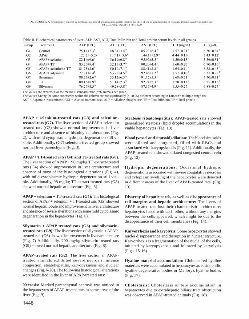
Biochemical parameters: Theanimals treated with APAP exhibitedsignificant elevations in the liverprofile parameters (P<0.05), i.e.,ALP, AST, ALT, TB and TP serumlevels, compared to those of the con-trol, TT, Se, and silymarin groups(Table II).
Histopathological studies
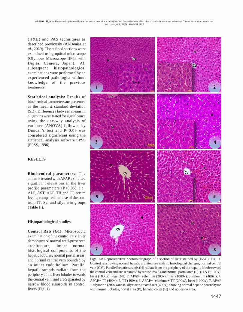
Control Rats (G1): Microscopicexamination of the control rats’ liverdemonstrated normal well-preservedarchitecture, intact normalhistological components of thehepatic lobules, normal portal areas,and normal central vein bounded byan intact endothelium. Parallelhepatic strands radiate from theperiphery of the liver lobules towardsthe central vein, and are Separated bynarrow blood sinusoids in controllivers (Fig. 1).
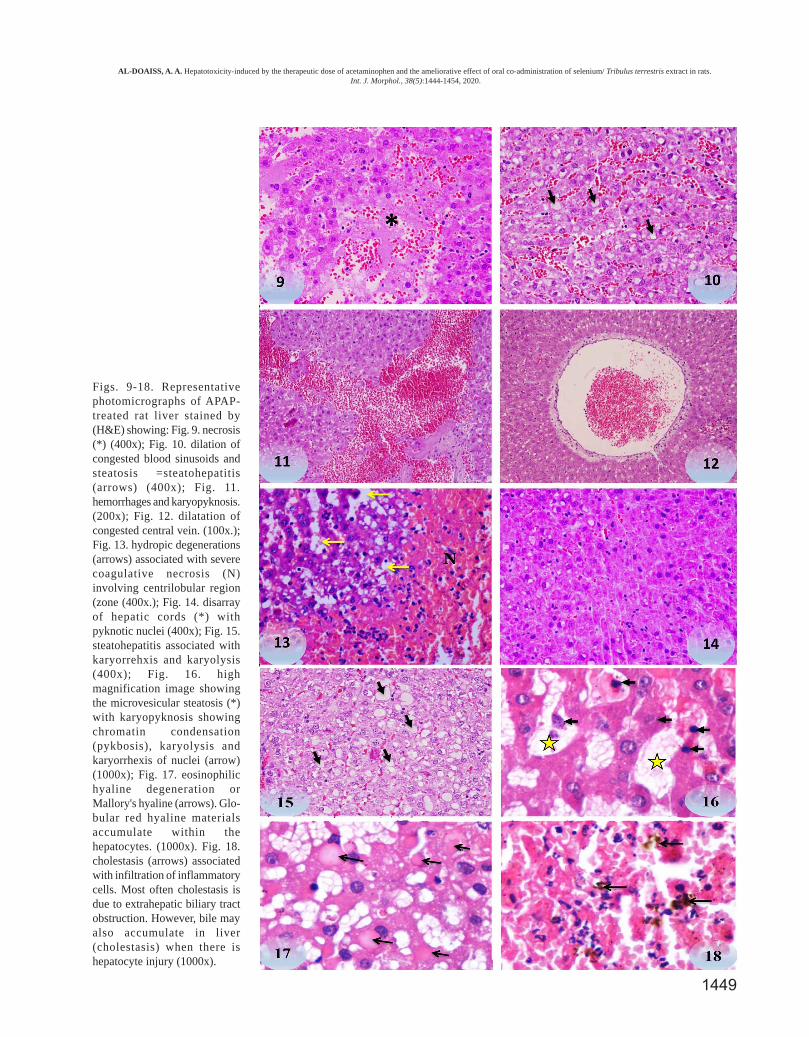
Figs. 1-8 Representative photomicrograph of a section of liver stained by (H&E): Fig. 1.Control rat showing normal hepatic architecture with no histological changes, normal centralvein (CV). Parallel hepatic strands (H) radiate from the periphery of the hepatic lobule towardthe central vein and are separated by sinusoids (S) and normal portal area (P). (H & E; 100x).Inset (1000x); Figs. 2-8. 2. APAP+ selenium (200x), Inset (1000x); 3. selenium (400x.); 4.APAP+ TT (400x); 5. TT (400x); 6. APAP+ selenium + TT (200x.), Inset (1000x); 7. APAP+ silymarin (200x) and 8. silymarin-treated rats (400x), showing normal hepatic parenchymawith normal lobules, portal area (P), hepatic cords (H) and no lesion area.
AL-DOAISS, A. A. Hepatotoxicity-induced by the therapeutic dose of acetaminophen and the ameliorative effect of oral co-administration of selenium / Tribulus terrestris extract in rats. Int. J. Morphol., 38(5):1444-1454, 2020.
1448
APAP + selenium-treated rats (G3) and selenium-treated rats (G7). The liver section of APAP + seleniumtreated rats (G3) showed normal improvement in liverarchitecture and absence of histological alterations (Fig.2), with mild cytoplasmic hydropic degeneration still vi-sible. Additionally, (G7) selenium-treated group showednormal liver parenchyma (Fig. 3).
APAP + TT-treated rats (G4) and TT-treated rats (G8).The liver section of APAP + 98 mg/kg TT extract-treatedrats (G4) showed improvement in liver architecture andabsence of most of the histological alterations (Fig. 4),with mild cytoplasmic hydropic degeneration still visi-ble. Additionally, 98 mg/kg TT extract-treated rats (G8)showed normal hepatic architecture (Fig. 5).
APAP + selenium + TT-treated rats (G5): The histologicalsection of APAP + selenium + TT-treated rats (G5) showednormal hepatic lobule and improvement in liver architectureand absence of severe alterations with some mild cytoplasmicdegeneration in the hepatocytes (Fig. 6).
Silymarin + APAP treated rats (G6) and silymarin-treated rats (G9): The liver section of silymarin + APAP-treated-rats (G6) showed improvement in liver architecture(Fig. 7) Additionally, 200 mg/kg silymarin-treated rats(G9) showed normal hepatic architecture (Fig. 8).
APAP-treated rats (G2): The liver section in APAP-treated animals exhibited severe necrosis, intensecongestion, steatohepatitis, karyopyknosis and nuclearchanges (Fig. 6-20). The following histological alterationswere identified in the liver of APAP-treated rats:
Necrosis: Marked parenchymal necrosis was noticed inthe hepatocytes of APAP-treated rats in some areas of theliver (Fig. 9).
Steatosis (steatohepatitis): APAP-treated rats showedgeneralized steatosis (lipid droplet accumulation) in theviable hepatocytes (Fig. 10).
Blood (vessel and sinusoid) dilation: The blood sinusoidswere dilated and congested, filled with RBCs andassociated with karyopyknosis (Fig. 11). Additionally, theAPAP-treated rats showed dilated congested central vein(Fig. 12).
Hydropic degenerations: Occasional hydropicdegenerations associated with severe coagulative necrosisand cytoplasm swelling of the hepatocytes were detectedin different areas of the liver of APAP-treated rats. (Fig.13).
Disarray of hepatic cords, as well as disappearance ofcell margins and hepatic architecture: The livers ofAPAP-treated rats lost their characteristic architecture;hepatocytes fused with each other, without any marginsbetween the cells appeared, which might be due to thedisappearance of their cell membranes (Fig. 14).
Karyorrhexis and karyolysis: Some hepatocytes showednuclei disappearance and disruption in nuclear structure.Karyorrhexis is a fragmentation of the nuclei of the cells,initiated by karyopyknosis and followed by karyolysis(Figs. 15-16).
Hyaline material accumulation: Globular red hyalinematerials were accumulated in hepatocytes as eosinophilichyaline degenerative bodies or Mallory's hyaline bodies(Fig. 17).
Cholestasis: Cholestasis or bile accumulation inhepatocytes due to extrahepatic biliary tract obstructionwas observed in APAP-treated animals (Fig. 18).
Table II. Biochemical parameters of liver: ALP, AST, ALT, Total bilirubin and Total protein serum levels in all groups.
The values are expressed as the means ± standard error (n=6 animals per group)The values having the same superscript within the column are not significantly (p <0.05) different according to Duncan’s multiple range test.AST = Aspartate transaminase, ALT = Alanine transaminase, ALP = Alkaline phosphatase, TB = Total bilirubin, TP = Total protein
AL-DOAISS, A. A. Hepatotoxicity-induced by the therapeutic dose of acetaminophen and the ameliorative effect of oral co-administration of selenium/ Tribulus terrestris extract in rats. Int. J. Morphol., 38(5):1444-1454, 2020.
Group Treatment ALP (U/L) ALT (U/L) AST (U/L) T.B (mg/dl) T.P (g/dl)
G1 Control 75.14±2.3b 48.34±3.4 b 85.23±6.4 b 1.57±0.21 b 6.50±0.34 b
G2 APAP 123.25±5.2a 117.15±5.6 a 144.17±2.9 a 4.44±0.15a 3.43±0.12a
G3 APAP+ selenium 82.11±9.6b 56.19±6.8 b 95.92±3.5 b 1.50±0.15 b 5.56±0.31 b
G4 APAP+ TT 85.20±9.4b 52.15±3.7 b 90.30±4.4 b 1.60±0.20 b 6.70±0.16 b
G5 APAP+ selenium+ TT 81.25±2.6b 50.10±7.1 b 88.41±2.2 b 1.64±0.11 b 6.33±0.43 b
G6 APAP+ silymarin 77.21±6.4b 53.72±4.5 b 92.46±1.2 b 1.57±0.10 b 5.37±0.22 b
G7 Selenium 80.23±2.6 b 53.12±6.3 b 91.17±5.5 b 1.68±0.21 b 5.70±0.31 b
G8 TT 89.16±8.9 b 51.14±2.3 b 92.24±2.3 b 1.70±0.13 b 6.25±0.15 b
G9 Silymarin 76.27±5.5 b 49.28±5.8 b 87.15±4.9 b 1.55±0.27 b 6.88±0.27 b
1449
Figs. 9-18. Representativephotomicrographs of APAP-treated rat liver stained by(H&E) showing: Fig. 9. necrosis(*) (400x); Fig. 10. dilation ofcongested blood sinusoids andsteatosis =steatohepatitis(arrows) (400x); Fig. 11.hemorrhages and karyopyknosis.(200x); Fig. 12. dilatation ofcongested central vein. (100x.);Fig. 13. hydropic degenerations(arrows) associated with severecoagulative necrosis (N)involving centrilobular region(zone (400x.); Fig. 14. disarrayof hepatic cords (*) withpyknotic nuclei (400x); Fig. 15.steatohepatitis associated withkaryorrehxis and karyolysis(400x); Fig. 16. highmagnification image showingthe microvesicular steatosis (*)with karyopyknosis showingchromatin condensation(pykbosis), karyolysis andkaryorrhexis of nuclei (arrow)(1000x); Fig. 17. eosinophilichyaline degeneration orMallory's hyaline (arrows). Glo-bular red hyaline materialsaccumulate within thehepatocytes. (1000x). Fig. 18.cholestasis (arrows) associatedwith infiltration of inflammatorycells. Most often cholestasis isdue to extrahepatic biliary tractobstruction. However, bile mayalso accumulate in liver(cholestasis) when there ishepatocyte injury (1000x).
AL-DOAISS, A. A. Hepatotoxicity-induced by the therapeutic dose of acetaminophen and the ameliorative effect of oral co-administration of selenium/ Tribulus terrestris extract in rats. Int. J. Morphol., 38(5):1444-1454, 2020.
1450
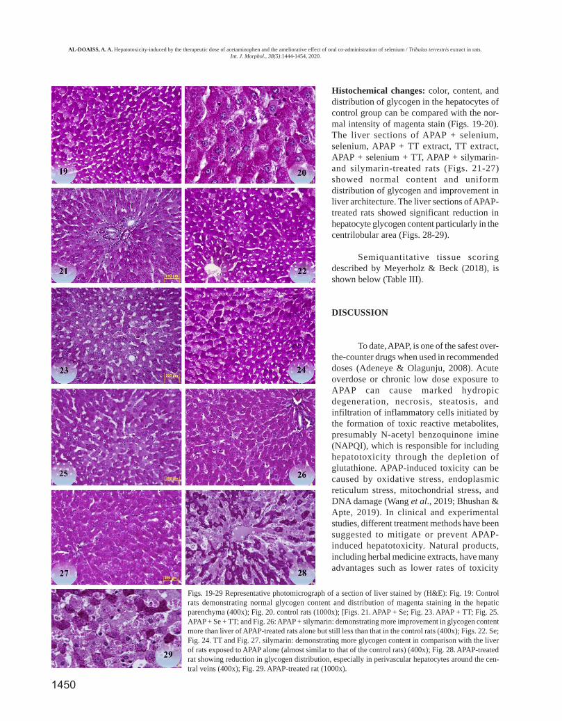
Figs. 19-29 Representative photomicrograph of a section of liver stained by (H&E): Fig. 19: Controlrats demonstrating normal glycogen content and distribution of magenta staining in the hepaticparenchyma (400x); Fig. 20. control rats (1000x); [Figs. 21. APAP + Se; Fig. 23. APAP + TT; Fig. 25.APAP + Se + TT; and Fig. 26: APAP + silymarin: demonstrating more improvement in glycogen contentmore than liver of APAP-treated rats alone but still less than that in the control rats (400x); Figs. 22. Se;Fig. 24. TT and Fig. 27. silymarin: demonstrating more glycogen content in comparison with the liverof rats exposed to APAP alone (almost similar to that of the control rats) (400x); Fig. 28. APAP-treatedrat showing reduction in glycogen distribution, especially in perivascular hepatocytes around the cen-tral veins (400x); Fig. 29. APAP-treated rat (1000x).
Histochemical changes: color, content, anddistribution of glycogen in the hepatocytes ofcontrol group can be compared with the nor-mal intensity of magenta stain (Figs. 19-20).The liver sections of APAP + selenium,selenium, APAP + TT extract, TT extract,APAP + selenium + TT, APAP + silymarin-and silymarin-treated rats (Figs. 21-27)showed normal content and uniformdistribution of glycogen and improvement inliver architecture. The liver sections of APAP-treated rats showed significant reduction inhepatocyte glycogen content particularly in thecentrilobular area (Figs. 28-29).
Semiquantitative tissue scoringdescribed by Meyerholz & Beck (2018), isshown below (Table III).
DISCUSSION
To date, APAP, is one of the safest over-the-counter drugs when used in recommendeddoses (Adeneye & Olagunju, 2008). Acuteoverdose or chronic low dose exposure toAPAP can cause marked hydropicdegeneration, necrosis, steatosis, andinfiltration of inflammatory cells initiated bythe formation of toxic reactive metabolites,presumably N-acetyl benzoquinone imine(NAPQI), which is responsible for includinghepatotoxicity through the depletion ofglutathione. APAP-induced toxicity can becaused by oxidative stress, endoplasmicreticulum stress, mitochondrial stress, andDNA damage (Wang et al., 2019; Bhushan &Apte, 2019). In clinical and experimentalstudies, different treatment methods have beensuggested to mitigate or prevent APAP-induced hepatotoxicity. Natural products,including herbal medicine extracts, have manyadvantages such as lower rates of toxicity
AL-DOAISS, A. A. Hepatotoxicity-induced by the therapeutic dose of acetaminophen and the ameliorative effect of oral co-administration of selenium / Tribulus terrestris extract in rats. Int. J. Morphol., 38(5):1444-1454, 2020.
1451
during prolonged use, antitussive, antibacterial, antifungal,anti-inflammatory, and anti-oxidant effects (Wang et al.).Recently, many experimental studies have demonstrated theprotective effects of several natural products against APAP-induced hepatotoxicity due to their actions as anti-inflammatory and anti-oxidant agents, and damage reparativeresponse (Lv et al., 2018).
The determination of the biomarkers (ALT, AST, andAL), TB, and TP are linked to hepatocyte function and integrityand can be used for the evaluation of hepatic injury (Rashid etal., 2016). Serum enzyme levels have been described aspotential indicators of necrosis and liver injury (Yoon et al.,2016). ALT is an effective biochemical marker for evaluatingliver function. Serum AST, ALP, and bilirubin are alsoassociated with liver injury and widely used as hepatic markersto evaluate hepatotoxicity or liver diseases (Girish et al., 2009).APAP-induced acute liver failure was characterized byhistopathological changes parallel with elevation of bloodchemistry parameters’ levels. Increase in enzyme levels areattributed to cellular leakage due to loss of selectivepermeability of cell membranes of hepatocytes. Hepaticenzymes are located in the cytoplasm but leak into circulatoryblood after hepatotoxicity (Wang et al.). Chronicadministration of acetaminophen produced a marked elevationof enzymes levels in APAP-treated rat.
In contrast, the co-administration of selenium/TTextract prevented the elevation of hepatic biochemical enzymeslevels, which were near the normal level, and repaired ofAPAP-damaged hepatic tissues. This corresponds with thecommonly accepted view that serum levels of liver enzymesreturn to normal after the hepatic tissue heals. ALP level isrelated to the response to cholestasis and biliary pressure afterAPAP administration. Serum bilirubin evaluation is one ofthe most sensitive tests employed for the diagnosis of hepaticdiseases. Hyperbilirubinemia has been previously observed
due to excessive heme destruction and blockage of biliary tract(Wolf et al., 1997). Administration of selenium/ TT extractincreased the level of protein, suggesting that it offeredprotection. The elevated levels of all liver enzymes and otherparameters were decreased to near normal levels after 30-daytreatment of selenium/ TT extract, indicating that it offeredprotection by preserving the structural integrity of thehepatocellular membrane against APAP and probably becauseof their membrane stabilizing activity which prevents leakageof intracellular enzymes.
In the current study, the histopathological investigationsrevealed the infiltration of inflammatory cells associated withnecrosis in the livers of APAP-treated rats, and this findingconcurs with that of Papackova et al. (2018). Hepatocytes ofAPAP-treated rats showed blood vessel & sinusoid congestion,hydropic degeneration, microvesicular fatty changevacuolation, eosinophilia, and scattered foci of necrosis withnuclear changes (pyknosis, karyolysis and karyorrhexis).
In the present study, co-treatment with selenium/TTextract inhibited all alterations of APAP- induced toxicity. Thisprotective role could be due to their ant oxidative activitymediated through free radical scavenging or production(Rashid et al.; Wang et al.). Moreover, enzyme levels in theserum and histopathological changes in the liver weremitigated and almost abolished in the APAP-treated groupcompared with the control group, after co-treatment withselenium/TT extract.
Histopathological assessments of tissues (tissue-basedpathology) are important tools that are considered acornerstone technique for evaluating the effects of medicationsor chemicals in biomedical studies, particularly those studyingexperimental animals related to human diseases. Histochemicalstaining is an essential method for tissue evaluation. Thereare several histological and histochemical staining techniques
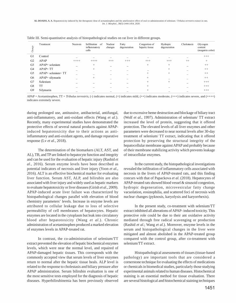
Table III. Semi-quantitative analysis of histopathological studies on rat liver in different groups.
APAP = Acetaminophen, TT = Tribulus terrestris, (-) indicates normal, (+) indicates mild, (++) indicates moderate, (+++) indicates severe, and (++++)indicates extremely severe.
AL-DOAISS, A. A. Hepatotoxicity-induced by the therapeutic dose of acetaminophen and the ameliorative effect of oral co-administration of selenium / Tribulus terrestris extract in rats. Int. J. Morphol., 38(5):1444-1454, 2020.
Treatment Necrosis Infiltration ofinflammatorycells
Nuclearchanges
Fattydegeneration
Congestion ofhepatic tissue
Hydropicdegeneration
Cholestasis Glycogencontent(magenta color)
G1 Control _ _ _ _ _ _ _ +++
G2 APAP ++++ ++++ +++ +++ +++ +++ ++ +G3 APAP+ selenium _ + _ _ + + _ ++G4 APAP+ TT _ + _ _ _ + _ ++G5 APAP+ selenium+ TT _ _ _ _ + + _ ++
G6 APAP+ silymarin _ + _ _ + _ _ ++G7 Selenium _ _ _ _ + + _ +++G8 TT _ _ _ _ + _ _ +++G9 Silymarin _ _ _ _ + _ _ +++
1452
that are used for detection of the chemical components oftissues. For example, H&E is a routine stain that is usedfor tissue examination in histology. Other specialhistochemical methods include periodic acid Schiff (PAS),which can bind to specific biochemical contents and serveas effective indicators used for the evaluation ofhistological alterations. PAS is evidently a specific stainused for detecting of glycogen in the liver and neutralmucins in other organs (Meyerholz et al., 2018).Administration of APAP at therapeutic dose for 30 dayscaused sever hepatocytes glycogen depletion in the liverof the treated rats. This histochemical change could be dueto the direct effects of APAP on the enzymes involved inthe processes of glycogenesis, glycolysis or glucoseabsorption. The liver cells in the perivascular zones weremore affected. This change refers to glycolysis, which wasmore affected than glycogenesis after APAP administration.Glycogen depletion in APAP-treated rats was associatedwith the harmful role of reactive metabolites. These resultscorrespond with the findings of a previous study on micetreated with APAP (500 mg/kg, i.p.) (Hinson et al., 1983).Selenium/TT extract treatment of rats prevents APAP-induced glycogen depletion by increasing detoxificationthrough reactive metabolite scavenging (Hinson et al.).Glycogen is an immediate fuel source found in thecytoplasm of cardiac (2 %), skeletal muscle (1–2 %), andliver cells (5–6 %). The glycogen particles in hepatocytescan be 10-fold larger than those in skeletal myocytes(Murray & Rosenbloom, 2018).
CONCLUSION
Our results demonstrate that therapeutic dose of APAPfor 30 days is capable of inducing marked hepatic histologicaland biochemical alterations in rats. Additionally, the presentresults provide strong evidence that the co-administration ofselenium/TT extract inhibits and ameliorates hepatotoxiceffects induced by APAP, and this role was possibly mediatedthrough free radical scavenging or inhibition of free radicalgeneration. Further studies are required, to assesshepatoprotective effects of medicinal plant extracts againstAPAP toxicity to confirm these protective effects.
ACKNOWLEDGMENTS
The author extends their appreciation to the Deanshipof Scientific Research at King Khalid University, Abha, KSAfor funding this work through Research Group Project undergrant number (R.G.P1/158/40).
AL-DOAISS, A. A. Hepatotoxicidad inducida por la dosis tera-péutica de acetaminofén y el efecto de mejora de la administraciónconjunta de extracto de selenio / Tribulus terrestris en ratas. Int. J.Morphol., 38(5):1444-1454, 2020.
RESUMEN: La dosis excesiva o el uso clínico a largoplazo de dosis terapéuticas de acetaminofeno (APAP) causahepatotoxicidad. Se ha descubierto que varias estrategias que in-tentaron mejorar la hepatotoxicidad por APAP no son adecuadaspara la práctica clínica. Este estudio tuvo como objetivo ilustrarlos cambios histopatológicos inducidos por la dosis terapéutica deAPAP e investigar el papel hepatoprotector de la administraciónconjunta de extracto de selenio / Tribulus terrestris (TT) simultá-neamente contra la hepatotoxicidad inducida por APAP en ratas.Cincuenta y cuatro ratas Wistar albino machos sanas sealeatorizaron en nueve grupos (G1 - G9) de seis ratas cada una, yse administraron con APAP y TT por vía oral durante 30 días de lasiguiente manera: Control (2 ml de solución salina normal), APAP(470 mg / kg), APAP (470 mg / kg) + selenio (2 mg / kg), APAP(470 mg / kg) + TT (98 mg / kg), APAP (470 mg / kg) + selenio (2mg / kg) + TT (98 mg / kg), APAP (470 mg / kg) + silimarina (200mg / kg), selenio (2 mg / kg), TT (98 mg / kg) y silimarina (200 mg/ kg). Los resultados demostraron que la exposición de las ratas ala dosis terapéutica de APAP durante 30 días causó cambioshistopatológicos significativos paralelos a parámetros elevados dequímica sanguínea. La administración conjunta de extracto deselenio / TT mostró lesiones histopatológicas significativamentereducidas y niveles restaurados o disminuidos de los parámetrosde química sanguínea. La histología hepática en el extracto deselenio / TT mostró una arquitectura hepática normal con cambiosleves y las ratas tratadas con silimarina no mostraron cambioshistopatológicos. La tinción histoquímica de PAS mostró que lahepatotoxicidad inducida por APAP se caracterizó por la pérdidade glucógeno de los hepatocitos. La suplementación con selenio /TT juega un papel potencial en la prevención de la pérdida deglucógeno inducido por APAP al aumentar la desintoxicación yeliminar los metabolitos reactivos. La administración conjunta deextracto de selenio / TT posee una propiedad hepatoprotectora sig-nificativa y mitiga la hepatotoxicidad inducida por APAP al mejo-rar su papel antioxidante y la integridad del tejido. Lasuplementación con selenio / TT podría representar un tratamientoefectivo contra la hepatotoxicidad inducida por APAP. Se necesi-tan más estudios para dilucidar el mecanismo exacto que subyacea la función protectora del extracto TT.
PALABRAS CLAVE: Acetaminofeno; Selenio; Tribulusterrestris; Hepatotoxicidad;Glucógeno.
REFERENCES
Abshenas, J.; Derakhshanfar, A.; Ferdosi, M. H. & Hasanzadeh, S. Protectiveeffect of Kombucha tea against acetaminophen-induced hepatotoxicityin mice: a biochemical and histopathological study. Comp. Clin. Pathol.,21:1243-8, 2012.
Adeneye, A. & Olagunju, J. Protective effect of oral ascorbic acid (vitaminc) against acetaminophen- induced hepatic injury in rats. Afr. J. Biomed.Res., 11(2):183-90, 2008.
AL-DOAISS, A. A. Hepatotoxicity-induced by the therapeutic dose of acetaminophen and the ameliorative effect of oral co-administration of selenium / Tribulus terrestris extract in rats. Int. J. Morphol., 38(5):1444-1454, 2020.
1453
Al-Doaiss, A. A.; Ali, D.; Ali, B. A. & Jarrar, B. M. Renal histologicalalterations induced by acute exposure of titanium dioxidenanoparticles. Int. J. Morphol., 37(3):1049-57, 2019.
Al-Taweel, A. M.; El-Deeb, K. S. & Al-Muhtadi, F. J. Chemicalcomposition and antimicrobial activity of the essential oil ofKleinia odora. Saudi Pharma. J., 12(1):47-50, 2003.
Almasi, F.; Khazaei, M.; Chehrei, S. & Ghanbari, A. Hepatoprotectiveeffects of Tribulus terrestris hydro-alcholic extract on non-alcoholic fatty liver- induced rats. Int. J. Morphol., 35(1):345-50,2017.
AlWahsh, M.; Othman, A.; Hamadneh, L.; Telfah, A.; Lambert, J.;Hikmat, S.; Alassi, A.; Mohamed, F. E. Z.; Hergenröder, R.; Al-Qirim, T.; et al. Second exposure to acetaminophen overdose isassociated with liver fibrosis in mice. EXCLI J., 18:51-62, 2019.
Bhushan, B. & Apte, U. Liver regeneration after acetaminophenhepatotoxicity: mechanisms and therapeutic opportunities. Am.J. Pathol., 189(4):719-29, 2019.
Chaudhary, S. A. & Akram, M. Weeds of Saudi Arabia and the ArabianPeninsula. Riyadh, National Herbarium, Regional Agriculture andWater Research Center, Ministry of Agriculture and Water, 1987.
Emmanuel, S.; Amalraj, T. & Ignacimuthu, S. Hepatoprotective effectof coumestans isolated from the leaves of Wedelia calendulaceaLess. in paracetamol induced liver damage. Indian J. Exp. Biol.,39(12):1305-7, 2001.
Freireich, E. J.; Gehan, E. A.; Rall, D. P.; Schmidt, L. H. & Skipper,H. E. Quantitative comparison of toxicity of anticancer agents inmouse, rat, hamster, dog, monkey, and man. Cancer Chemother.Rep., 50(4):219-44, 1966.
Gamal El-din, A. M.; Mostafa, A. M.; Al-Shabanah, O. A.; Al-Bekairi,A. M. & Nagi, M. N. Protective effect of Arabic gum againstacetaminophen-induced hepatotoxicity in mice. Pharmacol. Res.,48(6):631-5, 2003.
Ghaffari, T.; Nouri, M.; Irannejad, E. & Rashidi, M. R. Effect ofvitamin E and selenium supplement on paraoxonase-1 activity,oxidized low density lipoprotein and antioxidant defense indiabetic rats. Bioimpacts, 1(2):121-8, 2011.
Girish, C.; Koner, B. C.; Jayanthi, S.; Rao, K. R.; Rajesh, B. & Pradhan,S. C. Hepatoprotective activity of six polyherbal formulations inparacetamol induced liver toxicity in mice. Indian J. Med. Res.,129(5):569-78, 2009.
Hattori, A.; Yamada, N.; Nishikawa, T.; Fukuda, H. & Fujino, T.Protective effect of ajoene on acetaminophen-induced hepaticinjury in mice. Biosci. Biotechnol. Biochem., 65(11):2555-7, 2001.
Hinson, J. A.; Mays, J. B. & Cameron, A. M. Acetaminophen-inducedhepatic glycogen depletion and hyperglycemia in mice. Biochem.Pharmacol., 32(13):1979-88, 1983.
Hsu, C. C.; Lin, K. Y.; Wang, Z. H.; Lin, W. L. & Yin, M. C. Preventiveeffect of Ganoderma amboinense on acetaminophen-induced acuteliver injury. Phytomedicine, 15(11):946-50, 2008.
Hwang, H. J.; Kwon, M. J.; Kim, I. H. & Nam, T. J. Chemoprotectiveeffects of a protein from the red Algae porphyra yezoensis onacetaminophen-induced liver injury in rats. Phytother. Res.,22(9):1149-53, 2008.
Iwalokun, B. A.; Efedede, B. U.; Alabi-Sofunde, J. A.; Oduala, T.;Magbagbeola, O. A. & Akinwande, A. I. Hepatoprotective andantioxidant activities of Vernonia amygdalina on acetaminophen-induced hepatic damage in mice. J. Med. Food, 9(4):524-30, 2006.
Kadriya, E. D. S.; Abbas, F. A.; El Fishawy, A. & Mossa, J. S. Chemicalcomposition of the essential oil of Tagetes minuta growing in SaudiArabia. Saudi Pharm. J., 12(1):51-53, 2004.
Kale, R. H.; Halde, U. K. & Biyani, K. R. Protective Effect of aqueousextract of Uraria picta on acetaminophen induced nephrotoxicityin rats. Int. J. Res. Pharm. Biomed. Sci., 3(1):110-3, 2012.
Kanchana, N. & Sadiq, A. M. Hepatoprotective effect of Plumbagozeylanica on paracetamol induced liver toxicity in rats. Int. J.Pharm. Pharm. Sci., 3(1):151-4, 2011.
Khorsandi, L. & Orazizadeh, M. Protective effect of Curcuma longaextract on acetaminophen induced nephrotoxicity in mice. DARUJ. Pharm. Sci., 16(3):155-9, 2008.
Kurtovic, J. & Riordan, S. M. Paracetamol-induced hepatotoxicity atrecommended dosage. J. Intern. Med., 253(2):240-3, 2003.
Lv, H.; Xiao, Q.; Zhou, J.; Feng, H.; Liu, G. & Ci, X. Licochalcone Aupregulates Nrf2 antioxidant pathway and thereby alleviatesacetaminophen-induced hepatotoxicity. Front. Pharmacol., 9:147,2018.
Mahesh, A.; Shaheetha, J.; Thangadurai, D. & Rao, D. M. Protectiveeffect of Indian honey on acetaminophen induced oxidative stressand liver toxicity in rat. Biologia, 64:1225, 2009.
Manimaran, A.; Sarkar, S. N. & Sankar, P. Influence of repeatedpreexposure to arsenic on acetaminophen-induced oxidative stressin liver of male rats. Food Chem. Toxicol., 48(2):605-10, 2010.
Meyerholz, D. K. & Beck, A. P. Principles and approaches for repro-ducible scoring of tissue stains in research. Lab. Invest., 98(7):844-55, 2018.
Meyerholz, D. K.; Beck, A. P.; Goeken, J. A.; Leidinger, M. R.; Ofori-Amanfo, G. K.; Brown, H. C.; Businga, T. R.; Stoltz, D. A.;Reznikov, L. R. & Flaherty, H. A. Glycogen depletion can increasethe specificity of mucin detection in airway tissues. BMC Res.Notes, 11:763, 2018.
Mirone, M.; Giannetta, E. & Isidori, A. M. Selenium and reproductivefunction. A systematic review. J. Endocrinol. Invest., 36(10Suppl.):28-36, 2013.
Murray, B. & Rosenbloom, C. Fundamentals of glycogen metabolismfor coaches and athletes. Nutr. Rev., 76(4):243–259, 2018.
Papackova, Z.; Heczkova, M.; Dankova, H.; Sticova, E.; Lodererova,A.; Bartonova, L.; Poruba, M. & Cahova, M. Silymarin preventsacetaminophen-induced hepatotoxicity in mice. PLoS One,13(1):e0191353, 2018.
Pettie, J. M.; Caparrotta, T. M.; Hunter, R. W.; Morrison, E. E.; Wood,D. M.; Dargan, P. I.; Thanacoody, R. H.; Thomas, S. H. L.; Elamin,M. E. M. O.; Francis, B.; et al. Safety and efficacy of the SNAP12-hour acetylcysteine regimen for the treatment of paracetamoloverdose. E Clin. Med., 11:11-7, 2019.
Qazi, I. H.; Angel, C.; Yang, H.; Zoidis, E.; Pan, B.; Wu, Z.; Ming, Z.;Zeng, C. J.; Meng, Q.; Han, H.; et al. Role of selenium andselenoproteins in male reproductive function: a review of pastand present evidences. Antioxidants (Basel), 8(8):268, 2019.
Ramachandran, A & Jaeschke, G. Mechanisms of acetaminophenhepatotoxicity and their translation to the human pathophysiology.J. Clin. Transl. Res., 3(1):157-69, 2017.
Rashid, U.; Khan, M. R. & Sajid, M. Hepatoprotective potential ofFagonia olivieri DC. against acetaminophen induced toxicity inrat. BMC Complement. Altern. Med., 16(1):449, 2016.
Reagan-Shaw, S.; Nihal, M. & Ahmad, N. Dose translation from ani-mal to human studies revisited. FASEB J., 22(3):659-61, 2008.
Reshma, P. L.; Binu, P.; Anupama, N.; Vineetha, R. C.; Abhilash, S.;Nair, R. H. & Raghu, K. G. Pretreatment of Tribulus terrestris L.causes anti-ischemic cardioprotection through MAPK mediatedanti-apoptotic pathway in rat. Biomed. Pharmacother., 111:1342-52, 2019.
Saiyed, A.; Jahan, N.; Makbul, S. A. A.; Ansari, M.; Bano, H. & Habib,S. H. Effect of combination of Withania somnifera Dunal andTribulus terrestris Linn on letrozole induced polycystic ovariansyndrome in rats. Integr. Med. Res., 5(4):293-300, 2016.
Sener, G.; Omurtag, G. Z.; Sehirli, O.; Tozan, A; Yüksel, M.; Ercan,F. & Gedik, N. Protective effects of Ginkgo biloba againstacetaminophen-induced toxicity in mice. Mol. Cell. Biochem.,283:39-45, 2006.
Sener, G.; Sehirli, O.; Cetinel, S.; Yegen, B. G.; Gedik, N. & Ayanoglu-Dulger, G. Protective effects of MESNA (2-mercaptoethanesulfonate) against acetaminophen-induced hepatorenal oxidativedamage in mice. J. Appl. Toxicol., 25(1):20-9, 2005.
AL-DOAISS, A. A. Hepatotoxicity-induced by the therapeutic dose of acetaminophen and the ameliorative effect of oral co-administration of selenium / Tribulus terrestris extract in rats. Int. J. Morphol., 38(5):1444-1454, 2020.
1454
Taju, G.; Jayanthi, M. R. & Abdul Majeed, S. Evaluation ofhepatoprotective and antioxidant activity of Psidium guajava leafextract against acetaminophen induced liver injury in rats. Int. J.Toxicol. Appl. Pharmacol., 1(2):13-20, 2011.
Van Miert, A. S. J. P. A. M. The Use in Animals of Drugs Licensed forHuman Use Only. In: Van Miert, A. S. J. P. A. M.; Bogaert, M. G.& Debackere, M. (Eds.). Comparative Veterinary Pharmacology.Boston, MTP Press, 1986. pp.489-500.
Verma, P. K.; Raina, R.; Sultana, M.; Singh, M. & Kumar, P.Acetaminophen induced oxidative and histopathologicalalterations in hepatic tissue: protective effects of Alstonia scholarisleaf extracts. Pharmacogn. J., 8(4):385-91, 2016.
Wang, L.; Wei, W.; Xiao, Q.; Yang, H. & Ci, X. Farrerol amelioratesAPAP-induced hepatotoxicity via activation of Nrf2 andautophagy. Int. J. Biol. Sci., 15(4):788-99, 2019.
Wolf, A.; Diez-Fernandez, C.; Trendelenburg, C. F.; Prieto, P.; Hary,S. & Trammer, W. E. Paracetamol induced oxidative stress in rathepatocytes. J. Pharm. Exp. Ther., 280:1328-34, 1997.
Yemitan, O. K. & Izegbu, M. C. Protective effects of Zingiberofficinale (Zingiberaceae) against carbon tetrachloride andacetaminophen-induced hepatotoxicity in rats. Phytother. Res.,20(11):997-1002, 2006.
Yoon, E.; Babar, A.; Choudhary, M.; Kutner, M. & Pyrsopoulos, N.Acetaminophen-induced hepatotoxicity: a comprehensive update.J. Clin. Transl. Hepatol., 4(2):131-42, 2016.
Zhou, H. C.; Wang, H.; Shi, K.; Li, J.; Zong, Y. & Du, R.Hepatoprotective effect of Baicalein against acetaminophen-Induced acute liver injury in mice. Molecules, 24(1):131, 2019.
Corresponding author:Amin A. Al-DoaissDepartment of BiologyCollege of Science King Khalid UniversityP.O. Box: 9004 AbhaPostal Code 61413SAUDI ARABIA
Email: [email protected]
Received: 27-02-2020Accepted: 02-05-2020
AL-DOAISS, A. A. Hepatotoxicity-induced by the therapeutic dose of acetaminophen and the ameliorative effect of oral co-administration of selenium/ Tribulus terrestris extract in rats. Int. J. Morphol., 38(5):1449-1459, 2020.