mycophenolate mofetil hepatotoxicity associated with
TRANSCRIPT
INFOGRAPHICORIGINAL ARTICLE: HEPATOLOGY
Mycophenolate Mofetil Hepatotoxicity Associated With
Mitochondrial Abnormality in Liver Transplant Recipients
and Mice�Mikako Warren, yTania Mitsinikos, yGeorge Yanni, zMika Sasaki, zAtsuo T. Sasaki, and yDan Thomas
ABSTRACT
Copyright © ESPGHAN and NASPGHAN. All rig
Received July 31, 2020; accepted January 5, 2021.From the �Department of Pathology and Laboratory Medicine, the yDivision
Gastroenterology, and Nutrition, Children’s Hospital Los Angeles, Uni-versity of Southern California Keck School of Medicine, Los Angeles,CA, and the zDivision of Hematology and Oncology, Department ofInternal Medicine, University of Cincinnati College of Medicine, Cin-cinnati, OH.
Address correspondence and reprint requests to Mikako Warren, MD, Mail-stop #43, Children’s Hospital Los Angeles Department of Pathology andLaboratory Medicine, 4650 Sunset Blvd., Los Angeles, CA 90027(e-mail: [email protected]).
Supplemental digital conteappear in the printed teHTML text of this arti
The mouse study was supp(A.T.S.).
The authors report no consible for the content a
Copyright # 2021 by EuHepatology, and NutrGastroenterology, Hep
DOI: 10.1097/MPG.00000
JPGN � Volume 73, Number 4, October 2021
Objectives: Mycophenolate mofetil (MMF) is a widely used immunosup-
pressive agent. MMF hepatotoxicity has been reported in non-transplant and
renal transplant patients with minimal histologic description. This is the first
study describing detailed histology and ultrastructure of MMF hepatotoxicity.
Methods: Four liver-transplant recipients (Cases 1–4) were suspected to
have MMF hepatotoxicity. Cases 1–3 (two females and one male; 4–
17 years) had multiple biopsies for liver function test (LFT) abnormalities.
Case 4 (female; 16 years) had a surveillance biopsy. Electron-microscopic
examination (EM) was requested on Cases 1–3 for unexplained, persistent
LFT elevation and histologic abnormalities despite therapy and Case 4 for
unexplained histologic abnormalities despite a stable clinical course. To
confirm the pathologic changes in the human allografts, livers from MMF-
treated and untreated mice were also reviewed.
Results: While the allograft biopsies showed nonspecific histologic changes,
EM revealed unequivocal mitochondrial abnormalities similar to those seen in
primary and secondary mitochondrial disorders. In Cases 1 and 2, LFTs improved
after stopping and reducing MMF, respectively. In Case 3, pre- and post-MMF
treatment biopsies were performed and only the post-MMF biopsy demonstrated
mitochondrial abnormalities. Mitochondrial abnormality in Case 4 was subclinical.
The mouse study confirmed that MMF caused various stress changes in the
mitochondria; number of mitochondria/cell (mean� standard deviation; untreated
group: 58.25� 8.426; MMF-treated group: 76.37� 18.66), number of lipid
droplets/cell (untreated: 0.9691� 1.150; MMF-treated: 3.649� 4.143) and
sizes of mitochondria (mm, untreated: 0.8550� 0.3409; MMF-treated:
0.9598� 0.5312) were significantly increased in hepatocytes in the MMF-
treated mice compared with the untreated mice (P< 0.0001).
Conclusions: Although MMF is safe for the majority of patients, MMF can
cause mitochondrial stress, which may trigger more severe mitochondrial
abnormalities in a small subset. MMF hepatotoxicity should be considered
for MMF-treated patients with unexplained, persistent LFT abnormalities
and nonspecific histologic findings. EM should be requested for these cases.
Key Words: drug-induced liver injury, histopathology, liver transplant,
mitochondria, mycophenolate mofetil, ultrastructure (electron microscopy)
An infographic is available for this article at: http://links.lww.com/
MPG/C361.
(JPGN 2021;73: 463–470)
What Is Known
� Recognition of drug-induced liver injury (DILI) can bechallenging because DILI displays diverse, often non-specific laboratory and histopathologic changes.
� Rare cases with mycophenolate mofetil (MMF) hepa-totoxicity have been reported in non-transplant andrenal transplant patients.
What Is New
� This is the first study reporting detailed histopathol-ogy and ultrastructure of MMF hepatotoxicity.
� Despite nonspecific histologic abnormalities, electron-microscopic examination (EM) revealed unequivocalmitochondrial abnormalities similar to those seen inprimary and secondary mitochondrial disorders.
� MMF hepatotoxicity should be considered for MMF-treated patients with unexplained, persistent liverenzyme abnormalities and nonspecific histology.
� EM should be requested for these cases.
rug-induced liver injury (DILI) represents the leading cause
D of acute liver failure in the United States (1–3) with anestimated incidence of 1 in 10,000 and 1 to 100,000 patients (4).Recognition of DILI can be challenging clinically (3) and histolog-ically (5) because DILI can be present with highly diverse labora-tory and histologic changes. The histologic patterns of DILI includehepatitis, cholestasis, granulomatous inflammation, macro- and/ormicro-vesicular steatosis with or without steatohepatitis, hepato-cellular necrosis ranging from single cell drop-out to broad necrosis,sinusoidal obstruction/veno-occlusive disease, and any combina-tion of these injury patterns. Additionally, DILI frequently displaysnonspecific (unclassifiable) pathologic changes (5). These diverse,hts reserved.
nt is available for this article. Direct URL citationsxt, and links to the digital files are provided in thecle on the journal’s Web site (www.jpgn.org).orted in part by R21NS100077 and R01NS089815
flicts of interest. The authors alone are respon-nd writing of this article.ropean Society for Pediatric Gastroenterology,
ition and North American Society for Pediatricatology, and Nutrition00000003171
463
Warren et al JPGN � Volume 73, Number 4, October 2021
often nonspecific histologic patterns make it difficult to establishpractical diagnostic criteria for DILI.
To treat or prevent acute graft rejection in solid organ recip-ients, azathioprine, 6-mercaptopurine (6-MP), cyclophosphamide,and calcineurin inhibitors, such as cyclosporine and tacrolimus, havebeen used in combination with high-dose corticosteroids. Mycophe-nolate mofetil (MMF) and sirolimus have emerged recently asadditional immunosuppressive agents in managing solid organ trans-plants (6,7). These immunosuppressive agents are often used incombination with other medications and they may be also used totreat patients with pre-existing liver disease, such as autoimmunehepatitis. DILI can be associated with any of these immunosuppres-sive agents. Among these agents, azathioprine- and 6-MP-relatedhepatotoxicity are well-known and account for 1–2% of DILI. Theytypically show cholestatic, hepatocellular and mixed injury patterns(8). Otherwise, the injury is thought to be generally mild and only asmall number of cases have been reported (9). The number of casesmay be undercounted because it is often difficult to diagnose DILIassociated with immunosuppressive agents and determining a par-ticular causative agent is further challenging.
MMF is an immunosuppressive agent commonly used as anadjunctive agent to prevent and/or treat acute cellular rejection(ACR) in solid organ transplant recipients and as a therapeutic agentin non-transplanted patients with various diseases with immunedysregulation. Major adverse effects of MMF include bone marrowsuppression, gastrointestinal, neurological symptoms and teratoge-nicity. Rare sporadic cases with MMF-related hepatotoxicity havebeen reported in the native livers of non-transplant patients andrenal transplant recipients; however, these reports described no oronly minimal histopathology and ultrastructural analysis had notbeen performed (10–16).
Children’s Hospital Los Angeles (CHLA) performs 110–120transplant liver biopsies per year. Liver biopsy is routinely per-formed on transplant recipients with elevated liver function tests(LFTs) and as a surveillance biopsy for patients with normal LFTsin accordance with internal protocols. The predominant indicationfor allograft biopsy is to rule out ACR as it is the most frequentclinical concern and requires prompt treatment. ACR is diagnosedaccording to the standardized histologic criteria defined by theBanff Working Group (Banff criteria) (17); however, other possibleetiologies of liver dysfunction, which are often evident only asnonspecific histologic findings on liver biopsies, are common yetoften receive little consideration in pathology reports.
We herein present four liver transplant patients treated withMMF, who were clinically suspected to have MMF hepatotoxicity.This is the first study that shows the detailed histopathology andultrastructure of MMF hepatotoxicity in human liver allografts andmouse livers treated with MMF.
METHODS
Patients’ Transplant Liver BiopsiesThe study has been approved by our internal Institutional
Review Board (IRB) at CHLA. Liver biopsies were performed byinterventional radiology at CHLA. Biopsies were immediately fixedin 10% formalin (Medical Chemical Cooperation, Torrance, CA,USA) for light-microscopic examination (LM) and 2.5% bufferedglutaraldehyde (BCC Biochemical, Mount Vernon, WA, USA) forelectron-microscopic examination (EM).
All staining and histologic examination were performed atthe Clinical Laboratory Improvement Amendments (CLIA)-certi-fied laboratory at CHLA. For LM, we performed hematoxylin &eosin (H&E) and special staining (Periodic acid-Schiff [PAS], PASwith diastasis [PASD], reticulin, iron and trichrome) per biopsy
Copyright © ESPGHAN and NA
464
according to our operating procedure. Ultrastructural analysis wasperformed as previously described (18,19).
ACR was diagnosed and scored using the rejection activityindex (RAI), which grades: portal inflammation (score 1–3), bileduct damage (score 1–3), and venous endothelial inflammation(score 1–3). Each score was added and the degree of ACR wasscored as follows: RAI¼ 0–9; <3: borderline/indeterminate ACR,3–4: mild ACR, 5–7: moderate ACR and >7: severe ACR (17).
Nonspecific (unclassifiable) hepatocellular injury is oftenreferred to ‘‘reactive changes’’. Histologic features of ‘‘reactivechanges’’ include a combination of enlarged hepatocytes withhydropic changes (expanded, pale to clear cytoplasm) and coarseeosinophilic granules (eg, mega-mitochondria), nuclei with aniso-nucleosis and bi-/multi-nucleated forms, cholestasis, steatosis and/or necrosis. Necrosis can range from single cell necrosis (acidophilbodies) to rarely broad necrosis with collapsed lobules.
Mouse Liver Samples
The study has been approved by the Institutional AnimalCare and Use Committee (IACUC) at the University of Cincinnatiand CHLA. All of the animal experiments in the present study werecompliant with relevant ethical regulations regarding animalresearch. Mice were 7–8 months old female, which were house-bred from C57BL/6 mice (the Jackson Laboratory, Bar Harbor, ME,USA). For the MMF-treated group, mice (7–8 months of age) weregiven MMF by oral gavage. MMF was dissolved in 0.5% methyl-cellulose/0.1% Tween 80 solution and 120mg/kg/day was adminis-trated orally. For untreated group, 0.5% methylcellulose/0.1%Tween 80 solution was used for vehicle treatment. Two of the fiveMMF-treated mice died on days 12 and 13 of the treatment and wereexcluded from the experiment. The rest were sacrificed on day 14and necropsy was performed. Livers were harvested and immedi-ately fixed for LM and EM as described above.
H&E and trichrome staining were performed on each liversection from the three MMF-treated mice and the five untreatedmice at CHLA. Ultrastructural analysis was performed on themouse liver tissue from the three MMF-treated mice and threemice randomly selected from the untreated group, according to themethod described above. EM images were captured digitally at thesame magnification (8000�). Thirty hepatocytes per mouse liverwere randomly selected and numbers of the mitochondria and lipiddroplets were counted per hepatocyte using digital images. Thegreatest dimensions of randomly selected 50 mitochondria perhepatocyte from each mouse were measured using image analysissoftware, Cellsens (Olympus, Tokyo, Japan).
Data Analysis (Mouse Livers)
The number of mitochondria and lipid droplets per hepato-cyte (20 cells per mouse, total 60 cells per group) and size of thehepatocellular mitochondria (50 mitochondria per hepatocyte; total60 cells/3000 mitochondria per group) from the MMF treated anduntreated mice were compared by repeated measures mixed modelanalysis, with mice as a random effect and group as fixed, at a 0.005significance level, using Prism8 software (GraphPad Software, SanDiego, CA, USA).
RESULTS
Case ReportsThe study included three female and one male liver trans-
plant recipients. Clinical demographics of the patients are summa-rized in Table 1. Three MMF-treated liver transplant recipients
SPGHAN. All rights reserved.
www.jpgn.org
Copyright © ESPGHAN and NASPGHAN. All rights reserved.
TABLE
1.
Clin
icald
em
og
rap
hic
san
db
iop
syre
sults
of
Case
s1
–4
Bio
psy
EM
Pat
ho
log
yre
po
rt
Cas
en
o.
Ag
eS
exT
ran
spla
nt
Tim
eaf
ter
Tx
Rea
son
for
Tx
Bio
psy
no
.
Tim
ing
of
bio
psy
fro
mth
ein
itia
lb
iop
syo
fth
eev
ent
EM
per
form
edE
Mfe
atu
res
RA
Ire
port
edO
ther
fin
din
gs
rep
ort
edA
dd
itio
nal
test
ing
To
tal
Po
rtal
Du
ctE
nd
oth
elia
lF
ibro
sis
Lo
bu
lar
infl
amm
atio
nA
dd
itio
nal
feat
ure
s
11
2y
FC
adav
eric
wh
ole
liv
er
2m
oB
A,
end
-sta
ge
liver
dis
ease
,p
ort
alh
yp
erte
nsi
on
1D
ay1
No
N/A
31
11
No
ne
Rar
em
ild
N/A
C4
d:
neg
2D
ay8
No
N/A
21
10
No
ne
Fo
cal
neu
tro
ph
ilic
infi
ltra
tes
N/A
Vir
alst
ud
yan
do
ne
HS
V,
EB
ER
.):
CM
V,
EB
ER
,H
SV
1/2
:n
eg):
neg
3D
ay2
0Y
esM
ito
cho
nd
rial
ple
om
orp
his
m,
Cry
stal
loid
incl
usio
ns
21
10
Fo
cal
mil
dp
eris
inu
soid
alan
dp
erip
ort
al
Min
imal
scat
tere
dD
iffu
sere
acti
ve
chan
ges
,m
ild
sin
uso
idal
dil
atat
ion
C4
d:
neg
24
yF
Cad
aver
icw
ho
leli
ver
2m
oH
epat
ob
last
om
a1
Day
1N
oN
/A5
21
2n
on
eO
ccas
ion
alm
ild
N/A
CM
V,
aden
ovi
rus,
HS
V1
/2E
BE
R:
neg
2D
ay1
2N
oN
/A3
11
1n
on
eR
are
mil
dN
/AN
/A
3D
ay2
5N
oN
/A3
11
1M
ild
per
ipo
rtal
(sta
ge
1)
Sca
tter
edsi
ng
lece
lln
ecro
sis
N/A
CM
V,
aden
ovi
rus,
HS
V1
/2E
BE
R:
neg
4D
ay4
2Y
esM
ito
cho
nd
rial
ple
om
orp
his
mC
ryst
allo
idin
clus
ion
s
31
11
No
ne
No
ne
Rea
ctiv
ech
ang
esw
ith
lip
ofu
scin
,m
ild
sin
uso
idal
dil
atat
ion
CM
VE
BE
R,
C4
d:
neg
31
7y
MC
adav
eric
wh
ole
liv
er
3m
oC
DG
1D
ay1
No
N/A
31
11
Mil
dp
ort
alfi
bro
sis
(sta
ge
1)
Rar
em
ild
N/A
CM
V,
aden
ovi
rus,
HS
V1
/2,
EB
ER
,C
4d
:n
eg2
Day
6Y
esN
orm
al3
–4
1–
21
1P
eris
inu
soid
alfi
bro
sis
(sta
ge
0)
No
ne
Mil
dzo
ne3
dil
atat
ion
C4
d:
neg
3D
ay2
1Y
esM
ito
cho
nd
rial
ple
om
orp
his
m,
Cry
stal
loid
incl
usio
ns
31
11
Per
isin
uso
idal
fib
rosi
s(s
tag
e0
–1
)N
on
eD
iffu
sere
acti
ve
chan
ges
C4
d:
neg
4D
ay3
7N
oN
/A3
11
1N
on
eN
on
eR
eact
ive
chan
ges
and
scat
tere
dac
ido
ph
ilb
od
ies
CM
V,
aden
ovi
rus,
HS
V1
/2,
EB
ER
,C
4d
:n
eg4
16
yF
Cad
aver
icsp
lit
(lef
tla
tera
lse
gm
ent)
13
yB
A,
end
-sta
ge
liv
erd
isea
se1
Day
1N
oN
/A2
10
1P
ort
al,
per
isin
uso
idal
wit
hfo
cal
bri
dg
ing
(Sta
ge
2–
3)
No
ne
N/A
CM
V,
aden
ovi
rus,
HS
V1
/2,
EB
ER
:n
eg
2D
ay3
42
No
N/A
31
11
Po
rtal
,p
eris
inu
soid
alw
ith
foca
lb
rid
gin
g(S
tag
e2
–3
)
Fo
cal,
mil
dF
oca
lh
epat
ocy
ted
rop
ou
tN
on
e
3D
ay5
09
Yes
Mit
och
on
dri
alp
leo
mo
rph
ism
,C
ryst
allo
idin
clus
ion
s
21
01
Po
rtal
,p
eris
inu
soid
alw
ith
foca
lb
rid
gin
g(S
tag
e2
–3
)
No
ne
Mic
rov
esic
ula
rst
eato
sis
(30
%)
EB
ER
:n
eg
No
teth
at‘‘
add
itio
nal
feat
ure
s’’
inth
ep
ath
olo
gy
repo
rts
wer
en
ots
tan
dard
ized
amon
gp
ath
olo
gis
tsan
dd
idn
ota
ffec
tth
efi
nal
dia
gno
ses.
BA¼
bil
iary
atre
sia;
CD
G¼
con
gen
ital
dis
ord
ero
fg
lyco
syla
tio
n;
CM
V¼
cyto
meg
alo
viru
s;D
uct¼
bil
ed
uct
dam
age
sco
re;
EB
ER¼
Ep
stei
n-B
arr
enco
din
gre
gio
nin
situ
hy
bri
dis
atio
n;
En
do
thel
ial¼
ven
ous
end
oth
elia
lin
flam
mat
ion
sco
re;
HS
V¼
her
pes
sim
ple
xv
iru
s;K
asai¼
Kas
aip
roce
du
re;
mo¼
mo
nth
s;N
/A¼
no
tap
pli
cab
le;
neg¼
neg
ativ
e;P
ort
al¼
Po
rtal
infl
amm
atio
nsc
ore
;R
AI¼
reje
ctio
nac
tivit
yin
dex
;T
x¼
tran
spla
nt;
y¼
yea
rs.
JPGN � Volume 73, Number 4, October 2021 Mycophenolate Mofetil Hepatotoxicity
www.jpgn.org 465

Warren et al JPGN � Volume 73, Number 4, October 2021
(Cases 1–3; two females and one male; 4–17 years of age) hadmultiple allograft biopsies for unexplained, persistently elevatedLFTs. A biopsy was performed on an MMF-treated liver transplantrecipient (Case 4; a female; 16 years of age) for surveillancepurpose as per our internal CHLA protocol. All four patients hada history of good medication compliance.
EM was requested on Cases 1–3 because these patients contin-ued to have LFT abnormalities and nonspecific histologic abnormali-ties despite ongoing medical therapy, and on Case 4 as unexplainednonspecific histologic findings were evident in her surveillance biopsydespite having normal LFTs and a stable clinical course.
Case 1 was a 12-year-old female with a history of biliary atresia,status post-Kasai portoenterostomy with progression to end-stage liverdisease. Her post-transplant course was complicated by elevated LFTs,for which multiple liver biopsies were obtained. Given the findings of
Copyright © ESPGHAN and NA
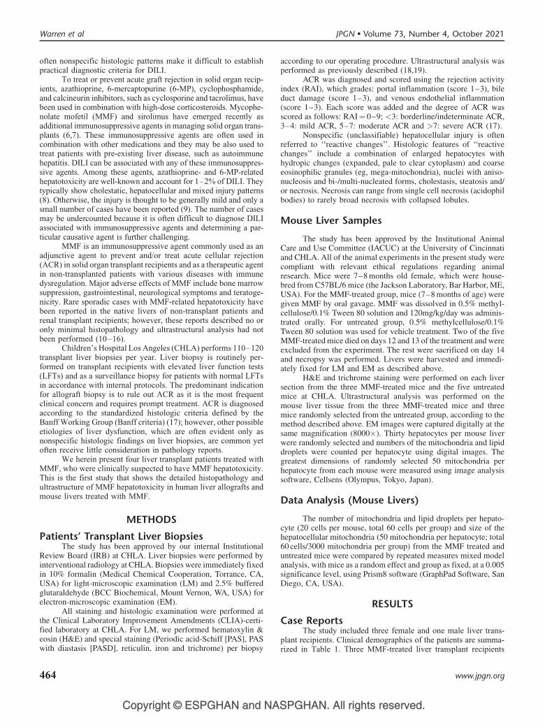
FIGURE 1. Clinical courses and medication history of Cases 1–4. Cases 1surveillancebiopsy as per our internal protocol. EM was requestedon Cases 1–
despite therapy and Case 4 for unexplained nonspecific histology despite n
reduction of MMF improved liver function. After 3 and 2 days after cessation
3 months after cessation and reduction. Case 3 underwent a pair of pre- andmitochondrialabnormalities.Case4 showedasubclinicalmitochondrial abno
to be the cause of the LFTabnormalities. After MMF hepatotoxicity became m
LFTs. Day 0 ¼ initial event or biopsy; EM ¼ electron-microscopic examinati
466
ACR as well as positive donor-specific antibodies and C4d staining, shewas aggressively treated with multiple courses of high dose intravenouscorticosteroids and increasing doses of tacrolimus. Because LFTs werenot normalized despite therapy, a third agent, MMF, was started as anadjunctive agent. However, her LFTs increased after MMF was started(Fig. 1). Given the mitochondrial abnormalities identified by EM,MMF was discontinued. Approximately 3 days after cessation ofMMF, LFTs started to improve (Fig. 1) and normalized at 2 monthsafter cessation (not shown in Fig. 1).
Case 2 was a 4-year-old female with a history of unresectablehepatoblastoma. Her post-transplant course was complicated byhemolytic anemia. Since tacrolimus-induced hemolysis was sus-pected, the medication was changed to cyclosporine, which improvedher hemolysis. Two months following her transplant she developedan acute rise in her LFTs due to ACR. She was aggressively managed
SPGHAN. All rights reserved.
–3 had multiple biopsies for persistently elevated LFTs. Case 4 had a3 for unexplained, persistent LFTabnormalities and nonspecific histology
ormal LFTs and a stable clinical course. In Cases 1 and 2, withdrawal or
and reduction of MMF, LFTs started to improve and normalized at 2 and
post-MMF treatment biopsies and only the post-MMF biopsy showedrmality. ForCase3and4, at the timeof theworkup,MMFwasnot thought
ore certain, MMF was discontinued. The patients currently have normal
on; LFT ¼ liver function test; MMF ¼ mycophenolate mofetil.
www.jpgn.org
JPGN � Volume 73, Number 4, October 2021 Mycophenolate Mofetil Hepatotoxicity
with high-dose corticosteroids and tacrolimus, which did not causehemolysis at that time. Her LFTs improved but did not normalize;therefore, MMF was added. However, her LFT abnormalities per-sisted after MMF was started (Fig. 1). Given the mitochondrialabnormalities identified by EM, MMF was reduced. Two days afterreduction of MMF, LFTs started to improve (Fig. 1) and normalizedat 3 months after reduction (not shown in Fig. 1).
Case 3 was a 17-year-old male with a history of congenitaldisorder of glycosylation Type Ib. His post-transplant course wasinitially uneventful, however he developed increased LFTs and giveninitial concerns for tacrolimus toxicity, his tacrolimus dosing wasdecreased and MMF was started; however, mild LFT abnormalitiespersisted after MMF was started (Fig. 1). At the initial diagnosticworkup MMF was not thought to the cause of hepatotoxicity. AfterMMF hepatotoxicity became more certain, it was discontinued andthe graft functions normalized (not shown in Fig. 1).
Lastly, Case 4 was a 16-year-old female with a history ofbiliary atresia status-post Kasai portoenterostomy, who presentedwith acute-on-chronic liver failure leading to end-stage liver dis-ease. Her post-transplant course was notable for EBV viremia, HSVstomatitis and an episode of ACR, all of which resolved before thecurrent presentation. Because her ACR was refractory to standardtherapy, MMF was started. Since then she had been treated withMMF for approximately one year. She underwent a protocolsurveillance liver biopsy as per the institutional protocol (Fig. 1).At the time of workup, MMF was not thought to be a cause ofhistologic abnormalities. After MMF hepatotoxicity became morecertain, it was discontinued. The patient currently has normal LFTs.
Figure 1 shows patients’ biopsy findings along with thelaboratory results and given medications. Multiple biopsies wereperformed for elevated LFTs, and the diagnoses established byboard-certified pediatric pathologists were generally consistentwith low-grade ACR (RAI ranging 2–4), except the initial biopsyof Case 1 with RAI of 5. Initial pulse corticosteroids resulted inlower LFTs; however, LFTs did not normalize despite aggressiveimmunosuppressive therapy.
In Cases 1 and 2, LFTs improved after stopping and reducingMMF, respectively; therefore, MMF hepatotoxicity was suspected.For Case 3, a pair of pre- and post-MMF treatment biopsies wereperformed and only the post-MMF biopsy demonstrated mitochon-drial abnormality. Case 4 was clinically stable but her surveillancebiopsy showed unequivocal mitochondrial abnormality.
Histologic and Ultrastructural Features of thePatients’ Liver Biopsies
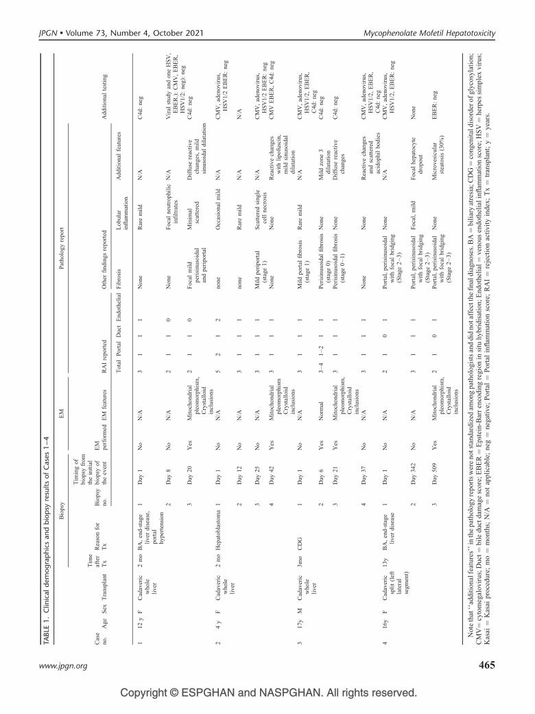
The histologic findings of each case are summarized inTable 1. Biopsies demonstrated features of mild ACR with patchyportal inflammation, some of which were accompanied by mildductal damage and/or mild subendothelial lymphocyte infiltrates. Inaddition, they showed mild, nonspecific hepatocellular injury(‘‘reactive changes’’). These ‘‘reactive changes’’ included mildlyenlarged hepatocytes with granular cytoplasm, anisonucleosis, andfocal areas with predominantly microvesicular steatosis (Fig. 2Aand B). For all of the four cases, EM revealed similar ultrastructuralfeatures including prominent mitochondrial pleomorphism (vari-ability in size and shape) and crystalloid inclusions (Fig. 2C and D),except for the pre-MMF biopsy in Case 3.
Histologic and Ultrastructural Features ofMouse Livers
Histology of the livers from the MMF-treated and untreatedmice showed no significant differences. MMF-treated mouse livers
Copyright © ESPGHAN and NA
www.jpgn.org
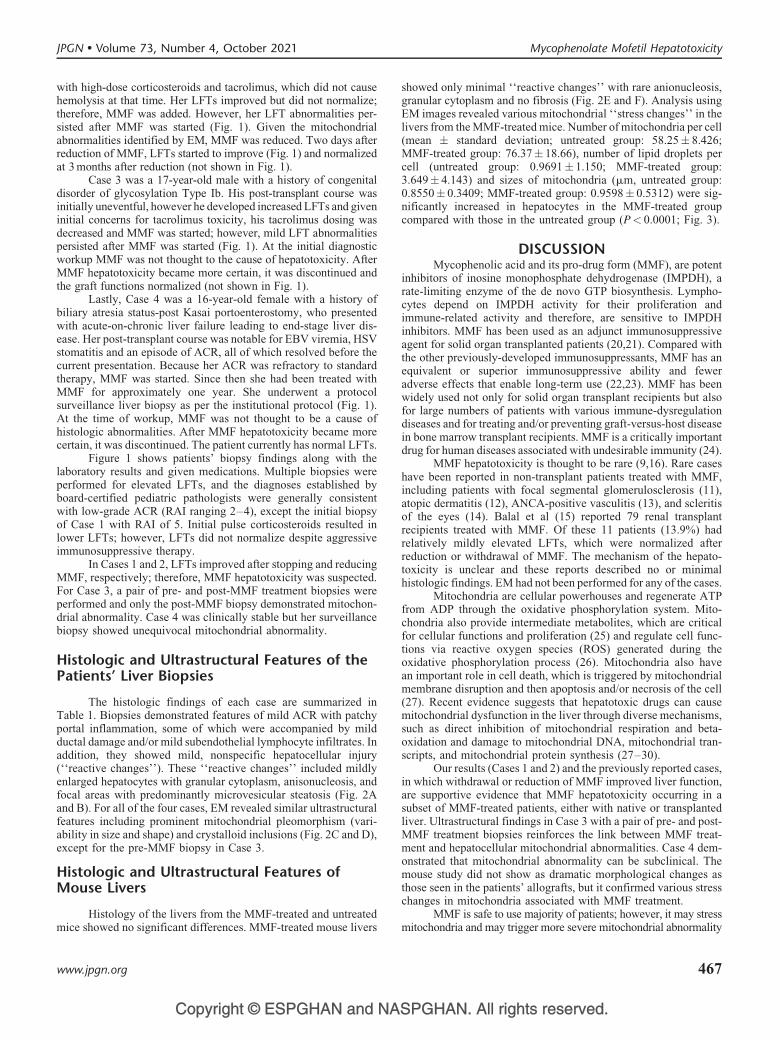
showed only minimal ‘‘reactive changes’’ with rare anionucleosis,granular cytoplasm and no fibrosis (Fig. 2E and F). Analysis usingEM images revealed various mitochondrial ‘‘stress changes’’ in thelivers from the MMF-treated mice. Number of mitochondria per cell(mean � standard deviation; untreated group: 58.25� 8.426;MMF-treated group: 76.37� 18.66), number of lipid droplets percell (untreated group: 0.9691� 1.150; MMF-treated group:3.649� 4.143) and sizes of mitochondria (mm, untreated group:0.8550� 0.3409; MMF-treated group: 0.9598� 0.5312) were sig-nificantly increased in hepatocytes in the MMF-treated groupcompared with those in the untreated group (P< 0.0001; Fig. 3).
DISCUSSIONMycophenolic acid and its pro-drug form (MMF), are potent
inhibitors of inosine monophosphate dehydrogenase (IMPDH), arate-limiting enzyme of the de novo GTP biosynthesis. Lympho-cytes depend on IMPDH activity for their proliferation andimmune-related activity and therefore, are sensitive to IMPDHinhibitors. MMF has been used as an adjunct immunosuppressiveagent for solid organ transplanted patients (20,21). Compared withthe other previously-developed immunosuppressants, MMF has anequivalent or superior immunosuppressive ability and feweradverse effects that enable long-term use (22,23). MMF has beenwidely used not only for solid organ transplant recipients but alsofor large numbers of patients with various immune-dysregulationdiseases and for treating and/or preventing graft-versus-host diseasein bone marrow transplant recipients. MMF is a critically importantdrug for human diseases associated with undesirable immunity (24).
MMF hepatotoxicity is thought to be rare (9,16). Rare caseshave been reported in non-transplant patients treated with MMF,including patients with focal segmental glomerulosclerosis (11),atopic dermatitis (12), ANCA-positive vasculitis (13), and scleritisof the eyes (14). Balal et al (15) reported 79 renal transplantrecipients treated with MMF. Of these 11 patients (13.9%) hadrelatively mildly elevated LFTs, which were normalized afterreduction or withdrawal of MMF. The mechanism of the hepato-toxicity is unclear and these reports described no or minimalhistologic findings. EM had not been performed for any of the cases.
Mitochondria are cellular powerhouses and regenerate ATPfrom ADP through the oxidative phosphorylation system. Mito-chondria also provide intermediate metabolites, which are criticalfor cellular functions and proliferation (25) and regulate cell func-tions via reactive oxygen species (ROS) generated during theoxidative phosphorylation process (26). Mitochondria also havean important role in cell death, which is triggered by mitochondrialmembrane disruption and then apoptosis and/or necrosis of the cell(27). Recent evidence suggests that hepatotoxic drugs can causemitochondrial dysfunction in the liver through diverse mechanisms,such as direct inhibition of mitochondrial respiration and beta-oxidation and damage to mitochondrial DNA, mitochondrial tran-scripts, and mitochondrial protein synthesis (27–30).
Our results (Cases 1 and 2) and the previously reported cases,in which withdrawal or reduction of MMF improved liver function,are supportive evidence that MMF hepatotoxicity occurring in asubset of MMF-treated patients, either with native or transplantedliver. Ultrastructural findings in Case 3 with a pair of pre- and post-MMF treatment biopsies reinforces the link between MMF treat-ment and hepatocellular mitochondrial abnormalities. Case 4 dem-onstrated that mitochondrial abnormality can be subclinical. Themouse study did not show as dramatic morphological changes asthose seen in the patients’ allografts, but it confirmed various stresschanges in mitochondria associated with MMF treatment.
MMF is safe to use majority of patients; however, it may stressmitochondria and may trigger more severe mitochondrial abnormality
SPGHAN. All rights reserved.
467
Copyright © ESPGHAN and NASPGHAN. All rights reserved.
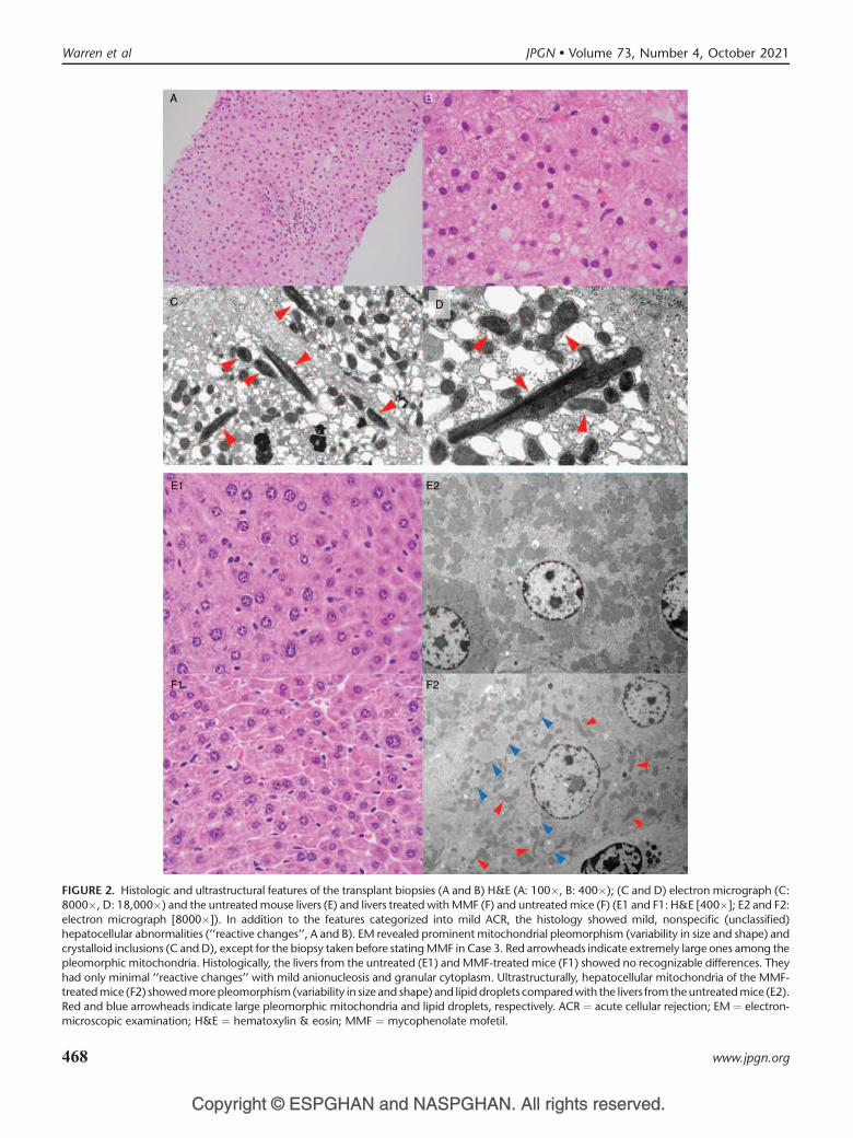
FIGURE 2. Histologic and ultrastructural features of the transplant biopsies (A and B) H&E (A: 100�, B: 400�); (C and D) electron micrograph (C:
8000�, D: 18,000�) and the untreated mouse livers (E) and livers treated with MMF (F) and untreated mice (F) (E1 and F1: H&E [400�]; E2 and F2:
electron micrograph [8000�]). In addition to the features categorized into mild ACR, the histology showed mild, nonspecific (unclassified)hepatocellular abnormalities (‘‘reactive changes’’, A and B). EM revealed prominent mitochondrial pleomorphism (variability in size and shape) and
crystalloid inclusions (C and D), except for the biopsy taken before stating MMF in Case 3. Red arrowheads indicate extremely large ones among the
pleomorphic mitochondria. Histologically, the livers from the untreated (E1) and MMF-treated mice (F1) showed no recognizable differences. They
had only minimal ‘‘reactive changes’’ with mild anionucleosis and granular cytoplasm. Ultrastructurally, hepatocellular mitochondria of the MMF-treated mice (F2) showed more pleomorphism (variability in size and shape) and lipid droplets compared with the livers from the untreated mice (E2).
Red and blue arrowheads indicate large pleomorphic mitochondria and lipid droplets, respectively. ACR ¼ acute cellular rejection; EM ¼ electron-
microscopic examination; H&E ¼ hematoxylin & eosin; MMF ¼ mycophenolate mofetil.
Warren et al JPGN � Volume 73, Number 4, October 2021
468 www.jpgn.org

A
B C
Untreated 1 Unteated 2 Untreated 3 MMF Treated 1 MMF Treated 2 MMF Treated 3
0.0
0.5
1.0
1.5
2.0
2.5
3.0
3.5
4.0
4.5
5.0
5.5
S iz e
of M
itoch
o ndr
i a (µ
m)
Samples: 3000 mitochondria Mean: 0.8550 SD: 0.3409
Samples: 3000 mitochondria Mean: 0.9598 SD: 0.5312
* p < 0.0001
FIGURE 3. Image analysis using EM revealed mitochondrial ‘‘stress changes’’; numbers of mitochondria and lipids and degree of mitochondrialpleomorphism (variability in size) were significantly increased in hepatocytes from the MMF-treated group compared with the untreated group
(P<0.0001). EM ¼ electron-microscopic examination; MMF ¼ mycophenolate mofetil.
JPGN � Volume 73, Number 4, October 2021 Mycophenolate Mofetil Hepatotoxicity
in a small subset. One should note that MMF’s pharmacokinetics andpharmacodynamics vary among individuals (31,32).
Diagnosing mitochondrial disorders is challenging by rou-tine LM alone, even for cases of genetically proven primarymitochondrial disorders with severe clinical manifestations,because the histologic features are diverse, ranging from normal,reactive changes, hepatitis, to various degrees of cellular necrosis,and are often nonspecific (18). EM plays an important role inidentifying morphological mitochondrial abnormalities at the ultra-structural level. Our EM results strongly suggest a correlationbetween MMF hepatotoxicity and mitochondrial abnormalities inhuman and mouse livers treated with MMF. The next important stepwould be to investigate a larger number of patients and to look forrisk factor(s), such as genetic alteration(s), that may make patientsmore susceptible to MMF hepatotoxicity. The role of IMPDH inmitochondrial morphology and function also needs to be studied.
In summary, this is the first study describing detailed histo-logic and ultrastructural features of MMF hepatotoxicity. Despitemild nonspecific histologic abnormalities, the allograft biopsiesrevealed unequivocal mitochondrial abnormalities similar to thoseseen in primary and secondary mitochondrial disorders. The mouse
Copyright © ESPGHAN and NA
www.jpgn.org
study confirmed that MMF caused various stress changes in themitochondria. Although MMF is safe for the majority of patients,MMF may stress mitochondria and the stress may trigger moresevere mitochondrial abnormality in a small subset. MMF hepato-toxicity should be considered for MMF-treated patients, either withnative or transplanted livers, who have unexplained, persistent LFTabnormalities and nonspecific histologic changes. EM can play acritical role in diagnosing these cases.
Acknowledgments: We would like to thank Alexander Navarrofor processing the ultrastructural materials. The mouse study wassupported in part by R21NS100077 and R01NS089815 (A.T.S.).
REFERENCES1. Navarro VJ, Senior JR. Drug-related hepatotoxicity. N Engl J Med
2006;354:731–9.2. Ghabril M, Chalasani N, Bjornsson E. Drug-induced liver injury: a
clinical update. Curr Opin Gastroenterol 2010;26:222–6.3. Fontana RJ, Hayashi PH, Gu J, et al., DILIN Network. Idiosyncratic drug-
induced liver injury is associated with substantial morbidity and mortalitywithin 6 months from onset. Gastroenterology 2014;147:96–108.
SPGHAN. All rights reserved.
469
Warren et al JPGN � Volume 73, Number 4, October 2021
4. Ostapowicz G, Fontana RJ, Schiødt FV, et al., Schiødt FV, et al., AcuteLiver Failure Study Group. Results of a prospective study of acute liverfailure at 17 tertiary care centers in the United States. Ann Intern Med2002;137:947–54.
5. Fisher K, Vuppalanchi R, Saxena R. Drug-induced liver injury. ArchPathol Lab Med 2015;139:876–87.
6. Mukherjee S, Mukherjee U. A comprehensive review of immunosup-pression used for liver transplantation. J Transplant 2009;2009:701464.doi: 10.1155/2009/701464.
7. Moini M, Schilsky ML, Tichy EM. Review on immunosuppression inliver transplantation. World J Hepatol 2015;7:1355–68.
8. Bjornsson ES, Gu J, Kleiner DE, et al., DILIN Investigators. Azathiopr-ine and 6-mercaptopurine-induced liver injury: clinical features andoutcomes. J Clin Gastroenterol 2017;51:63–9.
9. LiverTox. Clinical and research information on drug-induced liver injuryBethesda (MD): National Institute of Diabetes and Digestive and KidneyDiseases. https://www.ncbi.nlm.nih.gov/books/NBK547852/. 2012. [Ac-cessed 31 July 2020].
10. Chalasani N, Fontana RJ, Bonkovsky HL, et al. Causes, clinicalfeatures, and outcomes from a prospective study of drug-induced liverinjury in the United States. Gastroenterology 2008;135:1924–34.
11. Loupy A, Anglicheau D, Mamzer-Bruneel MF, et al. Mycophenolatesodium-induced hepatotoxicity: first report. Transplantation 2006;82:581.
12. Hantash B, Fiorentino D. Liver enzyme abnormalities in patients withatopic dermatitis treated with mycophenolate mofetil. Arch Dermatol2006;142:109–10.
13. Dourakis SP, Boki K, Soultati A, et al. Acute hepatitis followingmycophenolate mofetil administration for ANCA-positive vasculitis.Scand J Rheumatol 2007;36:237–9.
14. Sen HN, Suhler EB, Al-Khatib SQ, et al. Mycophenolate mofetil for thetreatment of scleritis. Ophthalmology 2003;110:1750–5.
15. Balal M, Demir E, Paydas S, et al. Uncommon side effect of MMF inrenal transplant recipients. Ren Fail 2005;27:591–4.
16. Hernandez N, Bessone F, Sanchez A, et al. Profile of idiosyncratic druginduced liver injury in Latin America: an analysis of published reports.Ann Hepatol 2014;13:231–9.
17. No author listed, Banff schema for grading liver allograft rejection: aninternational consensus document. Hepatology 1997;25:658–63.
Copyright © ESPGHAN and NA
470
18. Warren M, Shimada H. Cytologic and ultrastructural findings ofbronchoalveolar lavage in patients with chronic granulomatous disease.Pediatr Dev Pathol 2018;21:347–54.
19. Warren M, Shimura M, Wartchow EP, Yano S. Use of electron micro-scopy when screening liver biopsies from neonates and infants: experi-ence from a single tertiary children’s hospital (1991-2017). UltrastructPathol 2020;44:32–41.
20. Allison AC, Kowalski WJ, Muller CD, et al. Mechanisms of action ofmycophenolic acid. Ann NY Acad Sci 1993;696:63–87.
21. Bentley R. Mycophenolic acid: a one hundred year odyssey fromantibiotic to immunosuppressant. Chem Rev 2000;100:3801–26.
22. Molnar AO, Fergusson D, Tsampalieros AK, et al. Generic immuno-suppression in solid organ transplantation: systematic review and meta-analysis. BMJ 2015;350:h3163. doi: 10.1136/bmj.h3163.
23. El Hajj S, Kim M, Phillips K, et al. Generic immunosuppression intransplantation: current evidence and controversial issues. Expert RevClin Immunol 2015;11:659–72.
24. Downing HJ, Pirmohamed M, Beresford MW, et al. Paediatric use ofmycophenolate mofetil. Br J Clin Pharmacol 2013;75:45–59.
25. Owen OE, Kalhan SC, Hanson RW. The key role of anaplerosis andcataplerosis for citric acid cycle function. J Biol Chem 2002;277:30409–12.
26. Sena LA, Chandel NS. Physiological roles of mitochondrial reactiveoxygen species. Mol Cell 2012;48:158–67.
27. Pessayre D, Fromenty B, Berson A, et al. Central role of mitochondria indrug-induced liver injury. Drug Metab Rev 2012;44:34–87.
28. Labbe G, Pessayre D, Fromenty B. Drug-induced liver injury throughmitochondrial dysfunction: mechanisms and detection during preclini-cal safety studies. Fundam Clin Pharmacol 2008;22:335–53.
29. Begriche K, Massart J, Robin MA, et al. Drug-induced toxicity onmitochondria and lipid metabolism: mechanistic diversity and deleter-ious consequences for the liver. J Hepatol 2011;54:773–94.
30. Ramachandran A, Visschers RGJ, Duan L, et al. Mitochondrial dys-function as a mechanism of drug-induced hepatotoxicity: currentunderstanding and future perspectives. J Clin Transl Res 2018;4:75–100.
31. Kiang TKL, Ensom MHH. Population pharmacokinetics of mycophe-nolic acid: an update. Clin Pharmacokinet 2018;57:547–58.
32. Staatz CE, Tett SE. Pharmacology and toxicology of mycophenolate inorgan transplant recipients: an update. Arch Toxicol 2014;88:1351–89.
SPGHAN. All rights reserved.
www.jpgn.org