henoch-schönlein purpura in children: long-term outcome...
TRANSCRIPT
HENOCH-SCHÖNLEIN PURPURA IN CHILDREN: LONG-TERM OUTCOME AND TREATMENT
Tiivistelmä suomeksi
JAANARONKAINEN
Faculty of Medicine,Department of Paediatrics,
University of Oulu
OULU 2005

JAANA RONKAINEN
HENOCH-SCHÖNLEIN PURPURA IN CHILDREN: LONG-TERM OUTCOME AND TREATMENT
Academic Dissertation to be presented with the assent ofthe Faculty of Medicine, University of Oulu, for publicdiscussion in the Auditorium 12 of the Department ofPaediatrics, on November 25th, 2005, at 12 noon
OULUN YLIOPISTO, OULU 2005
Copyright © 2005University of Oulu, 2005
Supervised byDocent Matti NuutinenDocent Olli Koskimies
Reviewed byDocent Ilpo Ala-HouhalaDocent Kai Rönnholm
ISBN 951-42-7884-4 (nid.)ISBN 951-42-7885-2 (PDF) http://herkules.oulu.fi/isbn9514278852/
ISSN 0355-3221 http://herkules.oulu.fi/issn03553221/
OULU UNIVERSITY PRESSOULU 2005

Ronkainen, Jaana, Henoch-Schönlein purpura in children: long-term outcome andtreatment Faculty of Medicine, Department of Paediatrics, University of Oulu, P.O.Box 5000, FIN-90014University of Oulu, Finland 2005Oulu, Finland
AbstractThe aim of this work was to evaluate the outcome of childhood Henoch-Schönlein purpura (HSP),the effectiveness of Cyclosporine A (CyA) for treating severe HSP nephritis (HSN), and moreparticularly the possibility for influencing the course of HSP disease by early prednisone treatment.
A total of 47 adults who had had childhood HSP were evaluated after a mean of 24.1 years(16.4–35.6). The outcome was highly dependent on the renal symptoms at onset, since 7 out of 20adults (20%) who had severe renal symptoms at onset had renal impairment as adults, compared with2 out of 27 (7%) with mild or no renal symptoms at onset (relative risk 4.7; 95% CI 1.3–18.7). 70%of pregnancies in women after childhood HSN were complicated by hypertension or proteinuria.
The annual incidence of HSN with nephrotic-range proteinuria was 2 per million children under15 years. After a mean follow-up of 4.6 years, only three patients out of 19 were in completeremission. Kidney biopsy did not predict the outcome in these patients. CyA seemed to be promisingfor the treatment of severe HSN with nephrotic-range proteinuria, since four out of seven patientstreated with CyA achieved stable remission and three had preserved their renal function after a meanfollow-up of 6.0 years. Treatment at an early stage in the disease was associated with stable remission.
The efficacy of early prednisone treatment was evaluated in a randomized double-blind trialinvolving 171 patients (84 prednisone, 87 placebo). Prednisone, given at a dose of 1 mg/kg/day for 2weeks, with weaning over the next two weeks, was effective in reducing the intensity of abdominalpain (pain score 2.5 vs. 4.8; t-test p = 0.029) and shortening its duration (1.5 days vs. 2.7 days; t-testp = 0.028) compared with the placebo. The mean scores for joint pain were lower in the prednisonegroup (4.6 vs. 7.3; t-test p = 0.030) and the improvement from joint symptoms was faster (log rankp = 0.007).
Prednisone did not prevent the development of renal symptoms but it was effective in treatingthem, since renal symptoms resolved in 61% of the prednisone patients after treatment comparedwith 34% of the placebo patients (difference 27%, 95% CI 3–47%, p = 0.024). Prednisone was mosteffective for children aged 6 or more with renal symptoms at onset, since only two patients needed tobe treated in order to save one from renal involvement (95% CI's for NNT 2–6).
The long-term outcome of HSP is dependent on renal symptoms. Severe renal symptoms indicateearly immunosuppressive treatment for HSN, and patients with renal involvement at the acute phaseneed long-term follow-up, especially women during and after pregnancy. Early treatment withprednisone is effective in reducing the abdominal and joint symptoms involved in HSP and is alsoeffective in altering, but not preventing, the course of renal involvement.
Keywords: corticosteroid treatment, cyclosporine, end-stage renal disease, haematuria,immunosupressive treatment, nephrosis, proteinuria, toxaemia


Ronkainen, Jaana, Henoch-Schönleinin purppura lapsilla: pitkäaikaisennuste ja hoito Lääketieteellinen tiedekunta, Lastentautien klinikka, Oulun yliopisto, PL 5000, 90014 Oulunyliopisto2005Oulu
TiivistelmäVäitöskirjatyön tarkoituksena oli selvittää lapsuusiän Henoch-Schönleinin purppuran (HSP)pitkäaikaisennustetta, Siklosporiini-A:n (CyA) tehoa vaikean HSP-nefriitin hoidossa ja tutkia varhainaloitetun prednisonihoidon hyötyä HSP-taudin oireisiin.
HSP:n pitkäaikaisennustetta selvitettiin tarkastamalla 47:n lapsena HSP-taudin sairastaneenaikuisen terveystilanne keskimäärin 24.1 vuoden (16.4-35.6) seuranta-ajan jälkeen. HSP-taudinennuste oli vahvasti riippuvainen munuaisoireen vaikeusasteesta: 20 % niistä, joilla taudin alussa olivaikeat munuaisoireet, kärsi vielä aikuisiällä munuaisoireista; vastaava luku munuaisoireettomilla janiillä, joilla oli ollut vain lievää veri- tai valkuaisvirtsaisuutta, oli 7 %, (RR 4.7; 95 % CI 1.3–18.7).Raskauskomplikaatiot olivat yleisiä lapsuusiällä HSP-taudin sairastaneilla naisilla, sillä 70 %raskauksista komplisoi korkea verenpaine tai valkuaisvirtsaisuus.
Vuosittain 2 lasta miljoonasta sairastuu vaikeaan nefroottistasoiseen HSP-nefriittiin Suomessa.Vain kolme nefroottistasoiseen HSP-nefriittiin sairastuneesta 19 lapsesta oli 4.6 vuoden seurannanjälkeen parantunut oireettomaksi. Ensimmäisen munuaisbiopsian vaikeusaste ei ennakoinutselviytymistä. CyA näytti olevan lupaavan tehokas lääke vaikean HSP-nefriitin hoidossa, sillä neljäseitsemästä CyA-hoitoa saaneesta lapsesta, oli oireeton 6.0 vuoden seurannan jälkeen. Mitäaikaisemmin vaikean nefriitin hoito oli aloitettu, sen parempi hoitotulos oli.
Varhain aloitetun prednisonihoidon hyötyä HSP-taudin oireisiin selvitettiin satunnaistetullakaksoissokkotutkimuksella, johon satunnaistettiin 171 lasta (84 prednisoni, 87 lumelääke) saamaanjoko prednisonia 1 mg/kg/päivä 2 viikon ajan tai lumelääkettä. Prednisoni vähensi tehokkaasti vatsa-ja nivelkipuja ja se lyhensi merkitsevästi myös niiden kestoa. Prednisoni ei estänyt munuaisoireenkehittymistä lapselle, mutta niiltä, joille se kehittyi, oireet hävisivät merkitsevästi nopeamminlumelääkitykseen verrattuna (61 % versus 34 %, 95 % CI 3–47 %, p = 0.024). Kaikkein tehokkaintaprednisoni oli yli 6 vuotiaille lapsille, joilla oli munuaisoire heti taudin alussa (NNT 2, 95 % CI 2–6).
Tutkimuksen perusteella voidaan sanoa, että lapsuusiällä HSP-nefriitin sairastaneet lapsettarvitsevat seurantaa aikuisiällä, erityisesti naiset raskauden aikana. HSP-nefriitin varhainenhoitaminen on tärkeää. Varhainen prednisonihoito ei estä munuaisoiretta, mutta hoitaa jo kehittynyttänefriittiä ja vähentää vatsa- ja nivelkipuja tehokkaasti.
Asiasanat: immunosupressiivinen hoito, kortisoni hoito, munuaisen vajaatoiminta,nefroosi, raskausmyrkytys, siklosporiini, uremia, valkuaisvirtsaisuus, verivirtsaisuus


Menen kissan perässä kaksi kertaa kuperkeikkaa
enkä tiedä, kumpi on oikein. Ehkä toinen on väärin, kun muut niin inttävät.
Jospa molemmat ovat oikein. Minun tapani tehdä keikka.
Pentti Murto


Acknowledgements
This work was carried out at the Department of Paediatrics, University of Oulu, Finland, during the years 1999-2005. I am grateful to Professor Mikko Hallman, M.D., Head of the Department of Paediatrics, Professor Matti Uhari, M.D. and Professor Marjatta Lanning, M.D., who first gave me opportunity to study paediatrics and later allowed me to finish this slowly but surely progressing project in their clinic. This project has given me a chance to learn something about the scientific way of thinking, even though research work has been largely a secondary interest for me. I wish to express my gratitude to the people who have helped me over these six years.
Above all, I am deeply grateful to my energic and enthusiastic supervisor Matti Nuutinen, M.D. He is not only a skilful scientist but also a superb teacher, who always had time to help and advise me. The working atmosphere was never boring, thanks to Matti’s sense of humour and relaxed attitude. I also offer my warm thanks to my other supervisor, Olli Koskimies, M.D., for his support and encouragement during these years. Olli’s long perspective on Henoch-Schönlein purpura and his positive attitude towards clinical research have been of great value during this work.
I wish to thank all the people in the HSP-project who recruited and examined the patients during these years, and also my co-authors in the last paper in this thesis, Marja Ala-Houhala, M.D., Marjatta Antikainen, M.D., Jussi Merenmies, M.D., Jukka Rajantie, M.D., Juha Turtinen, M.Sc., and Timo Örmälä, M.D., who all received numerous telephone calls and e-mails last summer and who unselfishly helped me without caring about working hours. I also thank my co-authors Timo Jahnukainen, M.D., and Niilo-Pekka Huttunen, M.D., for their co-operation and help during the process. I am grateful to Helena Autio-Harmainen, M.D., who advised me as a co-author and who patiently guided me to understand something about renal biopsies. I have pleasant memories of several afternoons we spent over the microscope, discussing not only the biopsies but all aspects of life in general.
I wish to express my gratitude to the nurses Kaisu Koivunen, Riitta Tiitto, Raija Wilenius and Tuija Yliniitty and the secretaries Pirjo Haarahiltunen, Marja-Liisa Paavola, Seija Peltomaa and Päivi Murto at the Paediatric Clinic of Oulu University Hospital, who helped me in such a kind and friendly manner during this work and took such good care of the patients.

Docent Ilpo Ala-Houhala, M.D. and Docent Kai Rönnholm, M.D., the official referees of this thesis, are gratefully acknowledged for their valuable and constructive comments. Similarly, Marja Laitala, R.N., is thanked for her kind help during the last metres of this project.
I would like to thank Pentti Vuolukka, M.D., who gave me my very first, unforgettable paediatric lessons at Länsi-Pohja Central Hospital, and I am also very grateful to Brage Gustafsson, M.D., for his guidance and charming clinical lessons at the Malmi Hospital in Pietarsaari. I also thank all my colleagues in the Department of Paediatrics at Oulu University Hospital for their support and guidance.
Special thanks go to Lea Jämsä, who has been my friend from childhood, and Marjo Vilhola, with whom I have shared my most exciting sporting years. It has been a great pleasure to have such trustworthy friends.
My dear and patient parents, Raili and Antero, deserve great admiration and thanks for the support they have given me throughout life. I thank my brothers Jukka, who I know will stand by me what ever happens, and Jouko, who is not only a brother but also my best friend.
Finally, I owe my warmest thanks to my husband Markku, who keeps my feet on the ground and my three lovely and lively children, Sandra, Saku and Emma. There is nothing better for me than to be a mother.
This work was financially supported by Alma and K.A Snellman Foundation, Oulu, Finland, Foundation for Paediatric Research, Finland, Maud Kuistila Memorial Foundation, Finland Finnish Nephrology Association, and the Medical Research Funds of Tampere University Hospital and Oulu University Hospital.
Oulu, November 2005 Jaana Ronkainen

Abbreviations
ACE Angiotensin convertase enzyme ANCA Antineutrophil cytoplasmic antibodies APTT Activated partial thromboplastin time AST Antistreptolysin titre Az Azathioprine C3/C4 Complement 3/Complement 4 CI Confidence interval CP Cyclophosphamide CRP C-reactive protein CyA Cyclosporine A Dp Dipyridamole ECP Eosinophilic cationic protein ESR Erythrocyte sedimentation rate ESRD End-stage renal disease GFR Glomerular filtration rate HSP Henoch-Schönlein purpura HSN Henoch-Schönlein purpura nephritis IgA Immunoglobulin A IgAN IgA nephritis IL Interleukin ISKDC International Study of Kidney Diseases in Children MP/mp Methylprednisolone intravenous/per oral NNB Number needed to benefit NNH Number needed to harm NNT Number needed to treat NSAID Non-steroidal anti-inflammatory drug Pu Proteinuria TT Thromboplastin time TNF-α Tumour necrosis factor- α


List of original papers
This thesis is based on the following articles, which are referred to in the text by the Roman numerals:
I Ronkainen J, Nuutinen M & Koskimies O (2002) The adult kidney 24 years after childhood Henoch-Schönlein purpura: a retrospective cohort study. Lancet 360: 666–670.
II Ronkainen J, Ala-Houhala M, Huttunen NP, Jahnukainen T, Koskimies O, Örmälä T & Nuutinen M (2003) Outcome of Henoch-Schoenlein nephritis with nephrotic-range proteinuria. Clin Nephrol 60: 80–84.
III Ronkainen J, Autio-Harmainen H & Nuutinen M (2003) Cyclosporine A for the treatment of severe Henoch-Schonlein glomerulonephritis. Ped Nephrol 18: 1138–1142.
IV Ronkainen J, Koskimies O, Ala-Houhala M, Antikainen M, Merenmies J, Rajantie J, Örmälä T, Turtinen J & Nuutinen M. Early prednisone in treating childhood Henoch-Schönlein purpura; a randomized double-blind placebo-controlled trial. (Submitted)
Reprint with kind permission from Elsevier (I), Dustri-Verlag (II) and Springer Science and Business Media (III).


Contents
Abstract Tiivistelmä Acknowledgements Abbreviations List of original papers Contents 1 Introduction ...............................................................................................................17 2 Review of the literature .............................................................................................18
2.1 Definition of Henoch-Schönlein purpura ...........................................................18 2.1.1 Symptoms ....................................................................................................18 2.1.2 Influence of age on symptoms.....................................................................19
2.2 Epidemiology .....................................................................................................19 2.3 Aetiology ............................................................................................................20 2.4 Pathogenesis .......................................................................................................20
2.4.1 Role of IgA..................................................................................................20 2.4.2 Inflammatory processes ...............................................................................21 2.4.3 Genetic aspects ............................................................................................21
2.5 Laboratory findings ............................................................................................22 2.6 Biopsy findings...................................................................................................22
2.6.1 Skin and mucosal biopsies...........................................................................22 2.6.2 Renal biopsies..............................................................................................23
2.7 Differential diagnosis .........................................................................................24 2.7.1 Diseases with purpura..................................................................................24 2.7.2 IgA nephritis (IgAN) ...................................................................................25
2.8 Treatment of HSN...............................................................................................26 2.8.1 Immunosuppressive treatment .....................................................................26 2.8.2 Plasmapheresis and immunoglobulin therapy .............................................28
2.9 Prevention of HSN .............................................................................................28 2.10 Risk factors and prognosis for renal disease in HSP ........................................29
2.10.1 Influence of sex, age and extra-renal symptoms........................................29 2.10.2 Renal symptoms ........................................................................................29 2.10.3 Long-term prognosis for HSN ...................................................................30
3 Aims of the research ..................................................................................................31 4 Material and methods ................................................................................................32
4.1 Long-term outcome after childhood HSP (I) ......................................................33 4.2 Clinical outcome after severe HSN (II) ..............................................................34 4.3 Cyclosporine-A for the treatment of severe HSN (III) .......................................34

4.4 Effect of early prednisone on HSP (IV)..............................................................34 4.5 Statistical methods (I-IV) ...................................................................................36
4.5.1 Number needed to treat (IV)........................................................................37 5 Results .......................................................................................................................38
5.1 Long-term outcome after childhood HSP (I) ......................................................38 5.2 Clinical outcome after severe HSN (II) ..............................................................39 5.3 Cyclosporine-A for treating HSN (III) ...............................................................40 5.4 Effect of early prednisone on HSP (IV)..............................................................43
5.4.1 Effect of prednisone on extra-renal symptoms (IV) ....................................43 5.4.2 Effect of prednisone on renal symptoms (IV)..............................................45 5.4.3 Numbers needed to treat (IV) ......................................................................46
6 Discussion..................................................................................................................48 6.1 Study design and patient series (I-IV) ................................................................48 6.2 Outcome after HSP disease (I)............................................................................49
6.2.1 Renal symptoms predict the outcome (I, II) ................................................49 6.2.2 Role of renal biopsy in predicting the outcome (I, II, III) ...........................51
6.3 Long-term outcome after HSN versus IgAN......................................................53 6.4 Pregnancy after childhood HSP (I) .....................................................................54 6.5 Treatment of severe HSN (II, III) .......................................................................54
6.5.1 Timing of the treatment (II, III) ...................................................................55 6.5.2 CyA for the treatment of severe HSN (III) ..................................................55
6.6 Early treatment with prednisone (IV) .................................................................56 6.6.1 Safety of prednisone (IV) ............................................................................56 6.6.2 Effect of prednisone on extra-renal symptoms (IV) ....................................57 6.6.3 Effect of prednisone on renal symptoms (IV)..............................................57
7 Conclusions ...............................................................................................................59 References

1 Introduction
Henoch-Schönlein purpura (HSP) is the most common systemic vasculitis affecting children (1). The first reports verifying this syndrome characterised by purpura, abdominal pain, joint pain and renal involvement were produced by Heberden over 200 years ago (2). Subsequently, Schönlein described “Peliosis rheumatica” in a patient who had arthralgia and purpura, and his former student Henoch later reported gastrointestinal involvement and haemorrhagic nephritis as a possible complication of this syndrome (3, 4).
HSP is characterized by palpable purpura, oedema, abdominal pain, joint pain and renal symptoms. The prognosis is thought to be good as long as the patients have no renal symptoms (5). Renal symptoms vary from intermittent haematuria and proteinuria to rapidly progressive glomerulonephritis. Henoch-Schönlein nephritis (HSN) is not always a benign disease, since 1-3% of patients develop end-stage-renal disease (ESRD) and 20-35 % develop chronic renal disease according to long-term outcome studies (5-8). HSN is one of the most common reasons for a renal biopsy in children (9). Prevention of the development of glomerulonephritis would be important, since there is no specific treatment for HSN. This research was designed to investigate the long-term outcome of HSP and the treatment of severe HSN and to assess the efficacy of early corticosteroid treatment for alleviating the symptoms of HSP and preventing HSN.

2 Review of the literature
2.1 Definition of Henoch-Schönlein purpura
HSP is a vasculitis with IgA-dominant immune deposits affecting small vessels (10). This multisystem disease is most commonly characterized by involvement of the skin, joints, gastrointestinal tract and kidneys, but other organs may also be affected (11). The most common clinical manifestations of HSP are given in Table 1.
Table 1. Major clinical manifestations of HSP in children based on four studies
Clinical manifestation Blanco et al. (12)(n=116)
%
Calvino et al. (13)(n=78)
%
Allen et al. (14)(n=131)
%
Fisher et al. (15) (n=119)
%
All (n=444)
%
Purpura 100 100 100 100 100 Joint involvement 80 78 68 76 75 Gastrointestinal involvement
64 73 53 76 65
Renal involvement 25 54 41 54 43
2.1.1 Symptoms
Skin; All patients have skin symptoms, i.e. purpura and petechiae, mainly on the lower extremities and buttocks, but also on the upper extremities and sometimes the face (11, 16, 17). The reason for the purpura is minor bleeding from the small vessel walls due to leucocytoclastic inflammation. It is often combined with oedema, especially on the legs and hands, and scrotal oedema is also possible. The skin symptoms are the first sign of HSP-disease in 55-70% of patients (12, 13), but in some cases purpura initiates after the other symptoms, making diagnosis far more difficult (18-21).
19
Joints; 68-80% of patients have joint involvement, i.e. pain and oedema, usually in the ankles and knees, but also in the hands, wrists and elbows (12-15). Even when the oedema is periarticular the movements of the affected joint are often limited.
Gastrointestinal tract; 53-76% of patients have abdominal pain (12-15). Some patients can have severe colic pain requiring intensive care, since intussusception, obstruction and perforation are possible (21-25). 14-38% of patients have melaena (13-15, 26), and massive gastrointestinal bleeding is also possible (14). Severe gastrointestinal symptoms can lead to protein-losing enteropathy (15, 27). Intussusception has been reported in 1-3 % HSP patients (13, 14) and HSP-related intussusception is typically ileoileal (25, 28).
Kidney; 20-54% of patients in unselected materials have renal symptoms (12-15, 29, 30), which arise within a month of diagnosis in 85% of cases (31). Most patients have combined proteinuria and haematuria, but the renal symptoms vary from transient microscopic haematuria to severe nephrotic-nephritic syndrome (29).
Other symptoms; Neurological symptoms such as headache, convulsions, paresis and mental lability (32, 33), and also pulmonal symptoms such as bleeding (34-38) and interstitial lung disease (39, 40), have been reported in HSP patients. These symptoms are very rare, but can be fatal.
2.1.2 Influence of age on symptoms
The clinical spectrum of HSP in very young children (<2 years of age) and in adults differs slightly from that in the customary paediatric patients (2-15 years of age) (12, 14, 17, 41). Oedema and swelling form the prominent symptom in children under 2 years of age at onset, and the incidence of abdominal (8-29%), joint (17-56%) and renal symptoms (17-23%) is markedly lower than in older children (14, 17). In children under 2 years the skin rash frequently affects the face (17), which is seldom seen in older children. Scalp oedema is found in 59% of patients under 2 years of age at onset, compared with 19% of those aged 2 years or more (14).
Adults tend to have a lower frequency of abdominal pain as an onset symptom but a higher frequency of joint and renal involvement (12).
2.2 Epidemiology
According to a population-based survey, the estimated annual incidence of childhood HSP is 20.4/100 000 (1). The figure is highest in children between 4 to 6 years (70.3/100 000 per year). The incidence rate has been reported to be lower in a black population than in any other population (1). Boys are affected slightly more often than girls (1, 14), and the seasonal peak is in the autumn, winter and spring months (13-15), evidently related to infections. No rhythmic variation in annual incidence has yet been observed, but it is well known that the incidence of HSP in children varies between years (13).

20
2.3 Aetiology
70-80% of HSP patients have had a respiratory infection prior to onset of the disease (13, 14). Several triggering agents have been proposed (Table 2), especially streptococcal infections, confirmed by throat culture, which have been reported to precede the disease in 20-36% of cases (13, 14). The occurrence of the seasonal peak in the winter months (13-15) supports the notion of an infectious trigger, but drugs (antibiotics, ACE inhibitors, NSAIDs) and certain toxins (insect bites, vaccinations and food allergies) have also been implicated (13, 14).
Table 2. Triggering factors of HSP (14, 42-56).
2.4 Pathogenesis
2.4.1 Role of IgA
The pathogenetic mechanisms of HSP disease are poorly understood. It is clear that the main role in the immunopathogenic process is played by IgA, as increases in serum concentrations of IgA1, together with increases in circulating immunocomplexes containing IgA, have been described in patients with HSP (57, 58, 59). Production of IgA, like other humoral and immune responses, is tightly controlled by the B and T-lymphocytes, and it is suggested that dysregulation of this control in HSP patients, leads to an increase of IgA (57, 59, 60).
Altered O-glycosylation of IgA1due to an abnormal hinge region in the IgA1 molecule has been reported in HSN patients and in patients with IgAN, but not in HSP patients with only extra-renal symptoms (61). There are number of potential reasons why abnormalities in O-glycosylation may be pathogenic in IgAN and HSN. It may reduce the clearance of IgA1 molecules, resulting in an increase in circulating IgA, or it may increase the tendency of IgA1 to aggregate and form IgA immunocomplexes (59, 62-65).
Group Triggering factor
Bacteria
Streptococcus pyogenes, Staphylococcus aureus, Mycoplasma, Shigella, Yersinia, Legionella, Salmonella, Helicobacter pylori, Campylobacter
Viruses
Adenovirus, Parvovirus, Hepatitis B, Varicella zoster, Ebstein-Barr, Coxsackie, Herpes simplex, HIV
Drugs Thiazides, Antibiotics, ACE-inhibitors, NSAIDs
Others Insect bites, Food allergy, Toxocara canis
Vaccinations
Tuberculosis, Measles, Cholera, Yellow fever, Hepatitis B, Influenza, Pneumococcal, Meningococcal

21
Abnormally glycosylated IgA is possibly cleared less efficiently by the hepatocyte receptor for asialoglycoproteins (59, 64).
The role of IgA in the pathogenesis of HSP is supported by the fact that secretory IgA plays the major role in defence against exogenous antigens in the mucosal lining, and by the observation that respiratory tract infection precedes the onset of the disease in 70-80% of cases (66). Increased formation of circulating IgA by mucosal B cells is stimulated by the transmucosal penetration of exogenous antigens (66, 67). There is no evidence, however, of any single contagious agent against which serum IgA is raised in HSP patients (57).
2.4.2 Inflammatory processes
Leucocytoclastic vasculitis is the final immunopathological result when circulating IgA immunocomplexes are deposited in affected organs and provoke inflammatory lesions via the complement system and direct cell activation (68-70). The primary event is probably damage to the endothelial cells and invasion by leucocytes in a three-step process of rolling, sticking and firm adhesion to the endothelial cells followed by migration into the tissues (70). Complement breakdown products are chemoattractants for polymorpho-nuclear leucocytes (68), which are seen in the small vessel walls of HSP patients. Activation of an alternative pathway in the complement system has also been suggested in acute HSP, since degradation products of such a complement cascade have been demonstrated in the plasma and glomeruli (71, 72), but there are also studies that do not support any significant role for complement activation in the pathogenesis of HSP (73).
Proinflammatory cytokines such as endothelins, TNF α and interleukin β have been studied in HSP patients, and higher levels of these have been noted in the acute phase than in control patients or HSP patients in the remission phase (74-77). Cytokines probably play a role as a mediators of inflammation in HSP.
2.4.3 Genetic aspects
Lofters et al. reported the occurrence of HSP in three members of the same family on widely separated occasions, suggesting familiar predisposition to its development (78). Later, familial occurrence was observed in twins and siblings (45, 79, 80). An interesting finding of more common recurrences of HSN in kidneys from related living donors (9 out of 12 patients) than in cadaveric grafts (0 of 5 patients) supports the existence of a genetic aspect in the pathogenesis of HSP (81), even though the overwhelming majority of cases of HSP are sporadic (57).
Familial cases of primary IgA nephropathy (IgAN), a disease related to HSP, are well documented (80, 82), but efforts to identify a gene that causes this familial disease have failed. The findings of a study of 30 affected families support the hypothesis that familial IgAN is a multifactorial or “complex” disease in which one or more genes, probably combined with different environmental factors, may be responsible for its onset (82). Similarly, studies aimed at detecting a possibly inherited predisposition to HSP have

22
failed to show any single factor lying behind the disease, although an increased frequency of homozygous null C4 phenotypes (a gene producing no identifiable gene product) has been documented in HSP patients and in IgAN patients, causing a deficiency in C4 (83-85). The clinical importance of this finding remains unclear, although it has been assumed that C4 deficiency may reflect inadequate complement activity (85).
There are several reports of HSN and IgAN affecting the members of a single family, close relatives and even twins, either simultaneously or over a period of time (80, 86), and also a case report of a patient diagnosed earlier for IgAN who developed HSP later (87, 88). Several cases treated for IgA-nephropathy as adults have had evidence of typical HSP symptoms during childhood, and a debate is still going on over the similarities between the pathogenetic mechanisms of these two diseases (61).
2.5 Laboratory findings
The diagnosis of HSP is based on typical clinical signs appearing alongside purpura, and there is no special laboratory test. Thrombocytes must be normal to be able to make the diagnosis. Anaemia is rare as primary finding (13, 15), but it may develop if the patient has gastrointestinal bleeding or severe haematuria. 64% of patients have increased ESR (13), and serum IgA is elevated in 22–57 % (13, 89). IgE and eosinophil cationic protein (ECP) can be elevated (90, 91), and complement 3 (C3) and complement 4 (C4) levels are decreased in 4.2-20% of cases (12, 13). The IgA/C3 ratio has been suggested as a prognostic measure of developing HSN (92). Elevated antineutrophil cytoplasmic antibodies (ANCA) of the IgA isotype have been reported in HSP patients (93, 94), and elevated serum antistreptolysin titres (AST) have been noted in 30-35% of cases (67, 95).
CRP can be elevated, especially if the patient has signs of respiratory or bacterial infection at the same time, and serum albumin can be low due to proteinuria, although subnormal serum albumin levels have also been noted in patients without proteinuria, suggesting subclinical protein-losing enteropathy (15). Occult faecal blood is seen in 25% of HSP patients (14).
Activation of the coagulation system secondary to endothelial damage has been reported (96). D-dimer concentrations and von Willebrand factor antigen may be elevated and coagulation factor XIII activity decreased in HSP patients, but coagulation times (APTT, TT) are usually normal (96-98). In cases with severe abdominal pain preceding purpura, making the diagnosis of HSP difficult, coagulation factor XIII has been suggested as a useful diagnostic measurement (99).
2.6 Biopsy findings
2.6.1 Skin and mucosal biopsies
Leucocytoclastic vasculitis with vessel wall necrosis and perivascular accumulation of inflammatory cells surrounding the capillaries and postcapillary venules of the dermis

23
and IgA deposits in vascular walls have been found in skin biopsies of HSP patients (100). Dermal deposition of IgA is seen also in the non-purpuric skin (101), and similar findings are reported in biopsies taken from the intestinal mucosa (18, 102). The duodenum and small intestine are the most frequently involved sites in patients with abdominal pain (102).
Skin biopsies have been suggested as possible diagnostic criteria for HSP (100), and are commonly used as such in the case of adults.
2.6.2 Renal biopsies
Immunofluorescence findings from renal biopsies show IgA deposits alone or in association with less intense deposits of C3 and IgG in the mesangial area and also along the capillary walls in HSN. The deposits are distributed diffusely throughout the glomeruli, although the light-microscopy changes can be focal (103, 104). The histological lesion in HSN is variable in light microscopy, and there is no single pathognomonic lesion, although focal and local mesangial hypercellularity accompanied by an increase in the mesangial matrix is the most common lesion (29). 37-58% of HSN patients present with minimal alterations or mesangial proliferation, 23-36% with crescents in <50% of the glomeruli and 2-45% with crescents in >50% of the glomeruli (5, 6, 8, 105).
The HSN abnormalities observed in electron microscopy range from open capillary loops with slightly thickened basement membranes and fusion of the foot processes to almost totally sclerosed glomeruli with the loops occluded by basement-membrane type material (104). Sparse, small-sized electron-dense deposits are usually located at the endothelial aspect or within the basement membrane (29).
The ISKDC (International Study of Kidney Diseases in Children) histological grading system has been widely used to classify the severity of biopsy findings in HSN (Table 3) (106). This classification is based mainly on the presence of crescents, but does not take account of their maturity. A semiquantitative scoring system that grades the severity of acute and chronic changes based on abnormalities in the glomeruli, tubulointerstitium and vasculature has also been used to classify biopsy findings in HSP patients (107-110).

24
Table 3. ISKDC classification of kidney biopsies in Henoch-Schönlein purpura (106).
ISKDC grade Pathoanatomical findings
I Minimal alterations II Mesangial proliferation III A Focal proliferation or sclerosis with < 50% crescents III B Diffuse proliferation or sclerosis with < 50% crescents IV A Focal proliferation or sclerosis with 50 – 75% crescents IV B Diffuse proliferation or sclerosis with 50 – 75% crescents V A Focal proliferation or sclerosis with > 75% crescents V B Diffuse proliferation or sclerosis with > 75% crescents VI Membranoproliferative glomerulonephritis
ISKDC=International Study of Kidney Diseases in Children
2.7 Differential diagnosis
2.7.1 Diseases with purpura
Various types of vasculitis such as Wegener’s granulomatosis, polyarteritis nodosa, systemic lupus erythematosus, urticarial vasculitis, acute haemorrhagic purpura and hypersensitivity vasculitis can have symptoms similar to those of HSP (10, 111, 112). Immunoserological parameters (e.g. ANCA and antiphospholipid antibodies) can be used for differentiation with respect to some of these, but not all (10). According to the American College of Rheumatology (1990), the presence of any two or more of the following criteria will distinguish HSP from other forms of vasculitis (113): age under 20 years, palpable purpura, acute abdominal pain, or a biopsy showing granulocytes in the walls of the small arterioles or venules. HSP occurs most often in children between 3-10 years and presents classically with a rash on the lower extremities and in the buttocks area, so that a skin biopsy is rarely necessary for diagnosis. But the rash is not always classically distributed in the case of adults or very young children, and then a skin biopsy may be helpful (114).
Septic infections, leukaemia and idiopathic thrombocytopenic purpura can cause purpura symptoms, but the clinical condition of such patients is significantly worse than in HSP.
Acute post-streptococcal glomerulonephritis, which usually includes oedema and skin symptoms with throat infection, can mimic HSP disease, but the presence of low serum C3 and absence of abdominal and joint pain should help to differentiate these diseases (67).

25
Table 4. Diseases associated with diffuse mesangial IgA deposits (116).
Type Diseases Examples Primary IgA nephropathy Secondary Multisystem disease Henoch-Schönlein syndrome
Systemic lupus erythematosus Cystic fibrosis Celiac disease Crohn's disease Dermatitis herpetiformis Ankylosing spondylitis
Neoplasms Carcinomas of the lung and colon Monoclonal IgA gammopathy Mucosis fungoides Non-Hodgkin's lymphoma
Infectious diseases Mycoplasma infections Leprosy Toxoplasmosis
Others Chronic liver disease Thrompocytopenia Pulmonary hemosiderosis Mixed cryoglobulinemia Polycythemia Scleritis
2.7.2 IgA nephritis (IgAN)
IgA nephritis is one of the most common forms of glomerulonephritis and a well-known reason for ESRD worldwide (115). The diagnosis of IgA nephritis is based on an immunofluorescence finding of IgA deposits in the mesangial area of the glomeruli. These are observed secondarily in HSN and several other diseases (Table 4). As HSN and IgAN have several common features, the latter has generally been regarded as HSN without purpura. Whether these are in fact merely two phenotypes of a single disease remains a controversial matter (70). The main differences between them concern the age at the time of diagnosis and the visible symptoms, since HSN affects mainly children and always involves extra-renal symptoms, while IgA nephritis is usually diagnosed in young adulthood and the patients normally have only renal symptoms (117). Also, the hypersensitivity elements such as elevated IgE and ECP (eosinophil cationic protein) which are often described in HSP patients are not seen in IgAN cases (90, 91). The histological findings in the first kidney biopsy include more acute lesions in patients with HSN, and nephritic-nephrotic syndromes are more often seen at presentation (70). It is hard to decide when the initial onset of IgA nephritis has occurred in an individual patient, since in most cases there may have been silent microscopic haematuria for years before any kidney biopsy is taken, whereas the onset of the disease is more visible in HSP

26
patients due to the clear, abrupt extra-renal symptoms (purpura, abdominal pain and joint symptoms), and a kidney biopsy is generally performed if renal symptoms develop and continue for over 4-6 weeks. The immunofluorescence findings in HSP patients with renal symptoms are nevertheless similar with those in IgAN , and there are still mesangial IgA-deposits in 2/3 of patients 2-9 years after the acute phase of HSP nephritis (118).
In the long run, however, the renal disease in HSP is identical to IgA nephritis once the acute extra-renal symptoms have healed. HSN and IgA nephritis are generally called as IgA nephropathies.
2.8 Treatment of HSN
Several immunosuppressive and immunomodulative therapies have been used with HSN patients, but there is no evidence that any specific form of therapy can alter the course of the disease. Most publications up to now have been uncontrolled studies of variable patient series employing a range of treatment protocols, and it is therefore difficult to compare the findings. Anticoagulative and fibrinolytic agents has been combined with immunosuppressive agents to protect against the formation of thromboses or a hypercoagulable state (119), and factor XIII administration has also been used, as a few studies have documented decreased factor XIII levels in correlation with more severe clinical symptoms, including the development of nephritis (89, 97). ACE inhibitor is known to reduce proteinuria and to provide protection against deterioration in renal function in various renal diseases (120) and has recently been used for HSN patients in combination with immunosuppressive agents. Recent reports show encouraging results, especially when aggressive treatment is started early in the course of the disease (121). No firm recommendations have been accepted for the management of HSN.
2.8.1 Immunosuppressive treatment
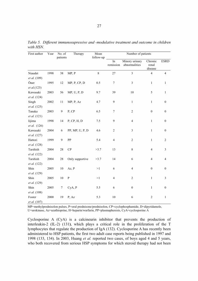
Early studies using per oral steroids for the treatment of HSN showed no benefit compared with subjects who did not receive any treatment (14). Counahan et al. found no differences in outcomes between patients receiving corticosteroids, immunosuppressives or both and patients with no treatment (106), and Tarshish et al. (2004) found in a recent prospective study that patients provided with only supportive treatment had a similar outcome to those treated with cyclophosphamide (122). On the other hand, Foster et al. (2000) reported in their retrospective analysis that patients with no treatment had a 5.9-fold relative risk of an unfavourable outcome relative to those treated with prednisone and azathioprine (107). Many non-randomized studies have pointed to benefits obtained from various types of immunosuppressive treatment, in most cases fairly aggressive (Table 5). Methylprednisolone pulses (MP) combined with per oral prednisolone alone or with additional immunosuppressive agents such as cyclophosphamide and azathioprine have been used effectively with HSN patients (109, 123, 130), and various fibrinolytic and anticoagulation drugs such as urokinase, heparin and dipyridamole has also been combined with immunosuppressive agents (123, 124, 126, 130).
27
Table 5. Different immunosupressive and -modulative treatment and outcome in children with HSN. First author Year No. of
patients Therapy Mean
follow-up Number of patients
In remission
Minory urinary abnormalities
Chronic renal
disease
ESRD
Niaudet et al. (109)
1998 38 MP, P 8 27 3 4 4
Öner et al.(123)
1995 12 MP, P, CP, D 0.5 7 3 1 1
Kawasaki et al. (124)
2003 56 MP, U, P, D 9.7 39 10 5 1
Singh et al. (125)
2002 11 MP, P, Az 4.7 9 1 1 0
Tanaka et al. (121)
2003 9 P, CP 6.5 7 2 0 0
Iijima et al. (126)
1998 14 P, CP, H, D 7.5 9 4 1 0
Kawasaki et al. (127)
2004 6 PP, MP, U, P, D 4.6 2 3 1 0
Hattori et al. (128)
1999 9 PP 5.4 4 2 1 2
Tarshish et al. (122)
2004 28 CP >3.7 13 8 4 3
Tarshish et al. (122)
2004 28 Only supportive >3.7 14 6 4 4
Shin et al. (129)
2005 10 Az, P >1 6 4 0 0
Shin et al. (129)
2005 10 P >1 4 2 1 3
Shin et al. (108)
2005 7 CyA, P 5.5 6 0 1 0
Foster et al. (107)
2000 19 P, Az 5.3 10 6 2 1
MP=methylprednisolon pulses, P=oral prednisone/prednisolon, CP=cyclophosphamide, D=dipyridamole, U=urokinase, Az=azathioprine, H=heparin/warfarin, PP=plasmapheresis, CyA=cyclosporine A
Cyclosporine A (CyA) is a calcineurin inhibitor that prevents the production of interleukin-2 (IL-2) (131), which plays a critical role in the proliferation of the T lymphocytes that regulate the production of IgA (132). Cyclosporine A has recently been administered to HSP patients, the first two adult case reports being published in 1997 and 1998 (133, 134). In 2003, Huang et al. reported two cases, of boys aged 4 and 5 years, who both recovered from serious HSP symptoms for which steroid therapy had not been

28
effective (135). The persistent renal disease lasting four months was successfully treated in one case, since the renal symptoms disappeared two weeks after CyA treatment was started. Later, Someya et al. reported a case of a 7-year-old boy who had nephrotic syndrome but did not respond to MP pulses with oral prednisolone (136). CyA was started 1 month after admission and proteinuria was reduced within two weeks (136). Recently, Shin et al. published a series of 7 patients with nephrotic-range proteinuria who had received CyA, six of whom were in complete remission at the latest observation after a mean follow-up of 5.5 years (2 – 9 years) and one had persistent renal disease (108).
2.8.2 Plasmapheresis and immunoglobulin therapy
Immunoglobulins have been administered to IgAN patients to downregulate the excessive production of IgA (137) and inhibit B cell differentiation and immunoglobulin production (138). Rostoker et al. used immunoglobulin therapy (IMIG) with 5 adult HSP patients and found that the extra-renal symptoms disappeared during the treatment and the level of proteinuria decreased, but discontinuation of the treatment was followed by a relapse (139, 140). Adverse renal effects have been reported after high-dose immunoglobulin therapy, and their use is controversial (141, 142). So far no reports have been published on immunoglobulin therapy used for children with HSP.
Hattori et al. used plasmapheresis as the sole therapy for 9 severe cases of HSN with nephrotic-range proteinuria in the early stage of the disease (128), with the result that 6 (67%) had a good outcome after 5.4 years of follow-up and all of them responded to the treatment at acute phase, as demonstrated by a reduced level of proteinuria (128). Schärer et al. and Gianviti et al. also found an immediate improvement in the course of the disease, but no long-term effect was seen, especially when the treatment was started late after the onset of renal disease (8, 143).
2.9 Prevention of HSN
Prevention of HSN could improve the prognosis for HSP, since the long-term outcome is highly dependent on the renal symptoms. Reports on the use of corticosteroids for treating HSP appeared in the literature around 1950 (144). Clinical experience and several uncontrolled retrospective studies (14, 26) have suggested that steroids may have a beneficial effect on abdominal pain, and it has been hypothesized that early administration of corticosteroids could prevent the development of nephritis (145). Retrospective uncontrolled studies have nevertheless given controversial results regarding the effect of corticosteroids in preventing late renal involvement (146, 147). In the first prospective study, by Mollica et al., none of the 84 patients who received prednisone treatment and 10 of the 84 without any treatment developed nephritis after 6 weeks of an acute episode (146), and a year later Saulsbury published his retrospective data on 50 patients, of whom 20 were treated with corticosteroids in the acute phase and 30 never received any corticosteroids (147). Delayed nephritis occurred in 4 of the former group and 6 of the latter, leading to the conclusion that early steroid treatment is not

29
effective (147). The first randomized, placebo-controlled study on the effect of corticosteroids was published by Huber et al. in 2004 (148). 40 children at a tertiary-care paediatric centre were randomized to receive prednisone (21) or a placebo (19), 2 mg/kg/day for first week, with weaning over the second week. After a year of follow-up 3 of the 21 prednisone patients and 2 of the 19 placebo patients had renal involvement, so that these authors concluded that the early administration of prednisone is not effective in preventing HSN (148). The severe limitation of that study was the sample size, which was calculated post hoc, assuming that the incidence of renal involvement at one year is 10.5% (the same as in their placebo group) (148).
2.10 Risk factors and prognosis for renal disease in HSP
The prognosis for HSP is usually excellent, since most of the patients recover spontaneously within weeks. Rare complications such as pulmonary or intestinal bleeding can cause severe morbidity and even mortality in the acute phase (14, 37), but the long-term outcome is predominantly related to the duration and severity of renal involvement (5, 7, 11, 13).
2.10.1 Influence of sex, age and extra-renal symptoms
There is mild predominance of boys among those affected by HSP (1, 14), but the risk of renal involvement is similar in both sexes (97). It is higher in patients over 4-7 years at onset, however, and also in those who have severe abdominal symptoms at onset and those with persistent purpura (89, 97). A 7.5-fold risk of renal involvement has been found in patients who have bloody stools (15). Adult patients have renal symptoms more often, the symptoms tend to be more severe and the prognosis after HSP tends to be worse than in children (12, 110).
2.10.2 Renal symptoms
20-54% of unselected HSP patients develop renal involvement during the acute phase (6, 14, 30), the majority of these (85%) doing so within the first 4 weeks and 97% within six months (31). Microscopic haematuria alone or combined with mild proteinuria is the most common manifestation of renal involvement (29). 10-30% of HSP patients with initial renal involvement develop nephritic or nephrotic syndrome (13, 30, 31), ESRD occurs in 1.1-1.5% and mortality is less than 1% in unselected patient series (6, 30).
A risk of persistent renal disease is associated with serious proteinuria and nephritic-nephrotic syndrome (5, 8, 31, 122, 149), whereas most of the patients with isolated haematuria with or without mild proteinuria recover completely (5, 30).
The prognostic factors for a poor outcome in adult patients are proteinuria (110, 150), hypertension (105) and renal impairment at onset (105, 110).

30
2.10.3 Long-term prognosis for HSN
A poor long-term outcome after childhood HSP is associated with persistent or chronic renal symptoms. 8-17% of patients with mild renal symptoms at onset, i.e. haematuria with or without mild proteinuria (<1g/day), and 44-47% of patients with severe renal symptoms at onset, i.e. nephritic or nephritic syndrome, proteinuria >1g/day or a renal biopsy with >50% crescents, have a poor outcome (5, 7, 8, 106). Clinical remission after severe disease and chronic renal disease in adulthood after apparent recovery and mild renal symptoms at onset are possible after childhood HSN (7). According to a 23.4-year follow-up by Goldstein et al., there is no prognostic factor that can reliably predict the individual outcome (7).

3 Aims of the research
The aims of the present work were
I to assess the long-term prognosis for childhood HSP disease, II to evaluate the outcome of severe HSN with nephrotic-range proteinuria and the effect
of Cyclosporine-A on severe HSN presenting with nephrotic-range proteinuria, and III to evaluate the clinical efficacy of early prednisone administration for treating the
symptoms of HSP and preventing renal involvement.

4 Material and methods
The population consisted of three patient groups collected retrospectively (I, II, III) and one prospectively (IV). The local ethics committees for the centres involved approved the protocol for the studies in which the subjects were personally contacted, and the protocol for paper IV was also approved by the National Agency for Medicines in Finland. Altogether 245 paediatric HSP patients (133 boys, 112 girls) were analysed, with a mean age of 8.4 years (range 1.7 – 15.6) at the time of diagnosis.
Long-term outcome after HSP (I). 52 adult subjects (26 male, 26 female) who had been treated for childhood HSP at Helsinki University Hospital in 1964-1983 were included in the analysis. Their medical histories were analysed retrospectively and 47 of them attended for a medical examination. The mean follow-up time was 24.1 years (range 16.4 – 36.5) and the mean age of the subjects at the last control visit was 32.1 years (range 18.8 – 45.3).
Outcome after severe HSN (II). 19 paediatric HSN patients (11 boys, 8 girls) with nephrotic-range proteinuria being treated at five university hospitals in Finland in 1990-1999 were included. The mean follow-up time was 4.6 years (range 0.75 – 9.1) and the mean age at the time of diagnosis 9.9 years (range 4.6 – 15.1). Medical histories were analysed retrospectively.
Cyclosporine A for treating severe HSN (III). 7 paediatric HSN patients (5 boys, 2 girls) with nephrotic-range proteinuria treated with CyA at Oulu University Hospital in 1994-1998 were included. The mean follow-up time was 6.0 years (range 4.4 – 8.9) and the mean age at the time of diagnosis 10.6 years (range 7.2 – 15.2). Medical histories were analysed retrospectively.
Early prednisone for treating HSP (IV). 171 paediatric HSP patients (93 boys, 78 girls) being treated at four university hospitals and 10 central hospitals in Finland in 1999-2005 were included in a prospective, randomized, placebo-controlled trial of early prednisone for the treatment of HSP. The mean age at the time of diagnosis was 7.0 years (range 1.7 – 15.6) and the patients were followed up for six months by means of four planned control visits.
The patient series and methods employed in all four studies are summarized in Table 6, and the basal characteristics of the patients in Table 7. Four patients are included in the series for both paper II and paper III.

33
Table 6. Materials and methods of the studies. (I-IV)
Study Design Setting and number of patients Purpose of the study
I Retrospective 52 patients with childhood HSP from Helsinki University Hospital treated in 1964–1983
Long-term outcome after HSP
II Retrospective 19 HSN-patients with nephrotic-range proteinuria from five University Hospitals in Finland treated in 1990–1999
Outcome after severe HSN
III Retrospective 7 pediatric patients with severe HSN from Oulu University Hospital treated in 1994–1998
Cyclosporine A in severe HSN
IV Randomized placebo-controlled
171 HSP-patients from 4 University Hospitals and 10 Central Hospitals in Finland treated in 1999–2005
Early prednisone in the HSP
Table 7. Base characteristic of patients in the four series. (I-IV)
Study Male/Female Mean age at diagnosis Mean follow-up years years (range) years (range)
I 26/26 5.9 (2.7 - 11.5) 24.1 (16.4 - 36.5) II 11/8 9.9 (4.6 -15.1) 4.6 (0.75 - 9.1) III 5/2 10.6 (7.2 - 15.2) 6.0 (4.4 - 8.9) IV 93/78 7.0 (1.7 - 15.6) 0.48 (0.08 - 0.79)
4.1 Long-term outcome after childhood HSP (I)
The medical histories of 67 patients with childhood HSP treated at Helsinki University Hospital in 1964-83 were obtained from the hospital registers, and a health questionnaire was mailed to the 65 patients whose address was available. Of the 52 (80%) who returned the health questionnaire, 47 (72%) were willing to attend for a medical examination. The health questionnaire asked about the patients’ current medication, health status and possible complications of pregnancies in the case of the women. The clinical outcomes for the 47 patients who were examined by a doctor were evaluated in terms of clinical findings (blood pressure, height, weight) and laboratory tests on urine and blood samples. The participants were divided into three groups according to clinical presentation at onset: 1) no renal symptoms, 2) proteinuria, haematuria, or both, and no biopsy specimen or ISKDC biopsy specimen grade I-II, and 3) renal symptoms lasting more than 1 month, nephritis or nephrosis, and ISKDC biopsy grade III or more. The outcome was classified as: A) healthy, no signs of renal involvement, B) minor urinary abnormalities, intermittent hypertension, haematuria, proteinuria, C) active renal disease, hypertension or constant proteinuria, and D) ESRD, dialysis treatment or renal transplantation. Outcomes A+B were judged to represent a good outcome and C+D a poor outcome.

34
4.2 Clinical outcome after severe HSN (II)
All children in Finland having HSP with nephrotic-range proteinuria (>40 mg/h/m2) in 1990-97 were surveyed by contacting the doctors treating paediatric kidney patients at five university hospitals and one central hospital. 19 patients (11 boys, 8 girls) were identified and their histories from the onset of HSP with proteinuria until the end of the follow-up were analysed retrospectively. A renal biopsy had been performed in all cases and repeated biopsies were available for 6 patients. Four patients had a grade II in first renal biopsy, 10 patients grade III (A/B), 4 patients grade IV and one patient grade V. The clinical outcomes at the end of the follow-up were graded into 4 categories: A) healthy, B) microscopic haematuria or proteinuria <1 g/day, C) proteinuria >1 g/day, and D) uraemia/ESRD or death. Grades A+B were considered favourable and C+D unfavourable.
4.3 Cyclosporine-A for the treatment of severe HSN (III)
Seven paediatric patients with severe HSN treated at Oulu University Hospital in 1994-98 were recruited on the criterion of nephrotic-range proteinuria (>40 mg/h/m2). Renal biopsies had been performed within three months (range 3 weeks – 3 months) of the initial diagnosis of HSP. Two patients had ISKDC grade II in their first biopsy, two grade III (A/B), two grade IV and one grade V. One patient received CyA as the first treatment modality and one CyA in combination with dipyridamole and prednisolone, while the other five had proved resistant to 1 – 3 types of immunosuppressive treatment before CyA was initiated. The initial dose of CyA (Sandimmun Neoral®, Novartis) ranged from 4 to 8 mg/kg/day, and the B-CyA level was kept at 150 – 200 μg/l for the first 6 months of the treatment. The dose during this maintenance phase was from 1 to 5 mg/kg/day and the B-CyA target level was 80 – 100 μg/l. All the patients also received the ACE inhibitor enalapril at a dose of 5 – 10 mg/day.
The patients were followed up at 1 – 6 month intervals for the duration of their HSP disease, and the laboratory results and patient histories were analysed from the onset of HSP until the end of the follow-up. Proteinuria <40 mg/h/m2 was taken as a response to treatment, and nephrotic relapse was documented if proteinuria increased to >40 mg/h/m2. Remission was documented when U-protein was <300 mg/l.
4.4 Effect of early prednisone on HSP (IV)
A total of 240 paediatric patients with newly diagnosed HSP were screened at four university hospitals and ten central hospitals in 1999-2005 for this prospective, double-blind, placebo-controlled trial of the effect of early prednisone on HSP. The scheme by which the 176 eligible patients were randomized to receive prednisone or a placebo is shown in Figure 1.
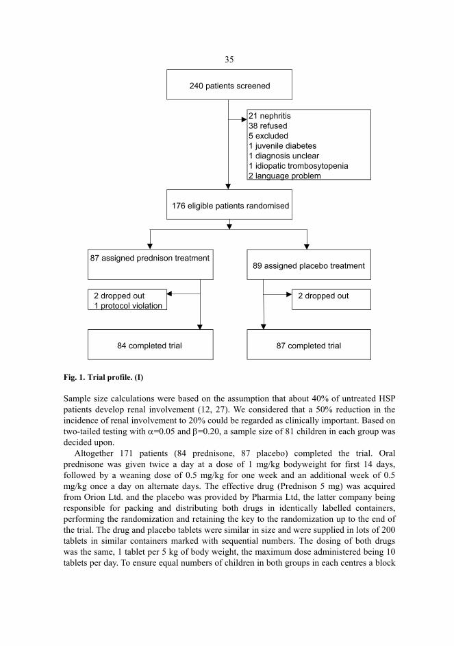
35
Fig. 1. Trial profile. (I)
Sample size calculations were based on the assumption that about 40% of untreated HSP patients develop renal involvement (12, 27). We considered that a 50% reduction in the incidence of renal involvement to 20% could be regarded as clinically important. Based on two-tailed testing with α=0.05 and β=0.20, a sample size of 81 children in each group was decided upon.
Altogether 171 patients (84 prednisone, 87 placebo) completed the trial. Oral prednisone was given twice a day at a dose of 1 mg/kg bodyweight for first 14 days, followed by a weaning dose of 0.5 mg/kg for one week and an additional week of 0.5 mg/kg once a day on alternate days. The effective drug (Prednison 5 mg) was acquired from Orion Ltd. and the placebo was provided by Pharmia Ltd, the latter company being responsible for packing and distributing both drugs in identically labelled containers, performing the randomization and retaining the key to the randomization up to the end of the trial. The drug and placebo tablets were similar in size and were supplied in lots of 200 tablets in similar containers marked with sequential numbers. The dosing of both drugs was the same, 1 tablet per 5 kg of body weight, the maximum dose administered being 10 tablets per day. To ensure equal numbers of children in both groups in each centres a block
240 patients screened
21 nephritis38 refused5 excluded1 juvenile diabetes1 diagnosis unclear1 idiopatic trombosytopenia 2 language problem
176 eligible patients randomised
87 assigned prednison treatment89 assigned placebo treatment
2 dropped out 2 dropped out 1 protocol violation
84 completed trial 87 completed trial

36
randomization system was employed with a block size of six. The observers and subjects in the trial were unaware of the randomization scheme.
Once informed consent had been obtained from the parents, the patients received their tablets and a symptom diary in which their parents marked scores for abdominal and joint pain and the results of the daily urinary dip stick test during the medication. The patients were examined by a doctor at inclusion and 7-10 days, 1 month, 3 months and 6 months after the start of medication. The results of the clinical examinations and laboratory findings were recorded on a structured data sheet.
Renal symptoms during treatment were recorded if readings of U-erythrocytes/U-protein +-++ were obtained by dip stick for three or more consecutive days or U-erythrocytes/U-protein +++ for two or more consecutive days. Renal involvement as an endpoint was defined as U-protein >200 mg/l, U-albumin >30 mg/l, or U-erythrocytes >5/vision field.
The primary endpoint was renal involvement at the 1-month, 3-month and 6-month control visits, and the secondary endpoints were the severity and duration of abdominal and joint symptoms during the treatment according to the symptom diary.
4.5 Statistical methods (I-IV)
SPSS (versions 10.0 – 12.0 for Windows) was used in the statistical analyses of the data. The differences in mean blood pressure and laboratory values between the outcome
groups in paper I were assessed by analysis of variance and the t test. Categorial variables such as sex, pregnancy, pregnancy complications, treatment and renal biopsy specimen grade were analysed with the χ2 test and Fisher’s exact test. Relative risks and their 95% confidence intervals were calculated.
The differences in the time when the CyA treatment was started between the patients who reached stable remission and those who became CyA-dependent were analysed in paper III using the t test.
Sum scores for (the severity of) abdominal pain and joint pain and the duration of abdominal and joint pain in days were calculated for each treatment group according to the symptom diary, and differences were tested with the t test. Differences in categorial variables (age, persistent purpura, severe abdominal pain) between the treatment groups were tested with the χ2 test and their 95% CI’s were calculated. The differences in proportions of renal involvement and their 95%CI's were calculated between the treatment groups.
The Kaplan-Meier method was used to analyse renal survival without an endpoint from the end of medication onwards, and the log rank test was used to compare the treatment groups. Improvement in extra-renal symptoms (abdominal an joint pain) was also analysed by the Kaplan-Meier method and the treatment groups were compared using the log rank test.

37
4.5.1 Number needed to treat (IV)
The number needed to treat (NNT) was calculated using the formula:
95% CIs were calculated for NNT and described as the number needed to harm (NNH) and number needed to benefit (NNB). A negative number needed to treat or NNH indicates that the treatment has harmful effects and one fewer patients receiving the new treatment (prednisone) would have a good outcome than if they all received the placebo (151). The result is statistically significant when 95% CI for NNT does not include infinity (∞).
NNT =100
Absolute risk reduction (ARR)
ARR = effect of prednisone treatment (%) – effect of placebo treatment (%)

5 Results
5.1 Long-term outcome after childhood HSP (I)
The outcome was highly dependent on the renal symptoms at onset, since 7 of the 20 adults who had severe HSN at onset (20%) had renal impairment as adults compared with 2 (7%) of the 27 with mild or no renal symptoms at onset (relative risk 4.7; 95% CI 1.3 – 18.7). The respective relative risks of a poor outcome were 5.0 for women (95% CI 1.1 – 32.5) and 2.0 for men (95% CI 0.2 – 17.5). All the patients with no renal symptoms at onset (n=9) had a good outcome after 24 years of follow-up. The results of the χ2 analysis showed a significant difference in onset grade between the outcome groups A+B and C+D (p=0.034) and a linear trend towards a worse outcome (C+D) with worsening onset symptoms (p=0.017) (Table 8).
Table 8. Clinical outcome 24 years after childhood HSP, by symptoms at onset. (I)
Clinical outcome (n=47) Healthy Minor urinary
abnormalitiesActive renal
disease ESRD
Good outcome
Poor outcome Onset grade
Onset symptoms
A B C D A+B C+D 1 No renal symptoms
(n=9) 8 (89%) 1 (11%) 0 0 9 (100%) 0
2 Proteinuria, hematuria, or both (n=18)
13 (72%) 3 (17%) 2 (11%) 0 16 (89%) 2 (11%)
3 Nephritis, nephrosis, or both (n=20)
9 (45%) 4 (20%) 5 (25%) 2 (10%) 13 (65%) 7 (35%)
Data are number (%). Good outcome (A+B) vs poor outcome (C+D) according to grade of onset (1,2,3): p=0.034 for chi2–test; p=0.017 for linear trend.

39
Table 9. ISKDC grade of first renal biopsy specimen and primary treatment in 47 patients with childhood HSP. (I)
Onset grade
Biopsy specimen taken
ISKDC grade of first biopsy specimen Treatment given
I II III IV None Steroids Other* 1 (n=9) 0 0 0 0 0 8 (89%) 1 (11%) 0 2 (n=18) 8 (44%) 3 (38%) 5 (62%) 0 0 13 (72%) 5 (28%) 0 3 (n=20) 20 (100%) 0 7 (35%) 11 (55%) 2 (10%) 6 (30%) 5 (25%) 9 (45%)
Data are number (%). * Cyclophosphamide, Azathioprine The severity of the kidney biopsies according to the ISKDC classification did not correlate with the risk of a poor outcome. Patients with a higher biopsy grade and more severe onset symptoms had more often received treatment than had those with low grades. No significant differences in outcomes were recorded between those who received treatment and those who did not (Table 9).
Of the 14 women who had been pregnant (54%), nine (64%) reported a history of proteinuria, hypertension or both during their pregnancies, and 16 of the 23 pregnancies (70%) had been complicated by one or both of these conditions. None of the five women with uncomplicated pregnancies had poor outcome, whereas 5 of the 9 women with complicated pregnancies (56%) did have such an outcome.
5.2 Clinical outcome after severe HSN (II)
According to our data, two new cases of HSN with nephrotic-range proteinuria are diagnosed in Finland annually, giving a yearly incidence of 2 per 1 million children under 15 years of age.
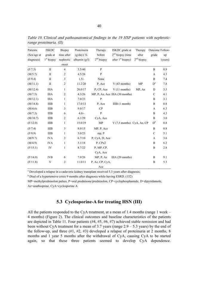
After a mean follow-up of 4.6 years 3 patients (15.7%) were healthy (A), 11 (57.9%) had microscopic haematuria or proteinuria < 1g/day (B), 2 (10.5%) had proteinuria > 1 g/day and 3 (15.7%) had developed renal failure or uraemia (D). One patient in outcome group D died of a hypertensive crisis after rapidly progressive glomerulonephritis (Table 10).
None of the 5 patients with grade IV-V in the first kidney biopsy had an unfavourable outcome (C+D), as opposed to 5 (36%) of the 14 patients with grade II-III. All 5 patients with grade IV-V in the first kidney biopsy had received combined immunosuppressive treatment after the severe findings in the biopsy.
40
Table 10. Clinical and pathoanatomical findings in the 19 HSP patients with nephrotic-range proteinuria. (II)
Patients (Sex/age at diagnosis)
ISKDC grade at
1st biopsy
Biopsy time after nephrotic
onset
Proteinuria (g/day)/ S-
albumin (g/l)
Therapy before
2nd biopsy
ISKDC grade at 2nd biopsy (time after 1st biopsy)
Therapy after
2nd biopsy
Outcome grade
Follow-up
(years)
(F/7.5) II 4 3.5/40 P B 0.9 (M/5.7) II 2 4.5/26 P A 4.3 (F/9.4) II 3 1.5/- None B 7.4 (M/11.1) II 2 11.2/20 P, Ace V (43 months) MP Da 7.8
(M/12.4) IIIA 1 20.0/17 P, CP, Ace V (11 months) MP, Az D 3.3 (M/7.5) IIIA 2 4.3/26 MP, P, Az, Ace IIIA (30 months) C 4.1 (M/12.1) IIIA 1 7.0/23 P B 3.1 (M/14.8) IIIB 1 17.0/13 P, Ace IIIB (1 month) B 0.8 (M/4.6) IIIB 3 9.0/17 CP A 6.3 (M/7.3) IIIB 6 4.0/- P B 4.3 (M/10.7) IIIB 2 6.1/29 CyA, Ace B 3.0 (F/12.0) IIIB 1 15.0/19 MP V (7,5 months) CyA, Az, CP Db 0.8
(F/7.4) IIIB 3 8.0/15 MP, P, Ace B 8.8 (F/9.9) IIIB 1 3.0/23 mp, P C 5.1 (M/9.7) IVA 2 6.7/10 P, CyA, D, Ace A 3.6 (M/4.9) IVA 1 3.1/18 P, CPx2 B 6.2 (F/15.1) IV 1 8.7/22 P, MP, CP,
CyA, Ace B 2.8
(F/14.0) IVB 6 7.0/26 MP, P, Az IIIA (20 months) B 9.1 (F/11.8) V 2 11.0/11 P, Az, CP, CyA,
Ace B 5.3
a Developed a relapse in a cadaveric kidney transplant received 5.3 years after diagnosis. b Died of a hypertensive crisis 9 months after diagnosis while having ESRD. (152) MP=methylprednisolon pulses, P=oral prednisone/prednisolon, CP=cyclophosphamide, D=dipyridamole, Az=azathioprine, CyA=cyclosporine A
5.3 Cyclosporine-A for treating HSN (III)
All the patients responded to the CyA treatment, at a mean of 1.4 months (range 1 week – 4 months) (Figure 2). The clinical outcomes and baseline characteristics of the patients are depicted in Table 11. Four patients (#4, #5, #6, #7) achieved stable remission and had been without CyA treatment for a mean of 3.7 years (range 2.9 – 5.3 years) by the end of the follow-up, and three (#1, #2, #3) developed a relapse of proteinuria at 2 months, 8 months and 1 year 5 months after the withdrawal of CyA, causing CyA to be started again, so that these three patients seemed to develop CyA dependence.

41
Fig. 2. Response of HSP-GN patients to CyA treatment during the first 6 months in terms of S-albumin and proteinuria. Patients #1, 2 and 3 became CyA-dependent and patients #4, 5, 6 and 7 achieved stable remission. Dark line= interruption or reduction of CyA-dose. (III)
Two patients (#2, #5) had notable but reversible side effects, with an increase in S-creatinine during CyA treatment, so that CyA was temporarily stopped in one case and the dose reduced in the other (Figure 2). None of the patients developed gingival hyperplasia during the treatment, but hirsutism was noted in four.
The only significant difference between the patients who became CyA-dependent and those who reached stable remission was the earlier commencement of CyA treatment in the latter group (p=0.045, t test). Neither the level of proteinuria at diagnosis or at the start of CyA treatment, nor the severity of the biopsy findings or the time required for a response to the treatment differed between the groups.
All the patients had their glomerular filtration rate, S-creatinine and P-Cystatin-C within the normal limits at the end of the follow-up (Table 11).
One of the three CyA-dependent patients (#2) achieved stable remission later and has been without CyA treatment for 1.7 years. A repeated kidney biopsy has been performed on the other two CyA-dependent patients, showing some improvement in their ISKDC grade. Recent follow-up data on the three CyA-dependent patients are given in Table 12 (unpublished data).
0
2000
4000
6000
8000
10000
12000
0 1 2 3 4 5 6
Months
Prot
einu
ria (m
g/d)
#1#2#3
0
2000
4000
6000
8000
10000
12000
0 1 2 3 4 5 6
Months
Prot
einu
ria (m
g/d)
#4#5#6#7
05
101520253035404550
0 1 2 3 4 5 6
Months
S-al
bum
in (g
/l)
#1
#2#3
0
5
10
15
20
25
30
35
40
45
0 1 2 3 4 5 6
Months
S-al
bum
in (g
/l) #4
#5
#6#7

42
Table 11. Clinical outcome of 7 HSN-patients treated with CyA. (III) Renal
findings at onset Laboratory findings
before CyA Laboratory findings at end of follow-up Patients
Sex/age at diagnosis Pu
(g/ day)
ISKDC
(crescents)
Treat-ment
before CyA Pu
(g/day)
S-alb
(g/l)
S-crea
(mmol/l)
Pu
(g/day)
S-alb
(g/l)
S-crea
(mmol/l)
GFR
(ml/min/1
.73 m2)
Plasma
Cystatin
-C
(mg/l)
Follow-up
(years)
1 (F/11.8)* 11.0 V (89%) P,Az, CP, Ace
9.8 14 101 3.5 37.9 69 105 0.80 8.9
2 (F/15.2)* 8.7 IV (79%) P, MP, CP, Ace
2.8 28 71 0.6 42.0 70 111 0.79 6.1
3 (M/7.2)* 2.3 IIIA (14%) MP, P, Ace
1.2 46 68 0.6 40.1 47 139 0.66 4.6
4 (M/9.8) 7.3 IV (57%) P, D, Ace 6.5 10 50 0.3 44.0 64 166 0.62 7.2
5 (M/8.7) 6.1 IIIB (30%) None 4.8 21 75 0.3 43.8 50 93 0.81 5.8
6 (M/13.8) 12.0 II MP, P, Ace
10.0 21 58 0.1 43.0 71 101 0.64 4.7
7 (M/7.5) 7.2 II MP, P, Ace
3.6 27 48 0.2 41.0 52 156 0.79 4.4
*CyA-dependent patients. MP=methylprednisolon pulses, P=oral prednisone/prednisolon, CP=cyclophosphamide, D=dipyridamole, Az=azathioprine, CyA=cyclosporine A
Table 12. Clinical and histopathological findings of CyA-dependent patients after mean 8.2 years follow-up. (Unpublished data)
1st Biopsy 2nd Biopsy Clinical outcome Patients
Sex/age at
diagnosis ISKDC
(crescents)
Time after
diagnosis
(years)
ISKDC
(crescents)
Time
after 1st
biopsy
(years)
Hemat-
uria
U-alb/U-
crea (g/mol
crea)
U-
albumin
(mg/l)
S-crea
(µmol/l)
Total time
of CyA-
treament
(years)
Follow-
up (years)
1 (F/11.8) V (89%) 0.19 II A-B (0%) 9.4 No 66 499.5 78 9.3 10.5
2 (F/15.2) IV (79%) 0.08 No 28 295.2 88 3.9 7.6
3 (M/7.2) IIIA (14%) 0.32 II (0%) 5.9 Yes 73 1174 51 5.4 6.7
43
Table 13. Clinical and laboratory features of participants at baseline. (IV)
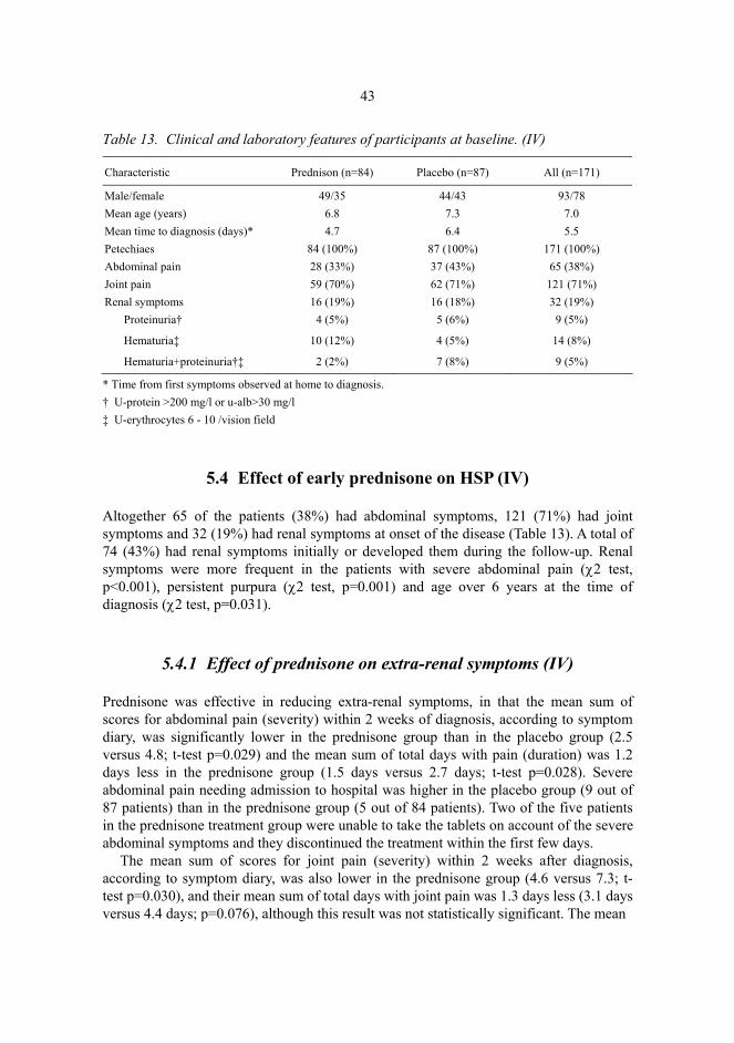
Characteristic Prednison (n=84) Placebo (n=87) All (n=171)
Male/female 49/35 44/43 93/78 Mean age (years) 6.8 7.3 7.0 Mean time to diagnosis (days)* 4.7 6.4 5.5 Petechiaes 84 (100%) 87 (100%) 171 (100%) Abdominal pain 28 (33%) 37 (43%) 65 (38%) Joint pain 59 (70%) 62 (71%) 121 (71%) Renal symptoms 16 (19%) 16 (18%) 32 (19%) Proteinuria† 4 (5%) 5 (6%) 9 (5%)
Hematuria‡ 10 (12%) 4 (5%) 14 (8%)
Hematuria+proteinuria†‡ 2 (2%) 7 (8%) 9 (5%)
* Time from first symptoms observed at home to diagnosis. † U-protein >200 mg/l or u-alb>30 mg/l ‡ U-erythrocytes 6 - 10 /vision field
5.4 Effect of early prednisone on HSP (IV)
Altogether 65 of the patients (38%) had abdominal symptoms, 121 (71%) had joint symptoms and 32 (19%) had renal symptoms at onset of the disease (Table 13). A total of 74 (43%) had renal symptoms initially or developed them during the follow-up. Renal symptoms were more frequent in the patients with severe abdominal pain (χ2 test, p<0.001), persistent purpura (χ2 test, p=0.001) and age over 6 years at the time of diagnosis (χ2 test, p=0.031).
5.4.1 Effect of prednisone on extra-renal symptoms (IV)
Prednisone was effective in reducing extra-renal symptoms, in that the mean sum of scores for abdominal pain (severity) within 2 weeks of diagnosis, according to symptom diary, was significantly lower in the prednisone group than in the placebo group (2.5 versus 4.8; t-test p=0.029) and the mean sum of total days with pain (duration) was 1.2 days less in the prednisone group (1.5 days versus 2.7 days; t-test p=0.028). Severe abdominal pain needing admission to hospital was higher in the placebo group (9 out of 87 patients) than in the prednisone group (5 out of 84 patients). Two of the five patients in the prednisone treatment group were unable to take the tablets on account of the severe abdominal symptoms and they discontinued the treatment within the first few days.
The mean sum of scores for joint pain (severity) within 2 weeks after diagnosis, according to symptom diary, was also lower in the prednisone group (4.6 versus 7.3; t-test p=0.030), and their mean sum of total days with joint pain was 1.3 days less (3.1 days versus 4.4 days; p=0.076), although this result was not statistically significant. The mean

44
Fig. 3. The disappearance of abdominal and joint symptoms during the treatment, by treatment group. (IV)
scores for abdominal and joint pain are given in Table 14 and the disappearance of abdominal and joint symptoms is depicted in Figure 3.
Skin symptoms were resolved more quickly in the patients with prednisone treatment (within 7-10 days) than in those receiving the placebo (χ2 test, p=0.021), but there were no differences in skin symptoms between the groups after 1 month (χ2 test, p=1.0), and 19 patients in the prednisone group (24%) and 16 in the placebo group (20%) had skin relapses later (χ2 test, p=0.354).
Table 14. Influence of prednisone and placebo treatment on body weight, blood pressure and the need for analgesics and on abdominal and joint pain. (IV)
Characteristics Means p value Prednisone Placebo
Mean difference
Confidence interval
(N=84) (N=87 Changes during treatment Increase of weight (kg)
1.4
0.4
1.1
0.5 - 1.6
<0.001
Systolic blood pressure* 109 106 3.2 -0.7 - 7.1 0.113 Diastolic blood pressure* 64 61 3.6 0.9 - 6.2 0.009 Days needing analgesic† 2.2 2.7 0.6 -0.7 - 1.9 0.369 Influence for abdominal pain Severity‡
2.5
4.8
2.4
0.3 - 4.5
0.029
Duration# 1.5 2.7 1.2 0.1 - 2.3 0.028 Influence for joint pain Severity‡
4.6
7.3
2.7
0.3 - 5.2
0.030
Duration# 3.1 4.4 1.3 -0.1 - 2.6 0.076 * Measured by taking the mean blood pressure from control visits at 7 to 10 days and 1 month. † Days when taking analgesic at home. ‡ Mean sum of scores for pain in symptom diary within two first weeks. # Mean sum of days with pain in symptom diary within two first weeks.
0 2 4 6 8 10 12 140
10
20
30
40
50
60
70
80
90
100
Disappearance of abdominal symptoms
Abdo
min
al p
ain
%
Day
Placebo (n=19) Prednisone (n=13)
Log rank p=0.124
0 2 4 6 8 10 12 140
10
20
30
40
50
60
70
80
90
100
Join
t pai
n %
Day
Placebo (n=30) Prednisone (n=32)
Log rank P=0.007
Disappearance of joint symptoms

45
The patients who received prednisone gained somewhat more weight and their diastolic blood pressure was higher during the treatment than among the patients receiving the placebo. The need for an analgesic was slightly lower in the patients with prednisone treatment, but not significantly so (Table 14).
5.4.2 Effect of prednisone on renal symptoms (IV)
Prednisone did not prevent the development of renal symptoms. Altogether 74 of the HSP patients (43%) had renal symptoms (38 prednisone, 36 placebo) during the 6 months follow-up, 96% of whom (71 of the 74 patients, 36 prednisone, 35 placebo) had these symptoms or developed them during the one-month treatment period. In order to analyse the effect of prednisone in altering the course of renal disease, we analysed the rate of resolution of renal symptoms in these 71 patients by treatment groups.
Renal symptoms had been resolved by the 1-month control visit in 22 of the 36 patients who received prednisone treatment (61%) and in 12 of the 35 (34%) who received the placebo treatment (difference 27%, 95% CI 3 – 47%, p=0.024), the corresponding figures at 3 months being 25 (69%) with prednisone and 17 (49%) with the placebo (difference 21%, 95% CI –2 – 42%, p=0.074) and those at 6 months 29 (81%) with prednisone and 20 (57%) with the placebo (difference 23%, 95% CI 2 – 43%, p=0.033) (Figure 4.).
Fig. 4. Renal symptoms after treatment in patients having renal symptoms at acute phase (36 prednisone, 35 placebo), by treatment groups. (IV)
0 %10 %20 %30 %40 %50 %
60 %70 %80 %90 %
100 %
At acutephase
1 month 3 months 6 months
PlaceboPrednisone
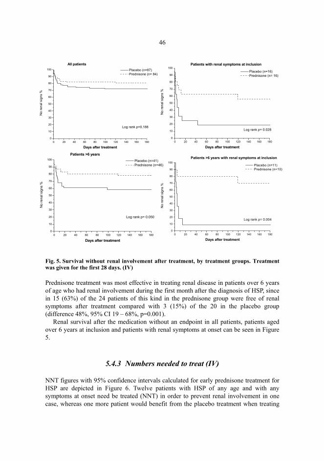
46
Fig. 5. Survival without renal involvement after treatment, by treatment groups. Treatment was given for the first 28 days. (IV)
Prednisone treatment was most effective in treating renal disease in patients over 6 years of age who had renal involvement during the first month after the diagnosis of HSP, since in 15 (63%) of the 24 patients of this kind in the prednisone group were free of renal symptoms after treatment compared with 3 (15%) of the 20 in the placebo group (difference 48%, 95% CI 19 – 68%, p=0.001).
Renal survival after the medication without an endpoint in all patients, patients aged over 6 years at inclusion and patients with renal symptoms at onset can be seen in Figure 5.
5.4.3 Numbers needed to treat (IV)
NNT figures with 95% confidence intervals calculated for early prednisone treatment for HSP are depicted in Figure 6. Twelve patients with HSP of any age and with any symptoms at onset need be treated (NNT) in order to prevent renal involvement in one case, whereas one more patient would benefit from the placebo treatment when treating
0 20 40 60 80 100 120 140 160 1800
10
20
30
40
50
60
70
80
90
100
No
rena
l sig
ns %
Days after treatment
Placebo (n=87) Prednisone (n= 84)
Log rank p=0.188
All patients
0 20 40 60 80 100 120 140 160 1800
10
20
30
40
50
60
70
80
90
100
No
rena
l sig
ns %
Days after treatment
Placebo (n=16) Prednisone (n= 16)
Patients with renal symptoms at inclusion
Log rank p= 0.028
0 20 40 60 80 100 120 140 160 1800
10
20
30
40
50
60
70
80
90
100
No
rena
l sig
ns %
Days after treatment
Placebo (n=41) Prednisone (n=46)
Patients >6 years
Log rank p= 0.050
0 20 40 60 80 100 120 140 160 1800
10
20
30
40
50
60
70
80
90
100N
o re
nal s
igns
%
Days after treatment
Placebo (n=11) Prednisone (n=10)
Log rank p= 0.004
Patients >6 years with renal symptoms at inclusion

47
24 (95% CI 2NNB to ∞ to 24 NNH). Early prednisone treatment seemed to be most useful for patients over 6 years of age with renal involvement at inclusion (NNT = 2), since none of them could be harmed by the treatment (95% CI 2NNB – 6NNB).
Fig. 6. Number needed to treat (NNT) with 95 percent confidence intervals (95%CI) in order to prevent renal involvement in one patient. (NNB = Number needed to benefit; NNH = Number needed to harm). (IV)
6 Discussion
6.1 Study design and patient series (I-IV)
The patients with renal symptoms were over-represented in our long-term outcome study (I) by comparison with other series of unselected patients (6, 13, 30), creating a follow-up bias which means that our results tend to overestimate the severity of HSP disease. As in the latest examination, 36% (17/47) of the patients discussed in paper I were admitted for nephrological examination two decades earlier and 64% (30/47) were uncomplicated cases referred by family doctors. This was nevertheless the first very long-term outcome assessment (mean follow-up over 20 years) in which all the patients were examined by a doctor and which included patients without any renal symptoms at the acute phase of the disease (n=9), thus giving the new perspective on the long-term outcome after childhood HSP.
Due the retrospective nature of the investigation reported in paper II, the patient series is highly variable, making it difficult to reach firm conclusions. Due to the small number of patients and different treatment protocols, the patients cannot be reliably compared. The figure quoted for the incidence of HSN with nephrotic-range proteinuria in Finland is reliable, however, since all the patients admitted to the five university hospitals were included.
The small number of patients is one limitation to be considered in connection with paper III. At the time when our patients were treated with CyA, there were no earlier results published on its administration to children with severe HSN, and this was the first published report. The patients were followed prospectively with a standardised scheme after the commencement of CyA medication, but the treatment given before CyA had been variable.
Paper IV fulfils all the criteria for evidence-based medicine, since the sample size was calculated in advance, the patients were collected prospectively, the treatment was placebo-controlled and based on randomization, and a double-blinded protocol was observed in the treatment and follow-up. The patients who dropped out were kept in the analysis (on an intention to treat basis) and those who refused to take part, did not differ from the participants in terms of their baseline characteristics. The patients were as

49
unselected as possible, since most of them were collected from health care centres as soon as a diagnosis of HSP disease had been reached at a mean interval of 5.5 days (0 - 63 days) after the first symptoms had been noticed at home. None of the patients had severe nephritis at the time of diagnosis, since we excluded cases of definite nephritis (U-erythrocytes >10 / vision field or U-protein >300 mg/l) in order to study the effect of prednisone in preventing it. Patients with mild proteinuria (U-protein 200-300 mg/l) and haematuria (U-erythrocytes 6-10/vision field) were included, however. The patients were followed up well, since 98% completed all the control-visits and the analyses were performed on an intention-to treat basis.
Neither the physician nor the parents knew whether a given child had received prednisone or placebo tablets, an although this was revealed in some cases at the one-month visit on account of weight gain in the prednisone group, this could not have affected the follow-up or management of the patient, and thereby our results, since the treatment was already over by that time. Moreover, all the data concerning renal involvement are based on the results of regular urine analyses during control visits, which were not dependent on the randomization or blinding.
6.2 Outcome after HSP disease (I)
Earlier outcome studies on unselected patient series (6, 13, 30) give a favourable prognosis for most patients, since 88%–98% of affected children do well after the acute phase of the disease. As long as the patient has no renal symptoms, the prognosis is excellent (5), even in the long term, as our results demonstrate.
6.2.1 Renal symptoms predict the outcome (I, II)
We analysed the outcomes of 394 cases of Henoch-Schönlein purpura, those described in our first paper (I) and those in four other reports (5, 7, 8, 30). The severity of renal symptoms at onset of HSP proved to be a prognostic factor, since patients with severe renal symptoms at onset more often had a poor outcome after 1-24 years of follow-up (38%, range 17-47%) than did those with mild renal symptoms (9%, range 0-17%; p<0.0001) (Table 15). This is also supported by the results of our paper II, where only severe cases with nephrotic-range proteinuria were included. Morbidity was high among these, since only one fifth of the patients had no signs of renal symptoms, 57% had microscopic haematuria or proteinuria and 26% had serious proteinuria or ESRD after a mean of 4.6 years of follow-up subsequent to severe HSN.
Bunchman et al. reviewed 16 patients who developed ESRD after HSN and compared them with HSN patients who had retained their renal function after 10 years of follow-up (153) and found that a creatine clearance (GFR) <70 ml/min/1.73m2 at three years of follow-up predicted progression to ESRD (153).

50
Table 15. Numbers of patients with poor outcome, by severity of renal symptoms at onset of Henoch-Schönlein purpura.
Series Number of patients reported
Follow-up (years)
Renal symptoms at onset
No Mild Severe
Ronkainen et al. (I) 47 24.1 (mean) 0/9 2/18 (11%) 7/20 (35%) Stewart et al. (30) 55 8.3 (mean) 0/37 (0%) 3/18 (17%) Goldstein et al. (7) 78 23.4 (mean) 5/39 (13%) 17/39 (44%) Niaudet et al. (5) 151 >1 3/18 (17%) 50/133 (33%) Schärer et al. (8) 63* 6.1 (mean) 2/25 (8%) 18/38 (47%) Total 394 0/9 12/137 (9%) 95/248 (38%)
Mild symptoms= hematuria, proteinuria (<1 g/day), or both. Severe symptoms= nephritic/nephrotic syndrome, proteinuria >1 g/day, biopsy specimen ISKDC grade III or more. *One patient who died of an extra-renal cause is not included in this table. The individual outcome is not always predictable, since there are children with mild renal symptoms at onset who develop renal impairment later, even after apparent recovery. This was seen both in the results of 78 patients followed up for 23.4 years by Goldstein and colleagues (7) and in our data on a subgroup of 19 patients from paper I who were analysed for the first time by Koskimies et al. after a mean follow-up of 6.0 years (6). 44 of these patients remained unchanged (56%), 17 (22%) improved and 17 (22%) deteriorated. The corresponding figures in our investigation were 11 (58%), three (16%) and five (26%) after a mean follow-up of 30 years (Table 16).
Table 16. Clinical outcome for 19 participants from study I in 1981 and 2000. (I)
Clinical outcome in 1981* Clinical outcome in 2000^ Onset grade Follow-up (years) A A II 34.9 A A II 30.3 A A II 24.5 A A III 34.4 A A III 32.6 A A III 27.9 A A III 26.1 A B III 33.1 A B III 30.2 A C II 34.0 B A II 30.3 B A III 30.7 B A III 29.7 B B II 36.5 B B III 27.7 B B III 27.9 B C III 25.6 B C III 27.0 C C III 26.6 A=healthy, B=minor urinary abnormalities, C= active renal disease, D=ESRD *Mean follow-up 6 years (SD 2.6). ^Mean follow-up 30 years (SD3.6)

51
6.2.2 Role of renal biopsy in predicting the outcome (I, II, III)
Although renal symptoms and their severity seem to be a prognostic factor, the results of a renal biopsy seem not to predict the outcome as well. In our two retrospective studies, as in other studies where the biopsy did not predict the outcome (7, 105), the biopsies had been assessed according the widely used ISKDC classification (106), and the poor correlation of biopsy grade with outcome may be due this system, which is mainly based on the amount of crescents but does not take account their maturity (154). This was seen in two of our patients treated with CyA, for example, who were re-biopsied 9.4 and 6.0 years after the first instance. No crescents were seen in the second biopsy (ISKDC grade IIA/B), even though over half the glomeruli were crescentic in both patients in the first biopsy (unpublished data), which means that the epithelial crescents had been reversible (Table 12). In adult patients, Pillebout et al. found that the degree of interstitial fibrosis, percentage of sclerotic glomeruli and presence of glomeruli with fibrinoid necrosis were factors associated with a poor renal prognosis (110). Adult patients are not always comparable with children, however, since ageing and concurrent illnesses can affect the biopsy findings (154). Moreover, Rauta et al., who evaluated a Finnish group of 42 adult HSN patients and found no histopathological features to predict the outcome in adult patients, attributed the fact that their histological findings were less severe than in most of the earlier reports due to possible differences in the indications for biopsy between centres (150).
The timing of the first biopsy may also be crucial, since an early biopsy with mild findings does not necessarily show the real severity of the disease. This was seen in our paper II, where three patients having only ISKDC grade II-III in the first biopsy, performed within 2 months of onset of the disease, developed a progressive renal disease later that led to an ESRD. A second biopsy 7.5 – 43 months later gave ISKDC grade IV-V findings.
Shin et al. and Niaudet et al. used a semiquantitative scoring system for HSN patients created by Andreoli and Bergstein for IgA nephropathy (108, 109, 155), a system in which the active, chronic and tubulointerstitial findings regarding the renal biopsy specimen is taken into account. Niaudet et al. reported that the best outcome results were obtained for patients whose initial biopsy showed a high activity index and a low chronicity index (109). All their patients were treated with MP pulses, and they found that the patients with recurrent symptoms still had an elevated activity index in the post-therapy biopsy while the chronicity index was either elevated or unchanged (109). Shin et al. also found that the activity index decreased after treatment but the chronicity and tubulointerstitial indices did not (108). In order to make comparisons, we later collected all the available biopsy specimens for our patients in paper I (27 out of 28 biopsies) and re-analysed them in terms of a semiquantitative classification modified from that used by Shin et al. (Table 17) (108). Although the mean scores for activity, chronicity, tubulointerstitial indices and total biopsy scores in the first biopsies were higher in the patients who developed a poor outcome, the results were not statistically significant (Table 18). Taking together, however, the semiquantitative scoring system for active and chronic changes in the kidney biopsy seems to be more informative and useful in clinical work than the generally used ISKDC classification.
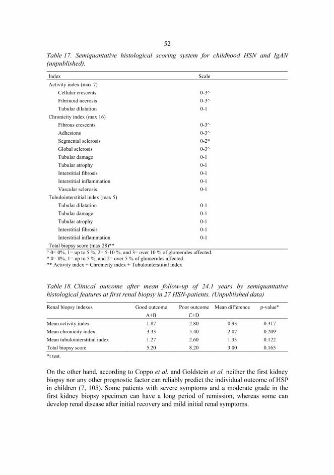
52
Table 17. Semiquantative histological scoring system for childhood HSN and IgAN (unpublished).
Index Scale Activity index (max 7)
Cellular crescents 0-3^ Fibrinoid necrosis 0-3^ Tubular dilatation 0-1
Chronicity index (max 16) Fibrous crescents 0-3^ Adhesions 0-3^ Segmental sclerosis 0-2* Global sclerosis 0-3^ Tubular damage 0-1 Tubular atrophy 0-1 Interstitial fibrosis 0-1 Interstitial inflammation 0-1 Vascular sclerosis 0-1
Tubulointerstitial index (max 5) Tubular dilatation 0-1 Tubular damage 0-1 Tubular atrophy 0-1 Interstitial fibrosis 0-1 Interstitial inflammation 0-1
Total biopsy score (max 28)** ^ 0= 0%, 1= up to 5 %, 2= 5-10 %, and 3= over 10 % of glomerules affected. * 0= 0%, 1= up to 5 %, and 2= over 5 % of glomerules affected. ** Activity index + Chronicity index + Tubulointerstitial index
Table 18. Clinical outcome after mean follow-up of 24.1 years by semiquantative histological features at first renal biopsy in 27 HSN-patients. (Unpublished data)
Renal biopsy indexes Good outcome Poor outcome Mean difference p-value* A+B C+D Mean activity index 1.87 2.80 0.93 0.317 Mean chronicity index 3.33 5.40 2.07 0.209 Mean tubulointerstitial index 1.27 2.60 1.33 0.122 Total biopsy score 5.20 8.20 3.00 0.165 *t test. On the other hand, according to Coppo et al. and Goldstein et al. neither the first kidney biopsy nor any other prognostic factor can reliably predict the individual outcome of HSP in children (7, 105). Some patients with severe symptoms and a moderate grade in the first kidney biopsy specimen can have a long period of remission, whereas some can develop renal disease after initial recovery and mild initial renal symptoms.

53
Fig. 7. Renal survival and morbidity (need of continual medication from the time of diagnosis) after childhood HSN and IgAN. (Unpublished data)
Table 19. The data and outcome after childhood IgA-nephropaties. (Unpublished data)
Clinical outcome Mean age at onset
Mean age at control
Mean follow-up years Healthy Minor urinary
abnormalities Chronic renal
disease ESRD or dialysis
All
HSP 9.3 32.5 25.1 19 (51%) 8 (22%) 7 (19%) 3 (8%) 37 (100%) IgA 13.8 34.4 18.7 9 (29%) 9 (29%) 10 (32%) 3 (10%) 31 (100%) All 11.3 33.5 22.0 28 (41%) 17 (25%) 17 (25%) 6 (9%) 68 (100%)
6.3 Long-term outcome after HSN versus IgAN
We recently performed a long-term follow-up study after biopsy-proven childhood IgAN employing exactly the same protocol as in paper I. After analysing the medical histories of the 41 patients whose diagnosis of IgAN was based on IgA deposits as the sole or main finding in the renal biopsy specimen, it became evident that in fact 10 of the patients had had extra-renal symptoms compatible with Henoch-Schönlein purpura (unpublished data). This finding and earlier reports of the close similarities between these two diseases (5, 70, 88) prove that many doctors do not distinguish clinically between these disease entities in adult patients. In order to make comparisons between the long-term outcomes of IgAN and HSN, we analysed a database on 37 HSN-patients (the 27 discussed in paper I and the 10 HSP cases in the IgA study) and 31 IgAN patients diagnosed during childhood (Table 19). Renal survival was closely similar, but the adults who had had childhood IgA nephritis more often needed continual medication after a mean follow-up of 22 years than those who had had childhood HSP nephritis (log rank p=0.030) (Figure 7). The patients with childhood HSN had more often been treated at the clinical onset of
0 5 10 15 20 25 300
10
20
30
40
50
60
70
80
90
100
Years
HSN IgAN
Renal survival
Log rank P=0.761
%
0 5 10 15 20 25 300
10
20
30
40
50
60
70
80
90
100
Years
HSN IgAN
Log rank p=0.034
Without continual medication%

54
the disease, however, since 23 of 37 the HSP patients had received any treatment as compared with 3 of the 31 IgAN patients and the mean activity index in the first renal biopsy had been significantly higher in the HSN patients (mean difference 1.1; 95% CI 0.2-2.0, p=0.013 [unpublished data]). Yoshikawa et al., comparing the clinical course of 128 HSN patients and 206 IgAN patients, noted that 72 (56%) of the former and 67 (32%) of the latter had no demonstrable abnormality after a mean follow-up of 5.0 years, 29 (23%) versus 103 (50%) had minor urinary abnormalities, 7 (5%) versus 26 (13%) had serious proteinuria and/or hypertension, and 20 (16%) versus 10 (5%) had developed chronic renal failure (156). They found that the worse outcome in cases of HSN was associated with more severe clinical and glomerular changes at presentation, whereas there was no relationship between the severity of clinical presentation and glomerular changes in the prognosis for IgAN (156). Both our findings and those of Yoshikawa et al. (156) support the conclusion that HSN is more of an acute disease and IgAN a slowly progressive, chronic glomerular disease. It is not clear for an individual patient when the initial onset of IgA nephritis has occurred, since in most cases the patients have had silent microscopic haematuria for years before the kidney biopsy was taken. In HSP the onset of the disease is more visible, with clear extra-renal symptoms, and a kidney biopsy is performed if the renal symptoms continue for more than 4-6 weeks.
6.4 Pregnancy after childhood HSP (I)
Pregnancy complications such as hypertension, proteinuria and toxaemia were frequent in women after childhood HSP, since 64% of the mothers and 70% of the pregnancies were affected by these symptoms. As the reported frequencies of pregnancy-associated hypertension, pre-eclampsia or both in a healthy population are 5-10% (157, 158), HSN increases the risk many-fold. The risk of pregnancy complications was not increased only among women who had had severe HSN (78%) but also among those who had had a mild renal disease at onset (40%). Two mothers with no renal symptoms at onset had complication-free pregnancies. Pregnancy complications are also common in women with idiopathic IgA nephritis (159, 160). Our data also suggest (I) that pregnancy could affect the outcome of HSP in women, since none of the five mothers with uncomplicated pregnancies had a poor outcome, whereas 56% (5/9) of those with complications did so, including two who had had active renal disease prior to pregnancy. It has been suggested that the hyperfiltration that occurs during pregnancy can overload the surviving nephrons (7, 161).
6.5 Treatment of severe HSN (II, III)
No single treatment protocol or medicine has proved to be the obvious choice for cases of severe HSN, and since there is a possibility of spontaneous recovery, some authors recommend treatment only for those with severe renal presentation and marked abnormalities in a renal biopsy (8, 109, 123). The indications for treatment should be well motivated, since immunosuppressive agents have several possible adverse effects. A risk
55
of fulminant infections and leukopenia has been reported with azathioprine, for example, infertility with cyclophosphamide, retardation of growth and cushinoid syndrome with high-dose clucocorticosteroids and nephrotoxicity with cyclosporine treatment (107, 162-165). In contrast, since the outcome is difficult to predict, as there is no single sign of severe sequelae and progression to an ESRD is possible, some authors find early aggressive treatment justified (121, 125). Our results presented in papers II and III also support early treatment despite the initial biopsy finding.
6.5.1 Timing of the treatment (II, III)
The timing and symptoms or findings indicating treatment seem to be the most important questions. Our results (papers II and III) suggest early aggressive treatment for severe symptoms (nephrotic-range proteinuria) regardless of the ISKDC classification at the early stage of the disease.
CyA treatment had been started significantly earlier in those who reached stable remission than in those who became CyA-dependent (III). Moreover, all five patients in paper II with a high ISKDC grade (IV-V) had more favourable renal survival than the 5 (36%) out of 14 with a lower ISKDC grade (II-III), possibly due to the early adoption of an aggressive treatment protocol for those with more severe biopsy findings. Tanaka et al. reported that early treatment may also prevent the histological progression of the disease, since the activity indices and tubulointerstitial scores in post-therapeutic biopsies of 9 patients treated with cyclophosphamide and oral prednisolone at an early stage in the disease were significantly lower (121). The chronicity indices did not change between the pre-therapy and post-therapy biopsies (107, 108). Shin et al. also reported that IgA, IgM, C3 and fibrinogen deposits decrease in post-therapy biopsies, thus indicating that it is perhaps possible to influence IgA accumulation/deposits and disease activity by means of early treatment (108).
Unfortunately, we had no opportunity to take post-therapy biopsies due to the retrospective nature of our outcome assessments (I and II) and the refusal of most parents and patients with CyA treatment (paper III), since four of them had been in remission without treatment for a mean period of 3.7 years (range 2.9-5.3). In the two CyA-dependent patients who were re-biopsied later (unpublished data), however, the amount of crescents had decreased, perhaps showing the influence of the treatment (Table 12).
6.5.2 CyA for the treatment of severe HSN (III)
Our evaluation of CyA treatment for severe HSN (III) was the first report to consider the efficacy of CyA in children with HSN. Earlier, CyA had been shown to be effective for treatment-resistant, steroid-dependent idiopathic nephrotic syndrome (164, 166-168). Shin et al. evaluated the efficacy of CyA treatment in seven patients with a follow-up time of 5.5 years (2-9 years) (108). All of them responded, and 6 out of the 7 had a favourable outcome (108). Taking together, our findings (III) and all the other published reports on the administration of CyA to children with HSP seem to suggest that CyA has

56
a considerable effect in reducing proteinuria in patients with HSN (Table 20). All our patients (III) received additional ACE inhibitor, however, which is known to reduce proteinuria (120), and thus we cannot discriminate between the two effects. At any rate, four out of 7 patients treated with CyA (57%) were symptom-free without any medication at the end of the follow-up.
CyA is known to be nephrotoxic (164, 169), and this effect is thought to be linked to total exposure to CyA treatment and the dose administered (164, 170, 171). It has been suggested that for patients requiring prolonged cyclosporine therapy, control biopsies are indicated because tubulointerstitial fibrosis may develop despite unchanged renal function (163). Shin et al. performed post-therapy biopsies on all their 7 patients treated with CyA for 8 – 12 months and did not report any nephrotoxicity (108). The re-biopsy of two CyA-dependent patients in our series (unpublished data) likewise showed no signs of nephrotoxicity.
Table 20. HSN patients treated with CyA in study III and three other studies. Outcome Series Number of
patients Proteinuria before
CyA Response
time Treatment
time Remission Incomplete remission
Follow-up
Ronkainen et al. (III)
7 1.2 -10 g/day 1 week - 4 months
1.2 - 5.7 years 4 3* 4.6 years
Shin et al. (108)
7 1.5 - 16 g/m2/day 3 months - 5 years
8 -12 months 6 1 5.5 years
Huang et al. (135)
1 300 mg/l 2 weeks 2 months 1 1 year
Someya et al. (136)
1 about 2g/day 2 weeks 6 months 1 1 year
* One has been in remission without CyA for 1.7 years. Two have still CyA treatment after 10.5 and 6.7 years follow-up. (Unpublished data)
6.6 Early treatment with prednisone (IV)
6.6.1 Safety of prednisone (IV)
There were no serious adverse effects of prednisone treatment (IV), although some patients with severe abdominal symptoms had problems in ingesting the medicine. Therefore, since prednisone is effective in reducing abdominal pain in the light of our results, intravenous administration would probably be more useful during the first days of illness in patients with the most severe abdominal symptoms. Patients receiving prednisone treatment gained somewhat more weight than those receiving the placebo. We discontinued the medication in two cases (1 in the prednisone group and 1 in the placebo group) who had had a suspected varicella zoster contact during treatment, since corticosteroid treatment during a varicella zoster virus infection can lead to fulminant disease (172).

57
6.6.2 Effect of prednisone on extra-renal symptoms (IV)
Early prednisone treatment was effective in reducing the severity and duration of abdominal and joint pain in HSP patients as compared with a placebo (IV). Earlier clinical experience, and also the uncontrolled studies, suggested an improvement in abdominal symptoms (14, 26), but the level of evidence has remained low due to the retrospective and uncontrolled nature of the findings. The authors were careful in making recommendations for the use of steroids to treat the symptoms, however, which in most cases heal spontaneously within 3 days (26). It has been suggested that steroid therapy may reduce abdominal complications such as invagination and intestinal bleeding (14, 148), but since these complications are very rare (3% for invagination, 5% for bleeding) the prescription of steroids for all patients has not been considered necessary (14). Probably due to the unselected nature of our patient series, these severe complications were not seen here. Severe abdominal pain needing admission to hospital was almost twice as high in the placebo group (9 out of 87 patients) than in the prednisone group (5 of 84 patients). Moreover, two patients out of these five with severe abdominal symptoms in the prednisone group could not take the tablets due to their severe symptoms. Abdominal pain is usually the most disturbing symptom in the acute phase of HSP and can lead to admission to hospital, even when no complications needing operative intervention are present. Our understanding of the effect of steroids on abdominal pain has until now been based on uncontrolled patient series and clinical experience. According to the present results, they can be effective in treating disturbing extra-renal symptoms of HSP and their use seems to be indicated.
6.6.3 Effect of prednisone on renal symptoms (IV)
Koskimies et al. reported that the presence and duration of urinary abnormalities had a clear prognostic value in HSP patients, since 37% (7/19) of their patients with persistent urinary abnormalities after 4 weeks had a poor outcome after 2 years of follow-up but none of the patients whose urine cleared in less than 4 weeks (29). It seems that early prednisone is effective in altering the course of renal disease, since in 61% of our prednisone patients (22/36) with renal symptoms at the acute phase were free of renal symptoms after one month of treatment as compared with 34% of the placebo patients (12/35) (p=0.024). Prednisone was most effective with patients aged 6 years or more who had renal symptoms at the time of diagnosis.
Early prednisone treatment did not prevent the occurrence of renal symptoms, however, since 36 patients in the prednisone group and 35 in the placebo group developed renal symptoms within one month of onset of the disease. In an earlier prospective but non-randomized study by Mollica et al., none of the 84 patients who received early prednisone treatment and developed renal involvement after 6 weeks but 10 of the 84 patients without prednisone treatment did so (146). Only patients without renal symptoms initially were included in their series (146). We found no preventive effect of early prednisone treatment in our placebo-controlled trial.

58
The only earlier placebo-controlled test of the effect of corticosteroid treatment on HSP is that performed by Huber et al., in which 40 children from a tertiary-care paediatric centre were randomized to receive prednisone (21) or a placebo (19) 2 mg/kg/day for the first week with weaning over a second week (148). After a year of follow-up, 3 (14%) of the 21 prednisone patients and 2 (11%) of 19 placebo patients had renal involvement, leading the authors to conclude that the early administration of prednisone is not effective in preventing HSN (148). Their sample size was too small to be able to test the efficacy of prednisone reliably in the treatment of HSP, since they calculated their sample size post hoc based on their own results in the 19 patients treated with a placebo (148). The respective figures in our work (IV) for renal involvement in all patients at 6 months were 9 (11%) when receiving prednisone and 15 (17%) when receiving the placebo.
Unfortunately, we cannot compare the positive findings regarding the effect of early prednisone in resolving renal symptoms, since Huber et al. do not give the individual outcomes for the patients with renal symptoms at onset (148).
Our findings (I, IV) are in accordance with earlier observations that HSN has a high incidence of spontaneous recovery (14, 147, 148). The risk of long-term renal impairment cannot be excluded, however, even though the initial symptoms may have disappeared, as shown by Goldstein (7) and also in our paper I.
It is evident that not all patients need early steroid treatment, but rather treatment should be targeted at selected patient groups who have a high risk of renal involvement. Age at onset has been suggested in earlier multivariate studies as a risk factor for renal involvement (89, 97), and this was also seen in our first paper (I), where the children presenting with serious renal symptoms at onset tended to be older than those whose symptoms were not serious. Persistent purpura and severe abdominal pain have been shown earlier to be risk factors for renal involvement (89, 97). Our results (IV) are in accordance with this.
The efficacy of prednisone was well documented by the fact that only three patients with renal symptoms at inclusion need to be treated in order to save one patient from renal involvement after the treatment, and this figure is reduced to two if only those who were over 6 years of age at onset are included. Thus prednisone treatment could be recommended in this selected patient group (age > 6 years and renal symptoms at onset), especially since no serious adverse effects of a short course of prednisone treatment were recorded. However, our results do not support the general use of prednisone treatment for children with HSP, and no general recommendations can be made until more placebo-controlled studies with sufficiently large patient series are available.

7 Conclusions
The conclusions to be reached from this work are:
1. The outcome after childhood HSP is dependent on the severity of renal symptoms at onset (I, II).
2. Long-term follow-up is needed after childhood HSN, especially for women during and after pregnancy (I).
3. Early treatment of HSN with nephrotic-range proteinuria is indicated regardless of the findings in the first biopsy (II, III).
4. Cyclosporine-A is a promising drug for treating severe HSN with nephrotic-range proteinuria (III).
5. Early prednisone treatment is effective in reducing and shortening the extra-renal symptoms and altering the course of the developing renal symptoms in paediatric patients with HSP (IV).
References
1. Gardner-Medwin J M M, Dolezalova P, Cummins C & Southwood T R (2002) Incidence of Henoch-Schönlein purpura, Kawasaki disease, and rare vasculitides in children of different ethnic origins. Lancet 360: 1197–1202.
2. Heberden W. Commentaria di morboriana: historia and curatione. London: Payne, 1801. 3. Schönlein J L (1837) Allgemeine und specielle pathologie und therapie. Wurzburg, Herisau. 4. Henoch E H (1868) Verhandlungen arztlicher gesellschaffen. Berliner Klin Wochenschr 5:
517–530. 5. Niaudet P, Murcia I, Beaufils H, Broyer M & Habib R (1993) Primary IgA nephropathies in
children: prognosis and treatment. Adv Nephrol 2: 121–140. 6. Koskimies O, Mir S, Rapola J, Vilska J (1981) Henoch-Schönlein nephritis: long-term
prognosis of unselected patients. Arch Dis Child 56: 482–484. 7. Goldstein A R, White R H, Akuse R & Chantler C (1992) Long-term follow-up of
childhood Henoch-Schönlein nephritis. Lancet 339:280–282. 8. Schärer K, Krmar R, Querfeld U, Ruder H, Waldherr R & Schaefer F (1999) Clinical
outcome of Schönlein-Henoch purpura nephritis in children. Pediatr Nephrol 13: 816–823. 9. Coppo R, Gianoglio B, Porcellini M G & Maringhini S (1998) Frequency of renal diseases
and clinical indications for renal biopsy in children (Report of the Italian registry of renal biopsies in children). Nephrol Dial Transplant 13: 293–297.
10. Jeannette J C & Falk R J (1997) Medical progress: small-vessel vasculitis. NEJM 337: 1512–1523.
11. Tizard E J (1999) Henoch-Schönlein purpura. Arch Dis Child 80: 380–383. 12. Blanco R, Martinez-Taboada V M, Rodriguez-Valverde V, Garcia-Fuentes M & Gonzalez-
Gay M (1997) Henoch-Schönlein purpura in adulthood and childhood; two different expressions of the same syndrome. Arthritis Rheum 40: 859–864.
13. Calvino M C, Llorca J, Garcia-Porrua C, Fernandez-Iglesias J L, Rodriguez-Ledo P & Gonzalez-Gay M A (2001) Henoch-Schönlein purpura in children from Northwestern Spain; a 20-year epidemiologic and clinical study. Medicine 80: 279–290.
14. Allen D M, Diamond L K & Howell D (1960) Anaphylactoid purpura in children (Schönlein-Henoch syndrome). Am J Dis Child 99: 833–854.
15. Fisher P J, Hagge W & Hecker W (1990) Purpura Schönlein-Henoch. Monatsschr Kinderheilkd 138: 128–134.
16. Szer I S (1996) Henoch-Schönlein purpura: When and how to treat. J Rheumatol 23: 1661–1665.

61
17. Al-Sheyyab M, El-Shanti H, Ajlouni S, Sawalha D & Daoud A (1995) The clinical spectrum of Henoch-Schönlein purpura in infants and young children. Eur J Pediatr 154: 969–972.
18. Guansekaran T S, Berman J & Gonzalez M (2000) Duodenojejunitis: is it idiopathic or is it Henoch-Schönlein purpura without purpura? J Pediatr Gastroenterol Nutr 30: 22–28.
19. Chesler L, Hwang L, Patton W & Heyman M B (2000) Henoch-Schönlein purpura with severe jejunitis and minimal skin lesions. J Pediatr Gastroenterol Nutr 30: 92–95.
20. Fitzgerald J F (2000) HSP – without the P? J Pediatr Gastroenterol Nutr; 30: 5–7. 21. Godkin A, Thompson M & Summerfield J (2000) Abdominal pain and melaena: an unusual
cause. Lancet 356: 562. 22. Martinez-Frontanilla L A, Haase G M, Ernster J A & Bailey W C (1984) Surgical
complications in Henoch-Schonlein purpura. J Pediatr Surg 19: 434–436. 23. Clark C V & Hunter J A (1983) Anaphylactoid purpura presenting as a medical and surgical
emergency. BMJ 287: 22–23. 24. Okano M, Suzuki T, Takayasu H, Harasawa S, Sato T, Tsutsumi Y, Ozawa A & Ohkido M
(1994) Anaphylactoid purpura with intestinal perforation: report of a case and review of the Japanese literature. Pathol Int 44: 303–308.
25. Lindenauer S M & Tank E S (1966) Surgical aspects of Henoch-Schönlein’s purpura. Surgery; 59: 982–987.
26. Rosenblom N D & Winter H S (1987) Steroid effects on the course of abdominal pain in children with Henoch-Schonlein purpura. Pediatrics 79: 1018–1021.
27. Reif S, Jain A, Santiago J & Rossi T (1991) Protein losing enteropathy as a manifestation of Henoch-Schönlein purpura. Acta Paediatr Scand 80: 482–485.
28. Glasier C M, Siegel M J, McAlister W H & Shackelford G D (1981) Henoch-Schonlein syndrome in children: gastrointestinal manifestations. Am J Roentgenol; 136: 1081–1085.
29. Koskimies O, Rapola J, Savilahti E & Vilska J (1974) Renal involvement in Schönlein-Henoch purpura. Acta Paediatr Scand 63: 357–363.
30. Stewart M, Savage J M, Bell B & McCord B (1988) Long term renal prognosis of Henoch-Schönlein purpura in an unselected childhood population. Eur J Pediatr 147: 113–115.
31. Narchi H (2005) Risk of long renal impairment and duration of follow-up recommended for Henoch-Schonlein purpura with normal or minimal urinary findings. A systematic review. Arch Dis Child 90: 916–920.
32. Östergaard J R & Storm K (1991) Neurologic manifestations of Schönlein-Henoch purpura. Acta Paediatr Scand 80: 339–342.
33. Chen C-L, Chiou Y-H, Wu C-Y, Lai P-H & Chung H-M (2000) Cerebral vasculitis in Henoch-Schönlein purpura: a case report with sequential magnetic resonance imaging changes and treated with plasmapheresis alone. Pediatr Nephrol 15: 276–278.
34. Vats K R, Vats A, Kim Y, Dassenko D & Sinaiko A (1999) Henoch-Schönlein purpura and pulmonary hemorrhage: a report and literature review. Pediatr Nephrol; 13: 530–534.
35. Wright W K, Krous H F, Griswold W R, Billman G F, Eichenfield L F, Lemire J M & Reznik V M (1994) Pulmonary vasculitis with hemorrhage in anaphylactoid purpura. Pediatr Pulmonol 17: 269–271.
36. Markus H S & Clark J V (1989) Pulmonary hemorrhage in Henoch-Schönlein purpura. Thorax 44: 525–526.
37. Kathuria S & Chejfec G (1982) Fatal pulmonary Henoch-Schönlein syndrome. Chest 82: 654–656.
38. Al-Harbi N N (2002) Henoch-Schönlein nephritis complicated with pulmonary hemorrhage but treated successfully. Pediatr Nephrol 17: 762–764.
39. Chaussain M, de Boissieu D, Kalifa G, Epelbaum S, Niaudet P, Badoual J & Gendrel D (1992) Impairment of lung diffusion capacity in Schönlein-Henoch purpura. J Pediatr 121: 12–16.

62
40. Cazzato S, Bernandi F, Cinti C, Tassinari D, Canzi A, Bergamaschi R, Corsini I, Capecchi V & Cacciari E (1999) Pulmonary function abnormalities in children with Henoch-Schönlein purpura. Eur Respir J 13: 597–601.
41. Amitai Y, Gillis D, Wasserman D & Kochman RH (1993) Henoch-Schönlein purpura in infants. Pediatrics 92: 865–867.
42. Fervenza F C (2003) Henoch-Schönlein purpura nephritis. Int J Dermatol 42: 170–177. 43. Reinauer S, Megahed M, Goerz G, Ruzicka T, Borchard F, Susanto F & Reinauer H (1995)
Schönlein-Henoch purpura associated with gastric Helicobacter pylori infection. J Am Acad Dermatol 33: 876–879.
44. Bjornberg A & Gisslen H (1965) Thiazides: A cause of necrotising vasculitis? Lancet 2: 982–983.
45. Kim C J, Woo Y J, Kook H, Choi Y Y, Ma J S & Hwang T J (2004) Henoch-Schonlein purpura nephritis associated with Epstein-Barr virus infection in twins. Pediatr Nephrol 19: 247–248.
46. Chan C H, Chong Y W, Sun A J & Hoheisel G B (1990) Cutaneous vasculitis associated with tuberculosis and its treatment. Tubercle 71: 297–300.
47. Maggiore G, Martini A, Grifeo S, De Giacomo C & Scotta M S (1984) Hepatitis B virus infection and Schonlein-Henoch purpura. Am J Dis Child 138: 681–682.
48. Veraldi S, Mancuso R, Rizzitelli G, Gianotti R & Ferrante P (1999) Henoch-Schonlein syndrome associated with human Parvovirus B19 primary infection. Eur J Dermatol 9: 232–233.
49. Goncalves R, Cortez Pinto H, Serejo F & Ramalho F (1998) Adult Schonlein-Henoch purpura after enalapril. J Intern Med 244: 356–357.
50. Meadow S R (1979) Henoch-Schonlein syndrome after chickenpox. Arch Dis Child 54: 564–565.
51. Kaneko K, Fujinaga S, Ohtomo Y, Nagaoka R, Obinata K & Yamashiro Y (1999) Mycoplasma pneumoniae-associated Henoch-Schonlein purpura nephritis. Pediatr Nephrol 13: 1000–1001.
52. Rasmussen N H (1982) Henoch-Schonlein purpura after yersiniosis. Arch Dis Child 57: 322–323.
53. Hamidou M A, Gueglio B, Cassagneau E, Trewick D & Grolleau J Y (1999) Henoch-Sconlein purpura associated with Toxocara canis infection. J Rheumatol 26: 443–445.
54. Hall T N, Brennan B, Leahy M F & Woodroffe A J (1998) Henoch-Schönlein purpura associated with human immunodeficiency virus infection. Nephrol Dial Transplant 13: 988–990.
55. Apostolopoulos P, Vafiadis-Zouboulis E, Delladetsima I, Charalambopoulos D, Archimandritis A & Katsilambros N (1999) Henoch-Schonlein purpura associated with Campylobacter enterocolitis. J Clin Gastroenterol 2: 346–347.
56. Youmbissi T J, Malik T Q, Ajit Kumar S, Rafi A, Al Khursanny A I & Karkar A (2001) Henoch-Schönlein nephritis and salmonella typhi septicaemia. Nephrol Dial Transplant 16: 1081–1082.
57. Knight J F (1990) The rheumatic poison: a survey of some published investigations of the immunopathogenesis of Henoch-Scönlein purpura. Pediatr Nephrol 4: 533–541.
58. Saulsbury F T (1992 ) Heavy and light chain composition of serum IgA and IgA rheumatoid factor in Henoch-Schonlein purpura. Arhritis Rheum 35: 1377–1380.
59. Coppo R & Amore A (2004) Henoch-Schönlein purpura. In Avner E D, Harmon W E & Niaudet P (editors): Pediatric Nephrology. 5th ed. Lippincott Williams & Wilkins, Philadelphia, p 851–863.

63
60. Kuno-Sakai H, Sakai H, Nomoto Y, Takakura I & Kimura M (1979) Increase of IgA-bearing peripheral blood lymphocytes in children with Henoch-Schoenlein purpura. Pediatrics 64: 918–922.
61. Allen A, Willis F R, Beattie J & Feehally J (1998) Abnormal IgA glycosylation in Henoch-Schönlein purpura restricted to patients with clinical nephritis. Nephrol Dial Transplant 13: 930–934.
62. Barrat J, Feehally J & Smith A C (2004) Pathogenesis of IgA nephropathy. Semin Nephrol 24: 197–217.
63. Moura I C, Arcos-Fajardo M, Sadaka C, Leroy V, Benhamou M, Novak J, Vrtovsnik F, Haddad E, Chintalacharuvu K R & Monteiro R C (2004) Glycosylation and size of IgA1 are essential for interaction with mesangial transferring receptor in IgA nephropathy. J Am Soc Nephrol 15: 622–634.
64. Tomana M, Novak J, Julian B A, Matousovic K, Konecny K & Mestecky J (1999) Circulating immune complexes in IgA nephropathy consists of IgA1 with galactose-deficient hinge region and antiglycan antibodies. J Clin Invest 104: 73–81.
65. Hiki Y, Kokubo T, Iwase H, Masaki Y, Sano T, Tanaka A, Toma K, Hotta K & Kobayashi Y (1999) Underglycosylation of IgA1 hinge plays a certain role for its glomerular deposition in IgA nephropathy. J Am Soc Nephrol 10: 760–769.
66. Davin J-C & Weening J J (2001) Henoch-Schönlein purpura nephritis: an update. Eur J Pediatr 160: 689–695.
67. Robson W L M & Leung A K C (1994) Henoch-Schönlein purpura. Adv Pediatrics 41: 163–194.
68. Szer I S (2003) Henoch-Schönlein purpura. In Hochberg M C, Silman A J, Smolen J S, Weinblatt M E & Weisman M H (editors): Rheumatology. 3rd ed. Mosby, Elsevier Ltd., p 1675 - 1681.
69. Novak J, Vu H L, Novak L, Julian B A, Mestecky J & Tomana M (2002) Interactions of human mesangial cells with IgA and IgA-containing immune complexes. Kidney Int 62: 465 - 475.
70. Davin J-C, ten Berge I J & Weening J J (2001) What is the difference between IgA nephropathy and Henoch-Schönlein purpura nephritis? Kidney Int 59: 823–834.
71. Kawana S & Nishiyama S (1992) Serum SC5b-9 (terminal complement complex) level, a sensitive indicator of disease activity in patients with Henoch-Schonlein purpura. Dermatology 184: 171–176.
72. Garcia-Fuentes M, Martin A, Chantler C & Williams D G (1978) Serum complement components in Henoch-Schonlein purpura. Arch Dis Child 53: 417–419.
73. Smith G C, Davidson J E, Hughes D A, Holme E & Beattle T J (1997) Complement activation in Henoch-Schonlein purpura. Pediatr Nephrol 11: 477–480.
74. Muslu A, Islek I, Gok F, Aliyazicioglu Y, Dagdemir A, Dundaroz R, Kucukoduk S & Sakarcan A (2002) Endothelin levels in Henoch-Schonlein purpura. Pediatr Nephrol 17: 920–925.
75. Wu T H, Wu S C, Huang T P, Yu C L & Tsai C Y (1996) Increased excretion of tumor necrosis factor alpha and interleukin 1 beta in urine from patients with IgA nephropathy and Schonlein-Henoch purpura. Nephron 74: 79–88.
76. Ha T S (2005) The role of tumor necrosis factor-alpha in Henoch-Schonlein purpura. Pediatr Nephrol 20: 149–153.
77. Besbas N, Saatci U, Ruacan S, Ozen S, Sungur A, Bakkaloglu A & ELNahas A M (1997) The role of cytokines in Henoch Schonlein purpura. Scand J Rheumatol 26: 456–460.
78. Lofters W S, Pineo G F, Luke K H & Yaworsky R G (1973) Henoch-Schönlein purpura occurring in three members of a family. Can Med Assoc J 109: 46–48.

64
79. Levy-Khademi F, Korman S H & Amitai Y (2000) Henoch-Schonlein purpura: simultaneous occurrence in two siblings. Pediatr Dermatol 17: 139–140.
80. Levy M (1989) Familial cases of Berger’s disease and anaphylactoid purpura: more frequent than previously thought. Am J Med 87: 246–248.
81. Hasegawa A, Kawamura T, Ito H, Hasegawa O, Ogawa O, Honda M, Chara T & Hajikano H (1987) Fate of renal grafts with recurrent Henoch-Shönlein nephritis in children. Transplant Proc 21: 2130–2133.
82. Schena F P, Cerullo G, Rossini M, Lanzilotta S G, D’Altri C & Manno C (2002) Increased risk for end-stage renal disease in familial IgA nephropathy. J Am Soc Nephrol 13: 453–460.
83. McLean R H, Wyatt R J & Julian B A (1984) Complement phenotypes in glomerulonephritis: increased frequency of homozygous null C4 phenotypes in IgA nephropathy and Henoch-Schonlein purpura. Kidney Int 26: 855–860.
84. Abe J, Kohsaka T, Tanaka M & Kobayashi N (1993) Genetic study on HLA class II and III region in the disease associated with IgA nephropathy. Nephron 65: 17–22.
85. Stefansson Thors V, Kolka R, Sigurdardottir S L, Edvardsson V O, Arason G & Haraldsson A (2005) Increased frequency of C4B*Q0 alleles in patients with Henoch-Schonlein purpura. Scand J Immunol 61: 274–278.
86. Meadow S R & Scott D G (1985) Berger disease: Henoch-Schönlein syndrome without the rash. J Pediatr 106: 27–31.
87. Hughes F J, Wolfish N M & McLaine P N (1988) Henoch-Schonlein syndrome and IgA nephropathy: a case report suggesting a common pathogenesis. Pediatr Nephrol 2: 389–392.
88. Paynter H E & Banks R A (1998) Evolution of IgA nephropathy into Henoch-Schoenlein purpura in an adult. Clin Nephrol 49: 121–123.
89. Sano H, Izumida M, Shimizu H & Ogawa Y (2002) Risk factors of renal involvement and significant proteinuria in Henoch-Schönlein purpura. Eur J Pediatr 161: 196–201.
90. Davin J-C, Pierard G, Dechenne C, Grossman D, Nagy J, Quacoe M, Malaise M, Hall M, Jansen F, Chantraine J M & Mathieu P R (1994) Possible pathogenic role of IgE in Henoch-Schönlein purpura. Pediatr Nephrol 8: 169–171.
91. Namgoong M K, Lim B K & Kim J S (1997) Eosinophil cationic protein in Henoch-Schönlein purpura and in IgA nephropathy. Pediatr Nephrol 11: 703–706.
92. Shin J I, Park J M, Shin Y H, Lee J S, Jeong H J & Kim H S (2005) Serum IgA/C3 ratio may be a useful marker of disease activity in severe Henoch-Schonlein nephritis. Nephron Clin Pract 101: 72–78.
93. Ozaltin F, Bakkaloglu A, Ozen S, Topaloglu R, Kavak U, Kalyoncu M & Besbas N (2004) The significance of IgA class of antineutrophil cytoplasmic antibodies (ANCA) in childhood Henoch-Schönlein purpura. Clin Rheumatol 23: 426–429.
94. Coppo R, Cirina P, Amore A, Sinico R A, Radice A & Rollino C (1997) Properties of circulating IgA molecules in Henoch-Schönlein purpura nephritis with focus on neutrophil cytoplasmic antigen IgA binding (IgA-ANCA): new insight into a debated issue. Nephrol Dial Transplant 12: 2269–2276.
95. Masuda M, Nakanishi K, Yoshizawa N, Iijima K & Yoshikawa N (2003) Group A streptococcal antigen in the glomeruli of children with Henoch-Schönlein nephritis. Am J Kidney Dis 41: 366–370.
96. Brendel-Müller K, Hahn A, Schneppenheim R & Santer R (2001) Laboratory signs of activated coagulation are common in Henoch-Schönlein purpura. Pediatr Nephrol 16: 1084–1088.
97. Kaku Y, Nohara K & Honda S (1998) Renal involvement in Henoch-Schönlein purpura: a multivariate analysis of prognostic factors. Kidney Int 53: 1755–1759.

65
98. De Mattia D, Penza R, Giordano P, Del Vecchio G, Aceto G, Altomare M & Schettini F (1995) Von Willebrand factor and factor XIII in children with Henoch-Schonlein purpura. Pediatr Nephrol 9: 603–605.
99. Kaneko K, Fujii S, Shono T, Matsumoto Y, Arii N & Kaneko K-I (2004) Diagnostic value of plasma factor XIII in Henoch-Schönlein purpura. Pediatr Nephrol 19: 702–703.
100. Davin J-C & Weening J J (2003) Diagnosis of Henoch-Schönlein purpura: renal or skin biopsy? Pediatr Nephrol 18: 1201–1203.
101. Giangiacomo J & Tsai C C (1977) Dermal and glomerular deposition of IgA in anaphylactoid purpura. Am J Dis Child 131: 981–983.
102. Esaki M, Matsumoto T, Nakamura S, Kawasaki M, Iwai K, Hirakawa K, Tarumi K-I, Yao T & Iida M (2002) GI involvement in Henoch-Schönlein purpura. Gastroeterol Endosc 56: 920–923.
103. White R H R, Yoshikawa N & Feehally J (1999) IgA nephropathy and Henoch-Schönlein nephritis. In Barrat T M, Avner E D & Harmon W E (editors): Pediatric Nephrology. 4th ed. Lippincott, Williams & Wilkins, Baltimore, p 691–703.
104. Meadow R (1992) Schönlein-Henoch syndrome. In Edelmann C M, Bernstein J, Meadow S R, Spitzer A & Travis L B(editors): Pediatric Kidney Disease. 2nd ed. Little, Brown and Company, Boston/Toronto/London, p 1525–1532.
105. Coppo R, Mazzucco G, Cagnoli L, Lupo A & Schena F P (1997) Long-term prognosis of Henoch-Schönlein nephritis in adults and children. Nephrol Dial Transplant 12: 2277–2283.
106. Counahan R, Winterborn M H, White R H R, Heaton J M, Meadow S R, Bluett N H, Swetschin H, Cameron J S & Chantler C (1977) Prognosis of Henoch-Schönlein nephritis in children. BMJ 2: 11–14.
107. Foster B J, Bernard C, Drummond K N & Sharma A K (2000) Effective therapy for Henoch-Schonlein purpura nephritis with prednisone and azathioprine: A clinical and histopathologic study. J Pediatr 136: 370–375.
108. Shin J I, Park J M, Shin Y H, Kim J H, Kim P K, Lee J S & Jeong H J (2005) Cyclosporin A therapy for severe Henoch-Sconlein nephritis with nephritic syndrome. Pediatr Nephrol 20: 1093–1097.
109. Niaudet P & Habib R (1998) Methylprednisolone pulse therapy in the treatment of severe forms of Schönlein-Henoch purpura nephritis. Pediatr Nephrol 12: 238–243.
110. Pillebout E, Thervet E, Hill G, Albert C, Vanhille P & Nochy D (2003) Henoch-Schönlein purpura in adults: outcome and prognostic factors. J Am Soc Nephrol 13: 1271–1278.
111. Sanchez N P, Winkelmann R K, Schroeter A L & Dicken C H (1982) The clinical and histopathologic spectrums of urticarial vasculitis: study of forty cases. J Am Acad Dermatol 7: 599–605.
112. Michel B A, Hunder G G, Bloch D A & Calabrese L H (1992) Hypersensitivity vasculitis and Henoch-Schonlein purpura: a comparison between the 2 disorders. J Rheumatol 19: 721–728.
113. Mills J A, Michel B A, Bloch D A, Calabrese L H, Hunder G G, Arend W P, Edworthy S M, Fauci A S, Leavitt R Y & Lie J T (1990) The American College of Rheumatology 1990 criteria for the classification of Henoch-Schoenlein purpura. Arthritis Rheum 33: 1114–1121.
114. Gedalia A (2004) Henoch-Schonlein purpura. Curr Rheumatol Rep 6: 195–202. 115. Simon P, Ramee M-P, Boulahrouz R, Stanescu C, Charasse C, Seng Ang K, Leonetti F,
Cam G, Laruelle E, Autuly V & Rioux N (2004) Epidemiologic data of primary glomerular diseases in western France. Kidney Int 66: 905–908.

66
116. Millner D S & Burke E C (1992) Abnormalities of immunoglobulin production. In Edelmann C M, Bernstein J, Meadow S R, Spitzer A & Travis L B (editors): Pediatric Kidney Disease. 2nd ed. Little, Brown and Company, Boston/Toronto/ London, p 1585–1593.
117. D’Amigo G (1987) The commonest glomerulonephritis in the world: IgA nephropathy. Q J Med 64: 709–727.
118. Algoet C & Proesmans W (2003) Renal biopsy 2-9 years after Henoch Schönlein purpura. Pediatr Nephrol 18: 471–473.
119. Miura M, Endoh M, Nomoto Y & Sakai H (1989) Long-term effect of urokinase therapy in IgA nephropathy. Clin Nephrol 32: 209–216.
120. Maschio G, Cagnoli L, Claroni F, Fusaroli M, Rugiu C, Sanna G, Sasdelli M, Zuccala A & Zucchelli P (1994) ACE inhibition reduces proteinuria in normotensive patients with IgA nephropathy: a multicentre, randomized, placebo-controlled study. Nephrol Dial Transplant 9: 265–269.
121. Tanaka H, Suzuki K, Nakahata T, Ito E & Waga S (2003) Early treatment with oral immunosuppressants in severe proteinuric purpura nephritis. Pediatr Nephrol 18: 347–350.
122. Tarshish P, Bernstein J & Edelmann C M (2004) Henoch-Schönlein purpura nephritis: course of disease and efficacy of cyclophosphamide. Pediatr Nephrol 19: 51–56.
123. Öner Ayse, Tinaztepe K & Erdogan Ö (1995) The effect of triple therapy on rapidly progressive type of Henoch-Schönlein nephritis. Pediatr Nephrol 9: 6–10.
124. Kawasaki Y, Suzuki J, Nozawa R, Suzuki S & Suzuki H (2003) Efficacy of methylprednisolone and urokinase pulse therapy for severe Henoch-Schönlein nephritis. Pediatrics 111: 785–789.
125. Singh S, Devidayal, Kumar L, Joshi K, Minz R W & Datta U (2002) Severe Henoch-Schönlein nephritis: resolution with azathioprine and steroids. Rheumatol Int 22: 133–137.
126. Iijima K, Ito-Kariya S, Nakamura H & Yoshikawa N (1998) Multiple combined therapy for severe Henoch-Schönlein nephritis in children. Pediatr Nephrol 12: 244–248.
127. Kawasaki Y, Suzuki J, Murai M, Takahashi A, Isome M, Nozawa R, Suzuki S & Suzuki H (2004) Plasmapheresis therapy for rapidly progressive Henoch-Schönlein nephritis. Pediatr Nephrol 19: 920–923.
128. Hattori M, Ito K, Konomoto T, Kawaguchi H, Yoshioka T & Khono M (1999) Plasmapheresis as the sole therapy for rapidly progressive Henoch-Schönlein purpura nephritis in children. Am J Kidney Dis 33: 427–433.
129. Shin J I, Park J M, Shin Y H, Kim J H, Lee J S, Kim P K & Jeong H J (2005) Can azathioprine and steroids alter the progression of severe Henoch-Schonlein nephritis in children? Pediatr Nephrol 20: 1087–1092.
130. Kawasaki Y, Suzuki J & Suzuki H (2004) Efficacy of methylprednisolone and urokinase pulse therapy combined with or without cyclophosphamide in severe Henoch-Schoenlein nephritis: a clinical and histopathological study. Nephrol Dial Transplant 19: 858–864.
131. Kahan B D (1994) Role of cyclosporine: present and future. Transplant Proc 26: 3082–3087. 132. Borel J F, Feurer C, Gubler H U & Stahelin H (1976) Biological effects of cyclosporin A: a
new lymphocytic agent. Agents Actions 6: 468–475. 133. Schmaldienst S, Winkler S, Breiteneder S & Hörl W H (1997) Severe nephritic syndrome in
a patient with Schönlein-Henoch purpura: complete remission after cyclosporine A. Nephrol Dial Transplant 12: 790–792.
134. Catalano C, Fabbian F, Bordin V & Di Landro D (1998) Failure of cyclosporine A in controlling Schoenlein-Henoch purpura. Nephrol Dial Transplant 13: 1605–1606.
135. Huang D-C, Yang Y-H, Lin Y-T & Chiang B-L (2003) Cyclosporin A therapy for steroid-dependent Henoch-Schönlein purpura. J Microbiol Immunol Infect 36: 61–64.
67
136. Someya T, Kaneko K, Fujinaga S, Ohtaki R, Hira M & Yamashiro Y (2004) Cyclosporine A for heavy proteinuria in a child with Henoch-Schönlein purpura nephritis. Pediatrics International 46: 111–113.
137. Emancipator SN (1990) Immunoregulatory factors in the pathogenesis of IgA nephropathy. Kidney Int 38: 1216–1229.
138. Kondo N, Ozawa T, Mushiake K, Motoyoshi F, Kameyama T, Kasahara K, Kaneko H, Yamashita M, Kato Y & Orii T (1991) Suppression of immunoglobulin production of lymphocytes by intravenous immunoglobulin. J Clin Immunol 11: 152–158.
139. Rostoker G, Desvaux-Belghiti D, Pilatte Y, Petit-Phar M, Philioppon C, Deforges L, Terzidis H, Intrator L, Andre C, Adnot S, Bonin P, Bierling P, Remy P, Lagrue G, Lang P & Weil B (1994) High-dose immunoglobulin therapy for severe IgA nephropathy and Henoch-Schönlein purpura. Ann Intern Med 120: 476–484.
140. Rostoker G, Desvaux-Belghiti D, Pilatte Y, Petit-Phar M, Philioppon C, Deforges L, Terzidis H, Intrator L, Andre C, Adnot S, Bonin P, Bierling P, Remy P, Lagrue G, Lang P & Weil B (1995) Immunomodulation with low-dose immunoglobulins for moderate IgA nephropathy and Henoch-Schönlein purpura. Nephron 69: 327–334.
141. Hansen-Schmidt S, Silomon J & Keller F (1996) Osmotic nephrosis due to high-dose immunoglobulin therapy containing sucrose (but not with glycine) in a patient with immunoglobulin A nephritis. Am J Kidney Dis 28: 451–453.
142. Blanco R, Gonzalez-Gay M A, Ibanez D, Sanchez-Andrade A & Gonzalez-Vela C (1997) Paradoxical and persistent renal impairment in Henoch-Schonlein purpura after high-dose immunoglobulin therapy. Nephron 76: 247–248.
143. Gianviti A, Trompeter R S, Barratt T M, Lythgoe M F & Dillon M J (1996) Retrospective study of plasma exchange in patients with idiopathic rapidly progressive glomerulonephritis and vasculitis. Arch Dis Child 75: 186–190.
144. Ashton H, Frenk E & Stevenson C J (1971) The management of Henoch-Sconlein purpura. Br J Dermat 85: 199–203.
145. Buchanec J, Galanda V, Belakova S, Minarik M & Zibolen M (1988) Incidence of renal complications in Schönlein-Henoch purpura syndrome in dependence of an early administration of steroids. Int Urol Nephrol 20: 409–412.
146. Mollica F, Li Volti S, Garozzo R & Russo G (1992) Effectiveness of early prednisone treatment in preventing the development of nephropathy in anaphylactoid purpura. Eur J Pediatr 151: 140–144.
147. Saulsbury F T (1993) Corticosteroid therapy does not prevent nephritis in Henoch-Schönlein purpura. Pediatr Nephrol 7: 69–71.
148. Huber A M, King J, McLaine P, Klassen T & Pothos M (2004) A randomized, placebo-controlled trial of prednisone in early Henoch-Schönlein purpura. BMC Medicine 2: 7. Published online 2004 April 2. doi: 10.1186/1741-7015-2-7.
149. Edström Halling S F, Söderberg M P & Berg U B (2005) Henoch Schönlein nephritis: clinical findings related to renal function and morphology. Pediatr Nephrol 20: 46–51.
150. Rauta V, Törnroth T & Grönhagen-Riska C (2002) Henoch-Schoenlein nephritis in adults – clinical features and outcomes in Finnish patients. Clin Nephrol 58: 1–8.
151. Altman D G (1998) Confidence intervals for the number needed to treat. BMJ 317: 1309–1312.
152. Nuutinen M, Lautala P, Remes M & Uhari M (2000) Nephrectomy in severe hypertension. Clinical Nephrology 54: 342–346.
153. Bunchman T E, Mauer S M, Sibley R K & Vernier R L (1988) Anaphylactoid purpura: characteristics of 16 patients who progressed to renal failure. Pediatr Nephrol 2: 393–397.
154. Thervet E, Pillebout E, Guillevin L & CESAR study group (2003) Outcome after childhood Henoch-Schonlein purpura. Authors reply. Lancet 361: 81.

68
155. Andreoli S P & Bergstein J M (1989) Treatment of severe IgA nephropathy in children. Pediatr Nephrol 3: 248–253.
156. Yoshikawa N, Ito H, Yoshiya K, Nakahara C, Yoshiara S, Hasegawa O, Matsuyama S & Matsuo T (1987) Henoch-Schoenlein nephritis and IgA nephropathy in children: a comparison of clinical course. Clin Nephrol 27: 233–237.
157. Magee L A, Ornstein M P & von Dadelszen P (1999) Management of hypertension in pregnancy. BMJ 318: 1332–1336.
158. Ros H S, Cnattingius S & Lipworth L (1998) Comparison of risk factors for pre-eclampsia and gestational hypertension in a population-based cohort study. Am J Epidemiol 147: 1062–1070.
159. Packham D K, North R A, Fairley K F, Whitworth J A & Kincaid-Smith P (1988) IgA glomerulonephritis and pregnancy. Clin Nephrol 30:15–21.
160. Jungers P, Forget D, Houillier P, Henry-Amar M & Grunfeld JP (1987) Pregnancy in IgA nephropathy, reflux nephropathy and focal glomerular sclerosis. Am J Kidney Dis 9: 334–38.
161. White R H R (1994) Henoch-Schönlein nephritis: a disease with significant late sequelae. Nephron 68: 1–9.
162. Foster B J, Shults J, Zemel B S & Leonard M B (2004) Interactions between growth and body composition in children treated with high-dose chronic glucocorticoids. Am J Clin Nutr 80: 1334–1341.
163. Habib R & Niaudet P (1994) Comparison between pre and post treatment renal biopsies in children receiving cyclosporine for idiopathic nephrosis. Clin Nephrol 42: 1446–1456.
164. Inoue Y, Iijima K, Nakamura H & Yoshikawa N (1999) Two-year cyclosporine treatment in children with steroid-dependent nephritic syndrome. Pediatr Nephrol 13: 33–38.
165. Pendse S, Ginsburg E & Singh A K (2004) Strategies for preservation of ovarian and testicular function after immunosuppression. Am J Kidney Dis 43: 772–781.
166. Niaudet P and the French Society of Pediatric Nephrology (1994) Treatment of childhood steroid-resistant idiopathic nephrosis with a combination of cyclosporine and prednisone. J Pediatr 125: 981–986.
167. Ponticelli C, Rizzoni G, Edefonti A, Altieri P, Rivolta E, Rinaldi S, Ghio L, Lusvarghi E, Gusmano R & Locatelli F (1993) A randomized trial of cyclosporine in steroid-resistant idiopathic nephritic syndrome. Kidney Int 43: 1377–1384.
168. Tanaka R, Yoshikawa N, Kitano Y, Ito H & Nakamura H (1993) Long-term cyclosporin treatment in children with steroid-dependent nephrotic syndrome. Pediatr Nephrol 7: 249–252.
169. Seikalay M G, Prashner H, Nolde-Hurlbert B & Browne R (2002) Long-term clinical and pathological effect of cyclosporine in children with nephrosis. Pediatr Nephrol 14: 214–217.
170. Iijima K, Hamahira K, Tanaka R, Kobayashi A, Nozu K, Nakamura H & Yoshikawa N (2002) Risk factors for cyclosporine-induced tubulointerstitial lesions in children with minimal change nephritic syndrome. Kidney Int 61: 1801–1805.
171. Kano K, Kyo K, Yamada T, Ito S, Ando T & Arisaka O (2001) Comparison between pre- and posttreatment clinical and renal biopsies in children receiving low doses cyclosporine-A for 2 years for steroid-dependent nephritic syndrome. Clin Nephrol 52: 19–24.
172. Dowell S F & Bresee J S (1993) Severe varicella associated with steroid use. Pediatrics 92: 223–228.