hematuria, stones and tumours. gross, painless hematuria is a malignancy until proven otherwise. 2....
TRANSCRIPT

UBC Urology
Hematuria, Stones and Tumours

Overview of Hematuria Objectives: 1. Take a relevant history from a patient with
gross hematuria.
2. Order relevant laboratory and radiologic tests for a patient with gross hematuria
3. Know who to refer to a urologist for further evaluation

Case #1
55 year old male with two days of gross hematuria.
What are the 4 general causes of gross hematuria in an adult?

Case #1 – Causes of Hematuria
1. Infections 2. Stones 3. Tumours 4. Trauma
Other conditions are uncommon; • pseudohematuria (ingestion of beets, red dyes, laxatives) • glomerulonephritis, hemoglobinuria, myoglobinuria • congenital AV malformations in the kidneys • GU endometriosis

Case #1
55 year old male with two days of gross hematuria.
What questions should you ask this patient?

Hematuria History
Localizing signs/symptoms • flank/abdominal pain vs. voiding symptoms • Initial vs. terminal vs. total hematuria Systemic condition • Bleeding elsewhere (nosebleeds, hematochezia) • Fever, chills, weight loss

Hematuria History
Risk Factors • Smoking
• Occupation (eg. painter, hairdresser)
• Medications (eg.: warfarin)
• Trauma
• Previous stones, malignancy, infections

Case #1 – Take Home Messages 1. Gross, painless hematuria is a malignancy
until proven otherwise. 2. Stones, infections and trauma are rarely
asymptomatic and are suspected from the history.
3. Anticoagulation and systemic coagulopathy are not sufficient explanation for gross hematuria.

Case #1
55 year old male with two days of gross hematuria.
What laboratory test should you order for this patient?

Case #1 – Lab Tests
• Urinalysis
• Urine culture
• WBC, Hgb, Platelets
• INR
• Creatinine


Dysmorphic RBCs
Glomerulonephritis may present as gross hematuria but urine microscopy will typically show “crenated” RBCs, RBC casts, granular casts and the dipstick will show heavy proteinuria.

Case #1
55 year old male with two days of gross hematuria.
What radiologic test(s) should you order for this patient?

Intravenous pyelogram (IVP) Historic relevance only – not routinely used Pro Good for detecting anything (stone or cancer) within the
collecting system (especially ureter/renal pelvis). Con Will miss small renal tumours. IV contrast allergy. IV contrast nephrotoxicity. Expensive. Radiation.

Ultrasound Pro • Good for renal tumours,
stones within the kidney and hydronephrosis.
• Inexpensive. • Safe. Con • Will miss ureteral stones, ureteral tumours and most
small or flat bladder tumours, small renal tumours. • May not differentiate blood clot from tumour in bladder or
renal pelvis. • No functional information.

CT - IVP Pro • Non-contrast CT for patients with renal colic. • Most sensitive for detecting any GU pathology
– Accurate staging of renal/ureteric tumours and renal trauma.
• May demonstrate other disorders (eg.: abd. aneurysm). • First choice for patients with gross hematuria. Con • Adverse reaction to IV contrast
(allergy and nephrotoxicity). • Expensive. • Radiation exposure.

Case #1 – Test Results
• Urinalysis: >100 RBC/hpf
• No UTI
• Normal creatinine (and other labs)
• Normal CT-IVP

Case #1
55 year old male with two days of gross hematuria.
Should this patient be referred to a Urologist?
Case #1 - Referral
YES !!!
• Gross hematuria in an adult (almost) always warrants cystoscopy
• Forgo cystoscopy only if risk of the procedure is greater than the risk of missing a bladder tumour
• Referral for gross hematuria is always appropriate!!!

Cystoscopy and retrograde pyelogram
© Ciba

Retrograde Pyelogram

Case #1 - Cystoscopy
• Cystoscopy is done as an outpatient, usually with local anaesthetic and no sedation.
• The procedure only takes a few minutes.
• Risk of cystitis after procedure.

Microhematuria
• Definition: – >3 RBC/hpf on
2 out of 3 UA
• Etiology: – Same as gross
hematuria; less likely malignancy
• Work-up: – Renal U/S
recommended for upper tract (or CT-IVP in higher risk patient)
– No cysto if no risk factors
– 3 year surveillance if negative work-up

Urinary Cytology
• Recommended for evaluation of gross or microhematuria
• Random urine sample but not first morning sample
• Can detect high grade carcinoma in situ or other high grade lesions that are easily missed on upper tract studies and cystoscopy

Summary - Hematuria
• Painless/asymptomatic gross hematuria is a malignancy until proven otherwise.
• Trauma, infections and stones are suspected based on the history.
• US or CT-IVP plus cystoscopy are appropriate in most patients with hematuria.

Renal Mass
Objectives; 1. Provide differential diagnosis for solid renal mass. 2. Describe the evaluation of a patient with a
suspected renal cell carcinoma. 3. Give three indications for a partial nephrectomy
rather than a radical nephrectomy for renal cell carcinoma.
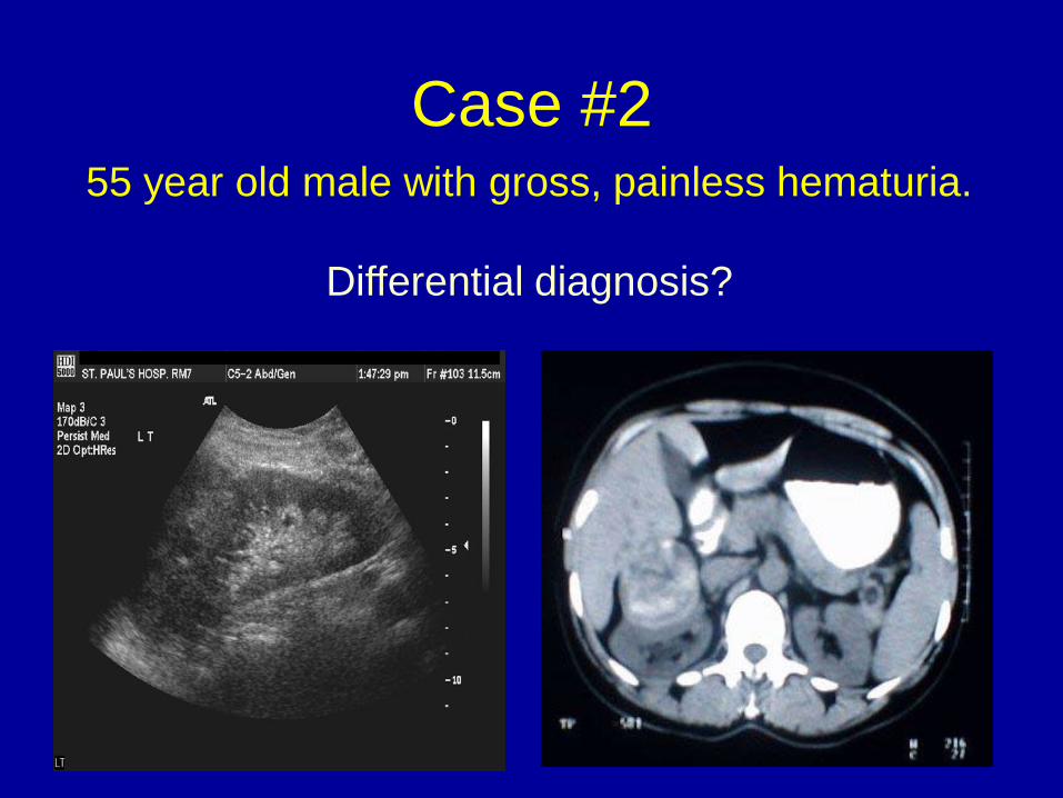
Case #2 55 year old male with gross, painless hematuria.
Differential diagnosis?

Solid renal mass – Differential diagnosis
Malignant - primary •Renal cell carcinoma •Transitional cell carcinoma Benign •Oncocytoma •Angiomyolipoma •Abscess •Pseudotumour (dromedary hump, hypertrophied column of Bertin, compensatory hypertrophy, etc…)
Malignant – secondary •Metastasis •Lymphoma

Fat within the mass is diagnostic of angiomyolipoma (-10 to -100 Hounsfield units)

Case #2
55 year old male with hematuria and a solid renal mass. What further tests are required before deciding on treatment?

Case #2 – Metastatic Evaluation
• Ca++, Alk. Phos. • LFT’s • CXR • review CT scan for:
1. local extension of tumour (eg. adrenal invasion) 2. regional lymphadenopathy 3. renal vein or IVC thrombus 4. liver mets 5. size/function of the opposite kidney

Case #2
If the serum Ca++ is elevated, what is likely cause?

Case #2 – Paraneoplastic Syndromes
renin - hypertension PTH-like peptide - hypercalcemia* erythropoietin - polycythemia abnormal liver function (Stauffer Syndrome) (*hypercalcemia can also be caused by bone
metastases)

Case #2
Treatment for locally confined renal cell carcinoma?

Radical vs. Partial Nephrectomy
Radical Partial


Indications for Partial Nephrectomy
Mandatory indication - solitary kidney
- bilateral tumours
- Hereditary syndromes (esp. von Hippel-Lindau syndrome)
- pre-existing renal impairment
Elective indication - implies contralateral
kidney is intact and functioning
- any small renal mass (< 4 cm diameter)
- especially if underlying disease such as HTN/DM
- trend toward partial for any tumor up to 7cm and larger tumors if technically feasible

Radiofrequency (RFA) vs. Cryoablation RFA Cryo

Renal Cell Carcinoma Histology
Sarcomatoid / Others 1-2%
Papillary 7-14%
Chromophobe 5-8%
Clear cell 75-80%

Renal Cell Carcinoma Staging • T1 less than 7 cm
– T1a <4cm – T1b 4-7 cm
• T2 >7cm but confined to kidney • T3 extends beyond kidney
– T3a adipose/adrenal – T3b renal vein, IVC below diaphragm – T3c IVC above diaphragm
• T4 invades neighbouring organ/side wall

RCC Survival Five year disease-specific survival T1 95% T2 90% T3a 60% T3b, c 25% (if completely resected) T4 20% N+ 10% – 20% M1 0%

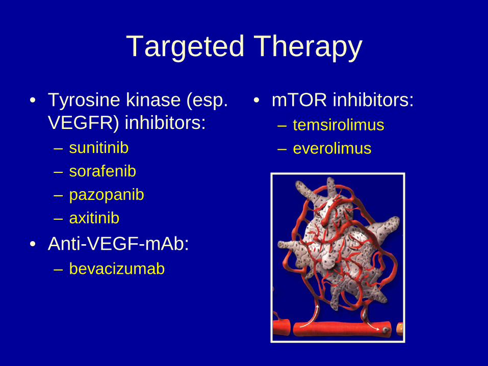
Targeted Therapy
• Tyrosine kinase (esp. VEGFR) inhibitors: – sunitinib – sorafenib – pazopanib – axitinib
• Anti-VEGF-mAb: – bevacizumab
• mTOR inhibitors: – temsirolimus – everolimus

Summary – Solid Renal Mass
• 70-90% likelihood of being renal cell carcinoma, depending on size
• Usually no biopsy – move straight to surgical intervention
• Try to preserve nephrons = partial nephrx
• Many new therapies improve survival of patients with metastatic renal cell carcinoma

Bladder Tumor
Objectives: 1. State 3 risk factors for transitional cell
carcinoma of the bladder
2. State the treatment options for superficial and invasive TCC of the bladder

Case #3
55 year old male with gross painless hematuria and a normal renal ultrasound. Cystoscopy shows this:

Case #3
What are the risk factors for transitional cell carcinoma of the bladder?

Risk factors for TCC
• smoking (R.R. 4X that of non-smokers) • occupational exposure to aniline dyes,
aromatic amines (eg.: textile manufacturing, dry cleaning, painting)
• previous exposure to cyclophosphamide (eg.: chemotherapy for lymphoma)
• previous radiotherapy (eg.: treatment of carcinoma of the cervix)

Case #3
Next steps?
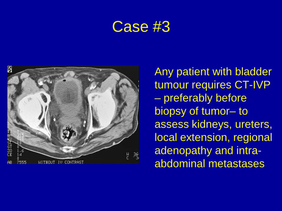
Case #3
Any patient with bladder tumour requires CT-IVP – preferably before biopsy of tumor– to assess kidneys, ureters, local extension, regional adenopathy and intra-abdominal metastases
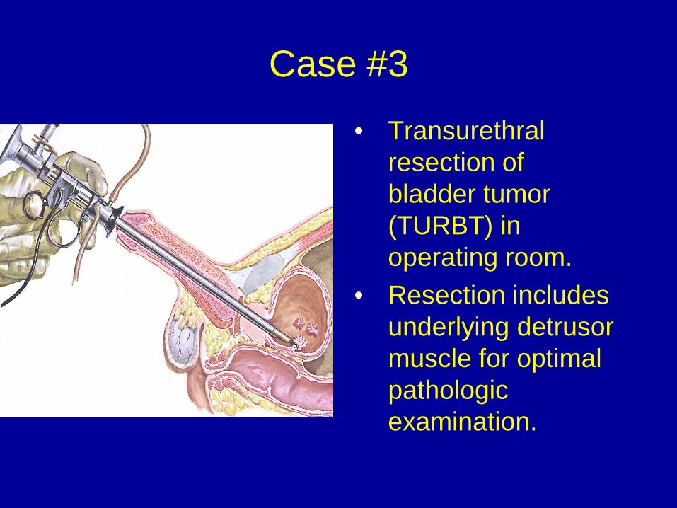
Case #3
• Transurethral resection of bladder tumor (TURBT) in operating room.
• Resection includes underlying detrusor muscle for optimal pathologic examination.

Case #3
How do you stage bladder tumours?

Non-muscle invasive bladder cancer (NMIBC)
Muscle invasive bladder cancer (MIBC)

Case #3
What treatments may be given for non-invasive TCC of the bladder after transurethral resection?

Indications for Intravesical Therapy
• any high grade tumour
• any lamina propria invasion (stage T1)
• carcinoma-in-situ
• multi-focal tumours
• rapid recurrence after initial resection
• (unable to completely resect transurethrally)

Intravesical Therapy
• Bacille Calmette-Guerin (BCG) - Only agent to demonstrate decreased rate of
progression in non-invasive bladder cancer - Requires induction and maintenance (36 months)
• Mitomycin - Especially effective as single dose after TURBT - Reduces short-term recurrence rate (within 2 years)
• Thiotepa (historical only) - Increased systemic toxicity due to small molecular
weight – therefore used only infrequently

Case #3
What are the indications for radical cystectomy for TCC of the bladder?

Indications for Radical Cystectomy
Curative • muscle invasive TCC (≥T2) • CIS or high-grade Ta/T1 that fails intravesical
therapy • extensive non-invasive tumours that cannot be
resected Palliative • control of hemorrhage in metastatic disease • extremely rare

Case #3
What do you do with the kidneys and ureters once the bladder is out?

Urinary Diversion – Ileal conduit
Pro • simplest to create • lowest risk of peri-operative complications • lowest risk of longer term metabolic complications
Con •abdominal stoma (“bag”) •incontinence
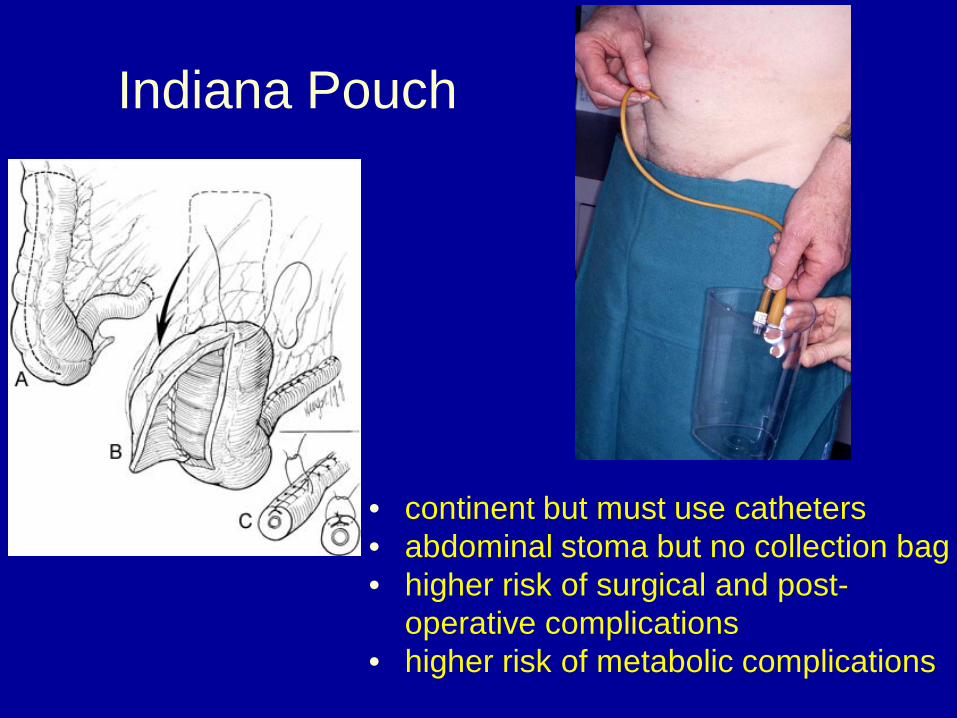
Indiana Pouch
• continent but must use catheters • abdominal stoma but no collection bag • higher risk of surgical and post-
operative complications • higher risk of metabolic complications

Orthotopic Neobladder
• continent and can void • 20% nocturnal incontinence • 1-2% need for
self-catheterization (higher in women)
• higher risk of surgical and post-operative complications
• higher risk of metabolic complications
• cannot be done if urethrectomy required

Chemotherapy for Bladder Cancer
• Gemcitabine/cisplatin most commonly used, although best evidence for MVAC (methotrexate/vinblastine/adriamycin/cispl)
• 60% response rate in metastatic disease – but rarely durable
• 5% survival benefit at 5 years when given before cystectomy (neoadjuvant)
• Questionable benefit after cystectomy (adjvuant)

TCC of the Upper Tract
• “filling defect” seen on retrograde pyelogram
• If opposite kidney is normal and there is no metastatic disease then treatment is a radical nephroureterectomy
• Can attempt endoscopic management if low volume and low stage/grade (need good biopsy)
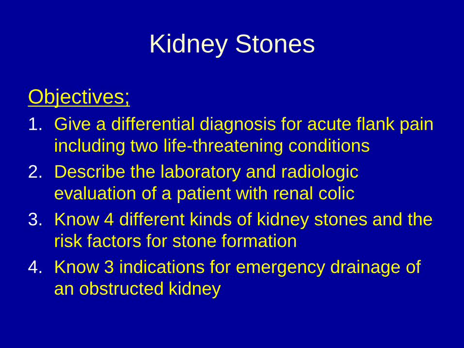
Kidney Stones
Objectives; 1. Give a differential diagnosis for acute flank pain
including two life-threatening conditions 2. Describe the laboratory and radiologic
evaluation of a patient with renal colic 3. Know 4 different kinds of kidney stones and the
risk factors for stone formation 4. Know 3 indications for emergency drainage of
an obstructed kidney

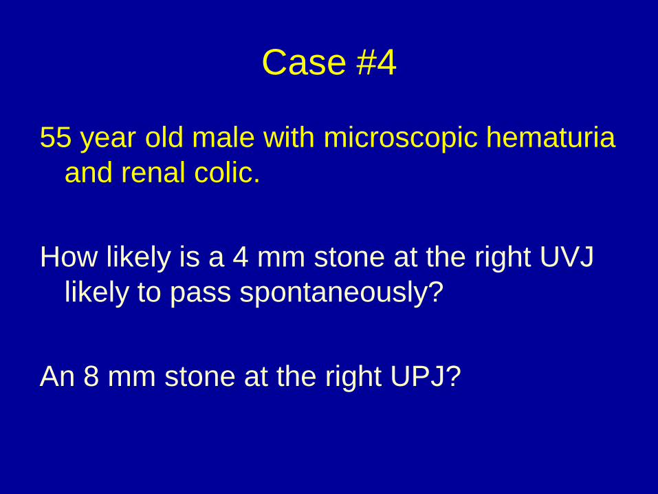
Case #4
55 year old man with microscopic hematuria and acute flank pain.
What is the differential diagnosis?

Differential diagnosis of renal colic
- abdominal aortic dissection - acute pancreatitis - abdominal aortic aneurysm rupture - renal abscess - cholecystitis - pyelonephritis - biliary colic - acute glomerulonephritis - appendicitis - renal vein thrombosis - diverticulitis - renal infarct - duodenal ulcer - ectopic pregnancy - viral gastroenteritis - pelvic inflammatory disease - inflammatory bowel disease - Fitz-Hugh-Curtis syndrome - splenic infarct - torsion/rupture of ovarian cyst - acute lumbar disc herniation - endometriosis - herpes zoster

Case #4
55 year old male with microscopic hematuria and renal colic.
Diagnostic evaluation?

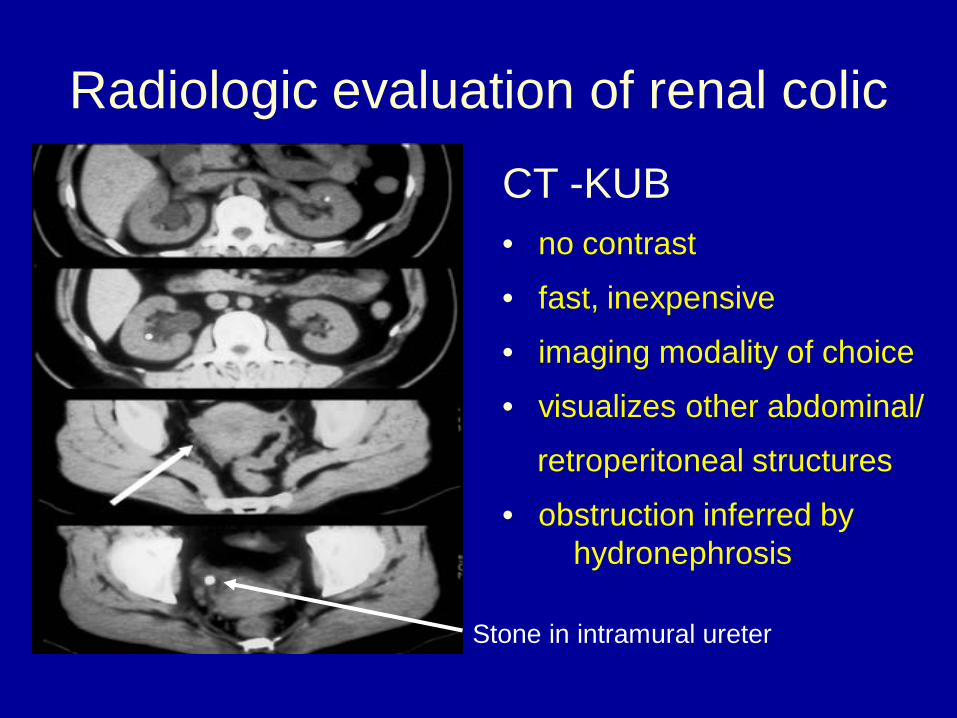
Radiologic evaluation of renal colic
KUB •“kidneys, ureters, bladder”
•plain X-ray of the abdomen
•85% of stones are visible
•no information regarding obstruction
Radiologic evaluation of renal colic
CT -KUB • no contrast
• fast, inexpensive
• imaging modality of choice
• visualizes other abdominal/
retroperitoneal structures
• obstruction inferred by hydronephrosis
Stone in intramural ureter

Case #4
Where are ureteric stones likely to be seen
in a patient with renal colic?

3 Physiologic Narrowings
Case #4
55 year old male with microscopic hematuria and renal colic.
How likely is a 4 mm stone at the right UVJ
likely to pass spontaneously? An 8 mm stone at the right UPJ?

Spontaneous Passage of a Ureteral Stone
In uncomplicated cases, spontaneous passage of the stone is safest; the likelihood of spontaneous passage is dependent on the size of the stone:
size likelihood
≤ 4 mm 90%
5 mm – 7 mm 50%
≥ 8 mm 20%

Case #4
55 year old male with microscopic hematuria and renal colic.
What other lab tests do you need before
making treatment decisions?

Lab Tests
• CBC (? elevated WBC ?)
• Creatinine (? impaired renal function ?)
• Urine microscopy (? bacteriuria or pyuria or pH ?)
• Ca++, uric acid, PO4
-- are often ordered but these will not influence the acute management

Case #4
55 year old male with microscopic hematuria and renal colic.
What are the indications for immediate
drainage of an obstructed kidney? When can you leave a kidney obstructed to
allow for spontaneous passage of a stone?

Immediate Referral to a Urologist
• obstructed ureter plus fever, chills, bacteriuria or elevated WBC (risk of urosepsis – may be life-threatening)
• obstructed ureter in an insulin-dependent diabetic (risk of papillary necrosis or emphysematous pyelonephritis)
• solitary kidney • pre-existing renal disease • significant co-morbid conditions
(congestive heart failure, pregnant …)

Case #4
Name 4 different kinds of kidney stones and the factors that predispose to stone formation?
Types of Stones
Calcium Oxalate • most common stone • most common predisposing factors:
1. dietary hyperoxaluria (chocolate, nuts, tea, strawberries, peanut butter, cabbage or excessive restriction of dietary calcium)
2. hypercalciuria (absorptive; inherited condition) 3. dietary hypercalciuria due to excess dietary sodium
or meat proteins

Types of Stones
Calcium Phosphate • second most common stone • often seen in patients with metabolic
abnormalities:
1. primary hyperparathyroidism 2. distal renal tubular acidosis 3. hypercalcemia due to malignancy or sarcoid

Types of Stones
Uric Acid • radiolucent on KUB but visualized on CT-KUB • predisposing factors:
– persistently acidic urine – low urine volumes (eg. from chronic diarrhea, excess
sweating, inadequate fluid intake) – gout (hyperuricemia) – excess dietary purine (meat) – chemotherapy for lymphoma, leukemia

Types of Stones
Struvite (infection stones) • magnesium, calcium and ammonium phosphate • urine pH > 8.0, therefore will only form in the
presence of urease-secreting bacteria • tend to form large stones or staghorn stones • urease-secreting bacteria include Proteus,
Klebsiella, Providentia, Pseudomonas and Staph. aureus – but not E. coli

Relief of Obstruction
Ureteric stents •can be placed retrograde via cystoscopy or antegrade via nephrostomy •indwelling “double-J” stents remain in place due to coiling of the ends in the renal pelvis and bladder

Relief of Obstruction
Percutaneous Nephrostomy • temporary drainage
into an external collecting bag
• inserted by radiologist under local anaesthetic

Describe three standard surgical therapies for renal and ureteral stones.
Case #4
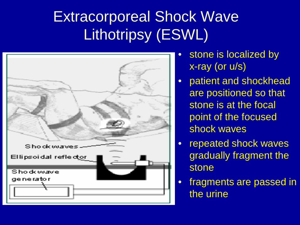
Extracorporeal Shock Wave Lithotripsy (ESWL)
• stone is localized by x-ray (or u/s)
• patient and shockhead are positioned so that stone is at the focal point of the focused shock waves
• repeated shock waves gradually fragment the stone
• fragments are passed in the urine

Extracorporeal Shock Wave Lithotripsy (ESWL)

Ureteroscopy

Holmium Laser Lithotripsy of a Ureteric Stone

Treatment of a Large Stone
Staghorn stone • ESWL will fragment the stone but the large stone burden is not likely to pass spontaneously • stones over 20 mm in diameter are better treated with PNL

Percutaneous Nephrolithotripsy

Testicular Tumors
Objectives
1.Differential diagnosis of a scrotal mass
– Caveat: recognize testicular torsion!
2.Classify testicular tumors
3.Treatment of testicular malignancies

Case #5
28 year old man with a right scrotal mass. Evaluation?

History
• onset • pain • firmness • history of undescended testis
• urethral discharge • STDs • LUTS

Physical Exam
• location of mass (testis,epididymis,scrotum) • reduces in supine position? • is mass tender? • transillumination? What test is next?

Diagnostic Tests
Urinalysis • pyuria with epididymitis Ultrasound • sensitive and specific for testicular tumour

Scrotal Mass - Differential Diagnosis
• hydrocoele • spermatocoele • varicocoele • epididymitis/orchitis • inguinal hernia • testicular tumor • paratesticular tumour (i.e.: adenomatoid
tumour or cystadenoma of the epididymis)

Hydrocoele
• fluid within tunica vaginalis • called “communicating
hydrocoele” if processus vaginalis is patent
• typically painless • transilluminates • cannot palpate testicle • no treatment required unless
for cosmetic reasons
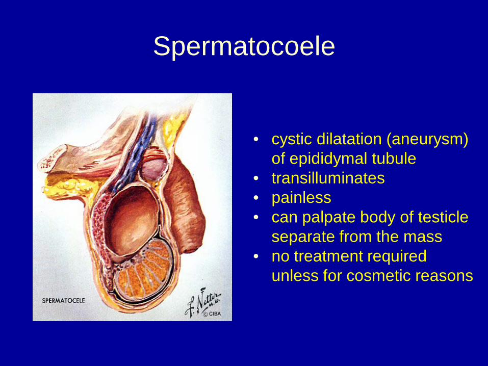
Spermatocoele
• cystic dilatation (aneurysm) of epididymal tubule
• transilluminates • painless • can palpate body of testicle
separate from the mass • no treatment required
unless for cosmetic reasons

Varicocoele
• varicosities of pampiniform plexus • 90% on left side • seen in 15% of post-pubertal male population • typically asymptomatic but may cause “achiness” • increases in size with valsalva or standing position. • associated with male factor infertility but most men with
varicocoeles can expect normal fertility. • surgical or angiographic correction of the varicocoele
results in improvement in semen parameters (number, motility, morphology) in 70% to 90% of cases

Varicocoele

Right Varicocoele
Not a normal variant – must consider
retroperitoneal mass/tumor with
obstruction of flow in right gonadal vein

“Acute Scrotum”
In adolescents and young men, with no history of trauma, the possibilities include:
• Testicular Torsion • Torsion of the Appendix Testis • Epididymitis
Testicular torsion and torsion of the appendix testis
are extremely uncommon in men >40 yrs.

“Acute Scrotum”
History: associated voiding symptoms or fever? (suggesting infection)
Physical examination: “lie” of the testicle (high-riding in torsion),
cremasteric reflex (absent in torsion), Prehn’s sign (relief of pain on lifting or supporting the scrotum – suggests epididymitis).
Urinalysis and urine culture should be done in all cases (pyuria suggests epididymitis)

Testicular Torsion
• Typically 12 to 18 years of age. • Presents with acute onset of severe scrotal pain and
swelling not associated with trauma. • Physical exam should include cremasteric reflex and
differentiate epididymis from testicle. • Urinalysis should be normal (pyuria suggests epididymitis) • Ultrasound with Doppler is most helpful ancillary test. • The most common misdiagnoses are torsion of the testicular
appendix and epididymitis.

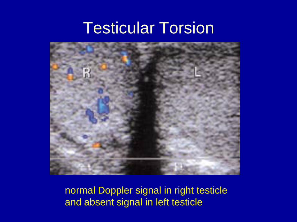
Testicular Torsion
normal Doppler signal in right testicle and absent signal in left testicle

Epididymitis
• Doppler signal is increased with inflammation
• thickened epididymis visualized

Epididymitis
• Men < 35 years of age – chlamydia, gonorrhea (STD) • Men > 35 years of age – coliforms (UTI)
• Diagnosis often made by physical exam, but pain and
swelling in advanced cases make the exam less helpful. • Complications include abscess formation, testicular
infarction and infertility. • Treatment is 4 weeks of antibiotics with rest, ice,
NSAIDs.

Torsion of Appendix Testis

Testicular Cancer
Special features: • It occurs in young men
who are otherwise in good health
• Even widely metastatic disease is potentially curable with multimodal chemotherapy

Testicular Tumours Classification
A. Primary 1. Germ cell tumours
- seminomatous - non-seminomatous
2. Non-germ cell tumours – Leydig cell tumour – Sertoli cell tumour
B. Secondary (lymphoma/leukemia)

Classification of Testicular Germ Cell Tumours
• Seminoma (classic/spermatocytic/anaplastic)
• Non-seminoma
– Embryonal Carcinoma
– Teratocarcinoma = immature teratoma
– Choriocarcinoma
– Yolk Sac Tumour (most common form in children)
– Teratoma (benign but can metastasize and grow)

Testicular Cancer
Usual presentation is a painless intratesticular mass discovered on self- examination. Typical age at diagnosis is 15 to 35 years, although smaller clusters of cases occur during infancy and over 60 years of age.

Self - Examination
Self – examination should be taught to young men They need to be shown the difference between the testicle and the epididymis They need to report any hard or suspicious lesions immediately

Tumour Markers
• β-HCG – produced by choriocarcinoma and in limited quantities by seminoma. Serum T1/2 = 24 hrs.
• α-fetoprotein – produced by yolk sac tumours, embryonal carcinoma and teratocarcinoma. Never found in patient with pure seminoma. Serum T1/2 = 3 – 5 days.
• LDH – correlates with tumour volume

Radical Orchiectomy
Testicle with the tunica vaginalis and spermatic cord are delivered through an inguinal incision Scrotal incisions or biopsies should never be done Check markers again after orchiectomy

Testicular Cancer - Metastasis
• Predictable pattern of spread: – Retroperitoneal lymph nodes first (along
spermatic vessels towards hilum of kidney) – Lung metastases second – Other organs after lungs
• Every patient requires CT-abd/pelvis, and either CXR or chest CT

Retroperitoneal Lymphadenopathy
Large retroperitoneal mass in patient with right testicular NSGCT

Testicular Cancer - Management
• Organ-confined, no mets (“clinical stage I”) – Active surveillance by far most commonly
practiced strategy – Sometimes chemo/XRT/RPLND
• Metastatic (stage II and III) – Almost every patient gets chemo (rarely XRT
for seminoma) – NSGCT patients often require subsequent
RPLND and resection of chest masses

Testicular Cancer - Management
• Differences for seminoma and NSGCT – both exquisitely sensitive to chemotherapy
• BEP or EP x 3-4 cycles – seminoma also responsive to XRT
• but rarely used because of potential toxicity – post-chemotherapy retroperitoneal lymph
node dissection rarely used for seminoma, but performed for every residual mass >1 cm in NSGCT (risk of teratoma)

Retroperitoneal Lymph Node Dissection